THE ROLE OF DOPAMINE D3
RECEPTORS IN THE CONTROL OF
ALCOHOL INTAKE
UNIVERSITY OF CATANIA
Ph.D. in Neuropharmacology
XXV Cycle
THE ROLE OF DOPAMINE D3
RECEPTORS IN THE CONTROL OF
ALCOHOL INTAKE
Doctorate Thesis
Giovanni Camillieri
Coordinator: Prof. Filippo Drago Tutor: Prof. Salvatore Salomone
UNIVERSITY OF CATANIA
Table of contents
Acknowledgements4 List of abbreviations 5 Preface 6 Introduction 1. Alcoholism 8
2. Dopamine pathways and receptors in the central nervous system 14
3. Alcoholism and dopamine 17
4. Experimental animal models of alcoholism 27
5. D3 dopamine receptor 40
6. Design of the present research 43
Chapter I 44
7. General Discussion
7.1 D3 KO mice show an aversion for the ethanol intake 87 7.2 Dopamine D3 selective antagonists counteract ethanol intake 89
Concluding Remarks 89
Acknowledgements
I wish to express my sincere gratitude to:
Professor Filippo Drago, my mentor, as he offered his continual informed and valuable support, discussion, his trust and for his example as a scientist and experienced researcher
Professor Salvatore Salomone, my tutor, for her precious suggestions during my doctorate studies
Also, I would like to thank Doctor Gian Marco Leggio, colleague and friend, for his support during my PhD studies
List of abbreviations
5-HT serotoninBDNF brain development neurotrophic factor
CAAs competing amino-acids
CNS central nervous system
D3-/- dopamine D3 receptor deficient mice
D3R dopamine D3 receptor
DA dopamine
DMSO dimethyl sulfoxid
DSM-IV Diagnostic and Statistical Manual of Mental Disorders, 4th edition
EPM elevated plus maze test
FST forced swimming test
GABA γ-aminobutyric acid
GABAA γ-aminobutyric acid receptor A
GAD generalized anxiety disorder
HF Familiar conditions
HU Unfamiliar conditions
i.p. intraperitoneal injection
ICD-11 Classification of Diseases, 11th edition
NGT novelty induced grooming test
PD Panic disorders
SIT Social Interaction Test
SSRI selective serotonin reuptake inhibitor
TCA tricyclic antidepressant drug
TRP tryptophan
Preface
Mesolimbic dopamine controls drug and alcohol seeking behavior. Stimulation of dopamine D3 autoreceptor reduces extracellular levels of
dopamine. We tested the hypothesis that dopamine D3 receptor (D3R) gene
deletion or its pharmacological blockade counteracts alcohol preference and intake in a long-term voluntary ethanol intake paradigm. Mice D3R-/- and their
wild type (WT) littermates, treated or not with the D3R antagonists U99194A
and SB277011A, were tested. The selectivity of the D3R antagonists was
further assessed by molecular modeling. Activation of dopamine (DA) transmission and D3R expression was assessed at the end of the experiment.
After 8 days, daily ethanol intake was negligible in D3R-/-and robust in WT;
this behavior was stably maintained for 44 days. Treatment with D3R
antagonists counteracted ethanol intake in WT and was associated to increased DA transmission (assessed as phosphorylation of DARPP-32 and GSK3β) in striatum and prefrontal cortex. Forced ethanol intake increased the expression
of RACK1 and BDNF in both WT and D3R-/-; in WT there was also a robust
overexpression of D3R. Thus, increased expression of D3R associated with
activation of RACK1/BDNF seems to operate as a reinforcing mechanism in voluntary ethanol intake. Taking into account that ethanol intake increases
overexpression would facilitate ethanol intake, and high levels of extracellular DA, from either gene deletion of D3Rblockade, would inhibit ethanol intake.
Thus, modulation of DA mesolimbic pathway by selective targeting of D3
Introduction
1. Alcholism
Although alcohol has been recognized as an agent of abuse since the 17th century, the disease model was accepted only in the last century. Alcoholism is defined as a chronic and progressive disease characterized by loss of control over the use of alcohol with subsequent social, legal, psychological and ethical consequences. Recently, monoamines were shown to have a predominant role in the etiology of alcoholism. Dopamine has been implicated by virtue of its actions on the reward center. Endogenous opioids which lessen stress and produce euphoria, are released upon alcohol intake, whereas increased serotonin facilitates tolerance and thereby fosters increased alcohol consumption. Alcohol has facilitative effects on inhibitory action of GABA, while the stimulatory effects of glutamate are decreased. Drugs used for treatment of alcohol dependence can be broadly classified into 4 groups: sensitizing drugs, opioid antagonists, drugs acting on serotonergic systems and acamprosate. Disulfiram has been shown to be most effective for patients who believe in its efficacy and remain compliant with the treatment. The opioid antagonist naltrexone lowers relapse rate, reduces drinking days and prolongs periods of abstinence, while
efficacy. The treatment of alcoholism should not be restricted to pharmacotherapy alone but should also be supplemented with interventions addressing the psychological, medical and social needs of patients.
In industrialized countries, alcohol use alone causes about 10% of total disability-adjusted life years lost (Rehm, Mathers et al. 2009), and a recent evaluation in the United Kingdom concluded that in aggregate, the harm to self and others inflicted by alcohol exceeds that caused by heroin or cocaine (Nutt, King et al. 2010). Alcohol consumption in the population is markedly skewed, and a large proportion of alcohol-related disability is due to alcohol addiction, hereafter equated with alcoholism. This is a condition that in the United States affects more than 12% of the population at some point in their life (Hasin, Stinson et al. 2007). Alcoholism is a chronic, relapsing disorder that shares many characteristics with other complex chronic conditions, such as diabetes or hypertension: it has a considerable component of genetic susceptibility, is under marked influence of environmental factors, and its onset and course are fundamentally shaped by behavioral choices (McLellan, Lewis et al. 2000; Goldman, Oroszi et al. 2005). This prompts the question of whether alcoholism can be tackled with medical treatments. Some efficacy of medications for alcoholism (Bouza, Angeles et al. 2004) as well as opiate (Amato, Davoli et al. 2005) and nicotine (Wu, Wilson et al. 2006) addiction has been documented and supports the feasibility of addiction pharmacotherapy. However, with the
exception of methadone or buprenorphine maintenance therapy for opioid addictions, the effect sizes of these treatments are small. Despite evidence-based guidelines that pharmacotherapy be considered in all patients with alcoholism, and in particular in those who are not successfully treated with behavioral interventions alone (Willenbring, Massey et al. 2009), only a small minority of patients receive medication for their alcoholism (Mark, Kranzler et al. 2003).
Clearly, extensive unmet medical needs remain in this therapeutic area.
A clinical diagnosis of alcoholism is currently made on the basis of diagnostic criteria that are standardized across addictive disorders by the Diagnostic and Statistical Manual of Mental Disorders, which is currently in its fourth edition (DSM IV). In the absence of reliable biomarkers, this approach eliminates some of the subjective judgment involved in making diagnoses, and has clinical utility. However, there is reason to believe that patients diagnosed using this approach are markedly heterogeneous. In fact, such heterogeneity was already proposed in the 1980s on the basis of clinical characteristics such as age of onset, but also on family history, which is a marker of genetic susceptibility (Cloninger 1987). Numerous other attempts at clinical subtyping of people with alcoholism have since followed. The use of genetic markers offers the possibility of more reliably and consistently capturing the heterogeneity of
Among individuals in the general population who fulfill diagnostic criteria for alcoholism, the majority — about three-quarters — never receive treatment (Hasin, Stinson et al. 2007). Available data indicate that those people who go on to enter treatment and those who do not are fundamentally different with regard to personality traits, alcohol use patterns and long-term outcomes (Vaillant 1983; Vaillant 1996; Fein and Landman 2005). Furthermore, classic longitudinal studies show that long-term outcomes and alcohol-related harm vary markedly between individuals in ways that do not seem to have a simple correlation with participation in treatment or the level of alcohol use (Vaillant 1983; Vaillant 1996). A clinical diagnosis of alcoholism is probably best viewed as an ‘end-stage disease’, similar to congestive heart failure. In this view, the diagnostic category of alcoholism consists of conditions that are phenotypically similar (or constitute ‘phenocopies’), but patients arrive at the disease state through fundamentally different trajectories. This is captured by a conceptualization that was first put forward for major depression (Kendler, Thornton et al. 2001), but is also likely to apply to addiction. In a kindling-like process, brain exposure to cycles of intoxication and withdrawal induces progressive neuro-adaptations that ultimately result in escalation of alcohol intake (Ballenger and Post 1978; Heilig and Koob 2007). In the absence of significant genetic susceptibility, escalation will only result following prolonged exposure to alcohol and the environmental factors with which it
interacts, such as stress. By contrast, when genetic risk factors are present, progression can be fast. These individuals can be viewed, in terminology borrowed from the depression literature (Kendler, Thornton et al. 2001), as ‘pre-kindled’, or ‘already there’.
Emerging evidence indicates that individuals with alcohol addiction who are on trajectories that are driven by different biological mechanisms or who are in different stages of addiction can be expected to respond to different treatments. Fundamentally, treatments for alcohol addiction must intervene with biological mechanisms that provide motivation for alcohol seeking and consumption (Heilig and Egli 2006). These mechanisms largely fall into two main categories. First, in a similar way to other drugs of abuse, alcohol can activate brain reward pathways, leading to positively reinforced alcohol seeking and use. Secondly, alcohol can acutely suppress negative emotions that result from stress or withdrawal from alcohol itself, such as anxiety and dysphoria, thus setting the scene for negatively reinforced alcohol use (Heilig and Koob 2007; Koob and Volkow 2010). To highlight the distinction between these two incentives for alcohol use, the terms ‘reward drinking’ and ‘relief drinking’ have been introduced (Heinz, Lober et al. 2003). It is reasonable to expect that these different types of excessive alcohol use will require different treatments.
Genetic and environmental factors in alcoholism can result in very different types of vulnerability, ranging from heightened impulsivity and reward from alcohol to enhanced stress responses and anxious personality traits (Cloninger 1987). Genetic variants that alter alcohol reward- or stress-related emotional processing are therefore probable modifiers of disease trajectories and of responses to treatments that target reward and stress systems.
2. Dopamine pathways and receptors in the central nervous
system
Four main dopaminergic pathways were identified within the CNS. The ventral tegmental area is the place of origin of two projection pathways towards the cortex (the mesocortical pathway) and the limbic area (the mesolimbic pathway); the hypothalamus is the place of origin of a projection towards the pituitary gland which controls prolactin secretion (the tubero-infundibular pathway) and a dopaminergic projection extends from the substantia nigra to the striatum (the nigrostriatal pathway) the degeneration of which is implicated in Parkinson’s disease. Using these pathways, dopamine receptors are located. Five genes encoding dopamine receptors were identified.
These receptors are divided in two subfamilies: the D1-like receptor subtypes (D1 and D5) coupled with the Gs protein activate adenylyl cyclase and the D2-like subfamily (D2, D3, and D4) coupled with G proteins inhibit adenylate cyclase (Missale, Nash et al. 1998). D1 and D2 dopamine receptors are the most abundant subtypes in the central nervous system, but D1 dopamine receptor is the most widespread. D1 mRNA was found in the striatum, nucleus accumbens, olfactory tubercule, hypothalamus and thalamus. In other areas such as
dopamine receptor is present in projections only (Jaber, Robinson et al. 1996). The D5 dopamine receptor is expressed at much lower level than the D1 dopamine receptor and its distribution is limited to the hippocampus and thalamus (the lateral mamillary nucleus and the parafascicular nucleus of the thalamus). The D2 dopamine receptors are located mainly in the striatum, olfactory tubercule, nucleus accumbens, the substantia nigra pars compacta, the ventral tegmental area and the pituitary gland. D2 dopamine receptors are pre- and post-synaptic receptors contrary to D1-like receptors which are mainly post-synaptic receptors (Jaber, Robinson et al. 1996). D4 dopamine receptors were found with a low expression in the basal ganglia and a higher expression in the frontal cortex, medulla, amygdala, hypothalamus and mesencephalon. However, this high expression is weak in comparison with other dopamine receptors (Jaber, Robinson et al. 1996). D3 dopamine receptors are expressed in the limbic area (nucleus accumbens, olfactory tubercule and islands of Calleja) and at a lower level in the striatum(Jaber, Robinson et al. 1996). The D3 dopamine receptors exist as autoreceptors that inhibit neuronal dopamine synthesis and post-synaptic receptors. These receptors by negatively regulating dopamine neuronal activity and/or by post-synaptic action exhibit an inhibitory influence on locomotor activity (Sibley 1999). The genetic techniques for negatively modulating dopamine receptor expression such as knockout animals and antisense technology showed that the disruption of D3, D4, D5 dopamine
receptor functions involved an increase or an improvement in the behavioral activity of animals contrary to the results observed with the disruption of D1, D2 dopamine receptor functions. Although these results have to be interpreted with caution as a compensatory mechanism could develop, these observations suggest that the most abundant dopamine receptors D1 and D2 are involved in positive regulation of behavioral activity whereas the D3, D4, D5 receptors are inhibitory by likely negative modulation of D1 and/or D2 receptor function in some cases (Sibley 1999).
3. Alcoholism and dopamine
Credit for discovering that striatal DA neurotransmission is fundamental to drug self-administration is due to two independent groups using different means: James Olds and colleagues in the 1950s and 60s, and the pharmacologists associated with Arvid Carlsson, including Nils Hillarp, Annica Dahlstrom, Kjell Fuxe, and Urban Ungerstedt, who were establishing DA as a neurotransmitter.
In 1954, Olds and Peter Milner introduced intracranial self- stimulation by implanting electrodes in the brains of rats and providing them with a lever that they could press to apply current(Olds and Milner 1954). Soon after, Olds introduced ‘‘intracranial self-administration,’’ a lever-operated device that would allow rats to inject drugs via a pipette directly into defined areas of the brain (Olds and Olds 1958). The self-stimulation paradigm provided the initial evidence of how activity in specific brain regions correlated with the fraction of the time the animals pressed the lever, while self-administration provided insights into drug effects at specific brain regions.
Initial experiments demonstrated that rats with electrodes in the septal area used as much as 92% of the time to bar press, in contrast to regions where ‘‘animals do everything possible to avoid stimulation.’’ By 1956, Olds and collaborators found that stimulation of the hypothalamus was even more
rewarding, eliciting as many as 5000 bar presses per hour. They soon suspected that the efficacy of stimulation of the lateral hypothalamus was due to activation of the medial forebrain bundle, through which DA neurons course from cell bodies in the midbrain to striatal and cortical targets.
They then attempted to interfere with bar presses by administering drugs(Olds, Killam et al. 1956). Successful inhibitors of self- administration included reserpine, which blocks uptake of catecholamines into synaptic vesicles(Carlsson, Hillarp et al. 1962; Kirshner 1962), and chlorpromazine, the antipsychotic, which Carlsson later showed blocks DA receptors.
By 1958, Olds concluded: ‘‘The cells which mediate primary rewarding effects are located in a midline system running from the midbrain through and into the subcortical and cortical groups of the rhinencephalon. The cell groups which mediate primary rewarding effects are different from those which mediate primary punishing effects’’(Olds and Olds 1958). They validated the role of medial forebrain projection from the midbrain to the cortical and subcortical areas in these rewarding effects in later lesion studies (Olds and Olds 1969).
Also in 1969, Olds and Phillips introduced the concept that salient stimuli are responsible for the firing of DA neurons by showing that ventral midbrain neurons fired at a higher frequency following a tone paired with food
(Phillips and Olds 1969). Remarkably, they conclude with essentially the contemporary understanding of the rules of DA neuronal firing: ‘‘Thus it was surmised that expectancy of reward, rather than response to the tones per se, accounted for the differing rates of firing in midbrain unit activity. These responses reflect an integration of sensory input with the internal state, where the response to tones which signified a reward appropriate to the submotivational state of the organism was amplified by the degree of that motivation.’’ These insights, along with a posthumous study that outlines a specific role for VTA neurons(Brauth and Olds 1977), led to subsequent explorations to decipher the rules by which reinforcement control the activity of DA neurons(Schultz 2011).
By 1976, the year that he died in an accident, Olds wrote a review on the state of self-stimulation and drive, concluding that ‘‘noradrenaline neurons might be the reward neurons addressed to negative drives and DA neurons to positive drives’’(Olds 1976). By introducing self-stimulation paradigms using electrical current and direct drug application, exploiting this to map the brain regions involved, and characterizing firing modes by these neurons to reward and stimuli associated with reward, Olds perhaps provided the strongest contribution to our under- standing of addiction.
Evidence that Addictive Drugs Enhance Striatal DA
histochemical fluorescent techniques showing that DA neurons originated in the ventral mesencephalon and projected to the cortex and striatum (Hillarp, Fuxe et al. 1966), in the pathway Olds had identified. Similarities between the behavioral response to AMPH and electric self-stimulation of this pathway were observed, and led Crow and colleagues to suggest that ‘‘the dopamine-containing system arising from the ventral mesencephalon may function as an activating system involved in the effects of positive reward on operant behaviour’’ (Anlezark, Arbuthnott et al. 1971). A variety of neurochemical experiments to measure catecholamine release during self-stimulation were conducted, as well as additional lesioning studies, as reviewed(German and Bowden 1974).
Experiments by Wise, Fibiger, Phillips, and others were influential in convincing the field that DA release was particularly important for reward, for example by showing that partial DA receptor blockade increased self-administration of AMPH by rats, while rats would self-administer direct DA receptor agonists(Yokel and Wise 1978). The memorably named De Wit and Wise (1977) showed that a DA D2 receptor antagonist, but not norepinephrine antagonists, blocked cocaine reinforcement.
An approach that convinced the field at large that DA was responsible for the actions of addictive drugs was in vivo micro-dialysis using electrochemical
this caused significant tissue damage, while voltammetry(Kissinger, Hart et al. 1973) measured catecholamine release and reuptake with extraordinary time resolution(Millar, Stamford et al. 1985; Rice, Oke et al. 1985) and was effective for studying cocaine and AMPH(Caviness and Wightman 1982; Ewing, Alloway et al. 1983), but could not differentiate between electroactive compounds (Gonon, Buda et al. 1980) including norepinephrine and DA. Microdialysis with HPLC electrochemical detection was pioneered by Ralph Adams and collaborators(Adams 1976; Plotsky, Wightman et al. 1977), who showed that AMPH released DA, and Ungerstedt’s lab(Ungerstedt and Pycock 1974), who confirmed this response in the striatum.
Microdialysis studies were extended by Imperato and Di Chiara and colleagues, who demonstrated that ethanol (Di Chiara and Imperato 1985), opiates and barbituates (Di Chiara and Imperato 1986), and nicotine (Imperato, Mulas et al. 1986) increased DA concentrations in striatum, particularly in the ventral striatum / nAc. Drugs with aversive properties decreased DA release, and non-abused drugs did not modify synaptic DA (Di Chiara and Imperato 1988).
Most recently, studies in human psychostimulant-naive individuals have shown that AMPH-mediated DA release as measured by D2 receptor availability is significantly associated with effects of the drug reported by the subject as ‘‘happiness’’ and ‘‘energy’’(Abi-Dargham, Kegeles et al. 2003).
In summary, a role for enhanced striatal DA neurotransmission in the addictive properties of drugs is supported by an increase in extracellular DA levels in the striatum measured following all of the classic addictive drugs; an increased level of drug self-administration when DA receptors are partially antagonized, with cessation at more complete blockade; inhibition of drug self-administration when catecholamines release is decreased by VMAT inhibition or when DA synthesis is blocked; cessation of self-administration when DA neurons or their axons are ablated; reports by AMPH naïve individuals providing a correlation between DA release and self-reported euphoria. Each mechanism of action essentially decouples DA transmitter levels from normal physiological control. From Olds’s studies, this would be expected to assign the stimuli that were associated with acquiring these drugs as reinforcements, providing a conceptual synaptic framework for addiction.
Voluntary drinking releases DA in humans (Boileau, Assaad et al. 2003), with recent human PET imaging showing that the equivalent of three drinks increased extrasynaptic DA in the striatum by 138% in men and 69% in women(Urban, Kegeles et al. 2010).
It is thus startling that we do not know how ethanol enhances DA transmission. One reason is because there are many possible receptor and channel targets for ethanol, as recently reviewed(Melis, Diana et al. 2009;
DA release at levels achieved by individuals who consume alcohol for its reinforcing properties. How Many Are Enough? Ethanol is typically measured as blood fraction (blood alcohol content [BAC], in the U.S. in units of g/100 ml written as a percentage). As there appears to be no blood/brain barrier to ethanol penetration, the extracellular levels in brain are close to those in blood (Robinson, Brunner et al. 2002). A glass of wine (150 ml, 2.6 M ethanol) yields 0.02% BAC (4.8 mM) in blood of non-alcoholic 68 kg men, with women achieving 34% higher levels(Frezza, di Padova et al. 1990). A typical level for legal intoxication is 0.08% BAC (17 mM), stupor occurs at 0.25% (54 mM), blackout at 0.35% (76 mM), and lethality at 0.4% (87 mM). Effects on DA transmission related to self-administration for reinforcing properties should therefore be present at 5–20 mM ethanol, lower than that often studied experimentally. Note however that alcoholics develop tolerance and can achieve extraordinary levels, as high as 1.20% BAC (260 mM)(Brick and Erickson 2009). How Does Ethanol Cause DA Release? It has been suggested that alcohol activates VTA neurons directly to release DA into nAc. However, some but not all dialysis experiments show that alcohol application into the nAc alone locally increases DA overflow while application of alcohol into the VTA does not(Yim, Schallert et al. 1998; Ericson, Molander et al. 2003), suggesting that effects at the axons may be required. In contrast, studies of ethanol using cyclic voltammetry in the striatal slice (Budygin, Phillips et al. 2001)
demonstrated effects of ethanol on evoked DA release only at very high (100– 200 mM) alcohol levels, which depressed release. Thus, the relevant brain sites in striatum are still unclear.
Ethanol might increase DA release by direct excitation of DA neurons. Some studies show an effect of alcohol to excite DA neurons in the VTA at concentrations of 20–320 mM(Brodie and Dunwiddie 1990; Okamoto, Harnett et al. 2006). This might involve an inhibition of potassium channels, including those that regulate after-hyperpolarizations and the rate of burst firing, as well as sustained K+ currents(Koyama, Brodie et al. 2007).
Alternatively, ethanol may act via disinhibition of DA neurons, most likely at GABA receptors. Ethanol effects on GABA are suspected to play a part in its effects, notably the motor-impairing and anxiolytic responses, in a manner related to the benzodiazepines and barbiturates. Recent evidence suggests a possible role for extrasynaptic GABAa receptors, as a population of extra-synaptic receptors containing a-4 subunits were found that provide a steady inhibition of thalamic neurons with sedative levels (50 mM) ethanol(Jia, Chandra et al. 2008). A recent paper showed that viral knockdown of the a-4 subunit in the nAc shell but not the core, decreased alcohol drinking and preference in the rat(Rewal, Jurd et al. 2009); perhaps the receptors in that area are more sensitive than those in the thalamus.
Several classes of striatal interneurons exert ‘‘veto’’ power on the ability of MSN neurons to fire(Tepper, Koos et al. 2004), and if either these or medium spiny neuron collaterals were inhibited, the net result of ethanol inhibition of GABAergic activity (or cholinergic) activity via GABAergic disinhibition could underlie a presynaptic component of enhanced DA release.
A similar pathway mediated by an ethanol-mediated GABA receptor disinhibition of DA neurons by ethanol could also occur in the ventral midbrain(Mereu and Gessa 1985), as found with opioids and sedatives. It should be noted that, in contrast, ethanol appears to enhance GABAb currents on midbrain DA neurons by activating GIRK currents(Federici, Nistico et al. 2009), which may reinstate some inhibition. Alternatively, opiate receptors on GABAergic VTA projection neurons have been implicated in the action of ethanol(Xiao and Ye 2008), perhaps via ethanol-mediated release of b-endorphin, which would similarly disinhibit DA neurons.
Other hypotheses have been offered for network effects via cannabinoids, serotonin, glycine, NMDA channels, nAChR, and NMDA receptors, but to date none have been shown to clearly cause enhanced firing of DA neurons at levels of 5– 20 mM ethanol, although the enhancement of burst firing seems to involve endocannabioids(Cheer, Wassum et al. 2007). In summary, while there is good evidence that levels of ethanol achieved during moderate drinking release DA, and while there are a multitude of potential targets, the means by
which ethanol releases DA during drinking at nominally rewarding levels remain unclear.
4. Experimental animal models of alcoholism
Researchers have known since 1940 that some rodents voluntarily consume alcohol in a laboratory setting. One can also assume that voluntary alcohol consumption by rodents and other mammals occurs in the wild, because some mammals, including rodents, occasionally consume large amounts of rotten fruits and exhibit abnormal behavioral patterns that may result from intoxication. Consequently, voluntary alcohol consumption, which is often observed in combination with palatable food or fluid intake, can be considered a part of the normal behavioral repertoire of rodents. These observations position rats and mice as ideal subjects for studying various aspects of human alcohol use, including alcohol reinforcement.
One commonly used approach to modeling human alcohol consumption in rodents are alcohol preference studies, in which the animals are given a choice between water and alcohol solutions and the investigators measure the amount consumed of each fluid. In comparison to other behavioral studies (e.g., anxiety tests), data on alcohol consumption levels obtained by such alcohol preference experiments show little variation, even when conducted in different laboratories(Crabbe, Wahlsten et al. 1999) and different settings. Moreover, because alcohol reinforcement is mediated by brain structures that have been strongly conserved during evolution (i.e., subcortical structures), rodent studies
have an enormous potential for further elucidating the neurobiological basis of alcohol consumption and alcohol reinforcement processes in humans.
This article presents several rodent models that have been used in recent years to study various aspects of alcohol addiction. The article first reviews traditional alcohol preference models and their limitations. It then describes newer models aimed at helping researchers investigate the rodent equivalent of complex human behaviors, such as craving, relapse, and loss of control over drinking. These models have been validated in pharmacological studies and have provided some insight into the neurochemical and cellular changes underlying addictive behaviors.
Alcohol Preference Models
As mentioned previously, researchers have conducted numerous alcohol preference studies in which the animals were offered a free choice between water and alcohol solutions of various concentrations. These studies found that when offered low alcohol concentrations (i.e., up to 6 percent weight/volume), which have a “sweet” taste, rats and mice generally drink more alcohol than water. At higher alcohol concentrations, however, at which the taste of the solution usually is aversive to rodents, large differences exist among individuals and among strains in alcohol preference. These observations suggest that
because of its stimulatory effect on the central nervous system. Only a few animals exhibit an alcohol preference that results from alcohol’s pharmacological (e.g., reinforcing) effects.
The large variability in alcohol preference among individual animals and strains has allowed researchers to selectively breed rats for differential alcohol preference, generating pairs of animal strains that are characterized by particularly low or high alcohol consumption levels. The best studied pairs of lines were generated in Finland, the United States, and Sardinia. The Finnish model—called Alko Alcohol (AA) and Alko Non-alcohol (ANA) rats— comprises two strains of albino rats that based on their selection or rejection of a 10-percent alcohol solution and water, were selectively bred starting in 1963(Eriksson 1968). The alcohol-preferring (P) rats, originally bred in Indiana, voluntarily consume 5–8 grams of alcohol per kilogram of body weight per day (g/kg/day), attaining blood alcohol concentrations of 50–200 mg/100 mL, whereas the non-alcohol-preferring rats (NP) consume less than 0.5 g/kg/day alcohol(McBride and Li 1998). The Sardinian alcohol-preferring (sP) rats also have been selectively bred for high alcohol preference and consumption for more than 20 years (Colombo 1997). These models have been used as a tool for characterizing the behavioral, neurochemical, and molecular correlates of differential voluntary alcohol consumption and preference.
A major limitation of these models is that alcohol preference alone does not necessarily indicate addictive behavior but often reflects controlled alcohol consumption. For example, animals from an alcohol-preferring strain of inbred mice called C57BL/6 have a high alcohol preference but do not meet important criteria of addictive behavior, such as loss of control over drinking. Thus, the animals’ alcohol intake decreases dramatically when they are offered diets augmented with sugar. Furthermore, the close correlation of food and alcohol consumption and the occurrence of alcohol consumption at normal times in the circadian cycle demonstrate that alcohol intake in C57BL/6 mice is controlled by normal behaviors(Dole, Ho et al. 1985). Consequently, the usefulness of alcohol-preferring inbred mouse strains as valid animal models of alcoholism is questionable.
Nevertheless, the aforementioned alcohol-preferring rat lines have allowed researchers to study numerous aspects of alcohol’s effects and their role in alcohol use. For example, studies have demonstrated clearly that these animals maintain voluntary alcohol consumption even in the presence of other palatable solutions(Lankford, Roscoe et al. 1991). Moreover, the alcohol-preferring rats find alcohol reinforcing, because they will orally self-administer alcohol even if they have to perform a task to obtain the alcohol (i.e., under operant conditions). Finally, an elegant set of experiments has shown that P- rats
those studies, the animals self-administered small amounts of alcohol via a special infusion device directly into a brain region called the ventral tegmental area(Gatto, McBride et al. 1994; Rodd-Henricks, McKinzie et al. 2000). This brain site is critically involved in initiating the reinforcing effects of drugs of abuse .
The Reinstatement Model
The main criterion of alcohol dependence is loss of control over drinking. Compulsive, uncontrolled alcohol-seeking and alcohol-taking behavior can occur even after long periods of abstinence and is usually associated with craving and relapse. Accordingly, animal models that measure relapse behaviors may allow researchers to investigate aspects of human alcohol dependence that are not easily addressed by preference models. One approach for measuring craving and relapse behaviors in animals is the reinstatement model (Stewart and de Wit 1987).
In a typical reinstatement experiment a rat is initially trained to press a lever for receiving alcohol or another drug. After the rat has learned this specific task, the drug is withheld, even if the animal presses the lever. After a while the rat stops pressing the lever, indicating that the lever-pressing behaviour has become extinguished. Following extinction, investigators present various stimuli and assess whether these stimuli reinstate the drug-seeking behavior— that is, if they cause renewed lever responding even if the animal does not
receive the drug. At least three types of stimuli can reinstate responding: (1) injection of a small dose of the drug (i.e., drug priming), (2) stress, and (3) conditioned stimuli that were previously paired during the initial training session with the delivery of the drug.
Although reinstatement models of intravenous self-administration of psycho-stimulants and opioids have existed for many years, few attempts have been made to transfer this paradigm to the alcohol field. Chiamulera and colleagues (1995) reported the first alcohol reinstatement study in rats. In that study, rats were trained over several months to press a lever in order to receive alcohol. After stable lever pressing was obtained, the rats were tested in extinction, receiving water instead of alcohol following lever pressing. After 8 to 10 extinction sessions, administration of a small quantity of alcohol reinstated previously extinguished alcohol-seeking behavior. These results are consistent with the widely reported
description of the “first - drink” phenomenon—that is, ingestion of a small alcohol amount may induce a strong subjective state of craving and, subsequently, relapse to drug-taking behavior in abstinent alcoholics(Ludwig, Wikler et al. 1974). This priming effect can occur even after years of abstinence (Besancon 1993)
to the animals’ feet (Le, Quan et al. 1998) as well as alcohol-associated olfactory cues (Katner, Magalong et al. 1999) could reinstate previously extinguished responding for alcohol. In conclusion, the characteristics of reinstatement of alcohol-seeking and -taking behavior are similar to those for other drugs of abuse. Furthermore, the reinstatement approach can be used to study the effects of putative anti- craving and anti-relapse medications.
Nevertheless, the usefulness of the reinstatement model in representing human alcohol dependence has two important limitations. First, researchers to date have not conclusively demonstrated that rats which go through a reinstatement procedure are truly alcohol dependent in the sense that they exhibit alcohol responding that is no longer controlled by normal behavioral mechanisms (i.e., is uncontrolled). Second, it appears that extinction of alcohol-seeking behavior usually plays only a minor role in alcoholic patients trying to achieve and maintain abstinence. With the exception of patients undergoing focused extinction therapy, alcoholics generally try to avoid exposure to external alcohol cues during abstinence. In most cases, alcoholics stay abstinent for a while but may experience craving and subsequent relapse if they are re-exposed to external cues (e.g., the sight of a bar or smell of alcohol), particularly if they are in a vulnerable internal state. Consequently, the animal reinstatement procedure may not accurately reflect the situation of abstinent alcoholics experiencing craving and relapse. This situation may be better mimicked by the
so-called alcohol deprivation effect (ADE), which is represented in an animal model in which long-term alcohol self-administration alternates with repeated alcohol deprivation phases.
Long-Term Alcohol Self-Administration With Repeated Alcohol Deprivation Phases: An Animal Model of Alcoholism
To model the compulsive, uncontrolled alcohol-seeking and alcohol-taking behavior characteristic of human alcoholics, Spanagel and Hölter (1999) developed a long-term model of alcohol self-administration with repeated alcohol deprivation phases. In this model, male Wistar rats have free access to food, water, and three alcohol solutions of 5, 10, and 20 percent (volume/volume) in their cage. After two months of continuous alcohol access, the rats are deprived of alcohol for several days before again being offered all alcohol solutions. This procedure is repeated monthly for the following year. The renewed availability of the alcohol solutions following a deprivation phase leads to a pronounced but temporary rise in alcohol intake and preference, the ADE. This pattern of relapse-like drinking is observed across several species, including rats, mice, monkeys and human social drinkers (Burish, Maisto et al. 1981).
In addition to the ADE, alcohol consumption behavior after long-term consumption followed by deprivation also is characterized by changes in the
alcohol but also consume large amounts of highly concentrated alcohol solutions at inappropriate times during their daily cycle (e.g., during the light phase when the animals are normally inactive and drinking activity is low). Interestingly, the ADE in chronically drinking rats can persist over long abstinence periods (i.e., several months), demonstrating that a specific memory for the drug exists. This persistence is similar to the behavior of human alcoholics, who can easily relapse even after years of abstinence.
An ADE can also be observed under operant conditions—that is, if the animals have to perform a task to receive the alcohol. In these experiments, the animals’ alcohol intake and preference increase significantly following an alcohol deprivation phase of 2 weeks (Holter, Landgraf et al. 1997), implying that a strong motivation exists for the drug. This strong motivation to drink a highly concentrated alcohol solution following deprivation is further demonstrated by the introduction of various progressive-ratio tasks, in which the animals have to work more and more (e.g., press a lever more often) in order to receive a rein- forcer (e.g., alcohol). In such studies the maximum number of consecutive lever responses the animals will per- form in order to receive one alcohol dose (i.e., the breaking point) is significantly higher following deprivation compared with baseline responding(Spanagel and Holter 2000). These findings suggest that at least in chronically drinking rats, the ADE represents a situation of increased
motivation to work for alcohol, which is compatible with the operational definition of craving(Markou, Weiss et al. 1993).
Such increased motivation to work for alcohol, however, is not the primary criterion for defining addiction in animals—the loss of control over drinking also must be demonstrated. In an attempt to assess uncontrolled drinking behavior, researchers have sought to influence the ADE by either adulterating the taste of the alcohol solution with quinine or by offering a highly palatable sugar solution instead of water. In the first experiment, the investigators added quinine hydrochloride to the alcohol solution, but not to the water(Spanagel, Holter et al. 1996). Quinine is a very bitter tasting substance that usually produces a strong taste aversion in rats. Despite the aversive taste, however, the long-term alcohol-drinking rats consumed large amounts of the quinine-containing alcohol solution following a deprivation phase. In fact, alcohol intake and preference, as well as the time course of the ADE in the quinine-exposed animals, were similar to those of control animals that had the same experimental history and which received unadulterated alcohol. It is important to note, however, that increasing quinine concentrations did affect the expression of the ADE. Thus, when the alcohol was adulterated with high quinine concentrations, alcohol consumption and preference after deprivation dropped even below baseline drinking and preference. These results show that
is relatively resistant to modification by taste adulteration; in other words, drinking behavior to a certain point becomes inflexible and uncontrolled.
These conclusions were further sup-ported by an experiment during which rats had a free choice between a sugar solution and alcohol after a period of alcohol deprivation (Spanagel and Weiss 1999). In general, rats have a high preference for the sugar solution over alcohol. Nevertheless, in this study, chronically drinking rats still consumed more alcohol following deprivation than before the deprivation period, indicating that the ADE was still present despite the availability of the sugar solution.
Thus, the two studies demonstrated that alcohol intake during the ADE remained unchanged after presentation of either an adulterated alcohol solution or a highly palatable sugar solution. These findings suggest that alcohol consumption in animals serves not only nutritional purposes but also is at least partly motivated by alcohol’s pharmacological effects. In other words, alcohol consumption during the ADE seems to involve compulsive, uncontrolled drug- seeking and drug-taking behavior and can clearly be dissociated from normal eating and drinking behaviors.
This conclusion is further supported by pronounced changes in the diurnal rhythm of drinking activity following alcohol deprivation in chronically drinking rats. For these experiments, the animals were tested in a fully automated electronic drinkometer device(Holter, Engelmann et al. 1998) that
allows researchers to monitor drinking patterns constantly on a computer. In the experiment, age matched control animals exhibited normal drinking activity— that is, high drinking activity during the active night phase and low, and for some hours no, drinking activity during the inactive light phase. In contrast, the pattern of drinking activity changed in the chronically drinking rats during the ADE.
In particular, most of the animals still showed high drinking activity during the inactive phase, and some animals even showed no differences in drinking activity during the dark and light phases of the daily cycle. Such a level of drinking activity is far beyond normal con trolled behavior seen in the appropriate control animals.
In summary, the results of the alcohol deprivation studies indicate a non-nutritional component of alcohol consumption and pharmacologically motivated drinking behavior in long-term alcohol self-administering rats. Moreover, because the animals’ drinking behavior was difficult to modify, alcohol drinking during the ADE appears to represent compulsive, uncontrolled drug-seeking and drug-taking behavior. Additional studies demonstrated that chronically drinking rats that underwent repeated alcohol deprivation phases exhibited tolerance, physical and psychological signs of withdrawal, and stress-induced drinking (Holter, Engelmann et al. 1998) (Spanagel and Holter 2000).
alcohol deprivation, similar to the anxiety attacks observed in human alcoholics undergoing withdrawal. This experience of anxiety after alcohol deprivation also might contribute to the relapse-like drinking behavior observed in the rats. Taken together these observations reflect some of the diagnostic criteria for alcoholism listed in the fourth edition of the Diagnostic and Statistical Manual
of Mental Disorders (DSM–IV) of the American Psychiatric Association
(1994). Thus, the alcohol deprivation model can serve as an animal model of alcoholism that covers most of the DSM–IV criteria. Any valid animal model of alcoholism, however, should have predictive value for the human situation. Therefore, anti-craving drugs that can effectively prevent relapse in human alcoholics also should be effective in the animal model.
5. Dopamine D3 receptor
The D3 receptor belongs to the family of G protein-coupled receptors, whose topography is characterized by the occurrence of seven transmembrane domains. Its primary sequence is close to that of the D2 receptor, and to a lesser extent, to the D4 receptor. Visualization of D3 receptor mRNA in rat brain by in situ hybridization indicated a predominant expression of the message in the ventral striatum and other limbic areas. In substantia nigra and ventral tegmental area, lesion studies showed that both D2 and D3 receptors are expressed by dopaminergic neurons and constitute autoreceptors. This lesion, downregulates postsynaptic D3 receptor in nucleus accumbens (Levesque, Martres et al. 1995), by deprivation of brain-derived neurotrophic factor (BDNF), an anterograde factor of dopamine neurons. The lesion also induced decrease in areas of dopamine cell bodies could reflect a similar processes occurring in non dopaminergic neurons. Dopamine release (Tang, Todd et al. 1994) and synthesis (O'Hara, Uhland-Smith et al. 1996) are inhibited by stimulation of the D3 receptor expressed in a transfected mesencephalic cell line and various agonists, with limited preference for the D3 receptor (Sautel, Griffon et al. 1995), inhibit dopamine release, synthesis and neuron electrical activity, giving support to the existence of D3 autoreceptors. Direct
immunocytochemical methods showing that D3R is expressed in all dopaminergic neurons (Diaz, Pilon et al. 2000). However, the selectively of agonists towards the D3 receptor in vivo has been strongly questioned, because they elicit similar inhibition of dopamine neuron activities in wild-type and D3 receptor-deficient mice (Koeltzow, Xu et al. 1998). In addiction, dopamine autoreceptor functions are suppressed in D2-receptor deficient mice (Mercuri, Saiardi et al. 1997; L'Hirondel, Cheramy et al. 1998). Nevertheless, dopamine extracellular levels in the nucleus accumbens (Koeltzow, Xu et al. 1998) and striatum (Joseph, Wang et al. 2002) are twice as high in D3 receptor-deficient as in wild-type mice, suggesting a control of dopamine neurons activity by the D3 receptor. A possible explanation for the increased dopaminergic tone in D3R-deficient mice might an increase in DA synthesis, release and /or decrease in DA uptake. The hypothesis of a control exerted by D3 autoreceptors is also supported by the observations that D3 receptor-deficient mice display signs reminiscent of hyperdopaminergia, presumably resulting from the lack of autoreceptors controlling DA neuron activity. These alterations may represent adaptative changes to increased DA tone (Le Foll, Diaz et al. 2005). Finally, D3 autoreceptors may mediate yet unrecognized control by DA of other activities of DA neurons, such as synthesis or release of neuropeptides co-expressed with DA in these neurons (neurotensin, cholecystokinin or neurotrophins. These convergent results suggested that D3 deficient mice seem to be more responsive
in several physiological situations compared to their W. T. littermates (Le Foll, Diaz et al. 2005).
6. Design of the present research
Based on the reviewed data of the literature, the aim of the present thesis was to asses the role of DA receptors, in particular D3 receptors in ethanol
addiction. The following aspects were investigated:
1. To determine the basal behavior of D3-/- and their WT littermates in an
experimental model of ethanol intake (Two Bottle Free Choice Paradigm)
2. To determinate the effect of different D3 selective antagonists in D3
-/-and WT mice in the long-term ethanol voluntary intake paradigm 3. To investigate the effect of buspirone in D3-/- and WT mice exposed to
Dopamine D
3receptor mediates alcohol intake
Gian Marco Leggio1*, Giovanni Camillieri1*, Alessandro Castorina2, ChiaraBianca Maria Platania1, Giuseppina Marrazzo1, Velia D’Agata2, Filippo Drago1, Claudio Bucolo1, Salvatore Salomone1
1Department of Clinical and Molecular Biomedicine, Section of Pharmacology
and Biochemistry, 2 Department of Bio-Medical Sciences; Catania University, Catania, Italy
*Equally contributing authors
Submitted to Biol. Psychiatry
Abstract
Mesolimbic dopamine controls drug and alcohol seeking behavior. Stimulation of dopamine D3 autoreceptor reduces
extracellular levels of dopamine. We tested the hypothesis that dopamine D3 receptor (D3R) gene deletion or its
pharmacological blockade counteracts alcohol preference and intake in a long-term voluntary ethanol intake paradigm. Mice D3R-/- and their wild type (WT) littermates, treated or not with the D3R antagonists U99194A and SB277011A,
were tested. The selectivity of the D3R antagonists was further assessed by molecular modeling. Activation of dopamine
(DA) transmission and D3R expression was assessed at the end of the experiment. After 8 days, daily ethanol intake was
negligible in D3R-/-and robust in WT; this behavior was stably maintained for 44 days. Treatment with D3R antagonists
counteracted ethanol intake in WT and was associated to increased DA transmission (assessed as phosphorylation of DARPP-32 and GSK3β) in striatum and prefrontal cortex. Forced ethanol intake increased the expression of RACK1 and BDNF in both WT and D3R-/-; in WT there was also a robust overexpression of D3R. Thus, increased expression of
D3R associated with activation of RACK1/BDNF seems to operate as a reinforcing mechanism in voluntary ethanol
intake. Taking into account that ethanol intake increases mesolimbic DA, low levels of extracellular DA resulting from D3Roverexpression would facilitate ethanol intake, and high levels of extracellular DA, from either gene deletion of
D3Rblockade, would inhibit ethanol intake. Thus, modulation of DA mesolimbic pathway by selective targeting of D3
Introduction
Alcohol is the most widely used addictive drug. Alcohol consumption in the population is underestimated, and a large portion of alcohol-related disability is due to alcohol addiction. The reinforcing properties of ethanol are mediated by the mesolimbic dopamine (DA) pathway1; both oral self-administration2 and systemic administration of ethanol increase the firing rate of mesolimbic dopaminergic neurons3, 4 and stimulate extracellular DA release in the striatum and in the nucleus accumbens5, 6. Moreover, reduced extracellular levels of DA have been observed in ethanol preferring rats and mice, suggesting that alterations in the mesolimbic DA pathway is an important factor predisposing to ethanol preference and intake7, 8.
DA exerts its action through five receptor subtypes (D1-5); the D3 receptor
(D3R) subtype plays an important role in the modulation of mesolimbic DA
pathway and in the control of drug-seeking behavior9, 10. D3R is located both at
pre- and post-synaptic level, primarily in the nucleus accumbens and granulate cells of the island of Calleja11-14; in these structures, stimulation of presynaptic D3R inhibits DA synthesis and release15. A number of different experimental
paradigms have explored the involvement of D3R in the mechanism of ethanol
addiction16-19. However, few studies have focused on voluntary ethanol intake16,
20, and only two21, 22 on voluntary ethanol intake in D
ethanol consumption16, 20; in contrast, genetic studies did not find a change in ethanol intake following D3R gene deletion21. These controversial results might
be, at least in part, accounted for by species differences (rat vs. mouse), and by the short duration of the experimental paradigm, limited to few days, a time not sufficient to establishing an ethanol preferring behavior.
The present study was therefore, designed to test the hypothesis that D3R gene
deletion or D3R pharmacological blockade counteracts the alcohol preference
and intake in mice tested in a long-term ethanol-drinking paradigm. Mice D3R
-/-and their wild type (WT) littermates, treated or not with D3R antagonists were
tested for 44 days in a voluntary ethanol intake paradigm21, 23. Activation of DA transmission and D3R expression was assessed at the end of the experiment.
Materials and Methods
Animals
Mice D3R null (D3R-/-) and WT littermates (males, 8–12 weeks old) were
individually housed, with free access to chow and water (except in the ethanol drinking procedure), in an air-conditioned room, with a 12-h light–dark cycle. Mice D3R-/- were 5th–8th generation of congenic C57BL/6J mice, generated by a
backcrossing strategy24. All experiments were carried out according to the Directive 2010/63/EU and to the Institutional Animal Care and Use Committee of the Catania University.
Alcohol-drinking procedures
Mice D3R-/- (n=30) and WT (n=30) received 24 h free access to tap water and
10% ethanol solution (v/v), contained in 100 ml graduated tubes with stainless steel drinking spouts; the position of tubes was interchanged (left/right) every 24 h, to prevent the acquisition of position. Ethanol and water intake was measured as daily consumption in grams. The experiment lasted 59 days. For the first 15 days (habituation period) animals received 24 h free access to two tubes containing only tap water (time 0 in Figure 1a). After the habituation period (from 15 to 59 day) 10% ethanol solution was available in one of the
18) received for the first 15 days (habituation period) tap water only (time 0), followed (from 15 to 59 day) by 10% ethanol only.
Drugs and treatments
Alcohol drinking solution (10% ethanol v/v) was prepared from 99 % ethanol
(Sigma, St Louis, MO) and tap water. The D3Rantagonists U99194A maleate
and SB277011A hydrochloride, both from Sigma, were dissolved in saline. Drugs solutions were freshly prepared and 10 mg/kg doses were intraperitoneally (i.p.) injected (in a volume of 10 ml/kg). Control animals received saline (vehicle) injections.
After 30 days of voluntary alcohol drinking procedure, both D3R-/- mice and
WT showed stable ethanol and water intake; at this time, they were randomly allocated to the 6 experimental groups (n=10 per group): WT/vehicle, WT/U99194A, WT/SB277011A, D3R-/-/vehicle, D3R-/-/U99194A, D3R
-/-/SB277011A. Animals were i.p. injected once a day, for 14 consecutive days, with a 23-gauge stainless steel needle of 31 mm length.
Analysis of mRNA expression by real-time quantitative RT-PCR
Total RNA was isolated by TRIzol (Invitrogen, Carlsbad, CA). Single-stranded
cDNA was synthesized with SuperScript III (Invitrogen), by priming with
external standards at known amounts, using specific primer pairs for D3R,
RACK1, BDNF and S18 ribosomal RNA (reference gene). Each PCR reaction
(20 μl final volume) contained 0.5 μM primers, 1.6 mM Mg2+, and 1× Light
Cycler-Fast Start DNA Master SYBR Green I (Roche Diagnostics, Indianapolis, IN). Amplifications were carried out in a Light Cycler 1.5 instrument (Roche Diagnostics). Quantification was obtained by the ΔCt
comparative method25.
Western Blot Analysis
Protein extracts from prefrontal cortex, striatum and cerebellum were run in SDS-PAGE, blotted and probed for non-phosphorylated and phosphorylated forms of DARPP-32 and GSK-3β, with primary antibodies (Cell Signalling Technology, Beverly, MA), diluted at 1:1,000, and secondary antibody (goat anti-rabbit IRDye; Li-Cor Biosciences, Lincoln, NE). Blots were scanned with an Odyssey Infrared Imaging System (Li-Cor Biosciences). And analyzed with ImageJ software (NIH, Bethesda, MD; http://rsb.info.nih.gov/ij/index.html).
Molecular modeling
Molecular docking studies were carried out to model the interactions of SB277011A and U99194A with D3R and D2R. Structures of ligands were
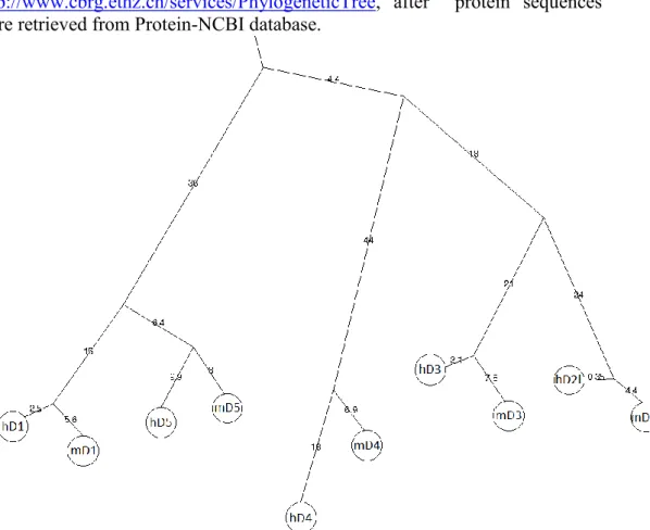
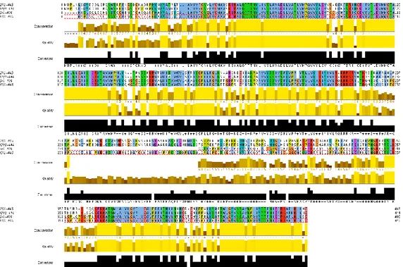
as .mol2 files26. When necessary, Open Babel was used to convert file formats. Protonation state of ligands was assigned at pH=7.4. Structure models of human D3R and D2LR, optimized by explicit molecular dynamics simulation in
water-lipid environment, were obtained as previously described27; human receptors were used instead of the murine ones because their crystallographic and/or theoretical structures are available; furthermore, their protein sequences are conserved between the two species, particularly in the transmembrane domain and in the binding pocket. For more details see Supplementary information.
Statistical analysis
Data were analyzed using one- or two-way analysis of variance (ANOVA). The post hoc Newman-Keuls test was used for multiple comparisons, taking P value less than 0.05 as significant.
Results
Dopamine D3R-/- mice exhibit lower voluntary ethanol intake as compared to
control
As shown in Figure 1a, WT preferred the ethanol-containing solution since the beginning. In contrast, during the first 8 days of observation, D3R-/- mice did not
show any preference for either ethanol or water (Figure 1a). During the entire period of observation (44 days) WT mice maintained their preferential intake of ethanol (p<0.01), whereas D3R-/- mice developed and maintained a preference
for water (p<0.01). Furthermore, there was no difference between WT and D3R
-/- in terms of total amount of fluid intake (ethanol + water) (Figure 1a).
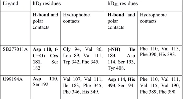
In silico analysis of U99194A and SB277011A interaction with D2R and D3R
shows their high selectivity for the D3R subtype.
To examine pharmacological antagonism at D3R, we docked U99194A and
SB277011A into models of D3R and D2LR receptors27. As shown in Table 1,
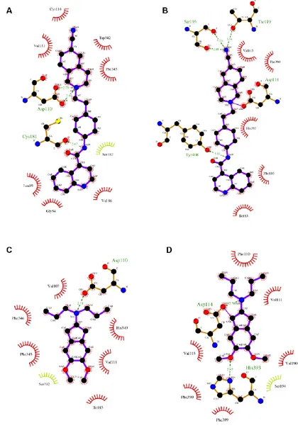
consensus-scoring of poses confirmed the higher affinity of docked compounds for the D3R in comparison to the D2LR subtype. The binding energy of
complexes was strictly related to ligand-protein interactions, each ligand bound in a different manner to D3R and D2LR receptors (Figure S3, Supplementary
Supplementary information). Ligands docked into D3R formed less polar
contacts than ligands docked into D2LR. Moreover, contacts of SB277011A
with hydrophobic residues were more numerous into D3R receptor than into
D2LR binding pocket; in contrast, U99194A interacted with the same number of
hydrophobic residues, though these residues were different in the two receptors.
Blockade of D3R counteracts voluntary ethanol intake
After 30 days of stable ethanol/water intake, mice received daily i.p. injections
of either vehicle or D3R antagonists (U99194A or SB277011A). As shown in
Figure 1b, treatment of WT with each D3R antagonist significantly decreased
voluntary ethanol intake as compared to the vehicle control group (p<0.01, for both U99194A and SB277011A). Treatment of D3R-/- with U99194A and
SB277011A did not change ethanol intake. Neither in WT nor in D3R-/- total
fluid intake was affected by treatment with D3R antagonists (Figure 1b).
Dopamine D3R is up-regulated in striatum and prefrontal cortex (PFC) of WT
mice following chronic ethanol intake
Figure 2 (a, b) shows the D3R gene expression profile in striatum and PFC of
control WT (not exposed to ethanol) or WT that had free access to water and ethanol, treated with vehicle or the D3R antagonist SB277011A. Chronic
PFC (P<0.01). Treatment with SB277011A induced a further increase of D3R
mRNA expression (P<0.05, P<0.01).
Expression of BDNF and RACK1 was increased following chronic ethanol intake in both WT and D3-/- mice
BDNF induces D3 receptor expression in nucleus accumbens, both during
development and in adulthood28. RACK1, a mediator of chromatin remodeling, regulates in an exon-specific manner the expression of BDNF gene29 and the
RACK1/BDNF pathway is activated upon exposure to ethanol30. We therefore
assessed BDNF and RACK1 mRNA expression in striatum and PFC of WT and D3R-/- that had free access to either water only or to both water and ethanol.
As shown in Figure 2, long-term free access to ethanol increased BDNF (panels c and d) and RACK1 mRNA (panels g and h) in striatum and PFC of WT mice (p<0.05, p<0,01; RACK1 increase in PFC did not reach statistical significance). The ethanol-dependent increase of BDNF and RACK1 expression was unaffected by chronic treatment with SB277011A. Mice D3R-/- showed an
increased basal expression of BDNF in the PFC as compared with their WT littermates (p<0.05, Figure 2f), that was not affected by long-term free access to ethanol or by the treatment with SB277011A. Surprisingly, long-term free access to ethanol increased BDNF and RACK1 expression also in the striatum
expression was found in the PFC of the of D3R-/- (Figure 2j).
Forced chronic ethanol intake results in increased BDNF and RACK1 expression both in WT and D3R-/- mice
Long-term free access to ethanol appeared to be associated with BDNF/RACK1 overexpression, but the interpretation of these data was made difficult by the different ethanol intake in the two genetic groups, being very high in WT and very low in D3R-/-. Furthermore, the finding that free access to ethanol was
associated with similar BDNF/RACK1 overexpression in striatum, regardless of genotype, suggested that this effect might be very sensitive to low levels of ethanol, while on the other hand, BDNF/RACK1 activation in PFC seemed less affected by ethanol exposure. To address these issues, some WT and D3R
-/-mice were subjected to forced ethanol intake, i.e. they had access to ethanol 10% solution only. As shown in Figure 3, forced ethanol intake induced a significant overexpression of BDNF and RACK1 mRNAs in striatum and PFC of both WT and D3R-/- mice (p<0.05, p<0.01). Of note, BDNF and RACK1
mRNA levels induced in D3R-/- mice were even higher than those induced in
WT; in these latter, treatment with SB277011A induced a further increase in BDNF and RACK1 (p<0.05 vs. ethanol alone), up to the level of ethanol-treated D3R-/- mice.
Dopamine receptor signaling is enhanced in PFC and striatum of D3R-/- mice
and of SB277011A-treated WT mice
Activation of D1 receptor results in activation of adenylyl
cyclase/cAMP/protein kinase A (PKA) signaling; a major substrate for PKA in
the striatum is DA and cAMP-regulated phosphoprotein (DARPP-32). D2-like
receptors regulate the activity of the protein kinases Akt and GSK3β; stimulation of either D2 or D3 receptors results in phosphorylation of Akt and
GSK3β31. In order to assess activation of dopaminergic transmission in different CNS areas, we determined, by immunoblot, the abundance of phosphorylated DARPP-32 (Thr 34) and of phosphorylated GSK3β (Ser 9). As shown in Figure 4, both phosphoDARPP-32 and posphoGSK3β were more abundant in PFC and striatum of D3R-/- than in WT mice. Treatment of WT
mice with SB277011A induced phosphorylation of DARPP-32 and GSK3β in both brain areas, up to the level of D3R-/- mice. In contrast, in cerebellum there
was no difference in the level phosphoDARPP-32 and posphoGSK3β between WT e D3R-/-, nor it was influenced by SB277011A-treatment in WT.