I
Rationale I
1Background I
41. Endocrinology of male reproductive system
61.1 The testis: general structure 6
1.2 Testicular functions and its regulation 6
1.3 The male hypotalamo-pituitary-gonadal axis 7
1.4 Endocrine regulation of the testis 10
1.5 Spermatogenesis 12
1.6 Steroid production 14
2. Testicular cancers
172.1 Introduction 17
2.2 Leydig cell hyperplasia and tumors 18
2.3 Relationship between estrogens and Leydig tumors 20
3. Estrogen regulation of testicular functions
233.1 Introduction 23
3.2 The aromatase gene: structure and regulation 24
3.2.1 The transcriptional factors CREB and SF-1 26
3.3 The estrogen receptors 27
3.4 Distribution of ERs and aromatase in male reproductive system 31
3.4.1 ERs and aromatase in rodent testis 32
3.4.2 ERs and aomatase in human testis 36
3.5 Role of estrogens in animal male reproduction 37
3.6 Role of estrogen in human male reproduction 39
3.7 Effect of eccessive estrogen on male reproduction 40
3.7.1 Exposure to eccess of estrogen in animals 40
3.7.2 Aromatase over-expression in rodents 41
3.7.3 Exposure to eccess of estrogen in human 42
3.7.4 Aromatase over-expression in humans 43
II
4. The IGF system
4.1 Introduction 44
4.2 effect of Insuline like Growth Factor I on testicular functions 47
4.3 IGF system and tumorigenesis 48
5. Materials and Methods I
505.1 Cell cultures 50
5.2 Radio immunoassay 51
5.3 Aromatase activity assay 51
5.4 Western Blot analysis 51
5.5 RNA extraction, reverse transcription and PCR 52
5.6 Cell proliferation assay 53
5.7 Transfection assay 53
5.8 Data analysis and statistical methods 54
6. Results
556.1 Nandrolone and stanazolol control Leydig cell proliferation through the induction of aromatase expression and estradiol production
55
6.1 Nandrolone or stanazolol in combination with IGF-I further induce Leydig cell proliferation and aromatase expression
57
6.3 Nandrolone and stanazolol activate rapid signalling through ERα transactivation
60
6.4 Nandrolone and stanazolol influence cell proliferation by increasing cyclin E expression levels
62
7. Discussion
64Rationale II
68Background II
718. Human adrenal Gland
728.1 The adrenal gland: general structure 72
8.2 Embriology and development 73
8.3 Histology 74
8.4 Adrenocortical steroidogenesis 75
III 8.5.2 P450SCC 78 8.5.3 P450C17 78 8.5.4 P450C21 78 8.5.5 P450C11-P450C18 79 8.5.6 3βHSD 79 8.5.7 β-steroid-sulfotransferase-sulfatase 79 8.5.8 17-chetosteroid-reductase 79
9. Adrenocortical cancers
81 9.1 Introduction 81 9.2 Epidemiology 81 9.3 Pathogenesis 82 9.4 Adrenocortical Adenoma 83 9.5 Adrenocortical Carcinoma 839.6 E2 and adrenocortical carcinoma proliferation 84
10 Aromatase expression in adrenal gland under physiological and
pathological condictions
86
11 IGF-II/IGFIR pathway role in ACCs
8712 The nuclear receptor coregulator PELP1
8812.1 Introduction 88
12.2 Protein domain structure 89
12.3 Interactions with NRs 89 12.4 PELP1-interacting proteins 90 12.5 Expression 91 12.6 Subcellular distribution 92 12.7 Post-translational modiications 92 12.8 Enzyme activity 93 12.9 Target genes 94
12.10 Chromatin remodelling and transcriptional activation 94
12.11 Nongenomic signalling 95
IV
12.13 PELP1 deregulation in hormonal cancers 96
12.14 Role of PELP1 in metastasis 98
13. Materials and Methods II
10013.1 Cell culture and tissue 100
13.2 RNA extraction, reverse transcription and PCR 101
13.3 Western Blot analysis 102
13.4 Assessment of cell proliferation 102
13.5 Chromatin immunoprecipitation assay 103
13.6 Immunoprecipitation assay 104
13.7 RNA interference 104
13.8 Data analysis and statistical metods 105
14. Results II
10614.1 IGF-II signaling is active in human ACCs tissues and in H295R cell and is invoved in cell proliferation
106
14.2 SF-1 and aromatase are highly expressed in human ACCs tissues and regulated by IGF-II/IGFIR pathway
108
14.3 Estrogens induce IGFIR expression through pCREB recruitment to IGFIR gene promoter
109
14.4 E2 and IGF-II increase cyclin D1 expression via ERα 111
14.5 ERα silencing blocks E2 and IGf-II dependent H295R cell proliferation 111
14.6 PELP1 is recruited to form a multiprotein complex in H295R after treatment with E2 and IGF-II
113
14.7 PELP1 knock down decreases ERK1/2 phosphorilation in H295R 114
14.8 PELP1 knock down decreases IGFIR expression in H295R 114
14.9 PELP1 knock down decreases aromatase expression in H295R 115
14.10 PELP1 knock down reduces cell proliferation in H295R 116
15. Discussion II
117References
122- 1 -
Rationale I
Substance abuse has become increasingly widespread among athletes at sub competitive and recreational level, raising concern for human health. In addition to the illicit use of substances to increase performance of athletes and to enhance the muscular mass and strength, more recently the use of some agents has been extended to non-athletes with the aim to combat ageing, obesity and improve appearance or libido (1). Since its discovery in 1935, numerous derivatives of testosterone have been synthesized, with the goals of prolonging its biological activity in vivo, producing orally active androgens, and developing products, commonly referred to as anabolic androgenic steroids (AAS), that are more anabolic and less androgenic than the parent molecule. AAS doping is undeniably rampant worldwide. The doses of testosterone or other androgens used by athletes are substantially larger than those prescribed for the treatment of androgen deficiency. In one survey (2), 50% of androgen users reported using at least 500 mg of testosterone weekly or an equivalent dose of another androgen; in another survey (3), almost one fourth of androgen users assumed 1000 mg of testosterone weekly or an equivalent dose of other androgens. It is becoming increasingly clear that the abuse of AAS is associated with serious adverse effects to the liver (4) and the cardiovascular (5) central nervous (6), musculoskeletal (7), endocrine (6) and reproductive (8, 9) systems. As a consequence of their effects on the endocrine and reproductive systems AAS cause suppressed spermatogenesis, gynecomastia and virilization. Clinical reports highlight a link between AAS abuse and various types of cancer, mainly to the liver such as hepatocellular adenomas and adenocarcinomas (10), however other types of cancer such as Wilms‘ tumors have been reported (11, 12).
Androgens exert their biological effect through an intracellular receptor, the androgen receptor (AR), that is present in the reproductive tract as well as in many non-reproductive tissues, including bone, skeletal muscle, brain, liver, kidney and adipocytes. Binding of androgens to AR determines receptor dimerization, nuclear translocation and binding to specific responsive elements (ARE) present in the promoter region of target genes (13). Androgens mechanism of action in skeletal muscle cells is well documented and includes up-regulation of markers of myogenic
Rationale I
- 2 -
differentiation, such as MyoD and myosin heavy chain II (14-16). However, androgens can be converted to estrogens through the action of the aromatase enzyme. In the human, aromatase is expressed in a number of cells including brain, skin fibroblasts, bone, adipose tissue, in steroidogenic tissues such as placenta and gonads (17), in particular in man aromatase is present in most of the testicular cells.
Estrogens are required for a normal spermatogenesis, which seems extremely sensitive to estrogen concentration. Transgenic mice lacking aromatase expression (ArKO mice) show an age-dependent disruption of spermatogenesis, a significant reduction in testis weight and compromised fertility (18, 19). Similarly, men with inactivating mutations of the aromatase gene, leading to the lack of the estrogen synthesis, are infertile (20). On the other hand, about half of the male transgenic mice over-expressing aromatase and presenting enhancement of circulating 17β-estradiol (E2) levels are infertile and/or have enlarged testis and show Leydig cell hyperplasia and Leydig cell tumors (21). Several studies indicated that estrogen produced locally can induce neoplastic changes in breast tissue (22), and that their excess in rodents is able to stimulate Leydig cell hyperplasia (LCH) associated with cryptorchidism, testicular cancer and alterations of spermatogenesis (23-25). In a previous study it has been shown that Leydig cell tumor is characterized by aromatase overexpression and consequent increased estrogen production, that contributes to inducing tumor cell proliferation (26). Aromatase activity is regulated mainly at the level of gene expression and is present throughout all maturational stages of the male gamete in humans (27, 28).
Recently it was shown that SF-1 (steroidogenic factor-1) is localized predominantly in Sertoli cells, LRH-1 (liver receptor homologue -1) is present in germ cells, while both transcription factors are present in the cells of primary rat Leydig and positively regulate the expression of the aromatase (29). Increased expression of transcription factors acting on the aromatase promoter PII, including SF-1, is at the basis of the mechanism of proliferation induced by IGF-1 on Leydig tumor cells (30) because it establishes an autocrine mechanism by which IGF-1 up-regulates aromatase through SF-1 activation. The consequent synthesis of estrogens is responsible for the induction of cyclins and cell proliferation.
- 3 -
Testosterone, nandrolone, stanozolol, methandienone, and methenolol are the most frequently abused androgens (2, 3, 31). These androgens can be differentially metabolized by aromatase, specifically nandrolone can be converted to estrogens, while stanozolol is a non-aromatizable androgen.
In addition to the use of androgens, athletes also abuse other drugs to purportedly enhance muscle building, muscle shaping or athletic performance (2). These accessory drugs include stimulants, such as amphetamine, clenbuterol, ephedrine, and thyroxine, anabolic agents such as growth hormone (GH), insulin and insulin-like growth factor-I (IGF-I) and drugs perceived to reduce adverse effects such as human chorionic gonadotropin (hCG), aromatase inhibitors or estrogen antagonists (2). In particular, IGF-I, which is the main effector for the action of GH, is a peptide physiologically produced by the liver. The potential benefits of IGF-I administration include increased muscle protein synthesis and the sparing of glycogenolysis with glycogen synthesis and increased fatty acid availability. IGF-I is known to have a role in testicular growth and development and in the control of Leydig cell number (32). IGF-I is produced locally in the testis, in Sertoli, Leydig and peritubular cells derived from the immature testis and cultured in vitro (33). The crucial role of IGF-I in the development and function of Leydig cells was highlighted by studies on IGF-I gene knockout mice (34, 35). The failure of adult Leydig cells to mature and the reduced capacity for testosterone production in IGF-I knock out (KO) mice are caused by deregulated expression of testosterone biosynthetic and metabolizing enzymes (36), expression levels of all mRNA species associated with testosterone biosynthesis are lower in the absence of IGF-I. Furthermore, IGF-I plays a central role in inducing aromatase expression in Leydig tumor cells, consequently IGF-I increases estrogen production that contributes to the induction of tumor cell proliferation (26).
Starting from these observations and taking into account that Leydig cell tumors are common in young men, the same age group commonly abusing AAS we wanted to investigate the effects of AAS and IGF-I on Leydig cell tumors. Our hypothesis is that AAS can induce Leydig cell tumor proliferation and that this effect could be potentiated by the concomitant use of IGF-I. To verify this hypothesis in the present study we evaluated on Leydig R2C cells the effects of commonly used ASS,
Rationale I
- 4 -
differentially metabolized by aromatase, such as nandrolone (aromatizable) and stanozolol (non-aromatizable), used alone or in association with IGF-I, on aromatase expression and Leydig cell tumor proliferation.
5
Background I: Endocrinology of male reproductive system
6
1. Endocrinology of male reproductive system
1.1 The testis: general structure
The human male reproductive system includes hypothalamic-pituitary-gonadals axis, epididymis, vas deferens, seminal vesicles, prostate and urethra. The testis is primarily composed by seminiferous tubules closely packed together and interstitial cells (37). The seminiferous tubules are composed by Sertoli cells that support germ cells during their maturation into spermatozoa and that create a blood-testis barrier, separate the germinal epithelium into basal and adluminal compartments. Sertoli cells are responsible for germ cells physical support and in addition they provide nutrients and growth factors. The major cell in the interstitial space outside the seminiferous tubule is the Leydig cell, which produces testosterone, a necessary component for germ cell maturation. Testes produce a large amount of spermatozoa through a complex process, known as spermatogenesis. Each seminiferous tubule is surrounded by mesenchymal cells. Among these are the peritubular myoid cells whose contractile elements generate peristaltic waves along the tubules, but do not present a tight diffusion barrier. Vascular smooth muscle cells, macrophages and endothelial cell types are also located in the interstitial space of the testis. The physiological role of macrophages has long been underestimated. In the rat, the number of macrophages is one quarter of the number of Leydig cells and the presence of macrophages is crucial for (re)population of Leydig cells during development and after experimental depletion (38, 39). Immune cells, known to secrete a number of growth factors and cytokines, are part of the intratesticular communication pathways (40).
1.2 Testicular function and its regulation
Testes are components of both the reproductive system (being gonads) and the endocrine system (being endocrine glands). The respective functions of the testicles are:
1. producing sperm (spermatozoa); 2. producing sex hormones.
7
These two functions occur into separate compartments within the testis: 1. the seminiferous tubules produce sperm;
2. the interstitial cells (i.e., Leydig cells) synthesize androgens (Fig. 1.1).
Figure 1.1. Schematic representation of functions of the testis.
Both functions, sperm-forming and endocrine, are under control of gonadotropic hormones produced by the anterior pituitary: luteinizing hormone (LH) and follicle-stimulating hormone (FSH).
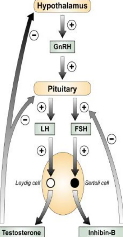
1.3 The male hypothalamo-pituitary-gonadal axis
Secretion of gonadotrophins from the pituitary gland is responsible for regulating hormonal control of the gonad in the male (Fig. 1.2). The male
Background I: Endocrinology of the male reproductive system
- 8 -
pituitary-gonadal (HPG) axis is active from fetal life and the level of hormones produced varies at different stages throughout life. The axis regulates the onest of puberty and the establishment of spermatogenesis (41), in addition to the production of gonadal androgens. Gonadotrophin releasing hormone (GnRH) is produced by the hypothalamus and stimulates the secretion of two gonadotrophins from the anterior pituitary. These glycoprotein hormones are LH and follicle stimulating hormone (FSH). LH binds to the LH/CG receptor on the Leydig cells of the testis to promote testosterone secretion from the Leydig cells, and FSH acts on the Sertoli cells. Testosterone secreted by Leydig cells diffuses into the seminiferous tubules and in them, only Sertoli cells possess receptors for testosterone and FSH and so these cells are the major targets of the ultimate hormonal signals that regulate spermatogenesis. Two important negative feedback loops exist to regulate the secretion of gonadotrophins. The testosterone negative feedback loop is established in fetal life and inhibits hypothalamic and pituitary production of GnRH and LH respectively (42). Negative feedback sensitivity of the HPG axis does not develop in the rat until late in gestation in (43) as LH is not present until 16.5 dpc in the rat (44), but after birth testosterone secretion is LH dependent in rat, human and marmoset (43). The other negative feedback loop results from production of Inhibin-B by the Sertoli cell, which exerts inhibitory effects on FSH secretion from the pituitary gland, however this negative feedback loop is only established at around puberty (45).
The profile of gonadotrophins and testosterone varies depending on age and development (Fig. 1.3). In the human during fetal life, the levels of testosterone are high with a peak at 14-17 weeks gestation (46). Following birth in humans and non-human primates there is an initial rise in gonadotrophins and testosterone that continues during early infancy, the so-called ‗mini puberty‘ (47). In humans the rise begins at 2 weeks of life and peaks between 1 and 3 months of age (48), falling to low levels at 6-8 months. This pattern of secretion has also been demonstrated in many other primates, including the rhesus monkey and the marmoset (49, 50).
- 9 -
Figure 1.2. The male HPG axis. Gonadotrophins (LH and FSH) released from the anterior pituitary
under the control of GnRH act on the testis to produce testosterone from the Leydig cell and Inhibin B from the Sertoli cell. Testosterone and Inhibin-B negatively feedback to the hypothalamus and/or pituitary. Stimulation (+) and inhibition (–) are indicated. Taken from (Mitchell et al., 2009).
In humans and non-human primates after the rise in gonadotrophins and testosterone during early infancy, there follows a period of relative ‗quiescence‘ during which levels of these hormones are relatively low (50). This period will be referred to as the ‗childhood period‘, which lasts from the end of infancy until the onset of puberty. Although this period has been described as a quiescent period it is clear that the testis is active. Sertoli cells actively express the FSH receptor, AMH and aromatase, which is the product of the CYP19 gene (51). In addition there are periods of germ cell proliferation (52) and the transient appearance of meiotic cells (51). Levels of the gonadotrophins and testosterone rise again peripubertally and remain high during adult life (Fig. 1.3). In rodents there is no equivalent childhood period of low gonadotrophin and testosterone (53).
Background I: Endocrinology of the male reproductive system
- 10 -
Figure 1.3. The profile of Gonadotrophin and testosterone secretion in primates. Taken from (Mitchell et al., 2009).
1.4 Endocrine regulation of the testis
The interaction of LH with its receptor initiates signalling through GTP binding proteins determining cyclic AMP (adenosine-3',5'-cyclic monophosphate) production (54) and signal transduction through protein kinase A pathway. Some data suggests that intracellular calcium concentration can be induced by the action of LH though the activation of the lipoxygenase pathway (55). In addition, changes in calcium concentration can also regulate adenylate cyclase through the protein kinase C pathway. FSH binding to its receptor is known to activate at least 5 signaling pathways in Sertoli cells: cAMP-PKA pathway, MAPK pathway, Phosphatidylinositol 3-kinase (PI3K) pathway, Calcium pathway, Phospholipase A2 (PLA2) pathway. Initially FSH binding to FSH receptor causes adenylate cyclase (AC) activation and increase in intracellular cAMP levels.
Multiple factors can be activated by cAMP in Sertoli cells including PKA that can phosphorylate a number of proteins in the cell and also regulate the expression and activity of numerous transcription factors including CREB.
During puberty, FSH activates MAPK cascade in Sertoli cells. ERK can activate transcription factors including SRF, c-jun and CREB. In granulosa cells, FSH also activates the p38 MAP kinase. FSH and cAMP also act through GEFs (guanine nucleotide exchange factors) to activate PI3K and then phosphoinositide dependant protein kinase (PDK1) and PKB in Sertoli cells.
- 11 -
Studies on granulosa cells identified Forkhead transcription factor (Forkhead), SGK (glucocorticoid-induced kinase) and GSK-3 (glycogen synthase kinase-3) as additional downstream targets of PI3K pathway.
Figure 1.4. Endocrine regulation of the testis. PMC, peritubular
myoid cell; CRE, cAMP responsive elements, ARE, androgen-responsive elements; ERE, estrogen-androgen-responsive elements.
FSH also mediates the induction of PLA2 and the subsequent release of arachadonic acid (AA) and the activation of eicosanoids such as PGE2 that may act as intracellular or extracellular signaling agents (56). However, gonadal steroids, i.e., androgen and estrogen, and other agents that bind or prevent binding to steroid hormone receptors (androgen receptor AR, ERα, and ERβ), which are present in Sertoli cells, germ cells and Leydig cells, also regulate testicular function (57). The pathway mediated by cAMP seems to be the primary intracellular signaling pathway
Background I: Endocrinology of the male reproductive system
- 12 -
in all testicular cells. However, several growth factors e.g., insulin like growth factor-1 (IGF-factor-1) and epidermal growth factor (EGF), acting their receptors, IGF-factor-1R and EGF-R, can modulate AR and ER-mediated pathways. In conclusion is possible to affirm that testicular functions are regulated by interactions between several signaling pathways, some directly, e.g., AR and ER-mediated pathways, and others indirectly by modulating hypothalamus-pituitary function. Hormonal activation of transcriptional gene activity results in changes in cell differentiation and function (Fig 1.4).
1.5 Spermatogenesis
Spermatogenesis is the process by which spermatogonia proliferate and then differentiate into mature spermatozoa. This process is initiated by FSH during puberty and both FSH and testosterone appear to be required for quantitatively normal spermatogenesis (41, 58, 59). Testosterone acts via the androgen receptor on the Sertoli cells, thereby exerting indirect effects on the germ cells, as evidenced by a reduced germ cell number and failure to progress beyond meiosis in mice with knockout of the androgen receptor in Sertoli cells (60). There are three phases of spermatogenesis that are common to all mammals. The first is a phase in which the spermatogonia are undergoing frequent cell divisions and differentiating into primary spermatocytes. The final mitotic division of B-spermatogonia gives rise to diploid preleptotene spermatocytes, which undergo meiotic division to produce haploid secondary spermatocytes. These cells are located in the adluminal compartment of the seminiferous tubule. The second meiotic division results in the formation of the spermatids, which will subsequently become mature spermatozoa following the process of spermiogenesis. The seminiferous tubule is organised with the spermatogonia adjacent to the basement membrane. As the germ cells differentiate they are directed towards the lumen (Fig. 1.5).
- 13 -
Figure 1.5. Schematic representation of a transverse view of a human seminiferous tubule. Sc –
spermatocyte, St – spermatid. Supporting the germ cells are the Sertoli cells, which form the ‗blood testis barrier‘ consisting of tight (occluding) junctions between adjacent Sertoli cells. Each Sertoli cell provides support for numerous germ cells at different stages of development and the function of the Sertoli cells at a given stage is determined by its germ cell complement. The patterns of germ cell association from basement membrane to the tubule lumen are classified into stages, which are based on the morphological development of the spermatids. Six stages have been described in the human.
In human adults the proliferation rate of the spermatogonia has been calculated as 26%, based on Ki67 staining. This is higher in type B spermatogonia (43%) compared to the type A spermatogonia (22%) (61). Each pachytene spermatocyte gives rise to four haploid spermatids and no further mitotic division occurs once the germ cells have reached this stage (41).
Germ cell numbers can be increased by administration of FSH in adult rhesus monkeys (62) and this has been shown to be due to proliferation of type Apale spermatogonia (59, 62). In these studies administration of hCG (62) or LH (59) did not increase cell proliferation, suggesting that the threshold for a maximal impact of androgens has already been met, whilst FSH administration can augment cell function. However the importance of testosterone can be inferred from studies in the hpg mouse which lacks gonadotrophins and therefore has an immature testicular phenotype without full spermatogenesis, but in which replacement of testosterone induces complete spermatogenesis despite undetectable FSH levels (63). In men with low FSH levels, hCG can re-initiate spermatogenesis, although the sperm
Background I: Endocrinology of the male reproductive system
- 14 -
concentrations do not return to normal (64). This suggests that both FSH and LH are required for normal spermatogenesis although it would appear that FSH is not essential to the process, in fact men with an inactivating mutation of the FSH receptor have varying degrees of spermatogenic impairment but are not infertile (65).
1.6 Steroid production
Testosterone is the major androgen secreted by the testis In addition to testosterone, in smaller amounts, is produced dihydrotestosterone through the actions of the enzyme 5 -reductase. Testis also contributes approximately for 25% of 17 -estradiol total daily production through the local action of the enzyme aromatase which converts androgenic substrates in estrogens (66). Cholesterol represents the major substrate for androgen production in Leydig cells and is derived by an uptake mechanism involving the binding of circulating low density lipoprotein to specific receptors on Leydig cells which, following internalisation, provides a significant source of cholesterol (67). In addition, Leydig cells can undertake de novo synthesis of cholesterol from acetate and relative contributions of these two sources is partly dependent on species and the state of stimulation of the Leydig cells.
The conversion of cholesterol to testosterone involves a number of steps that are catalyzed by enzymes predominantly belonging to cytochrome P450 family. The mobilization of cellular sources of cholesterol is achieved through the action of cholesterol ester hydrolase and subsequently, cholesterol is converted in pregnenolone through cholesterol side-chain cleavage enzyme (cytP450SCC) actions
- 15 -
Figure 1.6. Steps in steroidogenesis leading to androgens and estrogens production.
Cholesterol conversion in pregnenolone, being the key step of androgen production, is tightly controlled Availability of cholesterol can be rate-limiting and the intracellular trafficking of cholesterol, across mitochondrial membranes, is dependent on the steroidogenic acute regulatory protein (STAR) (69-71).
The role of this protein has been well demonstrated in patients with mutations in the gene encoding STAR in the disorder termed congenital lipoid adrenal hyperplasia wherein the mitochondria from the adrenals and gonads are unable to convert cholesterol to pregnenolone (72). Furthermore, the results of studies involving targeted disruption of the mouse gene encoding STAR, support the data observed in human (73). Testosterone production can be obtainded through two pathways. Pregnenolone can be converted in progesterone through 3 hydroxysteroid dehydrogenase enzyme activity (Δ4 pathway) or can be hydroxylated by
17-hydroxylase enzyme to form 17 -hydroxypregnenolone (Δ5 pathway). The relative importance of these two pathways vary with the species and the physiological status of the male (74). The further conversion of 17 -hydroxypregnenolone through the
Background I: Endocrinology of the male reproductive system
- 16 -
Δ5 pathway involves the formation of a C19 steroid, dehydroepiandrosterone, catalyzed by the enzyme 17,20 lyase and both steps appear to be catalyzed by a single microsomal enzyme, cytochrome P450C17, encoded by a single copy gene on
chromosome 10 (75, 76). The conversion of dehydroepiandrosterone in androstenediol is mediated by a microsomal enzyme, 17 -hydroxysteroid dehydrogenase (77, 78). Substrate conversions from the Δ5 to the Δ4 pathway are catalyzed by the enzyme 3 -hydroxysteroid dehydrogenase (79). In the Δ4 pathway, 17 -hydroxyprogesterone is converted in androstenedione and testosterone through the action of cytochrome P450C17. Starting from testosterone the enzyme 5
-reductase can produce dihydrotestosterone (80) while the enzyme aromatase con produce 17 -estradiol (66, 81).
- 17 -
2.Testicular cancers
2.1 Introduction
Although cancer of the testes is rare, accounting for only about 1 percent of all cancers in men of all ages and about 5 percent of all male genitourinary system cancers, it is the most common cancer in men between the ages of 15 and 35, and the second most common malignancy in men ages 35 to 39 (82-85).
Because the incidence of testicular cancer has risen markedly in the past 20 years, researchers focused their attention on possible environmental causes, including mother's diet during the pregnancy as well as the increasing presence of estrogen-mimicking pollutants in the environment.
The most consistent occupational association has been the elevated rate among men in professional occupation, which may be linked to an increased risk observed with lower levels of exercise. Other possible causes include hereditary factors, genetic anomalies, congenital defects involving the reproductive tract, testicular injury, and atrophy of the testes. Viral infections such as mumps, which cause inflammation of the testes, have not been proven to cause cancer.
Testicular cancer comprises a number of different diseases. Testicular germ cell tumours (TGCT) are the most common cancer of young men and represent 90% of testicular tumours (86). These tumours have increased in incidence during the last 50 years in many Western countries (87). TGCT have been postulated to arise during fetal life in humans (88), originating from the transformation of undifferentiated fetal germ cells (89). The exact origin and subsequent development of these pre-neoplastic germ cells is unknown. In addition there is a lack of suitable animal models of fetal and early postnatal testis development in which to investigate the origins of these tumours.
About 40 percent of germinal tumors are categorized as seminomas. Several other types of germinal tumors are referred to collectively as non-seminomas. Somatic cell tumors, known as sex cord-stromal neoplasms and Leydig cell tumors are relatively rare. However, deriving from endocrine active cells, they have endocrine manifestations.
Background I: Testicular cancers
- 18 -
2.2 Leydig cell hyperplasia and tumors
Although Leydig cells in adult men are considered to be a terminally differentiated and mitotically quiescent cell type, in various disorders of testicular function, focal or diffuse Leydig cell hyperplasia is very common. Micronodules of Leydig cells are frequently seen in certain conditions associated with severe decrease of spermatogenesis or germinal aplasia, such as the so-called Sertoli-cell-only syndrome (Del Castillo syndrome), cryptorchidism, or Klinefelter‘s syndrome (90). A term ―Leydig cell adenoma‖ is used when the size of a nodule exceeds several fold the diameter of a seminiferous tubule. It is unknown whether Leydig cell adenomas can further progress to form Leydig cell tumors. The mechanism determining Leydig cell hyperplasia in human male is still poorly understood. The disruption of hypothalamo-pituitary-testicular axis, leading to an excessive stimulation of Leydig cells by LH, can play a central role (90). In a small subset of cases structural changes of the LH receptor (91, 92) and G proteins (93, 94) were detected. Constitutively activating mutations of LH receptor cause early Leydig cell hyperplasia and precocious puberty (91, 95). Similarly, constitutively activating mutations of Gs-protein in Leydig cells lead into hyperplasia and endocrine hyperactivity (94, 96). However, Leydig cell hyperplasia is distinct from tumors that are usually solitary, and the role of the LH receptor and G protein mutations in the tumorigenesis may be limited to few cases (92, 94). Leydig cell hyperplasia and adenomas can be easily induced in rodents by administration of estrogens, gonadotropins and a wide range of chemical compounds.
Leydig cell tumors account for one to three percent of testicular neoplasms and occur in different age groups (96-98). Approximately the 20% is found before the age of 10, most often between five and ten years of age. Precocious puberty is the presenting symptom in these cases. Tumors produce androgens, mainly testosterone in a gonadotropin independent manner, and therefore LH and FSH remain low in spite of external signs of puberty. Approximately 10% of the boys also have gynecomastia that is caused by aromatase iper-activity and consequent excessive estrogen production. In adults, gynecomastia is found in approximately 30% of patients (98). The excessive androgen secretion rarely causes notable effects in adults.
- 19 -
Leydig cell tumors are always benign in children and can be treated with surgical enucleation when the tumor is encapsulated (84), whereas in adults malignant tumors have been found in 10-15% of patients, and inguinal orchidectomy remains the treatment of choice (97). The presence of cytologic atypia, necrosis, angiolymphatic invasion, increased mitotic activity, atypical mitotic figures, infiltrative margins, extension beyond testicular parenchyma and DNA aneuploidy are associated with metastatic behavior in Leydig cell tumors (98, 99). Malignant tumors are hormonally active only in exceptional cases. Benign tumors can be treated by orchidectomy, whereas an additional retroperitoneal lymphadenectomy should be considered when the gross or histological features suggest malignancy (98, 99). Malignant tumors have not responded favorably to conventional chemotherapy and irradiation (98, 99). Survival time has ranged from 2 months to 17 years (median, 2 years), and metastases have been detected as late as nine years after the diagnosis (98, 99). Therefore follow-up of patients with malignant Leydig cell tumors has to be life-long. The remaining testis may be irreversibly damaged by longstanding high estrogen levels, resulting in both permanent infertility and hypoandrogenism(98-100).
The most frequently encountered testicular neoplasm in mouse and rat is the Leydig cell adenoma. Incidence rates vary in different strains with the Sprague-Dawley SD rat ranging from 1 to 5% and the F-344 rat reaching nearly 100% (101). Early neoplasm are common in 1 yr old F-344 rats and become increasingly more frequent with age (102). Testicular neoplasia is less frequently observed in all strains of mice with incidence ranging from 1 to 2,5%. Leydig cell tumors in rodents generally occur in older animals, but in human can arise at any age, the majority between 20 and 60 years (103). The estimated incidence in man is 0.1-3 per million. The proliferative lesions in Leydig cells in rodents are similar and are observed as a continuous spectrum starting with smaller nodular foci of hyperplasia leading to large Leydig cell adenomas that can eventually replace the entire testis. The distinction between hyperplasia and adenoma is not always clear, with size being the major factor in the diagnostic criteria, with some debate over when focal hyperplasia becomes early neoplasia and there can be little morphological difference between a hyperplastic nodule and a small Leydig cell adenoma. The major difference between testicular
Background I: Testicular cancers
- 20 -
tumors observed in human and rodents (particularly the rat) are the high incidence of germ cell tumors in human and their occurrence in relatively young men. In rats, germ cell tumors are extremely rare, but Leydig cell tumors can be almost 100% in incidence in certain strains (e.g., Fisher F-344) and occur most frequently in older animals.
2.3 Relationship between estrogens and Leydig tumors
The biological significance of estrogen-induced testicular tumorigenesis has been suggested by the in vivo model overexpressing aromatase transgenic mice (21). Half of these males were infertile and some of them showed larger than normal testis and Leydig cell hyperplasia/Leydig cell tumor. Furthermore, aromatase was markedly immunolocalized in the cytoplasm of interstitial cells, and its immunoreactivity appeared to be strongest in the testes with more advanced stages of neoplasia. The same transgenic animals exhibited estrogen circulating levels at least twice higher than those of control animals and the levels of aromatase mRNA in their testicular tissues were fourfold higher when compared with controls. It is worth to mention how ERα protein in testicular tissue of aromatase transgenic animals was very high with respect to the undetectable levels of control animals. So the authors suggest how an enhanced synthesis of estrogens in tumoral tissues led to an upregulation of ERα expression. Human Leydig cell tumor is a rare testicular neoplasm where estrogen involvement in tumorigenesis process has scarcely been investigated. Recently, a strong aromatase expression in tumoral tissues was revealed by immunostaining and western blotting (104). This finding agrees with a single previous report (105) showing the aromatase immunolocalization in Leydig cell tumors. Furthermore, aromatase expression in control human testicular tissue confirmed Turner‘s report in normal testes (106). The enhanced endogenous synthesis of estrogens by Leydig cell tumor was reflected in both patients by a dramatic increase of estrogen circulating levels, resulting more than two fold higher than those of adult normal male, and by the low testosterone levels (at the lower limit of normal range) (104). Moreover, the ratio between the free fraction levels of the two steroids is furthermore increased in the target tissues. The diminished sperm count and motility of both patients may not only be related to altered testicular tropism, parenchymal compression, and increased
- 21 -
local temperature ipsilateral to the tumor (107) but also to the detrimental effects of high circulating estrogen levels on the counter-lateral gonad activity. In the adult normal male, 80% of the plasma estradiol originates from aromatization of testosterone and androstenedione in fat, striated muscle, and other tissues including bone and brain, while 20% in the circulation is secreted by the testis. So, it is reasonable to argue how the excessive increase of estradiol circulating levels, observed in the two patients with Leydig cell tumor, is the consequence of an enhanced rate of testicular secretion. This is confirmed by the evidence that estradiol, as well as E2/T circulating levels ratio, drops dramatically following surgical treatment, while for one of the two patients the persistence of a conspicuous bilateral gynecomastia led to bilateral mastectomia (104). Following orchidectomy, the two patients exhibited a moderate increase of sperm count and a remarkable augment of sperm motility (104). The latter event may be reconducted to the restored testosterone circulating levels likely affecting the entire male genital tract. The expression of ER isoforms in Leydig cell tumor is, to date, unknown. In fact, only a single work showed the ER immunolocalization in cryostat sections of Leydig cell tumor (77); recently, immunohistochemical and western blot analysis of tumoral tissues revealed the expression of ERα and of the two ERβ isoforms, ERβ1 and ERβ2, in neoplastic Leydig cells of both patients. So, the pattern of ERs expression in tumoral cells appears different from that of control Leydig cells, exhibiting only ERβ1 and ERβ2 as previously reported (108, 109) .
There is a growing body of evidence that ERα and ERβ can be expressed together in the same cell type and independently expressed in another. Therefore, homodimers (ERα–ERα/ERβ–ERβ) or heterodimers (ERα–ERβ) can be formed (81). The binding affinity of ERα–ERα/ ERα–ERβ dimers for a consensus DNA estrogen response element is reported to be higher than that of the ERβ –ERβ homodimer (110). Thus, the presence of ERα could reinforce the estradiol-induced tumor cell proliferation. Finally, has been demonstrated that neoplastic Leydig cells are potential estrogen biosynthesis sites and display a modified ER expression pattern. Therefore, it appears reasonable to suggest that the high estrogen levels, measured in the two patients, could play a role in the neoplastic transformation of Leydig cells, while the exclusive
Background I: Testicular cancers
- 22 -
presence of ERα in tumoral cells could amplify E2 signaling contributing to the tumor cell growth and progression.
- 23 -
3. Estrogen regulation of testicular functions
3.1 Introduction
Evidence supporting a role for estrogen in male reproductive tract development and function has been collected from rodents and humans. These studies fall into three categories:
1. aromatase localization and target protein for estrogen receptors (ERα and ERβ) in tissues of the reproductive tract;
2. analysis of testicular phenotypes in transgenic mice deficient in aromatase, ERα/ERβ gene;
3. investigation of environmental chemicals effects on male reproduction.
Estrogen is thought to have a regulatory role in the testis because estrogen biosynthesis occurs in testicular cells and the absence of ERs caused adverse effects on spermatogenesis and steroidogenesis (110). In males, estrogens derive from circulating androgens. Aromatization of C19 androgens, testosterone and androstenedione, to form estradiol and estrone, respectively, is the key step in estrogen biosynthesis, which is under the control of the aromatase enzyme (Fig. 3.1).
Background I: Estrogen regulation of testicular functions
- 24 -
3.2 The aromatase gene: structure and regulation
Aromatase is composed of two proteins: a ubiquitous NADPH-cytochrome P450 reductase and a cytochrome P450 aromatase (P450arom), which contains the heme and the steroid-binding pocket. In humans, P450AROM is the product of a single gene
located in region q21.1 of the chromosome 15 and called CYP19. The CYP19 gene is more than 123 kb in length with a coding region of 9 exons (II-X) and 9 nontranslated exons I (111) (Fig. 3.2).
Figure 3.2. Schematic presentation of the human aromatase gene. P = promoter.
Expression of the CYP19 gene is regulated by tissue-specific promoters producing alternate 5'-untranslated exons that are then spliced onto a common 3'- splice acceptor site in exon II, upstream of the translation starting site (17, 112, 113). Therefore, there is generation of CYP19 variants with different 5‘untranslated regions giving rise to different mRNAs; however, the coding sequences are identical and give rise in humans to a single protein composed of 503 amino acids with a molecular mass of 55 kDa. It is of note that P450AROM is encoded by a single CYP19
gene in most species except for pigs in which three distinct genes encode three aromatase isoenzymes (114) and for fish in which two genes (specifically expressed in the brain and gonads) have been identified (115). Different mechanisms of regulation of CYP19 gene expression have been described for various tissues. The synthesis of different aromatase isoforms between species and tissues may involve distinct aromatase genes and/or function of different promoter elements (116). In human adipose tissue, the primary promoter I.4 lies about 15 kb upstream of the start site of translation (117, 118) and is a TATA-less promoter driven by glucocorticoids and class I cytokines e.g. IL-6 and TNFα (113). The region of PII proximal to the translation start site regulates P450AROM expression in mammalian gonads (117, 119)
- 25 -
as well as in Leydig cell tumors (120). Numerous functional motifs have been identified in P.II (113) (Fig. 3.3).
Figure 3.3. Structure of the human Cyp19 gene showing the various untranslated first exons and their corresponding promoters. The region around promoter PI.4 and PII from human and PII from
rat are expanded to show the identified response elements. Sequences of these are shown in boxes.
In the testis, FSH and LH act by increasing concentrations of intracellular cyclic AMP to induce expression of P450AROM. Promoter PII activity is therefore regulated
by cyclic AMP and requires the transcription factors cAMP response element binding protein (CREB), cAMP response element modulator (CREM) and steroidogenic factor-1 (SF-1). SF-1 belongs to the nuclear orphan receptor superfamily and regulates steroidogenic gene transcription (e.g. P450AROM via its
interaction with numerous coactivators including CREB binding protein, DAX-1, SOX-9, WT1).
It has been shown that P450AROM mRNA level is increased in Leydig cells of mice
Background I: Estrogen regulation of testicular functions
- 26 -
homologue-1 (LRH-1), an SF-1 homologue, present in leydig and germ cells but not in sertoli cells, increases P450AROM gene expression in a mouse leydig cell line (29).
Moreover, it is now clear that not only P.II drives aromatase gene in rat testis but that two additional promoters, P.I.f (brain promoter) and a new one that we called P.I.Tr (testis rat) (122), are involved. It is also demonstrated that the nutritional status of fetuses (123) and aging (124) can modulate aromatase gene expression in male rats.
3.2.1 The transcriptional factors CREB and SF1
CREB is a transcriptional factor binding the cAMP responsive elements (CRE). From a structural point of view it is possible to identify two regions:
the N-terminal, setting the process of transactivation;
the C-terminal, involved in DNA binding and in the process of dimerization.
The human protein consists of 327 amino acids (CREB-327) while in rats it consists of 341 amino acids (CREB-341); both are expressed by homologous genes and are the product of exons subject to alternative splicing.
The phosphorylation site(P-box) or kinase-inducible domain (KID) is the critical region of CREB in response to the cAMP. The P-box contains several consensus phosphorylation sites for different kinases such as PKA, PKC, glycogen synthase kinase 3 and the casein kinase I and II (CK).
After activation of adenylate cyclase pathway, PKA phosphorylates CREB at the level of serine in position 133.
Phosphorylation induces a conformational change in the protein (125). In addition to PKA, other signal transduction pathways target the CREB protein, increasing or decreasing its transcriptional activity. An example is the kinase IV Ca2+ -calmodulin-dependent (CaMKIV), which phosphorylates CREB at serine 133 after membrane depolarization in neuronal cells.
CREB phosphorylation can also be induced by signal transduction pathways triggered by growth factors and inflammatory cytokines.
The kinase II Ca2+-calmodulin-dependent (CaMKII) phosphorylates CREB at serine
133 and serine 142, with a significant inhibition of CREB transactivation. In addition to calcium and cAMP, the phosphorylation of serine 133 also occurs through a transcriptional pathway Ras-dependent. PKC is able to phosphorylate CREB in vitro, but its role in vivo is doubtful; CREB phosphorylation in response to agents that
- 27 -
activate PKC may occur through the activation of mitogen-activated protein kinase (MAPK/RSK). SF-1 gene (steroidogenic factor 1) encodes for an orphan nuclear receptor that plays regulatory role in the endocrine function of the hypothalamic-pituitary-gonads, in adrenal gland and is an essential factor in sexual differentiation (126). SF-1 is present in the adrenal, Leydig, Sertoli and granulosa cells, in ovarian theca, in spleen, in anterior pituitary gland and in hypothalamus. It‘s well known that SF-1 plays an essential role in gonadal differentiation, because its expression in
primordial gonads assumes different characteristics in male and female, in
correspondence withthe divergence of the developmentof the testis from that of the ovary. In mice lacking SF-1 gene (SF-1 KO), were found: agenesis of the gonads (127) and of the adrenal gland, a sexual changefrom male tofemale and external and internal genitalis, a damaged function of the gonadotropins and ablation of specific regionsof the hypothalamus. Some studies suggest that SF-1 may also play a role in spermatogenesis as well as in steroidogenesis.The SF-1 DBD has two zinc-fingers and both, including also the P-box (Proximal box) in the first zinc-finger and the D-box (distal D-box) in the second zinc-finger, are highly conserved.
3.3 The Estrogen Receptors
Estrogen actions are mediated by binding to specific nuclear estrogen receptors (ERs), which are ligand-inducible transcription factors regulating the expression of target genes after hormone binding.
Two subtypes of ERs have been described: estrogen receptor α (ERα) and the more recently discovered estrogen receptor β (ERβ). The human gene encoding for ERα is located on the long arm of chromosome 6, while the gene encoding for ERβ is located on band q22-24 of chromosome 14. The two ER (α and β) proteins have a high degree of homology at the amino acid level (Fig. 3.4).
Background I: Estrogen regulation of testicular functions
- 28 -
Figure 3.4. ERs gene and its products.
While it is clear that estrogens regulate transcription via a nuclear interaction after binding to their receptors, a non-genomic action of estrogens has been recently demonstrated, suggesting a different molecular mechanism accounts for some estrogen actions.
In vitro studies showed a very short latency time between the administration of estrogens and the appearance of biological effects. These actions are thought to be mediated through cell-surface receptors, which are not believed to act via a transcriptional mechanism (128). The different types of estrogen action are summarized in Table 1.
- 29 -
Table 1. Estrogen actions and related biomolecular pathways and mechanisms.
Estrogen Actions
Receptors Mechanism Final effect Features
Genomic (nuclear actions) ERα Transcriptional: nuclear interaction with estrogen-responsive elements Modulation of estrogen target gene expression. Slow effects (minutes or hours) ERβ Transcriptional: nuclear interaction with estrogen-responsive elements Modulation of estrogen target gene expression. Slow effects (minutes or hours) Non Genomic (cell membranes actions) Estrogen receptors on cells membrane Cells membrane changes Changes in ionic transport through cell surface. Rapid effects (seconds)
ERs are members of the steroid/thyroid hormone super family of nuclear receptors, which share a common structural architecture, and consist of three independent but interacting functional domains: the N-terminal or A/B domains, the C or DNA-binding domain, and the D/E/F or ligand-DNA-binding domain (Fig. 3.4). Binding of a ligand to the ER causes a series of downstream events, including receptor dimerization, receptor-DNA interactions mediated by EREs present in the promoter region of target genes, recruitment of and interaction with transcription factors, and the formation of a preinitiation complex.
Ligand- receptor interactions ultimately cause changes in target gene expression (129). The N-terminal domain of nuclear receptors encodes an activation function
Background I: Estrogen regulation of testicular functions
- 30 -
called AF-1, which mediates protein-protein interactions to induce transcriptional activity. It is thought that this domain is highly active in ERα-mediated stimulation of reporter gene expression from a variety of ERE-constructs but its activity in the ERβ is limited (130). On the other hand, the C-terminal or ligand-binding domain contains the AF-2 interacting surface that mediates ligand binding and receptor dimerization to stimulate transcriptional activity (131). Thus, AF-1 and AF-2 are both involved in mediating the transcriptional activation functions of ERs. Although there is a high degree of homology in the DNA binding domains of ERα and ERβ (about 95%), only a partial homology exists in the ligand-binding domain (~60%) (132). Differences in ligand binding, in association with other factors, have the effect of altering the pattern of ER-mediated transcriptional activity. For example, some agonists bind both ER subtypes with the same affinity while others preferentially bind to ERα or ERβ (133-135). There is general agreement that ERs function as dimers, and co-expression of ERα and ERβ in the same cell causes the formation of homodimers (ERα/ERα and ERβ/ERβ) or heterodimers (ERα/ERβ), which affect ligand-specificity. The interactions between ERs and EREs are complicated by other factors, including the ability of ERβ to modulate ERα transcriptional activity and recruitment of several protein co-activators and repressors by both ER subtypes. Therefore, the relative amounts of ERα and ERβ in a given tissue are key determinants of cellular responses to estrogen and other ER agonists and antagonists (136). Moreover, ER and other steroid receptors have the ability to mediate biological effects through non-transcriptional mechanisms mediated by protein-protein interactions occurring between ERs and growth factors e.g., IGF-I and EGF (137). Furthermore, there is growing evidence for the presence of a small pool of ERs localized to the plasma membrane. For example, BSA-conjugated E2, which is unable to gain entry into the cytosol and acts at the plasma membrane, decreased testicular androgen production in vitro (138).
Recently, a large body of evidences has demonstrated that estrogens can function not only through ERs but can also trigger rapid responses that involve transduction pathways different from those activated ―classically‖ by ERs (139-141).
Estrogen rapid signaling involves a series of cell typedependent events that include mobilization of second messengers such as calcium, cAMP, nitric oxide, interaction
- 31 -
with membrane receptors such as insulin-like growth factor 1 receptor (IGF1R) and epidermal growth factor receptor (EGFR), and stimulation of effector molecules, such as the Src family of tyrosine kinases, the PI3K, the serine/threonine protein kinase AKT and the mitogen-activated protein kinases (MAPKs) (142-146). Several studies suggest that rapid responses to estrogens can be mediated by classical receptors localized to the plasma membrane (139, 147, 148), or by transmembrane proteins other than ERs. Recently, our and other studies identified a transmembrane estrogen-binding protein, the G-protein coupled estrogen receptor 1 (GPR30 from GPER gene), which is able to mediate estrogen action (149-152).
GPR30 has been identified in a variety of human and rodent estrogen target tissues (153-156). Immunocytochemical studies have identified the intracellular localization of GPR30 in the endoplasmic reticulum, Golgi apparatus (151) and plasma membrane (157). GPR30 activates MAPKs, via transactivation of EGFR through release of heparin-bound epidermal growth factor (HB-EGF) (158). It also facilitates stimulation of adenylyl cyclase and cAMP-mediated attenuation of the EGFR-to-MAPK signaling axis (149). Using a Gper-lacZ reporter mouse Isensee et al. (159) demonstrated extensive expression of GPR30 in several endocrine organs including the testis. This is in agreement with our studies that revealed GPR30 expression in a spermatogonia mouse cell line (GC-1 cells) (30), in adult rat pachytene spermatocytes (PS) (160) and in rat round spermatids (RS) (161) and suggests a role for this receptor in controlling spermatogenesis.
Moreover, it has also been recently demonstrated GPR30 expression in Sertoli cells (162).
3.4 Distribution of ERs and aromatase in male reproductive system
ERs and the aromatase enzyme are widely expressed in the male reproductive tract in both animals and humans, implying that estrogen biosynthesis occurs in the male reproductive tract and that both locally produced and circulating estrogens may interact with ERs in an intracrine/paracrine and/or endocrine fashion (128). The concept of a key estrogen action in the male reproductive tract is strongly supported by the fact that male reproductive structures are able to produce and respond to estrogens (163).
Background I: Estrogen regulation of testicular functions
- 32 -
3.4.1 ERs and aromatase in rodent testis
Aromatase and ERs are found at a very early stage of development in the rodent testis, thus suggesting a role for estrogens in influencing testicular development (59-61). ERα is expressed by Leydig cells in the rodent fetal testis at a developmental stage in which the androgen receptor is not yet expressed. The developing efferent ductules and epididymis also express ERα in the fetal rodent. By contrast, it is unclear whether ERα is present within the seminiferous tubules of the fetal testis, with variable results having been reported (60). ERα is abundant in the developing efferent ductules, which are the first male reproductive structures to express ERs during fetal development (62). ERβ is also found early in testis development in the gonocytes, Sertoli cells and Leydig cells, with the gonocytes showing the highest expression suggesting a role for estrogens in their maturation. In addition, ERβ is expressed by rat Wolffian ducts, the structures from which the efferent ductules and epididymis arise (60).
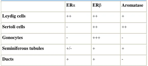
Aromatase is expressed in both Leydig and Sertoli cells in the rodent fetal testis, but not in gonocytes and immature structures of seminal tract. ERs and aromatase distribution in the fetal testes is summarized in Table 2.
Table 2. ERs and Aromatase distribution in the rodent fetal testis.
ERα ERβ Aromatase
Leydig cells ++ ++ +
Sertoli cells - ++ ++
Gonocytes - +++ -
Seminiferous tubules +/- + +
Ducts + + -
The finding of both aromatase and ERs in the developing fetal testis imply a possible involvement of estrogens in the process of differentiation and maturation of developing rodent testis from an early stage of morphogenesis (59;63). In the postnatal immature rodent testis ERα expression does not occur in the seminiferous
- 33 -
epithelium, remaining confined to the Leydig cells, rete testis, efferent ductules and epididymis (Table 3). In the neonatal rodent testis, ERβ is widely expressed by the rat seminiferous epithelium (Sertoli cells and germ cells) as well as by Leydig cells, efferent ductules and epididymis. At this stage ERβ seems to be the only ER in germ cells and is found in pachytene spermatocytes, round spermatids, and perhaps in elongated spermatids of rats and humans (58) (Table 3).
Table 3. ERs and Aromatase distribution in postnatal immature rodent testis.
ERα ERβ Aromatase
Leydig cells + + +
Sertoli cells - + +++
Gonocytes - + -
Seminiferous tubules - + +
Ducts + ++ (?)
Aromatase is expressed by the dividing Sertoli cells and is stimulated by FSH, with the levels of aromatase declining with age. Fetal Leydig cells also have the ability to produce estrogens in response to LH, but aromatase in this cell type is expressed to a lesser degree than during neonatal life. Interestingly the neonatal testis continues to show a greater degree of aromatase expression in the Sertoli cells than in the Leydig cells (the latter only express aromatase to a greater extent in the adult rat testis when they become one of the major sources of estrogens under the influence of LH) (Table 3). Germ cells in immature rats do not yet express aromatase. ERα is expressed in the Leydig cells of both adult rats and mice (64) but not in Sertoli cells. Studies on the precise cellular localization of ERs, however, are mainly based on immunocytochemistry, using different antibodies, and led to contradictory results. Whereas, it is generally agreed that both subtypes are expressed by the epithelial cells of the efferent ductules and epididymis, data concerning testicular expression differ between species, possibly due to different specificity characteristics of the
Background I: Estrogen regulation of testicular functions
- 34 -
antisera used. Knowledge of the distribution of ERα is of great importance in understanding estrogen action on the male reproductive tract. ERα is highly expressed in the proximal reproductive ducts (rete testis, efferent ductules, proximal epididymis) and its expression progressively decreases distally (corpus and cauda of the epidydimis, vas deferens). The highest degree of ERα expression is seen in the efferent ductules of the rat (65) and accounts for one of the most well-documented estrogenic actions on male reproductive system, fluid reabsorption from the efferent ductules. It has to be remarked that the concentration of ERα in the male reproductive tract is opposite to that of ERβ, which is more concentrated in the distal tract (Table 4).
Table 4. ERs and Aromatase distribution in the adult rodent testis.
ERα ERβ Aromatase
Leydig cells +/- +/- +++ Sertoli cells - + + Germ cells Spermatogonia Pachytene Spermatocytes Round Spermatids Spermatozoa +/- - (?) -/+ -/+ + (?) ++ + + + + (?) ++++ + (?) + ++ + Efferent ductules ++++ + - (?)
ERβ is expressed in Leydig, Sertoli and germ cells in adult rodents (66;67) and has also been detected in primate germ cells (68). There is now considerable evidence that germ cells contain both ERβ and aromatase (68). It should be noted that there are some controversies in terms of ERβ localization, with immunohistochemical studies showing some discrepancies, possibly due to methodological differences. It seems that the regulation of gonocyte multiplication, which is under the influence of growth factors and estradiol, may occur through the involvement of ERβ (69). By adulthood, rodent Leydig cells show higher aromatase activity compared to every other age and
- 35 -
in comparison to Sertoli cells (70). Aromatase is also expressed at high levels in germ cells throughout all stages of maturation, and its expression appears to increase as the germ cell becomes a mature spermatid (Fig. 3.5).
Figure 3.5. Aromatase and estrogen receptors (ER) in adult male rat gonad.
Aromatase has been demonstrated in terms of mRNA (RT-PCR), protein (Western blots) and enzyme activity (measurements of estradiol output in culture media) in the various testicular cells. ER: estrogen receptors localization.
Aromatase mRNA and activity, in fact, are found in germ cells from the pachytene spermatocyte stage in both rats and mice, and during their subsequent maturation into round spermatids (61;70;71). Aromatase seems to be present in higher levels in mature spermatids of the rat than in earlier germ cells (61;71;72). It is of interest that aromatase mRNA expression and enzyme activity is higher in germ cells when compared with Leydig cells, suggesting that germ cells may be a major source of estrogen in adult rodents. When fully developed spermatids are released from the epithelium, aromatase remaining in the residual body is subsequently phagocyted by the Sertoli cell. Some aromatase activity persists in the cytoplasmic droplet that remains attached to the flagellum as the sperm make its way through the epididymis, suggesting that mature spermatozoa are able to synthesize their own estrogen as they traverse the efferent ducts (73;74). The ability to synthesize estrogen gradually