UNIVERSITA’ POLITECNICA DELLE MARCHE
FACOLTA’ DI MEDICINA E CHIRURGIA
Dottorato di Ricerca XV CICLO
in
SALUTE DELL’UOMO
WHITE-PINK TRANSDIFFERENTIATION:
IN SEARCH OF THE MOLECULAR MECHANISMS
INVOLVED IN THE ADIPOEPITHELIAL
TRANSDIFFERENTIATION
IN THE MOUSE ADIPOSE ORGAN
Relatore: Chiar.mo
Dottoranda:
Prof. Antonio Giordano
Dr. ssa Samantha Acciarini
Index
1. Abstract______________________________________________________________pg. 3
2. Introduction__________________________________________________________ pg. 4
2.1. Adipose Organ___________________________________________________pg. 4 2.1.1. Brown Adipose Tissue (BAT) _________________________________ pg. 6 2.1.2. White Adipose Tissue (WAT) _________________________________ pg. 7 2.1.3. Mammary gland____________________________________________ pg. 9
2.1.3.1. Mammary gland development__________________________ pg. 10 2.1.3.2. Agents involved in mammary gland development___________pg. 13 2.2. Transdifferentiation______________________________________________ pg.20 2.3. Plasticity of the Adipose Organ_____________________________________pg. 21 2.3.1. Brown adipocytes transdifferentiate into white adipocytes__________ pg. 22 2.3.2. White adipocytes transdifferentiate into brown adipocytes__________ pg. 22 2.3.3. White-pink transdifferentiation in mammary gland________________ pg. 25 3. Aim of the Thesis_____________________________________________________ pg. 31
4. Materials and Methods________________________________________________ pg. 32
4.1. Animals_______________________________________________________ pg. 32 4.2. In vitro experiment: cultured cells___________________________________pg. 33 4.2.1. Isolation of MEO and cell culture, first phase____________________ pg. 34 4.2.2. Isolation of adipocytes, second phase___________________________pg. 37 4.2.3. Cocolture adipoepithelial cells, third phase______________________ pg. 38 4.3. Cellular Lysis__________________________________________________ pg. 39 4.4. RNA isolation__________________________________________________ pg. 39
4.5. Reverse Transcription____________________________________________ pg.41 4.6. Real-Time PCR_________________________________________________ pg.42 4.7. Western Blotting________________________________________________ pg.44 4.8. Statistical Analysis_______________________________________________pg.44 5. Results______________________________________________________________ pg.45
5.1. Gene expression analysis in adipocytes and MEO______________________ pg.45 5.1.1. Gene expression analysis in MEO_____________________________ pg. 46 5.1.2. Gene expression analysis in adipocytes_________________________ pg. 48 5.1.2.1. Adipocyte markers gene expression______________________pg. 48 5.1.2.2. Reprogramming markers gene expression_________________ pg. 49 5.1.2.3. Epithelial markers gene expression______________________ pg. 50 5.1.2.4. Pinking markers gene expression________________________ pg. 51 5.2. Protein expression analysis in adipocytes_____________________________pg. 53
5.2.1. E-Cadherin protein expression________________________________ pg. 53 5.2.2. ELF-5 protein expression ____________________________________pg. 54 5.2.3. β-Casein protein expression __________________________________pg. 55 5.3. Growth factors gene expression in MEO_____________________________ pg. 56 5.4. Growth factors receptors gene expression in adipocytes_________________ pg. 57 6. Discussion___________________________________________________________pg. 58
7. Future Prospects_____________________________________________________ pg. 60
1. Abstract
The mammalian adipose organ is characterized by great plasticity. After cold acclimation, for example, white adipocytes can convert into heat-producing brown adipocytes to sustain the thermogenetic needs of the body. Conversely, under lipid overload brown adipocytes transdifferentiate into lipid-storing white adipocytes to buffer the excess of nutrients introduced with the aliments.
We recently collected evidences that in the mammary gland of pregnant female mice, white adipocytes do not slim, dedifferentiate and acquire a pericytic position, as generally thought, but instead do transdifferentiate into milk-producing epithelial alveolar cells. Notably, such a transdifferentiation is reversible, because at the end of lactation alveolar epithelial cells quickly re-convert to lipid-storing white adipocytes.
In the attempt to detect the molecular cues involved in the adipoepithelial transdiffer entiation process we established a coculture system where we were able to reproduce to a some extent the adipoepithelial transdifferentiation. The analysis of the molecular players intervenying in our experimental setting stressed the possible role of the basic Fibroblast Growth Factor as a possible candidate directing adipoepithelial transdiffferentiation also in vivo.
2. Introduction
2.1. Adipose Organ
The concept of the adipose organ has been recently introduced [Cinti S. 1999]. In all mammals, including humans, the adipose organ is a single structure with a unitary function of dividing energy between thermogenesis and other metabolic needs of the body [Cinti S. 2001]. This organ contributes to other organism’s crucial survival needs like lactation, immune reponses and fuel for metabolism [Cinti S. 2012]. To perform its tasks, the adipose organ is endowed with high plasticity that implies, among other aspects, the reversible interconversion of brown and white adipocytes [Cinti S. 2001].
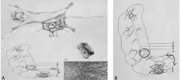
From a macroscopical point of view, the adipose organ is a multi-depot organ [Cinti S. 2005; Frontini A. & Cinti S. 2010]. In rodents for example depots are located in two compartments of the body: two are below the skin named subcutaneous depots (anterior and posterior), and some are inside the trunk, named visceral depots (mediastinic, mesenteric, retroperitoneal and addomino-pelvic composed, in females, by periovarian, perirenal, parametrial and perivescical fat) [Murano I. 2009; Vitali A. 2012]. The subcutaneous depots account for about 60-70% of the adipose organ [Cinti S. 2009 (b)] and it is mainly White Adipose Tissue (WAT) in adult mammals. In animals maintained at 28°C most of the adipose depots have a white-yellowish color, but, especially in young mammals, few brownish areas are in the anterior subcutaneous depot (i.e. cervical and interscapular) and in the visceral depot (i.e. perirenal depot) (Figure 1) [Cinti S. 2001, 2012].
Figure 1. A) Gross anatomy of the adipose organs of adult mice kept at 20°C. B) Schematic representation of
adipose organs anatomy of adult mice kept at 20°C [Cinti S. 2002].
Abdominal-pelvic depots are mixed depots, composed of WAT and Brown Adipose Tissue (BAT). The mediastinic depot is, especially in mouse, mainly brown; conversely the mesenteric and omental depots are mainly white. The epididymal depot is almost completely WAT [Cinti S. 2005; Murano I. 2009]. Thus, for practical convenience, most studies of adipose tissue have been limited to epididymal (eWAT) or posterior subcutaneous depot for WAT and interscapular BAT (iBAT) for BAT [Cousin B. 1993].
At histological level, the adipose organ is made up by two distinct tissues: WAT, consisting of unilocular white adipocytes, and BAT, consisting of multilocular adi pocytes. In many adipose depots the two tissues are intermingled (Figure 2) [Cinti S. 2001; Barbatelli G. 2010; Vitali A. 2012]. The composition as well as the color of the adipose organ varies not only in different anatomical locations but also changes with age and under different environmental and dietetic conditions [Cinti S. 2002, 2012]. The adipose organ is innervated [Bartness TJ. & Bamshad M. 1998; Giordano A. 2008] and nerve endings reach both the vasculature and adipocytes [Cannon B. 1986]. The sympathetic nervous system, controll, in part, the lipogenic and lipolytic pathways via noradrenergic fibers [Cousin B. 1993].
Figure 2. Light microscopy of mouse subcutaneous adipose tissue immunostained with anti-UCP 1 specific antibodies. White and brown are mixed together. Brown adipocytes show multilocular lipid depots, white
adipocytes show unilocular lipid depots. Only brown adipocytes stain for UCP1. Bar: 40 microns [Cinti S. 2006].
WAT and BAT exert opposite effects on the whole-body metabolism, which in turn reflect their different morphology and physiology. If WAT is the primary site of energy storage (white adipocytes accumulate lipids) and secrete hormones and cytokines that mainly modulate insulin resistance and food intake [Xu H. 2003], conversely BAT is important for both basal and inducible energy expenditure (brown adipocytes burn lipids) in the form of thermogenesis mediated by the expression of the tissue-specific uncoupling protein 1 (UCP1). Thus, excess accumulation of WAT causes obesity, while increased expression and activity of BAT improve insulin sensitivity and reduce susceptibility to weight gain [ Lowell BB. 1993; Cannon B. & Nedergaard J. 2004; Almind K. 2007; Cypess AM. 2009].
2.1.1. Brown Adipose Tissue (BAT)
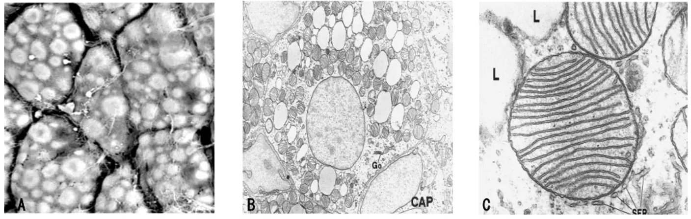
BAT is composed of polygonal cells (diameter about 30-50 μm) with a central roundish nucleus and several cytoplasmic lipid droplets. These cells are called multilocular adipocytes because their abundant cytoplasm contains numerous and large mitochondria packed with laminar cristae [Cannon B. & Nedergaard J. 2004; Ricquier D. 2005]. Peroxisomes, Golgi complex, rough and smooth reticolum, vesicles and other organelles are also visible by transmission electron microscopy [Cinti S. 2002, 2009; Barbatelli G. 2010] (Figure 3). Mitochondria are marked by the expression of uncouling protein 1 (UCP1) [Cannon B. & Nedergaard J. 2004; Ricquier D. 2005; Frontini A. 2007; Cypess AM. and Kahn CR. 2010].
Figure 3. Transmission electron microscopy. A) Scanning electron microscopy of brown adipose tissue (BAT).
Brown adipocytes are polyhedral with a multilocular lipid depot. Bar = 40 μm. B) Brown adipocyte of a neonatal rat filled with numerous small lipid droplets and typical mitochondria packed with cristae. L, lipid droplets.Go, Golgi apparatus; CAP, capillary. Magnification ×8700. C) High magnification of a typical brown adipocyte mitochondrion. L, lipid droplet; SER, smooth endoplasmic reticulum. Magnification 80000 [Cinti S. 2001; Cinti S. 2009].
BAT is important for thermogenesis but also regulates energy balance in small mammals, because its activation in mice promotes energy expenditure, reduces adiposity and protects from diet-induced obesity. Heat production is closely related to the presence of the UCP1 [Cannon B. & Nedergaard J. 2004; Frontini A. 2007; Cypess AM. & Kahn CR. 2010] and a large amount of nerves, especially in the interscapular area [Cinti S. 2001]. Exposure to temperatures below thermoneutrality is the physiological stimulus for BAT activation and thermogenesis [Himms-Hagen J. 2000]. To produce heat, brown adipocytes are activated by noradrenaline-releasing sympathetic nerves acting on ß3-adrenoreceptors (β3AR) to burn fatty acids, contained into the lipid droplets, in their motochondria. The UCP1 protein in the inner mitochondrial membrane uncouples oxidative phosphorylation resulting in energy dissipation in the form of heat [Cannon B. & Nedergaard J. 2004; Ricquier D. 2005; Cypess AM. & Kahn CR. 2010; Madsen L. 2010]. Given the large amount of lipid substrates and the
numerous, large and rich in ridges mitochondria, brown adipocytes oxidate considerable amounts of fat and sustain an intense thermogenesis [Klaus S. 1991; Cannon B. & Nedergaard J. 2004; Cinti S. 2006]. Furthermore, numerous adjacent brown adipocytes are able to simultaneously respond to a sympathetic sti mulus [Sheridan JD. 1971; Revel JP. 1971; Burke S. 2014] because they are electrically coupled by gap junctions [Schneider-Picard G. 1980]. Morphologically, the adipose organ of cold-exposed mice is browner, contains a higher number of brown adipocytes and is more densely innervated than the adipose organ of mice kept in a warm environment [Murano I. 2009; Vitali A. 2012].
If the environmental temperature is near thermoneutrality or in the absence of adrenergic stimulation such as, for example, in β-less mice [Bachman ES. 2002], the morphology of brown adipocytes is more similar to white adipocytes: they contain larger and fewer lipid droplets and a lesser number of mitochondria [Jimenez M. 2003].
2.1.2. White Adipose Tissue (WAT)
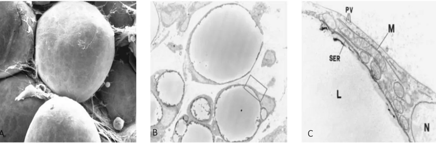
WAT is composed of roundish cells of variable size (60-80 μm), i.e. mainly large adipocytes are present in subcutaneous depots and more small adipocytes are present in visceral depots [Murano I. 2008; Barbatelli G. 2010]. They are also called unilocular adipocytes [Cinti S. 2012] because at light and electron microscopy a large lipid droplet, that occupies ~90% of the cell volume, flatten and squeeze the nucleus in the cell periphery. The lipid droplet is composed of triglycerides and the cytoplasm forms a very thin rim, with an electrondense barrier containing the structural Perilipin protein in between [Greenberg AS. 1991, Cinti S. 2002]. Cytoplasmic organelles (Golgi complex, smooth and rough reticolous and vesicles) are poorly developed and concentrated in the perinuclear area of white adipocytes. Mitochondria are small, elongated, slender and variable in number with tiny black ridges variously oriented. The outer surface of the cell membrane is characterized by a distinct basal membrane (or external lamina) (Figure 4) [Cinti S. 2002].
WAT is important for both short and long term energy storage [Bennett CN. 2002]. White adipocytes accumulate free fatty acids (FFA) supplying the organism with this substrate between one meal and another. When the time interval is the order of weeks, the energy stored in WAT as lipids becomes vital for survival. Conversely, white adipocytes can significantly increase their volume in situations that require the accumulation of triglycerides, as in genetic obesity and obesity induced by a high-fat diet [Cinti S. 2001]. Finally, WAT expresses β3 adrenoreceptors but is less innervated than BAT [Cinti S. 2001].
Figure 4. Transmission electron microscopy. A) Scanning electron microscopy and white (WAT) adipose tissue.
White adipocytes are spherical with a unilocular lipid depot. Bar = 25 μm. B) Epidydimal white adipose tissue of a 12-d-old rat. Magnification ×3300. [Cinti S. 2009]. C) Enlargement of the framed area in A, showing the cytoplasm of a white adipocyte containing few and small mitochondria (M) with randomly-oriented cristae. PV, pinocytosis vesicles; SER, smooth endoplasmic reticulum; L, lipid droplet. N, nucleus. Magnification ×32000 [Cinti S. 2001, 2009].
White adipocytes are also able to secrete hormones [Friedman JM. 1998; Trayhurn P. & Beattie JH. 2001], several cytokines [Rosen ED. & Spiegelman BM. 2006] and other factors like adiponectin and resistin [Bennett CN. 2002; Steppan CN. & Lazar MA. 2002]. The most important hormone produced and released by white adipocytes is Leptin that by acting on the central nervous system promotes satiety [Zhang Y. 1994]. Leptin influences both the eating behaviour of the individual and its energy expenditure, controlling, in this way, whole body energy homeostasis [Cinti S. 1997; Trayhurn P. 2007]. Fasting induces the adipocyte delipidizing with release of FFA whereas feeding induces leptin s ynthesis and secretion [Zhang Y. 1994; Friedman JM. 1998]. Mice with a genetic defect that inhibits the synthesis of leptin (ob/ob) or his receptor (db/db), become massively obese. The absence of this hormone massively stimulates food intake and for this reason leptin is regarded as the typical "satiety hormone". This condition provokes adipocyte hypertrophia, as observed in morbigen obesity, which has important consequences on the endocrine activity of adipocytes leading, for example, to a reduction in the secretion of adiponectin and an increase in the secretion of leptin [Matsubara M. 2002].
2.1.3. Mammary gland.
The mammary gland, which distinguishes mammals from all other animals, is a female anatomical structure that produces and secretes milk during pregnancy and lactation in order to nourish the offspring [Inman JL. 2015]. A useful model to study mammary gland development is the mouse gland [Brisken C. 1998]. The mammary gland (Figure 5A) of femal mice is located in the anterior and posterior subcutaneous depots [McCave EJ. 2010]. The adult mouse mammary gland has ten mammary glands, ten nipples, three symmetrical bilateral nipples in the anterior subcutaneous depot and two symmetrical bilateral nipples in the posterior subcutaneous depot [Cinti S. 2009(b); Smorlesi A. 2012; Giordano A. 2014]. In the gland both WAT and BAT are present [McCave EJ. 2010], but the realative proportion of brown and white adipocytes varies according to age, specie strain, environmental temperature and nutritional state [Cinti S. 2009(b)].
Mammary gland is also a unique glandular organ in that it reaches full development only after birth [Inman JL. 2015] and it expresses the maximum growth potential after ma turity following the onset of pregnancy and during lactation [Borellini F. 1989]. The resting mammary gland is composed of two tissues: the glandular epithelium and the stroma. Epithelium includes the ducts; the stroma is the adipose-rich connective tissue considered the scaffold of the mammary fat pad for the development of mammary epithelium [Richert MM. 2000; Hovey RC. 2004].
A variety of cell types are found in the mammary gland includi ng epithelial cells, adipocytes and other stromal cells i.e. fibroblasts, endothelial and lymphoid cells. Stromal cells support epithelial cell proliferation, differentiation and survival by the synthesis of type 1 collagen, an essential component of the extracellular matrix, as well as fibronectin, growth factors and cytokines within the ECM [Borellini F. 1989; McCave EJ. 2010], under the control of systemic hormones during puberty, pregnancy and lactation, also in humans [Medina D. 2004]. Adipocytes comprise a large portion of the stromal fat pad in adult virgin females and non-lactating gland. They are essential for ductal branching during puberty and for the maintenance of the ductal architecture in the adult mammary gland [Couldrey C. 2002; Landskroner-Eiger S. 2010].
2.1.3.1 Mammary gland development.
During the lifetime of the female mice, the mammary gland undergoes many changes in structure and function, including cyclic expansions corresponding to the hormonal changes induced by the estrous/menstrual cycle during puberty, as well as the dramatic changes that occur during pregnancy (Figure 5B), lactation and involution [Inman JL. 2015].
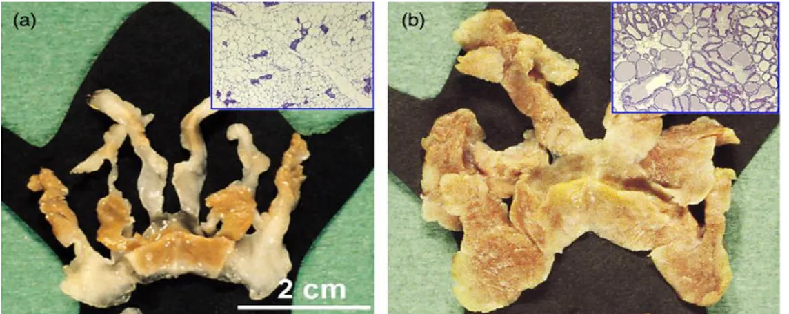
Figure 5. The mammary gland is a subcutaneous fat depot with extremely plastic morphology. a) Macroscopic
appearance of the anterior mammary gland of a virgin female mouse (histological appearance shown in the inset in the upper right corner). The scale bar in panel (a) applies also to panel (b). b) Macroscopic appearance of the same depot during lactation (histological appearance shown in the inset in the upper right corner of the panel) [Smorlesi A. 2012].
During embryogenesis in the female embryo a band of epithelial cells develops into the subcutaneous mesenchyme as a cord of cells partially canalized, opened at the apex (the future nipple) and beginning to branch at the distal end [Borellini F. 1989]. Up to day 22 to 23 of life, the development of the gland is mainly due to an increase in connective ti ssue and deposition of fat in a hormone-independent manner [Borellini F. 1989]. At birth the mammary gland is a rudimentary ductal system of only few ducts that develops in response to only estrogen [Fendrik JL. 1998]. The mammary epithelium grows extensively in a three-dimensional pattern and extends throughout the fat pad. Very few alveoli are formed and the gland is sexually mature at week 6 of postnatal life. At puberty (ten weeks of age) the adult mammary gland remains quiescent, the development of the ductal structures stops, in spite of the presence of estrogen produced by the ovaries [Borellini F. 1989], except for limited ductal side-branching during repeated estrous cycles [Brisken C. 2002; Neville MC. 2002; Ismail PM. 2003]. Under the influence of systemic hormones (estrogens, glucocorticoids and growth hormone) the ducts begin to expand into the surrounding stroma and develops in a basic arboreal network of ducts from the nipple [Borellini F. 1989; Ismail PM. 2003; Brisken C. & Rajaram RD. 2006] (Figure 6A). In the gland of virgin mice, the epithelium proliferates and apoptoses during each estrus cycle [Fata 2001].
During pregnancy, which in mice lasts 21 days, a “primed” ductal network became a secretory organ whose function is to produce milk [Elias JJ. 1993; Cinti S. 1999; Richert MM. 2000; Hovey MC. 2004]. An increase in the level of endocrine signals (serum prolactin and progesterone) and a network of transcription factors induce this dynamic process, definied alveologenesis [Fendrik JL. 1998; Brisken C. 2002; Neville MC. 2002]. During the first period, epithelial cells of the alveoli undergo extensive proliferation, leading to an increase in the number and size of alveolar lobules [Borellini F. 1989; Bussard KM. & Smith GH. 2011]. The ducts undergo further branching and, while the interstitial adipose tissue disappears progressively, proliferating epithelial cells fill the interductal spaces [Borellini F. 1989]. During the middle and late period of pregnancy (day 14.5), epithelial cells continue to multiply to form the lobuloalveolar structures. At least 90% of the organ is occupied by mature and fuctional lobulo-alveolar structures (Figure 6B) [Borellini F. 1989; Brisken C. and Rajaram R.D. 2006; Choi YS. 2009]. Alveoli are sphere-like anatomical structures composed of a single layer of secretory epithelial cells which are connected to the ductal network via a single small duct. Each individual alveolus is surrounded by contractile myo-epithelial cells [McManaman JL. 2003] that promote milk ejection, and a vascularized connettive-tissue stroma that provides the large quantities of energy and solutes required for milk production and contains lipid-depleted adipocytes and fibroblasts [Djonev V. 2001; McManaman JL. 2003].
Figure 6. Mouse mammary gland development during puberty and pregnancy shown are whole mount micrographs. A) At the onset of puberty the rudimental ductal system invades and eventually fills the fat pad. B)
With repeated estrous cycles, especially during mid pregnancy and lactation, the alveolar structures sprout from the expanded ductal tree. C) Section of lactating mammary gland. Arrow indicates the alveolar lumen filled with lipid droplets [Brisken C. and Rajaram R.D. 2006].
The mammary alveoli develop into clusters by da y 16.5 of pregnancy and start to show secretory activity. The epithelial cells become dilated at the end of pregnancy (days 18-21) because are filled of milk granules in the cytoplasm. Increased vascularization of the gland
and loss of fat in fat pad cells occur also [Borellini F. 1989; Neville MC. 2002; Rudolph MC. 2003; Morroni M. 2004;Brisken C. & Rajaram R.D. 2006].
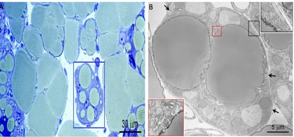
Functional differentiation of mammary epithelial cells culminates in lactogenesis. The first phase (called Lactogenesis I) involves an increased expression of some milk protein genes such as β-casein, lactalbumin and Whey Acidic Protein (WAP) and biosynthetic anzymes, and accumulation of neutral lipid droplets, visible in electron microscopy (Figure 6C) [Neville MC. 2002]. The Lactogenesis II is the secretory activation phase that occurs at the end of pregnancy in rodents and it occurs only in the presence of Prolactin and cortisol in appropriate concentrations [Nguyen DA. 2001]. Lactating tissue shows the typical features of a secretory epithelium (Figure 7A). Adipocytes became irregular and e xhibit smaller lipid droplets, an increased number of mitochondria and rough endoplasmatic reticulum (Figure 7B) [McManaman JL. 2003; Smorlesi A. 2012]. Cells are highly polarized, both functionally and morphologically, are connected by tight junctions, and the surfaces that face the cavity of the alveoli are covered with microvilli. A large part of the cytoplasm is occupied by rough endoplasmic reticulum and mitochondria; the well-developed Golgi apparatus is located in the apical region [Borellini F. 1989; Nguyen DA. 2001; Neville MC. 2002]. But they also contain some small droplets of fat in the cytoplasm. So, copious expression of milk protein genes by alveolar cells occurs [Nguyen DA. 2001; Neville MC. 2002] and c ytoplasmic lipid droplets and β-Casein move to the alveolar lumen [Brisken C. & Rajaram R.D. 2006].
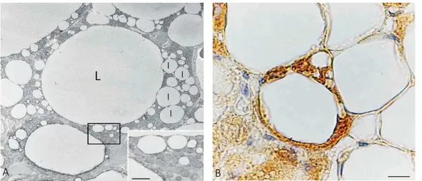
Figure 7. A) Histological appearance of mammary gland in late pregnancy showing developing alveoli with
unusual features (in the blue square) intermediate between adipocytes and glands. B) Electron microscopy of the adipoepithelial structure highlighted in A. To note, the big lipid droplets (droplets of this size are typical only o n white adipocytes) are surrounded by several nuclei owning to cells with cytoplasmic evidence o f milk protein containing granules (inset at the lower left corner) that are typical of milk-secreting mammary alveoli. Further evidence for the nature of this milk-secreting alveolar structure is the presence of myoepithelial cells (arrows and inset in the upper right corner) at the periphery underneath the basal membrane [Smorlesi A. 2012].
Giordano et al. (2014) proposed the name of “pink adipocytes” for these milk-secreting cells
with intermediate morphology. Pink adipocytes are capable of storing large amounts of lipid as well as adipocytes and are pink because arise esclusively in female pregnant mammary gland, which is pink at macroscopical appearance (Figure 7A-B) [Giordano A. 2014].
The epithelium and the surrounding myoepithelial cells are enclosed by the basal lamina, which provides a boundary between the epithelium and the stromal tissue [Borellini F. 1989]. If secretory activation take place in cultured cells and tissues, the milk offspring has been observed only in animal [Neville MC. 2002]. Suckling controlled the ejection of milk from the lactating mammary gland and is the appropriate stimulus for the release of oxytocin from the posterior pituitary. Oxytocin increases intramammary pressure by inducing contraction of the myoepithelial cells and thus aids in expelling the milk from the mammary glands [Inman JL. 2015]. In the mouse, milk yield is maximum at day 15 of lactation [Borellini F. 1989]. After weaning, milk production ceases and the gland involutes to a prepregnant immature state [Brisken C. 2002; Neville MC. 2002]. Alveoli progressively disappear and their “space” is replenished by adipocytes. As well as in the nonlactating adult breast, the stroma occupies the majority of the tissue, where the proportions of fibrous and adipose tissue vary with age [Richert MM. 2000; Hovey MC. 2004; Bussard KM. & Smith GH. 2011].
The cycle of proliferation-differentiation-regression is repeated at each gestation, and can be reproduced in culture systems in vitro [Borellini F. 1989].
2.3.1.2. Agents involved in Mammary gland development.
The regulation of mammary gland morphogenesis in vivo is a very complex phenomenon. The growth and differentiation of mammary cells can be induced in vitro [Borellini F. 1989]. A series of systemic hormones from several endocrine glands and environmental factors are involved and influence the glandular development, the epithelial proliferation and differentiation of the mammary gland as well as lactation through a complex network of intracellular communication between luminal cells, basal cells and the stroma [Borellini F. 1989; McCave EJ. 2010; Bussard KM & Smith GH 2011]. Three categories of hormones are involved: reproductive, metabolic and mammary hormones.
The levels of the reproductive hormones, estrogen, progesterone, prolactin and placental lactogen change during reproductive development and act directly on the mammary gland to bring about developmental changes or coordinate milk delivery to the offspring [Brisken C. 2000; Neville MC. 2002; Choi YS. 2009; Ercan C. 2011]. They modulate and interact with
growth factors and their receptors [Woodward TL. 1998, Woodward TL. 2000; Rodríguez-Cuenca S. 2006].
Progesterone (Pg) is a sex steroid endocrine hormone. The serum levels of Pg are
significantly attenuated during the later stages of pregnancy [Ismail PM. 2003]. At puberty in virgin animals Pg level is very low, increases at early pregnancy when Pg signaling initiates proliferative responses [Haslam SZ. 1988 (b)]. It is essential for alveolar morphogenesis [Brisken C. 1998] in the mammary glang of pregnant animals preparing the adipose tissue for the beginning of the late pregnancy [Lydon JP. 1995; Brisken C. 1998; Rodríguez-Cuenca S. 2006]. Pg is a negative regulators of lactogenesis [Nishikawa S. 1994], thus the fall in Pg levels at late pregnancy and after parturition, coinciding with the increase of prolactin and cortisol levels [Woodward TL. 2000; Rudolph MC. 2003] could serve for the fully differentiation of the mammary gland [Ismail PM. 2003] and activation of milk protein gene expression [Nguyen DA. 2001; Neville MC. 2002]. Pg receptor (PR-A and B isoforms) synthesis depends on estrogen [Beato M. 1995]. Pg also stimulates mammary epithelial cells to release paracrine signals on other nearby epithelial cells [Lydon JP. 1995; Brisken C. 1998] through Wingless-related MMTV integration site 4 (Wnt-4) and receptor activator of nuclear factor (NF)-κB ligand (RANKL) [Brisken C. 2000]. Wnt-4 and RANKL are transcription factors that stimulate side branching and alveologenesis during early pregnancy [Brisken C. 2000] by paracrine induction of β-catenin signaling in both luminal and basal cells. β-catenin is a component of cell-cell junctions and is associated with survival and more efficient self-renewal of mammary epithelial stem cells [Clarkson RW. 1999; Ercan C. 2011]. Its activation in basal mammary epithelial cells affects the growth, survival and differentiation of luminal cells altering the entire process of the postnatal mammary gland development [Teuliere J. 2005].
Prolactin (Prl) is a pituitary protein and a well-known master regulator of the lactating
mammary gland [Freeman ME. 2000]. Prl is necessary in epithelium for normal lobulo-alveolar morphogenesis and differentiation into milk-producing cells [Brisken C 1998; Ormandy CJ. 1997]. Added to a mouse mammary gland culture in combination with insulin and glucocorticoid, Prl induces the milk protein expression genes, like β-Casein [Guyette WA. 1979]. The responsiveness of mammary cells to prolactin during pregnancy is regulated by progesterone and prolactin levels. The number of prolactin receptors on mouse mammary gland from different developmental stages varies in inverse relationship to progesterone levels in serum. Conversely glucocorticoid increases prolactin receptors in mammary cells in culture. Prl also appears to stimulate both ribosomal and transfer RNA accumulation [Borellini F. 1989]. Acting on pancreatic β-cells Prl triggers cell proliferation and insulin
secretion thereby ensuring a series of metabolic adjustments required for pregnancy and milk secretion [Galsgaard ED. 2001]. Hypothalamic neuroendocrine dopaminergic neurons release dopamine in response to Prl. Dopamine inhibits prolactin release from the anterior pituitary, functioning as a negative feedback [Freeman ME. 2000]. Prl acts directly on the mammary epithelium, and indirectly by stimulating luteal progesterone secretion in rodents [Horseman ND. 1999].
Estrogen is the ovarian hormone considered the main effectors, also in vivo, of ductal
elongation in the mammary gland during puberty and pregnancy [Haslam SZ. 1988; Mallepell S. 2006]. The serum levels of estrogen, that are high during puberty and pregnancy, are down-regulated during late pregnancy and lactation stages [Choi YS. 2009]. Estrogen has a role in mediating the tissue sensitivity to the lactogenic stimuli [Borellini F. 1989]. In postlactation, when the breast is undergoing remodeling, the plasma estrogen levels are higher than in lactation [Saji S. 2000]. Estrogen acts in a paracrine fashion through two receptors (ER), ERα and ERβ [Beato M. 1995; Dupont S. 2000], both of which are expressed in the normal ductal epithelium of the human and mouse mammary gland [Jarvinen TAH. 2000, Saji S. 2000]. ERα signaling is essential for normal ductal elongation during puberty [ Mallepell S. 2006]. ERβ signaling may be required for fully functional lobuloalveolar development [Forster C. 2002]. Estrogen also induces PR expression and Pg ehances Insulin Growth Factor-I activity to stimulate mammary ductal morphogenesis, appearing to synergize with Growth Hormone [Ruan W. 1995; Saji S. 2000].
In addition to the female hormones, metabolic hormones appear to reinforce different stages of mammary gland development [Brisken C. & Rajaram RD. 2006]. These hormones are Growth Hormone, Thyroid hormone [Capuco AV. 1999], Insulin and Corticosteroids [Neville MC. 2002; Bussard KM. & Smith GH. 2011].
Growth Hormon (GH) or Somatotropin is a pituitary hormone that plays an important
role, but lesser than PRL, in mammary growth at puberty and development during pregnancy in rodents [Feldman M. 1993]. Epithelial GH receptor is required during late lobuloalveolar development also in vivo [Wintermantel TM. 2005]. GH may act in combination with Prl in
vitro to mediate alveolar proliferation and stimulate milk protein expression [Allan GJ. 2002].
The lactogenic activity of GH may be indirectly through insulin-like growth factor-1 expression [Feldman M. 1993; Ruan W. 1995]. Growth hormone and placental lactogen can substitute for prolactin in epithelial cell culture of mouse mammary gland [Borellini F. 1989].
Thyroid hormones are not necessary for ductal growth but seem to stimulate lobular
development, enhancing the tissue responsiveness to Prl in mouse mammary gland by activating the prolactin receptors also in vivo. They also regulate the level of EGF receptors at
several stages of development and are important for maintenance of alveoli during regression of the gland [Borellini F. 1989].
Insulin stimulates cell replication in serum-free culture but does not seem to be
necessary for ductal or alveolar growth. Like Prl, insulin also appears to stimulate both ribosomal and transfer RNA accumulation [Borellini F. 1989].
Also mammary hormones regulate the mammary gland development. This third category of hormones includes Prolactin, Parathyroid hormone-related protein, Glucocorticoids, Placental lactogens and Leptin [Neville MC. 2002].
Parathyroid hormone-related protein (PTHrP) is made in the epithelial cells of the
mammary bud. PTHrP and its receptor are necessary for survival and development of the embryonic mammary gland in mice as well as the formation of the nipple during embryonic mammary development. During lactation PTHrP release into the maternal blood circulation is associated with the increase of Prl [Foley J. 2001] and it is involved in calcium release from the bone during lactation in the newborn and the mother [Lippuner K. 1996]. PTHrP signaling is also involved in Wnt signaling pathways because it regulates the epidermal and mesenchymal expression of β-Catenin [Foley J. 2001].
Glucocorticoids act in a synergistic effect with Prl in cell and tissue culture of
mammary epithelia leading to enhanced transcription of milk protein genes [Rosen JM. 1998]. Glucocorticoids regulate mammary epithelial cell proliferation during late-lobuloalveolar development, but are not essential for the differentiation and function of secretory alveoli of the lactating mammary gland [Wintermantel TM. 2005].
Placental lactogen is released from the placenta during pregnancy and can fully
compensate for Prl. Together Prl and Pg, they stimulate the expansion and physiological differentiation of the lobuloalveolar system from the lobular buds during pregnancy and the final induction of milk protein expression and lactation [Horseman ND. 1999].
Leptin is secreted from the mammary fat deposits and interacts with the mammary
epithelial cells after Prl stimulation. Lactation, like other situations which impose a high energetic demand on the organism, is associated with a decrease in leptin signaling to the brain that is likely permissive for the increase in food intake seen in lactation [Woodside B. 2000].
Hormones and growth factors interact and modulate both growth factors and their specific receptors expression. Several stromal-derived growth factors, synthesized locally - and acting in a paracrine manner - or secreted by distant sources - and acting in a hormonal manner - also affect mammary gland development during pregnancy.
Epidermal Growth Factors (EGFs) Family is expressed during postnatal mammary
gland development and is localized in the ductal epithelium and in surrounding stromal cells [McCave EJ. 2010; Bussard KM. & Smith GH. 2011]. EGF stimulates proliferation of the epithelium in vitro [Borellini F. 1989] and its action is mediated by EGF receptors (ErbB1-4) [McCave EJ. 2010]. EGF signaling is essential for both alveologenesis (ErbB4) [Troyer KL. 2001; Long W. 2003], thus EGF-R levels are high during the proliferative phases of mammary gland development. EGF appears to be essential for the lobuloalveolar formation (ErbB2) and for the expression of differentiative potential of the mouse mammary gland during pregnancy [Borellini F. 1989; Jones FE. 1999; Troyer KL. 2001]. Its level decreases when the gland reaches functional differentiation. Amphiregulin is the only ligand of ErbB expressed on epithelial cells and required in the mammary gland [Yang Y. 1995].
Transforming growth factor-β (TGF-β) is a regulator of mammary gland development in vivo [Zangani D. 1999] produced by epithelium. TGF-β blocks adipocyte differentation in vitro [Rosen ED. & MacDougald OA. 2006] and act as a negative regulator in the ductal
epithelium affecting ductal growth and lateral branching during pregnancy as well as antagonizing the mitogenic effect of growth factors such as EGF [Daniel CW. 1996; Ercan C. 2011]. TGF-β isoforms are regulated by ovarian hormones, are highly expressed during pregnancy to maintain tissue homeostasis in terminal-end-buds and are dramatically down-regulated during lactation [Robinson SD. 1991].
Insulin-like Growth Factor (IGF) mediates glandular proliferation and development
during pregnancy in a paracrine manner [Feldman M. 1993; Brisken C. 2002]. The IGF pathway consists of three ligands (I, IGFII and insulin) and their receptors (IR, IGF-IIR, and insulin-receptor). IGF ligands play various roles in mammalian growth, development, and metabolism and act as endocrine factors that prevent apoptotic signaling. IGF-1 protein is localized in stromal cells in the mouse immature mammary gland, but with increasing age, it is also detected in the epithelium [Woodward TL. 1998]. IGF-1 expression in mammary tissue is regulated by pituitary GH [Ruan W. 1995] and it appears to synergize with estrogen in the induction of experimental normal ductal development [Ruan W. 1995].
Hepatocyte Growth Factor (HGF) is produced by mammary fibroblasts in vivo and in vitro. HGF is a potent mitogen for mouse and human mammary luminal epithelial cells,
increasing proliferation and branching morphogenesis [Niranjan B. 1995; Soriano JV. 1998]. The regulation of HGF synthesis in the mesenchyme during postnatal mammary gland development is under control of systemic hormones, and glucocorticoids downregulate HGF expression levels during pregnancy. Also EGF and TGF-β factors, produced in the epithelium, are suppressors of HGF expression of fibroblasts in cell culture [Yang Y. 1995].
Fibroblast Growth Factor (FGF) family proteins are expressed during mammary
ductal development, in both pubertal and adult mice and their expression decreased during pregnancy and lactation. Several members of the FGF family and their receptors (four known FGF receptor genes, Fgfr1-4) have been detected in the mammary gland [Coleman-Krnacik S. & Rosen JM. 1994]. Fgfs signalling have a function in both the embryo, in the formation of the mammary primordia, and in the adult during pregnancy. It is necessary and is specifically required in normal lobuloalveolar development of the mouse mammary gland during pregnancy, i.e. FGF2 (basic FGF) expression in stromal cells is induced only in the presence of epithelium [Coleman-Krnacik S. & Rosen JM. 1994; Jackson D. 1997; Dillon C. 2004; Zhang X. 2014]. FGF signalling during pregnancy is most likely regulated, directly or indirectly, by steroid, like estrogen and progesterone, and peptide hormones [Spencer-Dene B. 2001]
Cell-matrix interactions are critical for regulating the phenotype of many cells, including for successful alveologenesis and lactogenic differentiation of epithelial cells in the mammary gland. Matrix molecules, like β1-integrins, fibronectin and cadherins can also modulate responsiveness to ovarian hormones [Woodward TL. 1998].
β1-integrins are the cell surface receptor on luminal epithelial cells that mediate the
interaction of mammary epithelial cells with the extracellular matrix (ECM) [Klinowska TCM. 1999]. In mammary epithelium integrins are composed of α-subunit and β1 or β4-subunit and are more abundant at the basal surface than in the luminal portion [McCave EJ. 2010]. During pregnancy β1-integrins are important for normal alveologenesis, cooperating with HGF [Klinowska TCM. 1999], for fully functional differentiation (expression of the milk protein β-Casein) in vitro and in vivo and for maintainance of the integrity of mammary alveoli [Streuli CH. 1991; Klinowska TCM. 1999; Li N. 2005; Naylor MJ. 2005]. β1-integrin is essential also for Prl-mediated differentiation in mammary epithelium [Naylor MJ. 2005].
Fibronectin is a major stroma-derived component of the ECM and is important for
estrogen and progestin-induced epithelial proliferation in vitro and is developmentally and hormonally regulated in vivo [Woodward TL. 1998].
E-Cadherin (E-Cadh) is important for the epithelial integrity and terminal
differentiation of the mammary alveolar epithelium. E-Cadherin is the founder member of the cadherin superfamily of calcium-dependent cell adhesion molecules. The E-Cadherin–catenin adhesion complex is crucial for the polarization and function of epithelial cells and for the integrity of epithelial cell layers.E-Cadh-null mice show an involuting-like mammary gland at parturition and express β-Casein and WAP (milk proteins) at levels much lower compared to wild type control [Boussadia O. 2002].
This complex signaling network controls growth and differentiation of the normal mouse mammary gland in which prolactin signaling is the central hub. Prl, Pg and growth factors induce the transcription of genes via activation of target transcription factors, which induce the expression of genes involved in mammary gland development, like ELF-5 [Coletta RD. 2004] and GATA-3.
ELF-5 is an ephitelial specific (ETS) transcription factor family primarily expressed in
secreting epithelial cells of mouse and human mammary tissue [Lapinskas EJ. 2004]. In mammary gland its expression increases greatly during mid-late pregnancy, remains high during lactation and returns to baseline levels after involution [Zhou J. 2005, Harris J. 2006]. ELF-5 is a prolactin-regulated gene [Lapinskas EJ. 2004] and can substitute for prolactine signaling [Harris J. 2006]. During pregnancy is crucial for normal alveologenesis, functional lobulo-alveolar development [Zhou J. 2005; Harris J. 2006; Oakes SR. 2008; Choi YS. 2009] and milk protein expression, WAP and β-Casein [Zhou J. 2005; Harris J. 2006; Oakes SR. 2008; Choi YS. 2009]. ELF-5 knockouts exhibit a complete block in alveolar differentiation [Choi YS. 2009].
GATA-3 is the most highly enriched transcription factor in the mammary ductal
epithelium of pubertal mice [Kouros-Mehr H. 2006]. The expression is localized to the initial sites of mammary buds and is restricted to mature luminal ductal cells at later stages of mammary development [Kouros-Mehr H. 2006; Asselin-Labat M. 2007]. GATA-3 is necessary for the development and differentiation of luminal epithelial cells in the adult mammary gland, also in vivo. It is necessary also to maintain the integrity of the luminal epithelium [Kouros-Mehr H. 2006]. Deletion of GATA-3 at an early stage results in the impairment of ductal elongation; at a later developmental stage it leds to a block in differentiation resulting in a lactation-deficient phenotype [Kouros-Mehr H. 2006; Asselin-Labat M. 2007; Siegel PM. & Muller WJ. 2010]. High levels of GATA-3 expression are sufficient to induce the differentiation of a stem cell-enriched subpopulation towards the alveolar cell lineage and the expression of luminal differentiation markers, including WAP and β-Casein [Asselin-Labat M. 2007].
2.2. Transdifferentiation
Until a few years ago, it was a consolidated opinion that a differentiated cell of an adult organism, with a specific morphology and function could not change its phenotype. In recent years, however, numerous experimental settings in vitro showed that mature differentiated cells, under appropriate stimulation, exhibit the ability to transform, without going through dedifferentiation, into a different cell type by using a complex and still poorly understood process called "cell transdifferentiation" [Okada TS. & Clarendon 1991; Eguchi G. & Kodama R. 1993; Slack JM. & Tosh D. 2001; Tosh D. & Slack JM. 2002; Blelloch R. 2008; Eberhard D. & Tosh D. 2008]. The adult cells can reprogram their genetic expression and rearrange their morphological appearance to sustain different physiological variations depending on the environment in w hich they live [Tosh D. & Slack JM. 2002; Cinti S. 2009 (b)] (Figure 8).Zhou Q. et al 2008 provided an example of fully differentiated cells can be
directly reprogrammed in adult animals by a combination of transcription factors , without reversion to a pluripotent stem cell state [Zhou Q. 2008].
Figure 8. The regenerative-medicine toolbox. a) During
development, non-specialized cells with a broad developmental potential differentiate into various highly specialized cells that have limited developmental potential. b) Nonetheless, in the lab, these highly specialized cells can be induced to dedifferentiate, revert back to an earlier stem-cell fate with a broad developmental potential. The cells can then be triggered to differentiate into another cell type. c) In some circumstances a highly specialized cell can be induced to transdifferentiate into another specialized cell, bypassing the step of dedifferentiation [Blelloch R. 2008].
Studying transdifferentiation is important because it is useful for the identification of the master gene(s) (homeotic or selector gene) and the transcription factor(s) that are responsible for the switch in phenotype, i.e. changes in the cellular phenotype can predispose to the development of cancer. In the adipose organ, white adipocytes can transdifferentiate directly and reversibly into brown adipocytes under physiological stimuli in adult mice, old rats [Himms-Hagen J. 2000; Barbatelli G. 2010] and more recently in humans [Frontini A. 2013].
Thus, the adipose tissue is plastical, that is it adapts itself to different energy needs of the body.
2.3. Plasticity of Adipose Organ
Adipocytes show strong transdifferentiating plasticity, they can directly modify their specialized phenotype to respond to different nutritional and metabolic body status as well as changes in ambient temperature in a physiological and reversible manner. Thus, the adipose organ is a compex structure with highly plastic properties [Smorlesi A. 2012].
Moreover, the presence of both the brown and white adipocytes in the same depots of the adipose organ is related with the ability of these two tissues to transform from one to the other [Cinti 2009 (a)]. The adipose organ contains al least three cell types, white, brown and pink adipocytes; each has a unique morphology and expresses some different gene, i.e. Leptin is expressed by white and pink adipocytes [Cinti S. 1997; Cancello R. 1998; Smith-Kirwin SM. 1998], Perilipin 1 is expressed by white and brown adipocytes [Blanchette-Mackie EJ. 1995]. The three phenotypes correspond to three different physiological roles ( Figure 9): white adipocytes store lipids and secrete leptin and adiponectin, affecting eating behavior and metabolism [Zhang Y. 1994; Trayhurn P. 2013]; brown adipocytes produces heat and secrete hormone and growth factors [Fisher FM. 2012; Villarroya J. 2013]; pink cells produces milk and leptin also [Oliver P. 2002; Palou A. 2009].
Figure 9. Scheme of the three adipocytes forming the parenchyma of the adipose organ. The adipose organ parenchyma contains three cell types. White adipocyte stores and secretes lipids; brown adipocyte produces heat; pink adipocyte produces milk [Giordano A. 2014].
The well-kown reversible adipose tissue transformations induced by physiological stimuli are i) brown to white adipocytes, ii) white to brown adipocytes and iii) white adipocytes to pink cells [Giordano A. 2014]. The sympathetic nervous system seems to play a fundamental role in the plasticity of the adipose organ, i.e. a positive correlation between the density of
noradrenergic parenchymal fibres and the density of brown adipocytes in the adipose organ under different environmental temperatures has been shown [Murano I. 2009].
The first description in vivo of adipoepithelial transdifferentiation in healthy adult mammals was provided by our group [De Matteis R. 2009, Morroni M. 2004].
2.3.1. Brown adipocytes trandifferentiate into white adipocytes.
Ageing and the positive energy balance (energy intake is higher than energy expenditure), as well as warm exposure (34°C) or not adrenergically stimulated BAT lead to “whitening” [Cinti S. 1999, 2005; Frontini A. & Cinti S. 2010]. In these conditions brown adipocytes become hypertrophic and hyperplastic [Rothwell NJ. & Stock MJ. 1979; Lowell BB. 1993] and the white part of the adipose organ expands [Hausman DB. 2001]. Brown adipocytes directly transform into unilocular cells [Cinti S. 1997; Bachman ES. 2002], their typical big brown mitochondria transform into white smaller mitochondria with less-developed cristae, and vascular and nerve supply reduces [Cinti S. 1999, 2005; Frontini A. & Cinti S. 2010]. Brown to white transdifferentiation is accompained, at a genetic level, by inhibition of the UCP1 gene and activation of the leptin gene [Cinti S. 1997; Cancello R. 1998]. The “obese” adipose organ is infiltrated by macrophages [Weisberg SP. 2003; Xu H. 2003; Strissel KJ. 2007], which surround and absorb dead adipocytes and their remnant lipids (forming crown-like structures, CLS) and secrete many inflammatory cytokines [Murano I. 2008]. The presence of CLS turns the adipose tissue parenchyma into “morbigen” and inflamed tissue and can lead to obesity and insulin-resistance [Weisberg SP. 2003; Xu H. 2003; Cinti S. 2005, 2012; Giordano A. 2013]. Intra-abdominal adipose tissue of rodents and humans is highly susceptible to obesity-associated adipocyte death and macrophage infiltration [Gabriely I. 2002; Strissel KJ. 2007]. Blocking brown to white adipocyte transdifferentiation could cure obesity and diabetes [Ghorbani M. 1997; Strissel KJ. 2007].
2.3.2. White adipocytes trandifferentiate into brown adipocytes
White to brown transdifferentiation, named “browning” [Champigny O. 1991; Collins S. 1997; Ghorbani M. 1997; Ghorbani M. and Himms-Hagen J. 1998; Cinti S. 2001, 2002 (b); Seale P. 2008; Barbatelli G. 2010], is a transformation of white adipocytes into brown adipocytes. Browning is essential to increased heat production requirements during chronic cold exposure [Giordano A. 2014] and it is detectable even at macroscopic level in the adipose organ of a mouse kept at 6°C compared with one acclimatated at 28°C [Cinti S. 2005].
Treatment with β3 agonists, as well as cold exposure and hormonal stimuli, activates BAT by acting on the sympathetic nervous fibres that directly innervate brown adipocytes in the parenchyma through β3-adrenoreceptors activation [Jimenez M. 2003; Barbatelli G. 2010]. At histological level, the number of brown cells as well as brown characteristics in the white areas of the adipose organ increases [Cinti S. 2002], but the total number of cells remain unchanged [Murano I. 2009; Vitali A. 2012]. The density of vessels and sympathetic nerves activity increase in parallel with the increase of brown adipocytes [Garofalo MA. 1996; Cinti S. 2005, 2009, 2009 (b)]. The increase of BAT corresponds to a reduction of WAT, which is unrelated with apoptosis, and occurred in the absence of any marker of cellular proliferation [Himms-Hagen J. 2000; Ganneman JG. 2005; Barbatelli G. 2010]. These brown-like adipocytes arising in WAT in response to cold exposure are ontogenically different from those in the classical iBAT [Atit R. 2006; Seale P. 2008; Petrovic N. 2010], they are multilocular but show a variable immunoreactivity for the brown marker UCP1 [Himm-Hagen J. 2000; Cinti S. 2005; Granneman J.G. 2005; Barbatelli G. 2010]. The new adipocytes are called “paucilocular” adipocytes (Figure 10), but some groups refer to them as or “beige” [Ishibashi J. & Seale P. 2010] or “brite” (brown in white, regions of white adipose tissue containing brown or brown-like adipocytes) adipocytes [Petrovic N. 2010; Walden TB. 2012; Wu J. 2012]. However, these cells show a reduction in adipocyte size, an increase of their mithochondrial and lipid droplets content. Also mitochondria show an intermediate morphology [Barbatelli G. 2010; Giordano A. 2014].
Figure 10. Paucilocular adipocytes. A) Transmission electron microscopy of subcutaneous fat of a cold
acclimated adult mouse (6 8C for 5 days) showing a paucilocular adipocyte with an intermediate morphology between white and brown adipocytes. Note the predominant large central lipid droplet (L) an d several small cytoplasmic lipid droplets (l). Mitochondria are numerous and exhibit an intermediate morphology between those typical of white and brown adipocytes (inset: enlargement of squared area in (A). Scale bar: AZ5 mm and inset Z0.5 mm. B) UCP1-immunoreactive paucilocular adipocyte found in omental fat from a patient suffering for pheochromocytoma. Note the morphology corresponding to that described in (A). Surrounding (upper and right) white adipocytes are unilocular and UCP1 negative. Scale bar: 10 mm [Giordano A. 2014].
L l l l l A B
Cold-induced brite adipocytes, from inguinal depot, turned into unilocular adipocytes (morphology and gene expression pattern of white adipocytes) when the animals were exposed to warm and re-convert into brite adipocytes on additional cold stimulation [Rosenwald M. 2013] (Figure 11). Thus, browing is of remarkable pathophysiological interest because it could be exploited to fight obesity and metabolic syndrome (type 2 diabetes) [Kopecky J. 1996; Collins 1997; Rothwell NJ. & Stock MJ. 1979; Guerra C. 2001; Nedergaard J. 2011; Bachman ES. 2002; Almind K. 2007; Bartelt A. 2011; Vitali A. 2012; Stanford KI. 2013]. It is known that mice lacking functional BAT [Lowell BB. 1993] or β3-adrenoreceptors knockout [Bachman ES. 2002] are prone to diet-induced obesity and diabetes [Jimenez M. 2003; Feldmann HM. 2009; Barbatelli G. 2010].
β3-adrenoreceptor (β3-AR) agonists successfully curb obesity in genetically obese rats, as well as diet-induced obese rats [Ghorbani M. 1997; Ghorbani M. & Himms-Hagen J. 1997; Ghorbani M. & Himms-Hagen J. 1998] and in human BAT [Saito M. 2009]. As in experimental animals, also the human BAT (hBAT), detectable by positron emission tomography (PET) [Nedergaard J. 2007; Cypess AM. 2009; Saito M. 2009; Seale P. 2009; van Marken Lichtenbelt WD. 2009; Virtanen KA. 2009], is composed of UCP1-expressing adipocytes, is densely innervated [Zingaretti MC. 2009] and express β3-adrenoreceptors [Cinti S. 2005]. It displays outstanding plasticity due to ageing, obesity, metabolic desease [Huttunen P. 1981; Lean ME. 1986; Cinti S. 2006] and environmental conditions, i.e. cold exposure [van der Lans AJJ. 2013].
Thus, understanding the molecular mechanism of the adipose organ plasticity, in particular of browning, is important for the regulation of energy balance and the development of obesity in mice and humans [Cypess AM. & Kahn CR. 2010; Madsen L. 2010]. It might be of great interest for future treatment or prevention of obesity and type 2 diabetes [Carruba M. 1998; Cinti S. 2002 (b); Nedergaard J 2007;Cypess AM. 2009; van Marken Lichtenbelt WD. 2009; Virtanen KA. 2009; Zingaretti MC. 2009; Cypess AM. & Kahn CR. 2010; Enerback S. 2010]. Altough a pharmacological approach to obesity treatment has repeatedly been confirmed in experimental animals, yet it is not been approved in humans [Saito M. 2009].
The white-brown (and reverse) transdifferentiation is not the only physiologic reversible transdifferentiation that can occur in the adipose orga n of adult mice [Cinti S. 2016] (Figure 11).
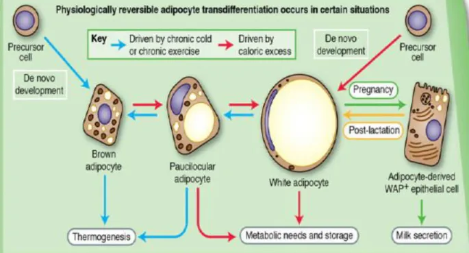
Figure 11. Evidence suggest that adipocytes transdifferentiation might underlie the changes in adipose organ
composition that are abserved in response to chronic cold or exercize, caloric excess or pregnancy/lactation. The facors driving these transdifferentiation pathways are under investigation [Cinti S. 2012].
2.3.3. White-pink transdifferentiation in mammary gland
The mammary gland is a unique organ of the body that continuously undergoes plastic and cycling changes during the reproductive life of femal mammals, especially during pregnancy and lactation when a progressive substitution of adipocytes by milk-secreting alveolar cells happen in the mammary glands [Elias JJ. 1973; Richert MM. 2000; Smorlesi A. 2012]. The alveolar mammary epithelium expands and differentiates during pregnancy, conversely the adipose tissue reduces [Elias JJ. 1973; Richert MM. 2000; Hovey RC. 2004]. Adipocytes progressively disappear during lobulo-alveolar development, but at the end of lactation the epithelial component of the gland is progressive substituted by adipocytes, reconstituting the pre-pregnancy anatomy of the mammary gland [Richert MM. 2000; Cinti S. 2009 (b)]. Our interpretation of this phenomenon is the reversible pregnancy-induced adipoepithelial transdifferentiation of the mammary gland. This hypothesis is supported by ultrastructural analysis [Morroni M. 2004], in vivo lineage tracing studies [Richert MM. 2000; Ercan C. 2011, Bussard KM. and Smith GH. 2011] and explant experiments, of both adipose tissue and isolated adipocytes, also from human adipose tissue [Morroni M. 2004; De Matteis R. 2009; Poloni AMG. 2012]. Thus alveolar epithelial cells can transdifferentiate into white adipocytes during mammary gland involution and white adipocytes can transdifferentiate into
milk-secreting alveolar epithelial cells during pregnancy [Morroni M. 2004 ; De Matteis R. 2009; Prokesh A. 2014].
It is well known that floating isolated adipocytes express mRNA encoding for specific mesenchymal stem cell markers (CD34, CD90, CD45 ans Sca-1) and for transcription factors that mediate the reprogramming of mature cells in pluripotent state (Oct-3/4, Sox-2, NANOG, c-Myc and KLF-4) [Takahashi K. & Yamanaha S. 2006; De Matteis R. 2009]. These results confirm that adipocytes are plastic and can adapt to environmental stimuli [Matsmoto T. 2008; McCave EJ. 2010; Poloni AMG. 2012], like the extracellular matrix composition [Howlett AR. 1993].
Interstingly and in line with the idea of adipoepithelial transdifferentiation, the early stages of alveolar development during pregnancy are characterized by pink cells, glandular cells (Figure 12) with an intermediate morphology [Giordano A. 2014].
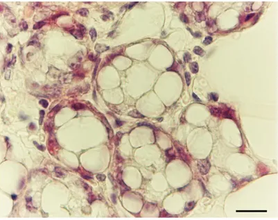
Figure 12. H&E Subcutaneous fat of mouse mammary gland at day 18 of pregnancy. Early stage of alveolar
glands formation. Epithelial cells show very abundant cytoplasmic lipids (pink adipocytes). Bar 12 mm [Cinti S. 2016].
In addition, in some areas of the late-pregnant mouse mammary gland (17 days of pregnancy) adipocytes showed morphologic aspect of transdifferentiation [Morroni M. 2004; Smorlesi A. 2012] (Figure 13 C-D). Some perialveolar adipocytes express Perilipin 1 (Plin 1), a typical adipocyte-specific protein [Greenberg AS. 1991] (Figure 13A), but also the Perilipin 2 (Plin 2, also named Adipose Related Protein, ADRP), an ubiquitous lipid droplet-associated protein playing an essential role in LD formation and maintainance in nonadipose cell, including mammary alveolar milk-secreting cells [Russell TD. 2007] (Figure 13B).
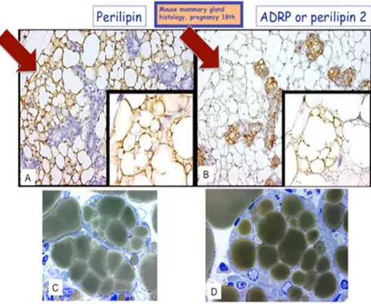
Figure 13. Immunohistochemical analysis Plin1-Plin2 of mouse mammary gland at 17 days of pregnancy. A)
Only adipocytes are Plin1 positive; B) only alveolar epithelial cells are Plin2 positive. A and B red arrows and magnification). During pregnancy, in some areas where adipocyte showed morphological aspect of adipoepithelial transdifferentiation, some structures with intermediate features between adipocytes and alveoli are also present. These compartmentalized adipocytes or early alveoli are marked for both proteins, see also C as an example of early alveolus and D as an example of compartmentalized adipocyte (stage of adipoepithelial conversion) [Morroni M. 2004]. C, D): High magnification of a resin embedded gland (allowing more detailed morphology than paraffin embedded tissue) comparable to that shown in A and B. Scale bar= (A, B) 70 mm (inset 23 mm); (C, D) 15 mm. Abbreviations: Plin 1, perilipin 1; Plin2, perilipin 2 [Prokesh A. 2014].
These cells are committed to the epithelial phenotype because the perialveolar adipocytes express the transcription factor ELF-5 (E74-like factor 5), highly specific for secreting epithelia (ductal and alveolar cells) and regarded as a master regulator of mammary alveolar differentiation [Lapinskas EJ. 2004; Oakes SR. 2008; Choi YS. 2009; Lee HJ. 2012]. Altought ELF-5 alone is not sufficient to change the adipocyte phenotype in culture, it is considered as one pioneering factor for the expression of mammary epithelial genes [Prokesh A. 2014], thanks to the fact that ectopic expression of ELF-5 even in virgin mice [Oakes SR. 2008] is able to induce an increase in the expression of WAP [Lee HJ. 2012] and K-18 in cultured mature adipocytes treated with an hormonal cocktail mimicking the hormone status during pregnancy [Zangani D. 1999] (Figure 14).
Figure 14: ImmunoHistoChemical analysis of ELF-5. IHC of mammary gland isolated from female mouse CD1
at 17th day of pregnancy: anti-ELF-5 antibody revealed ELF-5-positive nuclei in alveolar-glandular epithelial cells (lu in the lumen of glands) and in a group of small adipocytes nearby (some indicated by asterisks). Larger adipocytes (a) were negative as well as other cells in the tissue (see blue cores of capillaries) were negative. Transcription factor ELF-5 is considered a master regulator of alveologenesis. . Scale bar=12 mm. Abbreviations: ELF-5, ETS transcription factor 5 [Prokesh A. 2014].
Electron microscopy and gene expression data showed that adipoepithelial transdifferentiation occurs only in the ductal epithelium presence [Prokesh A. 2014]. In the same time, microarray studies, comparing the cleared fat pads [DeOme KB. 1959] and the controlateral one in virgin mice at different stages of pregnancy, have allowed to select a list of candidates potentially responsible for the induction of the trasdifferentiation process in the mammary gland during pregnancy [Prokesh A. 2014].
Transdifferentiation mechanisms could imply complex cell-cell and paracrine interactions between epithelial cells and adipocytes to form a distinctive microenvironment in the mammary gland during pregnancy [Howlett AR. 1993]. The normal mammary environmental is able to induce and direct cellular growth and differentiation, as well as tissue specific gene expression in the mammary gland [McCave EJ. 2010; Bussard KM. 2010]. During pregnancy the ductal epithelium can secrete paracrine factors that, by spreading among the adipocytes, are able to induce adipoepithelial transdifferentiation [Smorlesi A. 2012].
In order to shed more light on factors involved in adipoepithelial transdifferentiation, some important genes appear to be specific and crucial for study this phenomenon.
Adiponectin and Perilipin 1 as adipogenic markers.
Adiponectin (AdipoQ) expression is highly specific to both mouse and rat mature fat
cells. In cultured adipocytes, hormone-induced differentiation dramatically increases the level
lu lu lu lu a a a
*
*
*
*
*
*
of expression for AdipoQ whereas the expression of AdipoQ mRNA is significantly reduced in the adipose tissues from obese mice and humans [Hu E. 1996;Trayhurn P. 2007].
Perilipin 1 (Plin1). Perilipins are the only proteins known to associate exclusively with
intracellular lipid storage droplets, named for their location at the periphery of the droplets as determined by immunocytochemistry with light microscopy [Greenberg AS. 1991]. Plin 1 is a lipid droplet associated protein, highly expressed in mature adipocytes, also of lactating mammary gland, but not in milk lipid droplets of mammary alveolar epithelial cells that also contain intracellular stores of triacylglycerol. Levels of Plin 1 mRNA and protein begin to decrease at mid pregnancy, well before alveoli are significant cellular components of the mammary gland [Greenberg AS. 1991; Russell TD. 2007]. Plin 1 immunostaining is a tool for the identification of tissue adipocytes severely depleted of their triacylglycerol stores and thus without their characteristic spherical shape [Blanchette-Mackie EJ. 1995].
Krupple-like factor 4 (Kfl4), NANOG, Oct-3/4 and c-Myc as cellular reprogramming markers, they reside at the heart of the reprogramming process, stressing the potential risk of these new pluripotent cells [Ben-David U. & Benvenisty N. 2011]. They are expressed in adipose tissue and their expression pattern is variable during adipocytes differentiation [Rosen ED. & MacDougald OA. 2006].
KLF-4 is a protein of a large zinc-finger protein family that regulate cellular apoptosis,
proliferation and differentiation.
Oct-3/4 is a transcription factor mostly known through its involvement in the
inhibition self-renewal of undifferentiated embryonic stem cells [Ercan C. 2011]. E-Cadherin and Keratin 18 as epithelial markers.
E-Cadherin (E-Cadh) is an adhesion protein predominantly produced by epithelial
cells during embryonic stages [Daniel CW. 1995; Boussadia O. 2002]. E-Cadh is essential for the survival and function of lactating alveolar epithelial cells of the mammary gland [Daniel CW. 1995]. Deletion of E-Cadherin specifically affects terminal differentiation, alveoli fail to expand and histological sections reveal no fat droplets [Boussadia O. 2002].
Keratin 18 (K-18) is detected at all stages of mammary morphogenesis except during
lactation when it is weak or absent. K-18 is expressed in the postnatal mammary gland during lumen formation but decreases during mammary involution [Mikaelian I. 2006]. In mammary epithelial cells, K-18 maybe can be transcriptionally upregulated by ELF-5 [Choi YS. 2009]. ELF-5, GATA-3, β-Casein (or Casein 2) and WAP are ealy or late pinking markers. Synthesis of milk proteins is known to be highly up-regulated in pregnancy and differing profiles for the various milk proteins have been documented.