UNIVERSITÀ POLITECNICA DELLE MARCHE
Dottorato in Biologia ed Ecologia marina
Study of reproductive biology of European hake
(Merluccius merluccius) in Adriatic Sea
Phd student: Michela Candelma
Supervisor:
Prof. Oliana Carnevali
Co-Tutors:
Prof. Finn-Arne Weltzien Dr. Alberto Santojanni Dr. Sabrina Colella
Alla mia famiglia, che mi ha sempre sostenuto e incoraggiato ad andare avanti
Table of contents
1. Chapter I: Introduction………..1
1.1 Area of the study……….3
1.2 Biology of European hake……….………..3
2. Chapter II: Aim of the study………10
3. Chapter III: Gonadotropin characterization, localization and expression in European hake (Merluccius merluccius)………...13
4. Chapter IV: Reproductive biology of European hake (Merluccius merluccius): a multidisciplinary approach………...41
5. Chapter V: Reproductive studies of male European hake (Merluccius merluccius) in the Adriatic Sea………74
1
CHAPTER 1
________________________________________________________________________________
2
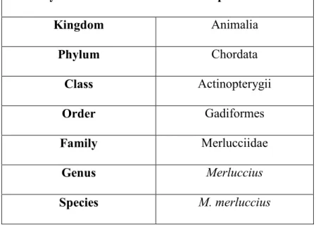
The European hake (Merluccius merluccius) is a nectobenthonic species widespread in the Eastern coast of Atlantic Ocean and in Mediterranean Sea. European hake, called in Italian “nasello” or improperly “merluzzo”, belongs to Gadiformes Order and to Merlucciidae Family, that includes 13 species spread in worldwide.
Systematic classification of European hake
Kingdom Animalia Phylum Chordata Class Actinopterygii Order Gadiformes Family Merlucciidae Genus Merluccius Species M. merluccius
The European hake has very commercial importance, it is one of the main target species of demersal commercial fishes in the Mediterranean Sea with total landings of 22547 tons in 2011 (Druon et al. 2015) as well as in the Atlantic Ocean, where the stock is an important sustenance of the fisheries (Cerviño et al. 2013). Because of the intense fishing of this stock, FAO included it in the list of overfished species (FAO 2011).
Figure 1. One of the biggest European hake sampled during three years in Adriatic Sea. The total length was 52 cm.
3
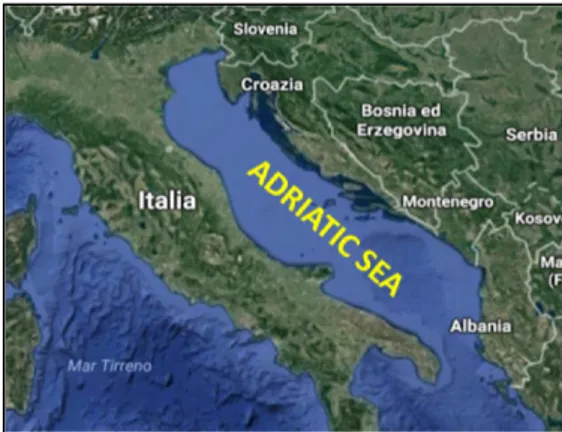
1.1 Area of the study
The European hake is equally distributed in Mediterranean Sea. While the Atlantic and Mediterranean population are considered as two distinct sub-species, the Mediterranean hake is considered a homogeneous stock, in fact the Adriatic stock was genetic coincident with North-western area (Orsi Relini et al. 2002; Lo Brutto et al. 1998). The Adriatic Sea is a semi-enclosed and shallow basin and the deeper areas are only the Jabuka/Pomo Pit and South Adriatic Pit with over 200 m of depth, while the rest of the sea is represented by
continental shelf (less 200 m deep). The our sampling area is 80-85 m deep and it is identified by the FAO- Geographical Sub-Area 17 (GSA17) between Northern and Central Adriatic Sea close to Ancona city. According to FAO (2016), also in Adriatic Sea the European hake stock status is in overexploitation with relative intermediate biomass. In this area, it represents an important fishing resource, shared by fisheries of several countries.
1.2 Biology of European hake
The body of hake is tapered with a grey-silver color.
The bathymetric distribution of hake in the Mediterranean Sea is between 25 to 1000 m depth. However, hake is mainly found at depths ranging from 100 to 400 m (Orsi Relini et al. 2002). The bathymetric distribution of European hake can change in relation to different physiological factors during life (feed availability, reproductive cycle, age, season) (Lloret et al. 2008).
The analysis of stomach contents evidenced that the diet characteristics of European hake change during life. Fishes smaller than 16 cm, feed crustaceans, instead with increase of their size, the
4
specimens prefer mainly teleosts (Sardina pilchardus, Sprattus sprattus and Engraulis encrasicolus) and rarely cephalopods (Vrgoč et al. 2004). Occasionally, also cannibalism event were confirmed for this species, when the density of the population is too high to give support to its conspecifics (Orsi Relini et al. 2002).
M. merluccius can live more than 20 years and it can reach 130 cm of total length (Jardas 1996). However, the size composition of exploited stock is constituted mainly of 0+, 1+ and 2+ year-old specimens (Vrgoč et al. 2004), represented by specimens shorter than 20 cm of total length (Zupanovic & Jardas 1986). The growth rate in hake varied with size and sex. Using tagging method and recapture techniques, Mellon-Duval and co-workers (2010) showed that from the second year of life, females grow faster than males and that hake became mature at the age of two years for both sexes, instead of 3 or 4 years as previously accepted. The length at which half population is sexual mature (L50) was estimated by different authors and it slightly changed based on the fishing areas and the sex. In fact, in Moroccan Atlantic coast it was 28.6 and 33.8 cm respectively for males and females respectively (El Habouz et al. 2011), in Iberian Atlantic waters 32.8 for males and 45.4 for females (Pineiro & Sainza 2003), in Alger 21.5 for males and 30.6 for females (Bouaziz et al. 1998), in both Catalan Sea and Tyrrhenian Sea was reported only female measure that was 35.8 and 35.1 respectively (Recasens et al. 2008). Finally, as reviewed by Vrgoc and co-warkers (2004), the length at which half population is sexual mature (L50) was estimated by different authors, in the forty years the range was 22-30 cm for males and 20-28 cm for females, subsequently in the eighty years the range of 20-28 cm for males and 23-33 cm for females is reported. In the low Adriatic areas Ungaro et al. (1993) found the same range of L50, 25-30 cm for both sexes.
European hake is a multiple spawner species with indeterminate fecundity, that spawns different times during reproductive season. Although the spawning females were found all year around (Murua & Motos 2006; Recasens et al. 2008; Recasens et al. 1998), the main reproductive season
5
was identified from Winter to Summer by many authors. In Bay of Biscay the main spawning season was intensified from January to March (Murua & Motos 2006; Lucio et al. 2000), in Galician Shelf in Winter and in Spring/Summer (Dominguez-Petit et al. 2010), in Tyrrhenian Sea from January to May, while in Catalan Sea from August to December (Recasens et al. 2008), in Eastern central Atlantic Ocean in Winter and in Spring with certain inter-annual variability (El Habouz et al. 2011), in Adriatic Sea, also, two spawning seasons were identified at different bathymetry, one in Winter in deeper waters and one in spring-summer in shallower waters (Zupanovic & Jardas 1986; Ungaro et al. 1993). M. merluccius spawns at 70-150 m of depth, at water temperature of 10°-13°C (Coombs & Mitchell 1982). The floating eggs of European hake are transparent, spherical with a single yellow oil globule (Bjelland & Skiftesvik 2006). As reported by Sánchez and co-workers (2012), that for the first time, provide information about spontaneous spawning of European hake, showed that eggs diameter was a range of 0.94–1.03 mm, while the total length of hatched larvae was about 3.0 mm. In rearing condition, the larvae started to feed 6 days after hatching and they approach to the surface (Bjelland & Skiftesvik 2006; Sánchez et al. 2012). In nature, after a first period of pelagic behavior of the larvae (estimated as 40 days post hatching), the animals have metamorphosis and move close to bottom sediments (Arneri & Morales-Nin 2000). The recruitment of young individuals to adult stock change based to reproductive period of the different area, for example in North-Western Mediterranean Sea and in Adriatic Sea is recorded in spring and autumn (Zupanovic & Jardas 1986; Arneri & Morales-Nin 2000; Orsi Relini et al. 2002), while in central Mediterranean Sea is recorded in late summer (Orsi Relini et al. 2002).
The economic importance of European hake is mainly due to the flesh quality (edible portion) that with low amount of fat, it is suitable for any type of human nutrition. Indeed, this species has the ability to accumulate the bulk of lipid (energy) especially in the liver (Lloret et al. 2008) while in
6
the muscle the lipid percentage is very low and the presence of perivisceral fat has not been found. Some authors studied whether the energy stored in the liver of hake is directly involved in reproduction, as typical for species temperate zones, or not. The results are different, while Lloret and co-workers (2008) reported that the energy reserves in the liver contribute to improve the estimation of the reproductive potential in this species, for Dominguez-Petit and co-workers (2010), the energy reserves stored in liver do not directly influence the egg production but rather the reproductive process can depend on environmental conditions and food availability.
Different fishing tool were used to catch the animals, the long-liners catch large adult female hake and trawlers and gill-netters targeted smaller size of specimens. In the Adriatic Sea, hake is mainly fished with bottom trawlers, while long-lines are less used (Vrgoč et al. 2004). High percentage of landings is represented by species under 20 cm of total length (Ungaro et al. 2003), impacting mainly juvenile hakes. Furthermore, the hake fishing has also negative effects on other species. As reviewed by Tsagarakis and co-workers (2014), in fact, such methods are not selective for hake and lead to produce a discard around 40% in several Mediterranean areas.
Despite the negative impact of fishing that led to a depletion of European hake stock in the world, the commercial aquaculture for this species has not been achieved. In the last years, the interest for this species as potential candidate for aquaculture production is increased, in fact its characteristics, as fast growth rate and the flesh quality which gives it a very high market value, contribute to the high potential value for aquaculture (Groison et al. 2010). Different authors tried to keep these animals in captivity. Bjelland and Skiftesvik (2006) caught live hakes for keeping in captivity but without success. However, the authors obtained eggs through stripping, providing advice about egg incubation, hatching and larvae growth. Afterwards, for the first time, Iglesias and co-workers (2010) bred wild hake for extended period, showing that it is possible to maintain hake specimens
7
in captivity. Only in 2012, Sánchez and co-workers obtained a spontaneous spawning from a hake broodstock kept in captivity for 2 years. Unfortunately, the larvae died during experiments and to date the complete hake life cycle in breeding was not achieved.
8
References
Arneri, E. & Morales-Nin, B., 2000. Aspects of the early life history of European hake from the central Adriatic. Journal of Fish Biology, 56(6), pp.1368–1380.
Bjelland, R.M. & Skiftesvik, A.B., 2006. Larval development in European hake (Merluccius merluccius L.) reared in a semi-intensive culture system. Aquaculture Research, 37(11), pp.1117–1129.
Bouaziz, A. et al., 1998. Reproduction du merlu Merluccius merluccius (Linnaeus, 1758) dans la région de Bou-Ismail. CIHEAM Cahiers Options Méditerranéennes, 35, pp.109–117. Lo Brutto, S. et al., 1998. Allozymic variation in Mediterranean hake Merluccius merluccius
(Gadidae). Italian Journal of Zoology, 65(SUPPL.), pp.49–52. Available at:
http://www.scopus.com/inward/record.url?eid=2-s2.0-0032461845&partnerID=tZOtx3y1. Cerviño, S. et al., 2013. Impact of egg production and stock structure on MSY reference points and
its management implications for southern hake (Merluccius merluccius). Fisheries Research, 138, pp.168–178.
Coombs, S.H. & Mitchell, C.E., 1982. The development rate of eggs and larvae of the hake,
Merluccius merluccius (L.) and their distribution to the west of the British Isles. ICES Journal of Marine Science, 40(2), pp.119–126. Available at:
http://icesjms.oxfordjournals.org/cgi/doi/10.1093/icesjms/40.2.119 [Accessed December 14, 2016].
Dominguez-Petit, R., Saborido-Rey, F. & Medina, I., 2010. Changes of proximate composition, energy storage and condition of European hake (Merluccius merluccius, L. 1758) through the spawning season. Fisheries Research, 104(1–3), pp.73–82.
FAO, 2016. Fisheries and Aquaculture Report. General Fisheries Commission for the Mediterranean. FAO, 890(890), pp.1–5.
FAO, 2011. Review of the state of world marine fishery resources. FAO Fisheries and Aquaculture Technical Paper, 569, p.334.
Groison, A.L. et al., 2010. Sperm motility in European hake, Merluccius merluccius, and
characterization of its spermatozoa concentration and volume, spermatocrit, osmolality and pH. Aquaculture, 301(1–4), pp.31–36. Available at:
http://dx.doi.org/10.1016/j.aquaculture.2010.01.026.
El Habouz, H. et al., 2011. Maturity and batch fecundity of the European hake (Merluccius merluccius, Linnaeus, 1758) in the eastern central Atlantic. Scientia Marina, 75(3), pp.447– 454. Available at:
http://scientiamarina.revistas.csic.es/index.php/scientiamarina/article/view/1268/1340. Iglesias, J. et al., 2010. Capture, transport and acclimatization to captivity of European hake,
Merluccius merluccius L: Preliminary data on feeding and growth. Aquaculture Research, 41(4), pp.607–609.
Jardas, I., 1996. Jadranska ihtiofauna, Školska knjiga.
Lloret, J., Demestre, M. & Sanchez-Pardo, J., 2008. Lipid (energy) reserves of European hake (Merluccius merluccius) in the north-western Mediterranean. Vie et Milieu-Life and Environment, 58(1), pp.75–85.
Lucio, P., Murua, H. & Santurtun, M., 2000. Growth and reproduction of hake (Merluccius merluccius) in the Bay of Biscay during the period 1996-1997. Ozeanografika, (3), pp.325– 354.
9
Mellon-Duval, C. et al., 2010. Growth of European hake (Merluccius merluccius) in the Gulf of Lions based on conventional tagging. ICES Journal of Marine Science: Journal du Conseil, 67(1), pp.62–70. Available at: http://icesjms.oxfordjournals.org/content/67/1/62.abstract. Murua, H. & Motos, L., 2006. Reproductive strategy and spawning activity of the European hake
Merluccius merluccius (L.) in the Bay of Biscay. Journal of Fish Biology, 69(5), pp.1288– 1303.
Orsi Relini, L. et al., 2002. Distribution of the Mediterranean hake populations (Merluccius merluccius smiridus Rafinesque, 1810) (Osteichthyes : Gadiformes) based on six years monitoring by trawl-surveys: some implications for management. Scientia Marina, 66, pp.21– 38.
Pineiro, C. & Sainza, M., 2003. Age estimation , growth and maturity of the European hake ( Merluccius merluccius ( Linnaeus , 1758 )) from Iberian Atlantic waters. ICES Journal of Marine Science, 60(3), pp.1086–1102.
Recasens, L. et al., 1998. Spatiotemporal variation in the population structure of the European hake in the NW Mediterranean. Journal of Fish Biology, 53, pp.387–401.
Recasens, L., Chiericoni, V. & Belcari, P., 2008. Spawning pattern and batch fecundity of the European hake (Merluccius merluccius (Linnaeus, 1758)) in the western Mediterranean. Scientia Marina, 72(4), pp.721–732.
Sánchez, F.J. et al., 2012. The first spontaneous spawning of European hake Merluccius merluccius L.: Characteristics of eggs and early larval stages. Aquaculture Research, 43(11), pp.1729– 1733.
Tsagarakis, K., Palialexis, A. & Vassilopoulou, V., 2014. Mediterranean fishery discards: review of the existing knowledge. ICES Journal of Marine Science, 71(5), pp.1219–1234.
Ungaro, N., Mannini, P. & Vrgoc, N., 2003. The biology and stock assessment of Merluccius merluccius (L.) in the Adriatic Sea: an historical review by geographical. Acta Adriatica, 44(1), pp.9–20.
Ungaro, N., Rizzi, E. & Marano, G., 1993. Note sulla biologia e pesca di Merluccius Merluccius (L.) nell’Adriatico Pugliese. Biologia Marina, suppl. al Notiziario S.I.B.M, 1, pp.329–334. Vrgoč, N. et al., 2004. Review of current knowledge on shared demersal stocks of the Adriatic Sea, Zupanovic, S. & Jardas, I., 1986. A contribution to the study of biology and population dynamics of
10
CHAPTER II
________________________________________________________________________________
11
Since the critic status of hake stock in the world and the commercial importance of this species, the protection of the population is a fundamental importance. To preserve the species and to improve fishing management, it is necessary increase the knowledge about the habits of the European hake and its stock conditions. For this purpose, the aim of present doctoral project was to assess the current state of the stock of European hake (Merluccius merluccius, L. 1758) in Adriatic Sea and overall to increase the knowledge about its reproductive biology. In addition, toxicological investigations were performed on the hake males to analyze the presence of endocrine disruptors in the environment.
In order to realize the project, a multidisciplinary study was carried out using macroscopic, histological and molecular approaches.
The macroscopic analysis consisted of sex determination and the measure of different body parameters of fish, total body length, the total and gutted weight and the liver and gonad weight of European hake in Adriatic Sea. Such information allowed to calculate the trend of morphometric indices, the Le Cren’s condition factor (Kn), the hepatosomatic index (HSI), the gonadosomatic index (GSI) in three years. Also, the size at first maturity (L50) was evaluated in males and in females. Furthermore, for the first time in Adriatic Sea, the fecundity, estimated as batch and relative fecundity, were analyzed in the female specimens and compared with values of other fishing areas in Mediterranean Sea and in Atlantic Ocean.
In addition to macroscopic analysis, also the histological analysis of the ovaries and testis were performed to validate the macroscopic scale that we used.
The molecular approaches were important tool to complete the scenario of the reproductive physiology of European hake. In fact, though the European hake is relevant commercial species, few studies focused on its reproductive physiology. So, the work was focused on studies regarding
12
the gonadotropin hormones, both the follicle stimulating hormone (Fsh), and the luteinizing hormone, (Lh).
In addition, also their relative receptors were analyzed. The sequences of such hormones and the receptors were sequenced and characterized. For Fsh and Lh, the mRNA expression were measured in pituitary gland in female specimens at different ovarian stages during natural reproductive cycle and their molecular localization was investigated in pituitary gland. Regarding the gonadotropin hormone receptors, their molecular levels were analyzed in oocytes at different maturity stages.
Finally, toxicological studies were performed through the analysis of molecular expression of vitellogenin A and B and estrogen receptor alpha in liver of males captured in the last three years.
13
CHAPTER III
________________________________________________________________________________
GONADOTROPIN CHARACTERIZATION,
LOCALIZATION AND EXPRESSION IN
14
Gonadotropin characterization, localization and expression in European
hake (Merluccius merluccius)
Michela Candelma¹, Romain Fontaine², Sabrina Colella³, Alberto Santojanni³, Finn-Arne Weltzien² and Oliana Carnevali¹
¹ Laboratory of Developmental and Reproductive Biology, DiSVA, Università Politecnica delle Marche, Ancona, Italy.
² Department of Basic Sciences and Aquatic Medicine, Norwegian University of Life Sciences, Oslo, Norway.
³ CNR-National Research Council of Italy, ISMAR -Marine Sciences Institute, Ancona, Italy. Corresponding author: Oliana Carnevali: [email protected]
ORCID ID Oliana Carnevali: 0000-0001-5994-0572
Manuscript published in Reproduction. DOI: http://doi.org/10.1530/REP-16-0377
Abstract
In vertebrates, the regulation of gametogenesis is under the control of gonadotropins (Gth), follicle-stimulating hormone (Fsh) and luteinizing hormone (Lh). In fish, the physiological role of Gths is not fully understood, especially in species with asynchronous ovarian development. In order to elucidate the role of Gths in species with asynchronous ovary, we studied European hake
15
(Merluccius merluccius) during the reproductive season. For this aim, we first cloned and sequenced both hormones. Then, we characterized their amino acid sequence and performed phylogenetic analyses to verify the relationship to their orthologues in other species. In addition, the quantification of gene expression during their natural reproductive season was analyzed in wild-caught female hake. Our results revealed that fshb peaked during the vitellogenic phase, remaining high until spawning. This is in contrast to the situation in species with synchronous ovary. lhb, on the other hand, peaked during maturation as is common also in species with synchronous ovarian development. Finally, combining double-labeling fluorescent in situ hybridization (FISH) for Gth mRNAs with immunofluorescence for Lh protein, we evidenced the specific expression of fshb and lhb in different cells within the proximal pars distalis (PPD) of the pituitary. In addition to gonadotrope cells specific to expression of either fshb or lhb, some cells showed co-expression of both genes. This suggests either that gonadotropes with co-expression are not yet specified, or they could have a plasticity that permit changes from one cell phenotype to another during certain life stages and in turn during different physiological states.
Keywords: Gonadotropin, European hake, Fsh, Lh, Fluorescence in situ hybridization, Immunofluorescence.
Introduction
The pituitary gonadotropins, follicle-stimulating hormone (Fsh) and luteinizing hormone (Lh), are hormones directly involved in regulation of gametogenesis in vertebrates (Nagahama 1994; Swanson et al. 2003; Weltzien et al. 2004). Both gonadotropins are heterodimeric glycoproteins and consist of a common α subunit shared with thyroid-stimulating hormone (Tsh) and a hormone specific β subunit important for their biological specificity.
16
Fsh and Lh are secreted by gonadotropes present mainly in proximal pars distalis (PPD) and some cells of pars intermedia (PI) of the pituitary gland in teleosts (Weltzien et al. 2003a). In mammals, the gonadotrope cells produce both gonadotropins (Nakane 1970) and rarely was found an overlapped expression. On the contrary, in teleosts, Fsh and Lh are synthesized by two different cell types (Zohar et al. 2010) and the colocalization of the two mRNAs in the same gonadotrope cell was only occasionally evidenced (García Hernández et al. 2002; Weltzien et al. 2014).
Because of the variety of reproductive strategies in different teleost species, the physiological roles of the two gonadotropins during the ovarian cycle do not seem to be the same for all species. Following Tyler and Sumpter (1996), reproductive strategies can be differentiated on the basis of the dynamics of ovarian development: ovaries can be classified as synchronous or asynchronous, even though such division could be reductive due to the wide number of reproductive strategies. The species with synchronous ovaries spawn eggs once within the reproductive season. These species are characterized by increasing plasma levels of Fsh with ovarian development (Tyler et al. 1997), while Lh surges during final maturation and spawning (Planas and Swanson 1995; Gomez et al. 1999; Yoshiura et al. 1999; Schmitz et al. 2005).
The regulatory mechanism of gonadotropins during gametogenesis is not so clear when we consider the teleosts with asynchronous ovaries, i.e. the species that spawn multiple batches of oocytes during the reproductive season. For instance in chub mackerel (Scomber japonicus), fshb mRNA levels increase during the first stages of ovarian cycle, peaking at the end of vitellogenesis, whereas lhb mRNA levels significantly increase during late vitellogenesis. Finally, the mRNA levels for both hormones significantly decrease during post-spawning (Nyuji et al. 2012). Also in other multiple batch spawners as Sparus aurata (Gen et al. 2000), Dicentrarchus labrax (Mateos et al. 2003), Hippoglossus hippoglossus (Weltzien et al. 2003b), Anguilla japonica (Han et al. 2003) and Paralichthys olivaceus (Kajimura et al. 2001) both hormones show the same fluctuation during gonadal cycle. Unlike these results, in the Atlantic cod (Gadus morhua) - a multiple batch spawner -
17
two peaks of fshb were observed, the highest before spawning and the second during spawning time together with lhb (Mittelholzer et al. 2009).
For this purpose the current study was aimed to better understand the functions of Fsh and Lh in the reproduction of European hake, a species with asynchronous ovarian development characterized by different phases of maturation (Sarano 1986; Murua et al. 1998). In the present study, the endocrine control of ovarian maturation of European hake was approached for the first time, by studying gonadotropin molecular characterization, expression and localization in the pituitary gland throughout the seasonal reproductive cycle in wild specimens captured in the Adriatic Sea.
Materials and methods
Monthly Sampling
During 2015, a total number of 129 European hake females were collected on board of bottom trawler fishing vessel in the Northern and Central Adriatic Sea (FAO- Geographical Sub-Area 17, according to GFCM division). The specimens were collected under the guidelines of the Data Collection Framework Regulation (EU Reg.199/2008) that established a Community system for the conservation and sustainable exploitation of fisheries resources under the Common Fisheries Policy (CFP). The procedures did not include animal experimentation and ethics approval are not necessary in accordance with the Italian legislation. We considered fish animals in a length range of
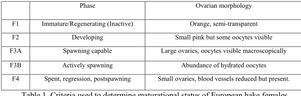
28-35 cm, that were macroscopically and microscopically classified in five different ovarian classes: immature/regenerating (F1); developing (F2); spawning capable (F3A); actively spawning (F3B), and spent, post spawning (F4) (Table 1), following the ovarian classification of Brown-Peterson et al. (2011), but adapted for European hake. The brief post-spawning phase make difficult the sampling of class F4 females, consequently only classes F1, F2, F3A, and F3B are included in this work. For the molecular analysis, we considered the animals sampled when the highest values
18
of gonadosomatic indices (GSI) were recorded during the year. The pituitary glands for qPCR analyses were immediately preserved in RNAlater (Ambion, USA) and stored at -20°C until further analysis. Pituitary glands for fluorescent in situ hybridization (FISH) and immunofluorescence techniques were fixed in 4% Paraformaldehyde (PFA) in PBST (PBS with 0,1% Tween-20, pH=7,4)and then dehydrated in increasing concentrations of ethanol before being preserved in pure methanol at -20°C until further processing. Body and gonad weights were recorded to calculate gonadosomatic indices (GSI): GSI (%) = gonad weight *100/gutted weight. Pieces of ovarian tissue from females collected in different maturity stages were fixed in 4% PFA for histological processing to confirm macroscopic classification of gonad development.
Phase Ovarian morphology F1 Immature/Regenerating (Inactive) Orange, semi-transparent F2 Developing Small pink but some oocytes visible F3A Spawning capable Large ovaries, oocytes visible macroscopically F3B Actively spawning Abundance of hydrated oocytes
F4 Spent, regression, postspawning Small ovaries, blood vessels reduced but present.
Table 1. Criteria used to determine maturational status of European hake females.
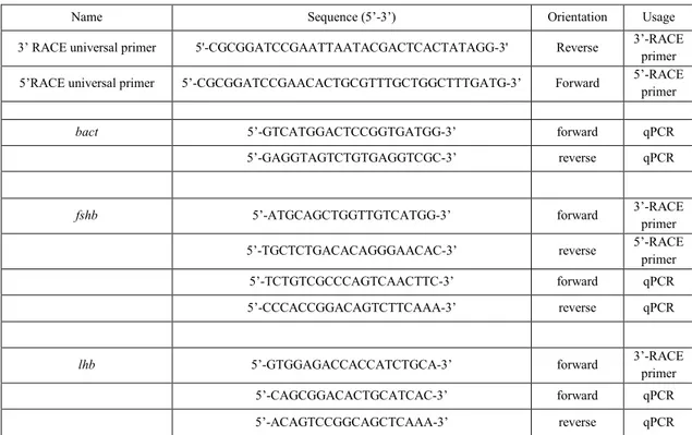
RNA extraction, cloning and sequencing of cDNA for European hake fshb and lhb
Total RNA was extracted from twenty pituitary glands using TRIzol Reagent, following the manufacturer’s protocol (Invitrogen Life Technologies, Milan, Italy). One total RNA was used for cDNA synthesis, employing iScript cDNA Synthesis Kit (Bio-Rad laboratories, USA). mRNA sequences for fshb and lhb were amplified using primers designed by Primer3 (http://bioinfo.ut.ee/primer3/) after alignment (ClustalW2 software, at EMBL-EBI, Cambridge, UK) with sequences from other teleost species to localize well conserved regions (Table 2). PCR amplifications were run using annealing temperatures between 52-62°C and 1 min extension time on an iCycler Thermocycler (Bio-Rad, San Diego, California, USA). Each gene product was cloned into the pGEM T Easy vector system (Promega, Milan, Italy) in accordance with the manufacturer’s
19
protocol. From the obtained partial sequences, 5’-RACE and 3’-RACE (Rapid Amplification of cDNA Ends) primers were designed (Table 2). When a single band was obtained following RACE PCR, it was isolated and cloned. All the cloned inserts were sequenced by BMR Genomics of Padova (Italy).
Name Sequence (5’-3’) Orientation Usage
3’ RACE universal primer 5'-CGCGGATCCGAATTAATACGACTCACTATAGG-3' Reverse 3’-RACE primer 5’RACE universal primer 5’-CGCGGATCCGAACACTGCGTTTGCTGGCTTTGATG-3’ Forward 5’-RACE primer
bact 5’-GTCATGGACTCCGGTGATGG-3’ forward qPCR
5’-GAGGTAGTCTGTGAGGTCGC-3’ reverse qPCR
fshb 5’-ATGCAGCTGGTTGTCATGG-3’ forward 3’-RACE primer
5’-TGCTCTGACACAGGGAACAC-3’ reverse 5’-RACE primer 5’-TCTGTCGCCCAGTCAACTTC-3’ forward qPCR 5’-CCCACCGGACAGTCTTCAAA-3’ reverse qPCR
lhb 5’-GTGGAGACCACCATCTGCA-3’ forward 3’-RACE primer 5’-CAGCGGACACTGCATCAC-3’ forward qPCR 5’-ACAGTCCGGCAGCTCAAA-3’ reverse qPCR
Table 2. Primer sequences of European hake used for 5’- and 3’-RACE and qRT-PCR. RACE, rapid amplification of cDNA ends; bact, β-actina; fshb, follicle-stimulating hormone beta subunit; lhb, luteinizing hormone beta subunit
Protein prediction, identification and multiple alignments
Primary structures were predicted and characterized by web-based bioinformatics tools such as
Expasy Translate (http://web.expasy.org/translate/), ORF Finder (http://www.ncbi.nlm.nih.gov/projects/gorf/), the BLAST suite (http://blast.ncbi.nlm.nih.gov/Blast.cgi). The open reading frames were aligned with Clustal Omega
v. 1.2.1 (Sievers et al. 2011 - http://www.ebi.ac.uk/Tools/msa/clustalo/) from the Multiple Sequence Alignment tools provided by the EBI website. Gonadotropins were aligned with MUSCLE v. 3.8
20
(Edgar 2004 - http://www.ebi.ac.uk/Tools/msa/muscle/). The printing and shading of the alignment
files was obtained with BOXSHADE v. 3.21 (http://www.ch.embnet.org/software/BOX_form.html), with a consensus threshold set at 0.5 to be
represented by symbols.
Real time PCR
qPCRs were performed with the SYBR green method in an iQ5 iCycler thermal cycler (Bio-Rad). Triplicate PCRs were carried out for each sample analyzed following Santangeli et al. (2016). β-actin (bact) was used as housekeeping gene to standardize the results by eliminating variation in mRNA and cDNA quantity. As shown in Raingeard et al. (2009), the bact was chosen like reference gene because the mRNA levels were close to those of our target genes and the levels did not vary through different stages of reproduction. No amplification product was observed in non-template controls and no primer-dimer formations were observed in the control samples. The data obtained were analyzed using the iQ5 optical system software version 2.0 (Bio-Rad) including GeneEx Macro iQ5 Conversion and Genex Macro iQ5 files.
Fluorescence In Situ Hybridization (FISH)
RNA probes for European hake fshb and lhb were synthesized using pGEM-T easy vector (Promega, Madison, WI). Antisense and sense RNA probes were labeled with digoxigenin-11-UTP for fshb and fluorescein-12-UTP for lhb (Roche Diagnostics, Indianapolis, USA). Probes were purified using Nucleospin RNA clean-UP kit (Macherey-Nagel, Hoerdt, France). Regarding the tissue preparation, the pituitary glands were separated from brain. After the samples were rehydrated by a descending series of ethanol solutions (75%, 50% and 25% ethanol), they were placed in 3% agarose and cut (120 μm sagittal sections) with a VT1000S Leica vibratome. FISH was performed according to Fontaine et al. (2013). Briefly, tissues were permeabilized by 45
21
minutes treatment with proteinase K (10 μg/ml; P6556, Sigma) and prehybridized in hybridization buffer for 4 hours at 65ºC. Hybridization was performed at 65ºC for 18 h in hybridization buffer containing a mixture of probes (150 ng/ml each). Samples were washed with wash buffer composed of 25 ml formamide, 12,5 ml SSC 20x, 50 μL Tween20, 770 μL of 1 M Citric acid and water at different concentrations (50% formamide/50% 2x SSC [saline sodium citrate buffer]; 2x SSC; 0.2x SSC; PBST), treated for 30 minutes with 3% H2O2 to inactivate endogenous peroxidases. They
were then incubated overnight at 4°C with antibody. For digoxigenin-labeled fshb probe, an anti-digoxigenin-peroxidase conjugated antibody (Roche Diagnostics) and a Cy5-conjugated tyramide (Perkin Elmer) were used. Fluorescein-labeled lhb probe was recognized by an antiperoxidase conjugated antibody (Roche Diagnostics) and revealed by a homemade fluorescein-conjugated tyramide. After extensive washes, samples were mounted between slide and coverslip in Vectashield H-1000 Mounting Medium (Vector, Eurobio/Abcys). The sense probes were used for negative control (data not shown).
Combined FISH and immunofluorescence
In order to identify the phenotype of lhb-expressing cells in the hake pituitary gland, a triple labeling protocol was achieved by combining two-color FISH and immunofluorescence. After FISH, the sections were incubated for 2 hours at room temperature in a blocking solution (normal goat serum 4%, dimethylsulfoxide 1%, Triton 0.3% in PBST) and subsequently incubated over night at 4ºC with a rabbit antibody (1/1000) directed against the β subunit of medaka Lh (Oryzias
latipes). The secondary antibody used was a goat antirabbit IgG coupled to DyLight 549 (Jackson
ImmunoResearch Europe Ltd, Newmarket, Suffolk, England). Negative control for the immunofluorescence was verified in section of different part of pituitary gland (data not shown).
22
Fluorescent images were acquired using a Zeiss LSM 710 laser scanning confocal microscope. For fluorescent tyramides used in two-color FISH and for fluorophore-coupled secondary antibodies used in immunofluorescence studies, lasers with a wavelength of 488 (FITC; Alexa-488), 555 (DyLight 549), and 633 (Cy5; Alexa-633) nm, respectively, were used. Channels were acquired sequentially to avoid cross talk between the different filters. The focal planes were recorded using Zeiss ZEN 2009 software. Z-projections were obtained using Image J software distributed by Fiji (Schindelin et al. 2012). Composites were assembled using Adobe Illustrator CC (Adobe Systems, San Francisco, California).
Ovarian histological processing
Female gonads were maintained in 4% of PFA for fixation overnight and stored in 70% ethanol until further processing. Analysis of ovarian stages were conducted on histological paraffin sections (6-7 μm thickness) stained by Harris Hematoxylin and Eosin method following Caputo et al. (2001) and examined by Zeiss Axioscop (Oberkochen, Germany) light microscopy with phase contrast optics at a 200x magnification.
Phylogenetic analysis
Alignments and similarity matrices were calculated using Basic Local Alignment Search Tool (BLAST). The structure of deduced amino acid sequences of cloned fragments was predicted by comparison with known structures. Phylogenetic analyses of deduced amino acid sequences of Fshb and Lhb for European hake and other fishes were performed by MEGA version 6.0 (Tamura et al. 2013) using maximum-likelihood and default settings. A rooted consensus phylogenetic tree was generated by means of the Neighbor-Joining algorithm, using as outgroup the glycoprotein hormone
23
beta subunit-related protein of Drosophila melanogaster for Fsh and Lh. The robustness of the nodes was carried out by a bootstrap analysis from thousand data set replicates.
Statistical analysis
Results were expressed as the mean ± s.d. Statistical differences were determined using one-way ANOVA, followed by Bonferroni's multiple comparison test. All statistical analyses were performed using Prism 6 (GraphPad Software, San Diego, CA, USA). P-values <0.05 were considered to be significant. Exclusively for GSI data, P-values <0.1 were considered significant and the results were expressed as the mean ± SEM.
Results Molecular characterization of gonadotropin beta subunits
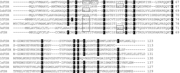
The European hake cDNA encoding the fshb consisted of 550 nucleotides with an open reading frame (ORF) of 372 nt and with 5’ and 3’ untranslated regions (UTRs) of 108 and 70 nucleotides, respectively (Accession number of Genbank, KX377614). The consensus polyadenylation signal (AATAAA) was 15 nt upstream to the poly(A) tail. The similarity of fshb nucleotide sequence of European hake to the sequences of other fishes was in a range from 65% to 85%. The deduced amino acid sequence of Fshβ was 123 amino acid long. In silico analysis of the mature peptide showed a signal peptide of 18 amino acid, two N-linked glycosylation sites (NFT) and 12 conserved cysteine residues (fig. 1). A fragment of lhb of 509 nt was amplified, cloned and sequenced (Accession number of Genbank, KX377615). The nucleotide fragment showed high homology with
lhb of other teleosts in a range from 68% to 82%. The partial deduced amino acid sequence was 89
aa long and although partial, we recognized some conserved clusters, like 9 cysteine residues highly conserved.
24
(A)
EhFSH ---MQLVVMAAVL-AMTVGGQGCRFVCRPVNFTINVT--SCDKHRSIVTTICEGQCYQMDPIYK--LYRPQQKTC 67 AcFSH ---MQLVVMAAVL-AMTWADQPCSFTCRPTPTTIAVK--SCVRTESINTTMCEGQCYQEDPMDP--GERPQQYTC 67 EsbFSH ---MQLVVMVAVL-ALARAGQGCSFGCHPTNISIQVE--SCGLTEVIYTTICEGQCYHEDLVYLSHYERPEQRIC 69 AhFSH ---MQLVVMATVLAAVAGAGQGCSFDCRPTNVRIPVE--SCGSTEYIDTTVCAGQCYNKDPVYISKEGPDKQNSC 70 ZfFSH ---MRMRVLVLALLLPVLMSAESECRCSCRLTNISITVESEECGSCVTIDTTACAGLCWTMDRVYPSSMAQHTQKVC 74 CsFSH MYCTHLMTLQLVVMAMLWVTPVRAGTECRYGCRLNNMTIIVEREDCHGSITI--TTCAGLCETTDLNYQSTWLPRSQGVC 78 JeFSH ---MHLAVTALCLTL---APVLARASTSCGLANISISVENEECGGCITFNTTACAGLCFTQDSVYKSSLKSYPQQAC 71 hFSH ---MKSLQFCFLF---CCWKAICCNSCELTNITITVEKEECNFCISINTTWCAGYCYTRDLVYKDPARPNIQKTC 69 EhFSH N-GDWSYETKHFEDCPVGF----SYPVARSCNCAMCQGGNTQCEMFLDEVPTCHPLANLIQ--- 123
AcFSH S-GDWAYEVKHFEGCLEGV----LYPVARSCKCSLCQSSNTDCERVLWDVC--- 113 EsbFSH N-GDWSYEVKHIKGCPVGV----TYPVARNCECTTCNTENTDCGRFPGDIPSCLSF--- 120 AhFSH N-GDWSYEVKHINGCPVAV----TYPVARHCHCSICNLDDTDCSPFPGDIPGCLTTLHSLSLSTLD 131 ZfFSH NFKNLMYKSYEFKGCPAGVDSVFVYPVALSCECNQVNSDTTDWGAISPQTTSCSIH--- 130 CsFSH NFKEWSYEKVYLEGCPSGVEPFF-IPVAKSCDCIKCKTDNTDCDRISMATPSCIVNPLEM--- 137 JeFSH NFRDVVYETVHLPGCPSGMDLHFTYPVALSCECSKCNTDSTDCGPLNTEVSGCLTH--- 127 hFSH TFKELVYETVKVPGCAHHADSLYTYPVATECHCGKCDSDSTDCTVRGLGPSYCSFSEMKE--- 129
(B)
EhLH ---VETTICSGHCITMDPVMVAPR 21 AcLH --MASSSFLRLLP-LLSVALG---ALAP--LGAAYQLPLCQLVNQTVSVEKKGCPGCHPVETTICSGHCITMDPSRVPPR 72 EsbLH --MAVQASRVMFPLVLSLFLGATSSIWPLATAEAFQLPPCQLINQTVSLEKEGCPKCHPVETTICSGHCITKDPVIKIPF 78 AhLH METEQISVRVKLPLTLIFF---LSSMWPLAPAVAFQLPKCQLIKQMVSLEKEGCPKCHTVETTICSGHCNTKDPVIKIPF 77 ZfLH --MLLAGNGV----FFL---FSLFFLLAAAQSLVFPRCELVNETVSVEKEGCPKCLVFQTTICSGHCVTRDPVYKSPF 69 CsLH --MLGLHVGT----LIS---LFLCILLEPVEGSLMQPCQPINQTVSLEKEGCPTCLVIQTPICSGHCVTKEPVFKSPF 69 JeLH -MSVYPECTW----LLF---VCLCHLLVSAGGSLLLPCEPINETNSVEKDGCPKCLVFQTSICSGHCITKDPSYKGPL 70 hLH MEMLQG---L--LLLLLLSMG---GAW---ASREPLRPWCHPINAILAVEKEGCPVCITVNTTICAGYCPTMMRVLQAVL 69 EhLH L-KVFKKVCTYRELQYRLFELPDCPPGVDPVVQYPA-LSCSCSHCAMATSDCTVDSLQPDYCTSQTLNYY---- 89 AcLH LSKVVQKVCTYQELQYRPLELPGCGPGVDPVVHYPAALSCSCSRCSMETSDCTVESLPPDFCTSTSLNYY---- 142 EsbLH -SNVYQHVCTYRNSHYKTFELPDCPPGVDPTVTYPVAQSCHCGRCAMDTSDCTFESLQPNFCMNDIPFYY---- 147 AhLH -LNVYQHVCTYQELYYKTFELPDCPPGVDPTVSYPVAVSCYCGRCALNTSDCTFESLQPDFCMNDIPFYD---- 146 ZfLH -STVHQTVCTYRDVRYETINLPDCSAGVDPQITYPVALSCDCSLCTINTSDCTIQSLQPDFCMSQREDFSAY-- 140 CsLH -STVYQHVCTYRDVRYETIRLPDCPPWVDPHVTYPVALSCDCSLCNMDTSDCTIESLQPDFCITQRVLTDGDMW 142 JeLH -STVYQRVCTYRDVRYETVRLPDCRPGVDPHVTFPVALSCDCNLCTMDTSDCAIQSLRPDFCMSQRASLPA--- 140 hLH -PPLPQVVCTYRDVRFESIRLPGCPRGVDPVVSFPVALSCRCGPCRRSTSDCGGPKDHPLTCDHPQLSGLLFL- 141
Figure 1. Amino acid sequence alignments of beta subunits of follicle-stimulating hormone, Fshb (A) and luteinizing hormone, Lhb (B) of European hake and other fishes. The cysteine residues are highlighted and potential N-glycosylation sites are shaded. The sequences were extracted from GenBank databases and their abbreviation and accession numbers are: Merluccius merluccius (Eh KX377614, KX377615), Gadus morhua (Ac: ABD62883, ABD62884), Dicentrarchus labrax (Es: AAN40506, AAN40507), Danio rerio (Zf: AAV31152, AAV31153), Anguilla japonica (Je: Q9YGK3, BAD14302), Oncorhynchus keta (Cs: AAA49408, AAA49409), Hippoglossus hippoglossus (Ah: CAD10501, CAD10502) and human (P01228, P01229).
25
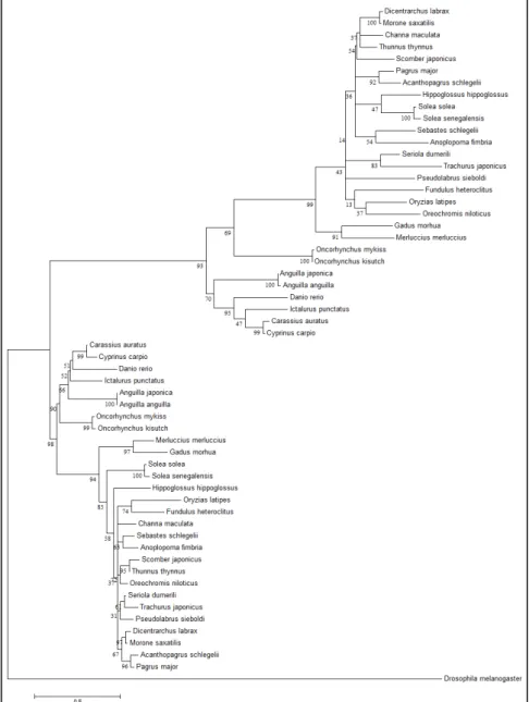
Phylogenetic analysis of gonadotropinsPhylogenetic analyses showed that the vertebrate Gths were divided into Fsh and Lh clusters in the phylogenetic tree rooted using the glycoprotein hormone beta subunit-related protein of D.
melanogaster as outgroup (fig. 2). The percentage of trees in which the associated taxa clustered
together is shown below the branches. Both gonadotropins of M. merluccius are closely associated to gonadotropins of G. morhua (fig. 2).
Ovarian histology and gonadosomatic index (GSI) variation
Figure 2. Phylogenetic comparison of fish full-length follicle-stimulating hormone (Fshb) and luteinizing hormone (Lhb) amino acid sequences. The analysis was performed by MEGA6 using maximum likelihood and default settings. A rooted consensus phylogenetic tree generated by means of the Neighbor-Joining algorithm, glycoprotein hormone beta subunit-related protein of D. melanogaster as outgroup. Bootstrap values from 1000 replicates are indicated for each tree node.
26
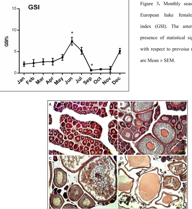
The analysis of GSI of hake females through the year evidenced a significant increase (P<0.1) in June compared to precedent months (fig. 3), suggesting the summer season as reproductive period for this species in Adriatic Sea. While, the GSI peak in December was not significant compared to other months. The histology confirmed the asynchronous development of oocytes for this species (fig. 4), meaning that the ovaries exhibit multiple oocyte stages (fig. 4b,c,d).
Figure 3. Monthly seasonal variation of European hake females gonadosomatic index (GSI). The asterisks indicate the presence of statistical significance (P<0.1) with respect to prevoius month. The values are Mean ± SEM.
Figure 4. Tissue sections of the ovarian follicles at different developmental stages of European hake females. (A) F1, immature/regenerating; (B) F2, developing; (C) F3A, spawning capable; (D) F3B, actively spawning. Scale bar = 50 μm for A, B, C; scale bar = 100 μm for (D). Different stages of ovarian cells were reported in figure. PO, Primary Oocytes (unyolked oocyte); Vtg1, Vitellogenin 1; Vtg2, Vitellogenin; Vtg3 Vitellogenin 3; Hydrated oocytes (present only in actively spawning individuals); POF (Post-ovulatory follicle).
27
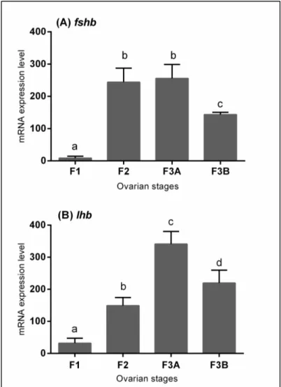
Gene expression profile analysisIn this study, the expression of fshb and lhb in the pituitary of females during oocyte maturation were examined by qPCR (fig. 5). Four stages were analyzed, from immature to actively spawning stages. The mRNA level of fshb was low in immature phase (F1) and raised in developing phase (F2) (P<0.05). It remained at an high level in spawning capable phase (F3A) and significantly decreased in the last phase considered (F3B) (P<0.05). The levels of lhb showed a significant increasing trend with a significant peak at beginning of maturation (F3A) (P<0.05). The lhb mRNA levels were declined during actively spawning stage (F3B) (P<0.05) as fhsb.
Localization of fshb and lhb mRNA and Lhb protein
Figure 5. Relative mRNA expression levels of (A) fhsb and (B) lhb in pituitary gland of European hake in specimens with different ovarian stages. Abundance of fshb and lhb transcript was determined by qRT-PCR and normalized with b-actin. The values are Mean ± SD.
28
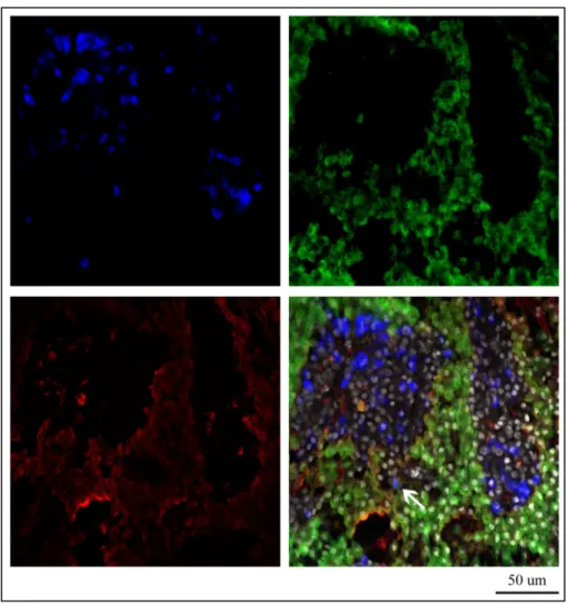
The double-labeling FISH and immunofluorescence techniques were coupled to detect the localization of fshb and lhb expression simultaneously and Lhβ protein on female adult pituitary tissues during reproductive cycle. Contrary to sense probes (data not shown), antisense probes revealed the expression of fshb and lhb mRNA in distinct cells in proximal pars distalis (PPD) and in the edge of pars intermedia (PI). fshb and lhb were expressed mostly in adjacent pituitary cells, but some cells showed co-expression of both hormones (fig. 6d). Comparing the gene expression of both gonadotropin β subunits with that of Lhb protein, we observed that the anti-Lhb serum merged completely with the cells positive to lhb mRNA expression. Also, anti-Lhb stained the cells expressing both hormones, in addition to few cells expressing only fshb (fig 7d). During the reproductive cycle the expression of gonadotropins mRNAs and Lhb protein was always detectable.
Figure 6. Differential expression of the fshb (A), lhb (B) transcripts and Lh protein (C) in adult European hake pituitary gland. The picture 6D shows the merging of three pics and the arrow evidences the fshb and
29
DiscussionThe characterization and expression studies of gonadotropin genes have been investigated in several teleost species. As already reported in the Introduction, the mRNA expression of gonadotropins during the reproductive cycle has not been well defined in teleosts. The main matter is definitely linked to the wide variety of reproductive strategies of different species. For this purpose the current study was aimed to better understand the functions of Fsh and Lh in the reproduction of European hake, a species with asynchronous ovarian development characterized by different phases of maturation (Sarano 1986; Murua et al. 1998). In the present study, the endocrine control of ovarian
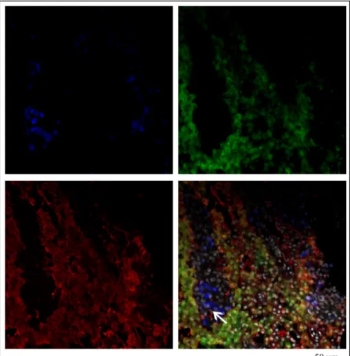
Figure 7. Differential expression of the fshb (A), lhb (B) transcripts and Lh protein (C) in adult European hake pituitary gland. The picture 7D shows the merging of three pics and the arrow evidences the presence of Lh protein in the fshb cells.
30
maturation of European hake was approached for the first time, by studying gonadotropin molecular characterization, expression and localization in the pituitary gland throughout the seasonal reproductive cycle in wild specimens captured in the Adriatic Sea.
The fshb and lhb nucleotide sequences were obtained by the RACE method. While the approach was successfully performed for fshb, same approach failed to amplify the 5’ terminal part of lhb, obtaining a partial sequence of 509 nt. The two nucleotide sequences were used to predict the relative amino acid sequences. Analyzing the Fshb deduced polypeptide, it had 63% sequence homology to another gadoid fish, Atlantic cod (Gadus morhua) and 35-59% identity range to other teleosts. It is notable that the N-glycosylation site conserved the position through teleosts, though in the most of fishes the specific consensus sequence of N-linked glycosylation consists in Asn-X-Ser whereas in hake was Asn-X-Thr as in chum salmon (Sekine et al. 1989) and in tetrapods. Also like tetrapods and ancient teleosts (Yoshiura et al. 1999; Quérat et al. 2001), the European hake showed two potential N-glycosylation sites in contrast to other modern teleosts. The presence of two sites ensures the biological potencies of the hormone and stabilize the interactions with its receptor (Bousfield et al. 1994). Also as tetrapods and ancient teleosts, European hake showed 12 cysteine residues associated with ‘seatbelt’ region. The same numbers of cysteine residues were found also in other teleosts, except for siluriformes and cypriniformes, that present an additional residue at the N-terminus, the Cys3 (according to the numbering in tetrapods). The lack of Cys3 could increase the stability on the tertiary structure of the heterodimer (Xing et al. 2004; Levavi-Sivan et al. 2010). Regarding the partial amino acid sequence of Lhb, it had 77% homology with Atlantic cod and 57-70% identity range to other fishes. We found nine of the expected twelve cysteine residues that are typical for teleosts, and their position was highly conserved. Together with Fshb protein, the Lhb amino acid sequence was used for the construction of the phylogenetic tree that was inferred from various teleost Fshb and Lhb protein sequences. Both hormones were distinctly separated in
31
independent order and both showed high similarity to respective homolog hormone genes of Atlantic cod, confirming that they belong to Gadiformes order.
The monthly trend of the mean GSI showed two different peaks, one in December and the highest in June, where the reproductive activity was concentrated. The increase of GSI, excluding the spawning period, confirmed that hake spawns continuously throughout the year (Murua and Motos 2006).
Considering the expression profiles of gonadotropin genes in the pituitary gland of European hake during their reproductive cycle, the novel finding is consistent with detectable levels of fshb along all the reproductive phases - from Immature/Regenerating to Actively Spawning period as classified by Brown-Peterson et al. (2011). Previous studies concerning the expression pattern of fshb during gonadal maturation suggested that the hormone is mainly involved in the first phases of the gonadal cycle and the temporal profile of mRNA expression was distinct from that of lhb, which was expressed especially during the last phases of reproduction (Planas and Swanson 1995; Tyler et al. 1997; Gomez et al. 1999; Yoshiura et al. 1999; Schmitz et al. 2005). Unlike species with synchronous ovarian development, the European hake displayed high expression of fshb during F2 and F3A phase, suggesting a continues role of this hormone due to the presence of different oocyte stages into the ovary at the same time. Indeed, while some oocytes are involved in final maturation and ovulation, other oocytes in the same ovary are still in vitellogenesis. A similar trend was detected in other multiple spawners (Gen et al. 2000; Kajimura et al. 2001; Mateos et al. 2003; Weltzien et al. 2003b). Remarking the typical reproductive pattern reported and consistent with other teleost species,the gene expression of lhb in the pituitary showed a significant peak in F3A as in all teleosts. The low fshb and lhb expression in actively spawning stage females (F3B) may be caused by a negative feedback exerted on the gonadotropic cells by the high levels of gonadal steroid hormones (Kajimura et al. 2001), though other authors (Asturiano et al. 2002) evidenced that changes in gonadal hormones could lead to the spermiation and ovulation.
32
Finally, the present study provided for the first time the information about the specificity of hormone production by gonadotropic cells by combining double-labeling fluorescent in situ hybridization of fshb and lhb with Lhβ protein immunofluorescence. The distribution of gonadotropic cells evidenced several differences with other teleosts. Differently to So et al. (2005) where the cells expressing fshb were arranged as small cell clusters or single cells and lhb like large clusters in zebrafish, there was no apparent difference in the distribution pattern between fshb- and
lhb-cells in European hake, indeed both gonadotrope cell types were arranged like large clusters
throughout PPD during all ovarian stages. Although it was difficult to quantitate accurately the numbers of the two types of gonadotropic cells, they seemed to be similar. In addition Cao et al. (2009) detected fshb cells in the middle area of PPD and lhb in the middle and in periphery of PPD and in PI, whereas in European hake the fshb and lhb cells were homogenously distributed through the whole PPD and in PI. Previous studies in Senegalese sole and Atlantic halibut (Cerdà et al. 2008; Weltzien et al. 2003b) evidenced mainly the fshb and lhb expression in distinct gonadotropes of the PPD, although the authors did not exclude the possible presence of the co-expression of both gonadotropins in a single gonadotropic cell. The use of confocal imaging associated with fluorescent labeling allowed us to establish that a limited number of cells co-express both hormones. It is still unclear whether the cells that produced both hormones have always presented such ability or, instead, if this is an acquired function during reproductive cycle. From previous results, Golan et al. (2014) found few cells that showed co-localization of Fshb and Lhb proteins in tilapia and zebrafish, hypothesizing that these gonadotropes are bipotent cells that subsequently undergo the differentiation into either fshb- or lhb- cells. In addition to these hypotheses, we suggest that the gonadotropes could have a plasticity that permit changes during lifetime and in turn during different physiological states. The presence of Lhb protein merged mainly with lhb-cells and also in cells expressing both hormones. Surprisingly, it was also recorded in a few fshb cells. Since we could not investigate the presence of Fshb protein due to the lack of suitable antibodies, it remains
33
difficult to clarify if the presence of Lhb protein in those few cells is the result of a changing of phenotype or those cells are not completely differentiated.
In conclusion, we have used the European hake, an important commercial species, like model to
elucidate the role of Fsh and Lh in species with asynchronous ovary. We report the sequences and molecular information on gonadotropin subunits in European hake. In addition, the specific expression of fshb and lhb in different cells in pituitary gland confirmed the gonadotropin localization similar to other teleost fish, but the additional small number of cells that showed co-expression could provide important bases for further investigations on fsh and lh cell differentiation.
Declaration of interest
The authors declare no conflict of interests.
Funding
This research did not receive any specific grant from any funding agency in the public, commercial or not-for-profit sector.
Acknowledgements
Michela Candelma had a grant from COST Office (Food and Agriculture COST Action FA1205: Assessing and improving the quality of aquatic animal gametes to enhance aquatic resources. The need to harmonize and standardize evolving methodologies, and improve transfer from academia to industry; AQUAGAMETE) for this project. The authors wish to thank the Dr. Filippo Domenichetti, and Dr. Camilla Croci of Marine Science Institute – CNR and the Captain Giordano
34
and crew of “Orizzonte” vessel for their support in sampling. Also we are grateful to Susann Burow from Weltzien’s laboratory for the use of medaka Lhb antibody in our work.
References
Asturiano JF, Sorbera L, Ramos J, Kime D, Carrillo M & Zanuy S 2002. Group-synchronous ovarian development, spawning and spermiation in the European sea bass (Dicentrarchus labrax L.) could be regulated by shifts in gonadal sterodiogenesis. Scientia Marina 66 273– 282.
Bousfield G, Perry WM & Ward DN 1994 Gonadotropins: chemistry and biosynthesis. In The Physiology of Reproduction, edn 5, pp. 1749−1792. Eds E Knobil and JD Neill. New York: Raven Press.
Brown-Peterson NJ, Wyanski DM, Saborido-Rey F, Macewicz BJ & Lowerre-Barbieri SK 2011 A Standardized Terminology for Describing Reproductive Development in Fishes. Marine and Coastal Fisheries 3 52–70.
Cao H, Zhou L, Zhang YZ, Wei QW, Chen XH & Gui JF 2009 Molecular characterization of Chinese sturgeon gonadotropins and cellular distribution in pituitaries of mature and immature individuals. Molecular and Cellular Endocrinology 303 34–42.
Caputo V, Candi G, Colella S & Arneri E 2001 Reproductive biology of turbot (Psetta maxima) and brill (Scophthalmus rhombus) (Teleostei, Pleuronectiformes) in the Adriatic Sea. Italian Journal of Zoology 68 107-113.
Cerdà J, Chauvigne F, Agulleiro MJ, Marin E, Halm S, Martinez-Rodriguez G & Prat F 2008 Molecular cloning of Senegalese sole (Solea senegalensis) follicle-stimulating hormone and luteinizing hormone subunits and expression pattern during spermatogenesis. General and
35
Comparative Endocrinology 156 470–481Edgar RC 2004 MUSCLE: a multiple sequence alignment method with reduced time and space complexity. BMC Bioinformatics 5 113.
Fontaine R, Affaticati P, Yamamoto K, Jolly C, Bureau C, Baloche, S., Gonnet F, Vernier P, Dufour S & Pasqualini, C. 2013 Dopamine Inhibits Reproduction in Female Zebrafish. Neuroendocrinology 154 807–818.
García Hernández PM, García Ayala A, Zandbergen MA & Agulleiro B 2002. Investigation into the duality of gonadotropic cells of Mediterranean yellowtail (Seriola dumerilii, Risso 1810): Immunocytochemical and ultrastructural studies. General and Comparative Endocrinology 128 25–35.
Gen K, Okuzawa K, Senthilkumaran B, Tanaka H, Moriyama S & Kagawa H. 2000 Unique expression of gonadotropin-I and -II subunit genes in male and female red seabream (Pagrus major) during sexual maturation. Biology of Reproduction, 63 308–19
Golan M, Biran J & Levavi-Sivan B. 2014 A novel model for development, organization, and function of gonadotropes in fish pituitary. Frontiers in Endocrinology 5 182.
Gomez JM, Weil C, Ollitrault M, Le Bail PY, Breton B & Le Gac F 1999 Growth hormone (GH) and gonadotropin subunit gene expression and pituitary and plasma changes during spermatogenesis and oogenesis in rainbow trout (Oncorhynchus mykiss). General and Comparative Endocrinology 113 413–428.
Han YS, Liao IC, Huang YS, Tzeng WN & Yu JYL 2003 Profiles of PGH-α, GTH I-β, and GTH II-β mRNA transcript levels at different ovarian stages in the wild female Japanese eel Anguilla japonica. General and Comparative Endocrinology 133 8–16.
36
subunits (GTH-Ibeta and -IIbeta) and their expression profiles during gametogenesis in the Japanese flounder (Paralichthys olivaceus). General and Comparative Endocrinology 122 117–129.
Levavi-Sivan B, Bogerd J, Mañanós EL, Gómez A & Lareyre JJ 2010 Perspectives on fish gonadotropins and their receptors. General and Comparative Endocrinology 165 412–437.
Mateos J, Mañanós E, Martinez-Rodriguez G, Carrillo M, Querat B & Zanuy S 2003 Molecular characterization of sea bass gonadotropin subunits (α, FSHβ, and LHβ) and their expression during the reproductive cycle. General and Comparative Endocrinology 133 216–232.
Mittelholzer C, Andersson E, Taranger GL, Karlsen Ø & Norberg, B. 2009. Quantification of gonadotropin subunits GPα, FSHβ, and LHβ mRNA expression from Atlantic cod (Gadus morhua) throughout a reproductive cycle. Comparative Biochemistry and Physiology - B Biochemistry and Molecular Biology 153 288–295.
Murua H & Motos L 2006 Reproductive strategy and spawning activity of the European hake Merluccius merluccius (L.) in the Bay of Biscay. Journal of Fish Biology 69 1288–1303.
Murua H, Motos L & Lucio P 1998 Reproductive modality and batch fecundity of the European hake (Merluccius merluccius L.) in the Bay of Biscay. California Cooperative Oceanic Fisheries Investigations Reports 39 196–203.
Nagahama Y 1994 Endocrine regulation of gametogenesis in fish. International Journal of Developmental Biology 38 217–229.
Nakane PK 1970 Classifications of anterior pituitary cell types with immunoenzyme histochemistry. The Journal of Histochemistry and Cytochemistry 18 9–20.
Nyuji M, Selvaraj S, Kitano H, Ohga H, Yoneda M, Shimizu A, Kaneko K, Yamaguchi A & Matsuyama M 2012 Changes in the expression of pituitary gonadotropin subunits during
37
reproductive cycle of multiple spawning female chub mackerel Scomber japonicus. Fish Physiology and Biochemistry 38 883–897.
Planas JV & Swanson P 1995 Maturation-associated changes in the response of the salmon testis to the steroidogenic actions of gonadotropins (GTH I and GTH II) in vitro. Biology of Reproduction 52 697–704.
Quérat B, Tonnerre-Doncarli C, Géniès F & Salmon C 2001 Duality of gonadotropins in gnathostomes. General and Comparative Endocrinology 124 308–314.
Raingeard D, Bilbao E, Sáez-Morquecho C, de Cerio OD, Orbea A, Cancio I & Cajaraville MP 2009 Cloning and transcription of nuclear receptors and other toxicologically relevant genes, and exposure biomarkers in European hake (Merluccius merluccius) after the Prestige oil spill. Marine Genomics 2 201–213.
Santangeli S, Maradonna F, Gioacchini G, Cobellis G, Piccinetti CC, Dalla Valle L & Carnevali O 2016 BPA-Induced Deregulation Of Epigenetic Patterns: Effects On Female Zebrafish Reproduction. Scientific Reports 6 21982-21982.
Sarano F 1986 Ovarian cycle of the hake, Merluccius merluccius, as a partial spawner fish. Revue Des Travaux de l’Institut Des Pêches Maritimes 48 65–76.
Schindelin J, Arganda-Carreras I, Frise E, Kaynig V, Longair M, Pietzsch T, Preibisch S, Rueden C, Saalfeld S, Schmid B, Tinevez JY, White DJ, Hartenstein V, Eliceiri K, Tomancak P & Cardona A 2012 Fiji: an open-source platform for biological-image analysis. Nature Methods 9 Schmitz M, Aroua S, Vidal B, Le Belle N, Elie P & Dufour S 2005 Differential regulation of
luteinizing hormone and follicle-stimulating hormone expression during ovarian development and under sexual steroid feedback in the European eel. Neuroendocrinology 81 107–119.
38
Analysis of Chum Salmon Gonadotropin cDNAs. Proceedings of the National Academy of Sciences of the United States of America 86 8645–8649.
Sievers F, Wilm A, Dineen D, Gibson TJ, Karplus K, Li W, Lopez R, McWilliam H, Remmert M, Söding J, Thompson JD & Higgins DG 2011 Fast, scalable generation of high-quality protein multiple sequence alignments using Clustal Omega. Molecular Systems Biology 7 539.
So W, Kwok H & Ge W 2005 Zebrafish Gonadotropins and Their Receptors: II. Cloning and Characterization of Zebrafish Follicle-Stimulating Hormone and Luteinizing Hormone Subunits. Their Spatial-Temporal Expression Patterns and Receptor Specificity. Biology of Reproduction 72 1382–1396.
Swanson P, Dickey JT & Campbell B 2003 Biochemistry and physiology of fish gonadotropins. Fish Physiology and Biochemistry 28 53–59.
Tamura K, Stecher G, Peterson D, Filipski A & Kumar S 2013 MEGA6: Molecular evolutionary genetics analysis version 6.0. Molecular Biology and Evolution 30 2725–2729.
Tyler CR & Sumpter JP 1996. Oocyte growth and development in teleosts. Reviews in Fish Biology and Fisheries 6 287–318.
Tyler C, Pottinger T & Coward K 1997 Salmonid follicle-stimulating hormone (GtH I) mediates vitellogenic development of oocytes in the rainbow trout, Oncorhynchus mykiss. Biology of Reproduction 57 1238–1244.
Weltzien F-A, Norberg B, Helvik JV, Andersen Ø, Swanson P & Andersson E 2003a Identification and localization of eight distinct hormone-producing cell types in the pituitary of male Atlantic halibut (Hippoglossus hippoglossus L.). Comparative Biochemistry and Physiology - A Molecular and Integrative Physiology 134 315–327.
39
characterization and expression of FSH beta, LH beta, and common alpha-subunit in male Atlantic halibut (Hippoglossus hippoglossus). General and Comparative Endocrinology 131 87–96.
Weltzien F-A, Andersson E, Andersen Ø, Shalchian-Tabrizi K & Norberg, B 2004 The brain-pituitary-gonad axis in male teleosts, with special emphasis on flatfish (Pleuronectiformes). Comparative Biochemistry and Physiology - A Molecular and Integrative Physiology 137 447– 477.
Weltzien F-A, Hildahl J, Hodne K, Okubo K & Haug TM 2014 Embryonic development of gonadotrope cells and gonadotropic hormones - Lessons from model fish. Molecular and Cellular Endocrinology 385 18–27.
Yoshiura Y, Suetake H & Aida K 1999. Duality of gonadotropin in a primitive teleost, Japanese eel (Anguilla japonica). General and Comparative Endocrinology 114 121–131.
Zohar Y, Munoz-Cueto JA, Elizur A & Kah O 2010 Neuroendocrinology of reproduction in teleost fish. General and Comparative Endocrinology 165 438–455.
Xing Y, Myers RV, Cao D, Lin W, Jiang M, Bernard MP, Moyle WR 2004. Glycoprotein hormone assembly in the endoplasmic reticulum: III. The seatbelt and its latch site determine the assembly pathway. Journal of Biological Chemistry 279 35449–35457.
Gene bank accession numbers of species used for phylogenetic analysis
Merluccius merluccius (KX377614;KX377615); Gadus morhua (ABD62883; ABD62884); Dicentrarchus labrax (AAN40506; AAN40507); Acanthopagrus schlegelii (ADX31689; ADX31690); Pagrus major (BAB18563; BAB18564); Channa maculata (AAS01610; AAS01609); Seriola dumerili (BAR79709; BAR79710); Hippoglossus hippoglossus (CAD10501; CAD10502);
40
Scomber japonicus (AEN14604; AEN14605); Thunnus thynnus (ABP04057; ABP04050); Sebastes schlegelii (AAU14141; AAU14142); Anoplopoma fimbria (AGS55583; AGS55584); Acanthopagrus schlegelii (AAX18926; ABQ96864); Solea solea (ABW81403; AHZ13200); Oryzias latipes (BAK61761; BAK61762); Oreochromis niloticus (AAP49575; AAP49576); Pseudolabrus sieboldi (BAF81900; BAF81901); Trachurus japonicus (AGO59024; AGO59025); Oncorhynchus mykiss (BAB17686; BAB17687); Oncorhynchus kisutch (AAO72299; AAO72300); Carassius auratus (BAA13530; BAA13531); Anguilla japonica (Q9YGK3; BAD14302); Fundulus heteroclitus (P30971; P30972); Cyprinus carpio (CAA42542; CAA42543); Danio rerio (AAV31152; AAV31153); Ictalurus punctatus (AAG32155; AAG32156); Morone saxatilis (AAC38035; AAC38019); Drosophila melanogaster (AAM53262).
41
CHAPTER IV
________________________________________________________________________________
REPRODUCTIVE BIOLOGY OF EUROPEAN HAKE
(MERLUCCIUS MERLUCCIUS): A MULTIDISCIPLINARY
APPROACH
42
Reproductive biology of European hake (Merluccius merluccius): a multidisciplinary approach
Michela Candelma1, Sabrina Colella2, Luisa Dalla Valle3, Alberto Santojanni2, Daniela Bertotto4, Giuseppe Radaelli4, Oliana Carnevali1
1 Laboratory of Developmental and Reproductive Biology, DiSVA, Università Politecnica delle Marche, Ancona, Italy
2 CNR-National Research Council of Italy, ISMAR-Marine Sciences Institute, Ancona, Italy
3 Department of Biology, University of Padua, 35131 Padua, Italy
4 Department of Experimental Veterinary Sciences, Faculty of Veterinary Medicine, University of Padua, Italy
Corresponding author: Oliana Carnevali, email: [email protected], telephone number: 00390712204990
ORCID ID Oliana Carnevali: 0000-0001-5994-0572
Submitted paper
Abstract
The fecundity estimation in fish and the study of the size at first maturity are important factors involved in the evaluation of reproductive potential of the stock. In addition, studies regarding the spawning season of a species, the reproductive physiology and the physiological indicators involved in the energy state are other fundamental tools that allow to improve the knowledge of the state of