I want to dedicate this work to my family who always supported me. I would like to thank all my friends who always have a smile that makes things easier.
Finally I really would like to thank Professor Franco Giorgi and Professor Roberto Maggio
that tutoring me along these years. Thanks to everyone
UNIVERSITY of PISA
BIOS-Research Doctorate School in Biomolecular Sciences
Research Doctorate Course in Molecular Biotechnologies XXI cycle 2006-2008
RESEARCH PROJECT:
EFFECT OF DDT AND ANALOGUES ON THE ACTIVITY OF GLYCOPROTEIN HORMONE RECEPTORS
Tutors:
Prof. Franco Giorgi Prof. Marcella Camici
President of the Course
in Molecular Biotechnologies: Candidate:
CONTENTS
ABSTRACT ….……… Pag. 4
INTRODUCTION ……….……… Pag. 7
MATERIALS AND METHODS ..……… Pag. 12
RESULTS ………..……… Pag. 19
DISCUSSION ……… Pag. 45
ABSTRACT
In the first part of this study, we aimed at establishing whether two previously identified thyroid disruptors, the insecticide 1,1,1-trichloro-2,2- bis(p-chlorophenyl)ethane (DDT) and Aroclor 1254 (a complex mixture of polychlorinated water), may inhibit the thyrotropin (TSH) receptor (TSHr) activity. DDT and Aroclor 1254 were shown to inhibit both the basal and bovine TSH (bTSH)-stimulated accumulation of cAMP in Chinese hamster ovary (CHO)-K1 cells stably transfected with the TSHr. Furthermore, both DDT and Aroclor 1254 did indeed prevent cAMP accumulation, as induced by the constitutive activity of a point mutant TSHr(A623I) transiently transfected in African green monkey kidney fibroblast COS-7 cells. Neither trypsin digestion of the extracellular domain (ECD) nor deletion of the ECD in a mutant TSHr trunk transiently transfected in COS-7 cells counteracted the inhibitory activity of DDT and Aroclor 1254. DDT exerted a weak inhibitory activity against forskolin in both CHO-K1 and COS-7 cells, whereas it was nil against the agonists dopamine and 5-(N-ethyl-carboxamido)-adenosine (NECA) in CHO cells stably transfected with the dopamine D1 receptor and in COS-7 cells transiently transfected with the adenosine type 2a receptor (A2a receptor). Furthermore, DDT was inactive against the stimulation by isoproterenol of the endogenously expressed β2 adrenergic receptor in COS-7 cells. Conversely, Aroclor 1254 inhibited completely forskolin activity in CHO-K1 cells, but not in COS-7 cells. Furthermore, it did not prevent accumulation of cAMP as induced by NECA in A2a transfected cells. We interpreted these results as indicating that DDT and possibly Aroclor 1254 may have an uncompetitive inverse agonist activity for the TSHr. The following experiments was performed to test whether other compounds that are structurally correlated to DDT may also behave as potential TSH receptor inhibitors.
Amongst the compounds structurally homologous to DDT, quercentin, kanferol and stirbestrol showed the strongest inhibitory effect on cAMP accumulation in TSH-stimulated CHO-TSHr transfected cells. Since several of these compounds, like DDT itself, have an estrogenic activity, it is likely that their cell binding sites may be physiologically relevant for estrogens. As a matter of fact, thyroid stimulating hormone
(TSH), luteinizing hormone (LH) and follicle-stimulating hormone (FHS) receptors are all members of the glycoprotein hormone receptor family and there are indications that estrogens can inhibit the activity of both LHr and/or FSHr. In line with this view, there are a number of functional experiments showing that DDT may inhibiting the activity of TSHr, LHr and FSHr. Furthermore, the estradiol can also inhibit the LHr activity at concentrations that are considered physiological in the in ovarian follicle, thus indicating that it might play a role in oocyte maturation. These experiments support the concept that DDT may act through an hypothetical “estrogenic binding site” conserved throughout evolution in the glycoprotein hormone receptor family.
To further deepen our knowledge on the effects induced by DDT, we examined how DDT alter the process of TSHr internalization in CHO-TSHr transfected cells. When these cells were probed immunocytochemically with anti-TSH receptor antibodies, the receptor appeared to be mostly expressed on the plasma membrane. By comparison, the overall antibody fluorescence became primarily restricted to a juxta-nuclear position when CHO-TSHr cells were exposed to [1 mU/ml] TSH for 1 or 5 min. However, in the presence of DDT, this fluorescence maintained a peripheral location along the cell plasma membrane, even when CHO-TSHr cells were simultaneously stimulated with TSH for 1 and 5 minutes. To verify the specificity of DDT on TSH receptor, CHO cells transfected with the A2a receptor were used as a control. Following
1 min stimulation with the agonist Neca, A2a receptors were gradually internalized
regardless of the presence of DDT in the culture medium, thus confirming the specificity of DDT for the TSHr.
In the last part of the project, we aimed at setting up an assay to perform intramolecular fluorescence resonance energy transfer (FRET) to studying directly the mechanism by which DDT and its analogues interact with the TSHr. To this purpose, a tetracystein motif acting as a binding site for the fluorescein Arsenical Hairpin (FlAsH) compound was inserted into the carboxyl terminal domain of the TSHr. FlAsH is known to become fluorescent only when bound to this tetracystein motif. Due to the small size of the FlAsH coumpound, this modified TSH receptor may prove more adequate for intramolecular FRET than other chromophores that are known to cause large steric
hindrance or even interact with the receptor function (GFP, YFP, etc). In this study the TSHr-FlAsH mutantion was initially characterized in several cell lines, and eventually the best results were obtained with HeLa cells. It is my intention to continue these experiments by trying to introduce the FlAsH motif in the third intracellular loop of the TSHr together with the cyan fluorescent protein at its carboxyl terminal. In principle, these modifications should allow us to perform an intramolecular FRET of the TSHr and hopefully to investigate in more detail the effect exerted by estradiol on the LH receptor.
INTRODUCTION
G-protein coupled receptors (GPCRs) constitute the largest family of seven-trans-membrane (7TM) receptors. Their evolutionary success is due to the extreme versatility by which they bind a variety of signaling molecules such as hormones and neurotransmitters (Lefkowits et al., 2004). Their ubiquitous distribution in the human body, along with the capacity to regulate virtually all known physiological processes has made this family of receptors the most important target for drug discovery (Bockaert et al., 1999).
In vertebrates, the pituitary control on the development and physiology of the thyroid and gonads is exerted through the secretion of three glycoprotein hormones: the thyroid-stimulating hormone (TSH), the follicle-stimulating hormone (FSH), and the luteinizing hormone (LH). These hormones bind to specific receptors, namely TSHR, FSHR and LHR, that are expressed in the thyroid gland and in the gonads, respectively. These receptors belong to the superfamily of cell-surface, G protein-coupled receptors. TSHR, FSHR and LHR receptors constitute the subfamily of glycoprotein hormone receptors (GpHRs) which differ from other members of the family for having a very large extracellular domain amounting to about half of the whole protein itself. This domain contributes to the ligand specificity by an appropriate lack of homology in these receptors (Kohn, et al., 1995; Simoni et al., 1997; Dufau, 1998).
Schematic representation of the structure of the glycoprotein hormone receptors and their genes. ECL and ICL indicate the extracellular and intracellular loops of the transmembrane domain (dotted portion of the protein). Exons of the genes are enumerated and the ‘extra intron’ of the LHR is indicated with an arrow.
These receptors have an extracellular domain composed of a cluster of nine leucine-rich repeats (LRRs) that is flanked by cysteine-rich sequences on both proximal and distal sides. The LRRs form a succession of beta-sheets and alpha-helices organized into a horseshoe-shaped structure. It has been proposed that this structure may act as a binding and recognition site for the glycoprotein hormones by assuming a concave surface during ligand interaction (Smits et al., 2003).
Vertebrate GpHRs express 11 conserved cysteine residues. The presence of disulfide bonds between these conserved cysteine residues is known to help maintaining the stability of the tertiary structure (Simoni et al., 1997; Dufau, 1998). The distal cysteine-rich sequence is the least conserved of all segments of GpHR proteins. While there are several potential N-linked glycosylation sites in the extracellular domain of GpHRs that are not strictly conserved, there is only one site in the leucine-rich repeat 6 that is conserved in all three receptors.
A percentage of G-protein coupled receptors may elicit a cell response in the absence of any ligand. This capacity to signal in the absence of a ligand is referred to as ligand-independent or constitutive-receptor activity (Costa and Cotecchia, 2005). Experimental evidence demonstrating the existence of this constitutive activity comes from studies on heterologous expression systems, where receptors are expressed with high densities or mutated in such a way as to make their effector pathways more efficiently activated. Certain ligands may decrease this basal effector activity for having an intrinsically negative efficacy or for producing responses opposite to those of the agonist. These ligands are termed negative antagonists or inverse agonists, that are, in turn, classifiable as either competitive or non-competitive (Costa and Cotecchia, 2005). Competitive inverse agonists are known to interact with the orthosteric sites of the receptor, while uncompetitive inverse agonists interact with allosteric sites of the receptor. One of major effects caused by administration of competitive or uncompetitive inverse agonists is the suppression of a natural agonist input. This is likely to have a minor negative effect, unless the receptor constitutive activity is so relevant that only antagonists with negative effect can extinguish its activity. Such cases include natural activating mutations (the list of which is presently increasing; see Birnbaumer, 1995;
Parnot et al., 2002), but also virally encoded G-protein coupled receptors (Rosenkilde et al., 2001; Smit et al., 2003).
TSHr has been found to be activated above its natural constitutive activity by a wide spectrum of gain-of-function mutations (Cetani et al., 1996). Germline TSHr mutations cause hereditary toxic thyroid hyperplasia, while a number of somatic mutations have been found to be responsible for the majority of autonomous thyroid adenomas (Tonacchera et al., 1998). Moreover, the TSH receptor may be activated by autoantibodies responsible of the hyperthyroidism (Greave’s disease), but also by the autoantibodies acting as TSH antagonists in hypothyroid patients (Hashimoto thyroiditis) (Davies et al., 2002). Many researchers have tried to isolate these autoantibodies that inhibit the thyrotropin receptor, but so far unsuccessfully.
One of the main hypotheses explaining the TSHr function assumes that the ectodomain might exert an inhibitory effect on a noisy rhodopsin-like serpentine domain. This hypothesis is supported by a number of early data showing that TSHr-expressing cells undergo partial activation of the receptor when mildly treated with trypsin (Van Sande et al., 1996). That such an effect may really occur was actually demonstrated by Zhang et al. (2000), who found that TSHr activation is secondary to “beheading” in N-terminal truncated mutants. Recently, Chen et al. (2003) have narrowed down the inhibitory effect of the ectodomain to its C terminus, indicating a cluster of lysine (Lys287, Lys290, Lys291) and arginine (Arg293) residues as possible targets for trypsin to exert its activation effect. The fact that little is known about the mechanisms governing glycoprotein hormone action is partly due to lack of pharmacological tools capable of interacting directly with these receptors. During a screening of several environmental factors that alter thyroid homeostasis, Santini et al. (2003) found that some of these compounds disrupt TSHr function and were consequently defined as thyroid disruptors. Aroclor 1254 (a complex mixture of polychlorinated biphenyls) and the insecticide 1,1,1-trichloro-2,2-bis(p-chlorophenyl)ethane (DDT) are two of the several compounds they tested that are thus further analyzed in this work. Both Aroclor 1254 and DDT were previously shown to prevent TSH-induced accumulation of cAMP in Chinese hamster ovary (CHO)-K1 cells
stably transfected with TSHr. However, in spite of this inhibitory effect, they did not interfere with 125I-bovine TSH (bTSH) binding to the receptor. These results suggested that Aroclor 1254 and DDT may inhibit TSHr function by interacting with receptor sites other than the TSH recognition site. In this first part of the study we aimed at verifying whether the inhibitory activity exerted by these thyroid disruptors on the thyroid homeostasis may occur via interaction with the TSH receptor.
On the long run, our aim is to use DDT as a lead compound to search for TSH receptor inhibitors potentially capable of acting as pharmaceutical drugs counteracting hyperthyroidism. According to this expectation, we tested several structural analogous of DDT in order to find compounds with higher potency. Among them we tested: rosmarinate, pelargonide, quercetin, kanferol, rutine, galangine, estradiol, T3 hormone, diphenylethylen and stirbestrol. So far Kanferol, quercetin and Stilbestrol proved capable of being more active at lower concentrations than DDT. Evidence supporting a role for quercetin as TSHr inhibitor comes from several epidemiological studies. Populations which are fed on a diet rich in onions, which contain a large amount of quercetin, tend to develop hypothyroidism.
From a chemical point of view, DDT is structurally correlated with the synthetic estrogen stilbestrol, as demonstrated by the observation that it has an estrogenic activity by its own. Moreover, the observation that DDT can inhibit the cAMP accumulation in CHO-LHr and CHO-FSHr cells stimulated by their respective agonist led to the hypothesis that DDT and its analogues might bind a conserved binding site shared by all these receptors. To test this hypothesis, the influence of estradiol was assayed directly on CHO cells transfected with the LHr. Our results showed that LHr was inhibitied by estradiol at a concentration- and time-dependent manner suggesting a possible physiological role of this interaction. (Wright et al., 2005 )
Additional experiments were carried to determine how DDT may affect the receptor localization and turn-over in TSHr transfected cells. To this aim, the TSH receptor was localized immunocytochemically in CHO cells using specific anti-TSHr antibodies. TSHr was found to vary in location depending on the time of DDT and/or
TSH exposures. By contrast, DDT was found not to affect internalization of the A2a receptor under the same experimental conditions.
Moreover, the TSH receptor was modified by introducing a short tetracystein sequence along its C-terminal domain (FlAsH binding motif) to test the feasibility of the FlAsH technique to detect the receptor. Through this procedure we were able to confirm the inhibitory effect exerted by DDT on the internalization of the TSHr in TSH-FlAsH receptor transiently transfected HeLa cells. Once transiently expressed in HeLa cells, this modified TSH receptor was also localized in vivo by dynamical confocal microscopy. This approach was set up in view of the possibility of performing intramolecular fluorescence resonance energy transfer (FRET) with the TSHr (Hoffmann et al., 2005). In fact, one of the major problems encountered with this technique is due to the actual dimension of the inserted chromophores, that can modify the protein so drastically to make it no longer functionally active. The FlAsH technique makes the intramolecular FRET more feasible by employing much smaller chromophores.
MATERIALS and METHODS
Materials. Tissue culture media and sera were from Sigma-Aldrich (St. Louis,
MO) and Celbio S.p.A. (Milan, Italy). [3H]Adenine was from PerkinElmer Life and Analytical Sciences (Boston, MA). Forskolin, Ro 20-1724, 3-isobutyl-1-methylxanthine (IBMX), 5’-(N-ethyl-carboxamido)-adenosine (NECA), 1,1,1-trichloro-2,2-bis(p-chlorophenyl)ethane (DDT) and 1,2-Ethanedithiol (EDT), human-recombinant-FSH (hrFSH), human-chorionic-gonadotropin (hCG)and the T3 Hormone were from Sigma-Aldrich. DDT analogues: rosmarinate, pelargonide, quercetin, kanferole, rutine, galangine, estradiol, diphenylethylen, stirbestrol were a kind gift from prof. Dibari (Department of Chemistry, University of Pisa). DDT and analogues were dissolved in 99.5% dimethylsulfoxide (DMSO) and further diluted in Dulbecco's Modified Eagle's Medium (DMEM). Drug concentrations were restricted to a maximum of 100 µM since higher concentrations resulted in the solvent, dimethylsulfoxide, to affect TSHr activity.
Plasmid DNA transfections were carried out using either the DEAE-dextran or the lypofectamine (Sigma-Aldrich, St. Louis, MO) procedures. Primary antibodies for the localization of the TSH receptor, either monoclonal or polyclonal, and the polyclonal anti-adenosin A2a receptor were purchased from Santa Cruz Biotechnology,
Inc. CA.
FlAsH kit for the detection of the FlAsH binding motif were from Invitrogen.
Eukaryotic Expression vectors. Wild type and truncated human TSH receptor
cDNA inserted into pcDNA3.1Zeo(-) vector were a kind gift from Dr. M. Szkudlinski (Trophogen, Inc. Rockville, MD). The truncated TSH receptor (TSHr-trunk) was modified by deletion of the entire extracellular domain until aminoacid E409, except for the 22 aminoacids of the signal peptide that were joined to E409. Both the wild type and the TSHr-trunk contain an HA-epitope inserted at the N-terminus adjacent to the signal peptide. The construction of wild type and truncated TSH receptors has been described by Zhang et al., (2000). The construction of the TSHr(A623I) receptor mutant has been described by Parma et al., (1993). The human adenosine A2a receptor cloned in a
for type V adenyl cyclase, subcloned into a pXMD1 vector, was kindly provided by Dr Zvi Vogel (Weizman Institute of Sciences, Israel).
Cell Culture. In this study severalcell types were employed to test the effect of DDT on the activity of different receptors: African green monkey kidney (COS)-7 cells, Chinese Hamster Ovary (CHO)-K1 cells and CHO-K1 cells stably transfected with the TSH, the FSH or the LH wild type receptor (CHO-TSHr, CHO-FSHr and CHO-LHr) and with the dopamine D1 (CHO-D1) or the adenosine A2a (CHO-A2a) receptor. COS-7
cells were transiently transfected with one of the following receptors: TSHr, TSHr-trunk, TSHr (A623I) or adenosine A2a. COS-7 and CHO-K1 cells were incubated at
37°C in a humidified atmosphere (containing 5% CO2) in Dulbecco’s modified Eagle’s
medium which was supplemented with 10% Fetal Bovine Serum, 100 units/ml penicillin, 100 µg/ml streptomycin, 2% L-glutamine and 1% non-essential aminoacids.
HeLa cells transiently- or CHO-K1 cells stably-transfected with the TSH receptor wilde type or the modified receptor (TSH-FlAsH receptor) were routinely employed in the second part of this study (Perret et al., 1990). Control cells were either CHO-K1 cells or CHO-K1 stably transfected with the adenosine A2a (CHO-A2a)
receptor. HeLa cells were incubated in 24-well plates (1.0 x10⁵ cells for each well) with a DMEM Mixture F12 HAM supplemented with glutamine. CHO-TSHr and CHO-A2a
cells were constantly maintained in the presence of the selecting agent G-418 (Sigma Aldrich, St. Louis, MO) at the concentration of 0.6 mg/ml of culture medium. HeLa-, CHO-TSHr and CHO-TSHr-FlAsH cells were seeded on round cover glass slips placed at the bottom of each well tray. By the end of each culture period, cover glass slips were mounted on glass slides using the Anti-fade kit (Invitrogen, Carlsbad, CA).
Construction of TSHr-FlAsH pcDNA 3.1 plasmid. The TSH-FlAsH receptor
was generated by inserting an exapeptide containing a tetracysteine motif (CCPGCC) along the C-terminal of the TSH receptor downstream of the leucine 764 and the resulting TSHr-FlAsH cDNA was subcloned in the pcDNA3.1Zeo vector between EcoRI and XhoI sites.
The TSHr-pcDNA3.1 plasmid was cut with the restriction enzymes EcoRI and BstEII to obtain a 2277 bp fragment and with Ecori and XhOII to obtain a 5935 bp fragment. A 177 PCR fragment was amplified from the TSHr in order to insert the tetracystein domain. These three fragments were joined together in order to construct the TSHr-FlAsH-pcDNA3.1(-) mutant bearing the tetracystein domain. Transfection was performed in E.coli by elettroporation and colonies were allowed to grow in ampicillyn selective LB agar. The right sequence was tested by restriction analysis with SmaI and by automated sequencing (Fig.19).
Stable transfection of TSH-FlAsH receptor into CHO-K1 cells. Exponentially growing cells were first treated with 1x trypsin , then seeded at 5 x 10⁵ cells in 10-cm Petri dishes and eventually incubated overnight in 10 ml of growth medium. The following day, 20 to 30 µg of plasmid DNA were mixed with 0.5 ml of 0.25 M CaCl2 plus 0.5 ml of 2x PBS, and allowed to stand for 10 to 20 min at room
temperature. One ml of this calcium phosphate-DNA solution was added drop wise to the Petri dish and gently swirled to attain an even distribution. Petri dishes were then incubated for 15 to 24 h at 37°C under 5% CO2. By the end of this incubation period,
medium was removed, cells rinsed twice with fresh growth medium and further incubated for 24 h at 37°C under 5% CO2. Cells were then seeded for a few additional
days into 96-well trays at the density of 1 cell per well in the presence of G418 medium (0,8 mg/ml). Clones were selected for the presence of the TSH-FlAsH receptor using the adenylyl cyclase assay (Johnson and Salomon, 1991).
Transient transfection of COS7 cell by the DEAE-Dextran method. Exponentially growing COS7 cells were trypsinized, seeded at 5 x 10⁵ cells in 10-cm Petri dishes and incubated overnight in 10 ml of growth medium. Then 2 to 3 µg of plasmid DNA were mixed with a 0,05% DEAE-Dextran (Sigma Aldrich, St. Louis, MO) solution in PBS, swirled gently and added dropwise to the culture for 30 minutes at 37 °C under 5% CO2. By the end of this period, the medium was removed and cells
rinsed twice with fresh medium and incubated again for 2 h in the presence of 80 µM chloroquine (Sigma Aldrich, St. Louis, MO). Cells were eventually re-fed by incubation in a new medium for 24 h at 37 °C under 5% CO2. The following day, cells were seeded
at the appropriate ratio (70 x 103 – 100 x 103) in 24-well Petri dishes and incubated for additional 24 h prior to being tested for adenylyl cyclase.
cAMP Accumulation. cAMP accumulation in intact cells was measured in
quadruplicate experiments through a two-step column separation procedure (Johnson and Salomon, 1991). The experimental protocol for this functional assay was as follows: CHO-K1, CHO-TSHr or CHO-A2a cells were preincubated for 2 h with 250 µ l
of medium supplemented with 1.25 µCi of [3H]adenine or/and other compounds to test at 37 °C in a humidified atmosphere. By the end of this preincubation period, cells were stimulated with their respective agonist or/and other compounds to test for 10 additional minutes at the indicated concentrations in the presence of the phosphodiesterase inhibitors Ro 20-1724 (0.5 mM) and IBMX (1 mM). Reaction was then blocked with perchloric acid containing 0.1 mM unlabeled cAMP, followed by neutralization with KOH. The amount of [3H]cAMP formed was determined as described by Johnson (1991) Dpm values of [3H]cAMP recovered from the columns were normalized to their respective protein contents, on average being equal to 50.3 ± 3.4 µg.
RIA cAMP. COS-7 cells transiently transfected with the TSHr or the
TSHr(A623I) receptor (and no AC5 plasmid), were seeded at the concentration of 25,000 cells/well into 96-well plates containing 50 µl of medium and used for the assay the following day. Cells were washed with Hanks’ balanced salt solution and incubated for 1 h at 37°C in hypotonic buffer. The cell lines were then exposed for 2 h to different concentrations of Aroclor 1254 and DDT. Assays were performed in triplicate for each experiment. Extracellular cAMP accumulation in the medium was determined by a in house RIA (Vitti et al., 1993), using a commercially available anti-cAMP antibody. Extracellular cAMP was measured because cell lysate was shown to interfere with RIA. Vitti et al., (1993) have shown that extracellular and intracellular cAMP equilibrate rather quickly when cells are allowed to swell in hypotonic buffer.
Adenylate cyclase assay on membranes. Confluent CHO-TSHr cells were
scraped off the plate in a hypotonic solution (Hepes 1 mM/EDTA 2 mM) containing a cocktail of protease inhibitors (1.5 µM pepstatin, 4 µM leupeptin, 0.01 M aprotinin and
were then resuspended in 50 mM HEPES/NaOH pH 7.4 plus the protease inhibitor cocktail and used in AC assay. AC activity was measured by monitoring the conversion of [α-32P] ATP to [α-32P] cAMP, using a previously reported method (Johnson and Salomon, 1991). Briefly, enzyme activity was routinely assayed in 100 µl reaction mixture containing 50 mM HEPES/NaOH, pH 7.4, 0.1 mM ATP, 0.1 mM cAMP, 1 mM DTT, 2 mM MgCl2, 0.1 mg/ml bacitracine, 0.5 mg/ml creatine phosphate, 0.1 mg/ml creatine phosphokinase, 1 mM EGTA, 0.5 mM Ro 20-1724 and 1 mM IBMX. (as phosphodiesterase inhibitors), 10 µM GTP, 1 µCi [α-32P] ATP and tested compounds. Incubation started by addition of CHO-TSHr membranes (20 µg proteins) and carried out for 10 min at 30°C. The reaction was terminated by placing assay tubes in an ice bath followed by addition of 0.5 ml stop solution containing 120 mM Zn(C2H3O2)2/[3H]cAMP (10,000 cpm/sample) and 0.5 ml of 144 mM Na2CO3. The total radiolabeled cAMP was purified on columns of Dowex 50 ion-exchange resin and alumina (Johnson and Salomon, 1991). In these experiments, we tested the ability of both compounds, DDT (1-100 µM) and Aroclor (1-100 µM), to modulate AC activity in the absence or in the presence of TSH (1 mU/ml). The compounds were dissolved in DMSO and diluted in Hepes buffer so that the final DMSO concentration never exceeded 1 % of the total volume.
Statistical analysis. ANOVA + Scheffe’s post hoc tests were employed to ascertain the significance of data belonging the same experimental group. In every experiment, the significance level was set at p < 0.05.
Transient trasfection of HeLa cell by the Lypofectamine method. HeLa cells
were cultured at a density of 7 x 10⁴ cells/well on 14 mm diameter round cover slips (sterilized and de-greased with 80% EtOH and absolute EtOH). The following day, cells were transfected with the lipofectamine solution and incubated for additional 24 h in 5% of CO2 at 37 °C. The lipofectamine solution was prepared by mixing 0.5 µ l
lipofectamine and 0.4 µg TSH-FlAsH plasmid in 250 ul DMEM mixture F12 HAM, supplemented with glutamine and non essential amino acids. The following day, the lipofectamine solution was replaced with fresh medium containing FBS and antibiotics and maintained until the FlAsH assay was performed.
FlAsH assay. HeLa cells were assayed for FlAsH following exposure to 1µM
FlAsH, 12,5 µM EDT and 0,05% DMSO in 1g/l glucose in PBS. Initially, 667 µ M FlAsH, 8,3 mM EDT and 33,3% DMSO were allowed to equilibrate for 15 min at room temperature in the darkness. Subsequently, this solution was diluted with 1g/l glucose/PBS to yield a final concentration of 1µM FlAsH, 12,5 µM EDT and 0,05% DMSO. Following 10 additional minutes of equilibration at room temperature, 300 µl of this mix were added to HeLa cells. Cells were further incubated for an additional 1h at 37 °C in 5% CO2 in the presence of either DMSO alone as in control cells, or DMSO
containing 100 µM DDT as in experimental HeLa cells. Cells were then washed three times with 300 µM EDT, 25 µM BAL wash buffer containing either 100 µM DDT or 1% DMSO. Following the last wash, TSH was eventually added and maintained in the culture medium for 10 minutes at room temperature (Hoffmann et al., 2005). The reaction was blocked by fixing HeLa cells for 1h with 1% glutaraldehyde, 2% paraformaldehydein cold (4 °C) phosphate buffer at pH 7.2. Following fixation, cover slips were transferred to cold phosphate buffer and eventually mounted on rectangular glass slides with 20 µ l of Anti fade solution. Mounted glass slides were sealed with nail polish, stored overnight at -20 °C and then analyzed by scanning confocal microscopy. To detect the tetracysteine-bound FlAsH, fixed HeLa cells were excited at 488 nm with an Argon laser and the resulting signal filtered by scanning confocal microscopy at 515-530 nm emission was evaluated with an image analyzer program.
Fluorescence immunocytochemistry. CHO-K1, CHO-TSHr and CHO-A2a
cells were cultured on glass cover slips at 37 °C in a humidified atmosphere (containing 5% CO2) in Dulbecco’s modified Eagle’s medium supplemented with 10% fetal bovine
serum, 100 units/ml penicillin, 100 µg/ml streptomycin, 2% L-glutamine and 1% non essential amino acids. On the third day of culture, the entire medium was substituted with fresh medium with no serum to enhance receptor expression. Following an additional day, cells were exposed for 100 minutes to DDT (Sigma Aldrich, St. Louis, MO) at the final concentration of 100 µM. Controls were run with CHO-K1 cells exposed to the same DDT concentration, with CHO-TSHr or CHO-A2a cells not exposed to DDT. In some experiments, both experimental and control cells were exposed for 30 minutes to the fluorescent probe Lysotracker (InVitrogen, Carlsbad, CA)
at the concentration of 50 nM to label acidified lysosomal or late endosomal vesicles. The TSH hormone was eventually added at the concentration of 0.01 U/ml and allowed to act on experimental and control cells for either 1 or 5 minutes. The agonist NECA was used under the same experimental conditions at the final concentration of 1µM. By the end of each incubation period, cells were washed with phosphate buffer saline (PBS) and fixed with 4% paraformaldehyde in 0.05 M phosphate buffer at pH 7.2. Cells were permeabilized with 0.05% Tween-20 and 0.5% Bovine Serum Albumin (BSA) in PBS and then incubated for 90 minutes at room temperature in a PBS solution containing either mouse anti-TSHr (125 µg/ml) diluted 1:20 or rabbit anti-A2a receptor
(200 µg/ml) diluted 1:150, respectively. Following incubation with primary antibodies, cells were washed three times with PBS containing 0.1% Tween-20, plus an additional time with PBS alone and incubated for 1h at room temperature with a secondary antibody FITC-labeled goat anti-mouse IgG or anti-rabbit IgG (InVitrogen, Carlsbad, CA). Cells incubated with secondary antibodies alone were used as negative controls. After the second incubation, cells were washed with PBS containing 0.1% Tween-20, and with PBS alone. After completion of all incubation steps, cells were extensively washed with H2O and finally mounted on glass slides with anti-fading agents. Staining
was assessed with a Zeiss fluorescence microscope equipped with a colour video camera (Axio Cam MRC, Milano Italy) controlled by an AxioVision software.
Laser scanning Fluorescence microscopy A few cell preparations were treated
and fixed as specified above and eventually observed by confocal microscopy. To this end a number of glass slides were first screened by conventional microscopy and then examined in a fluorescence microscope connected to a TCS 4D confocal scanning system (Leica Microsystems, Heidelberg, Germany), equipped with a Ar/Kr laser. Fluorescent signals were then detected at 488/568 nm emission wave length.
RESULTS
Effect of Aroclor 1254 and DDT on bTSH dependent and independent accumulation of cAMP in CHO-TSHr cells. In this first set of experiments we
characterized the effect of the Aroclor 1254 and DDT thyroid disruptors on bTSH dependent and independent accumulation of cAMP in CHO-TSHr cells. The dose response curve of bTSH in CHO-K1 cells stably transfected with human TSHr provided an EC50 value of 0.34 ± 0.015 mU/ml (Fig.1A).
We tested the effect of Aroclor 1254 and DDT on CHO-K1 cells stably transfected with human TSHr. According to Santini et al. (2003), these two compounds should prevent cAMP accumulation in CHO-TSHr cells when administered together with bTSH (1 mU/ml per 10 min). At 10 µM concentration, DDT already began to inhibit cAMP accumulation, while at 100 µM concentration the inhibition was 45.8%. In contrast, the effect of Aroclor 1254 was significant only at 100 µM with a 62.7% inhibition (Fig.1B). The IC50 of DDT against bTSH (1mU/ml) stimulated cAMP
accumulation was 17.5 ± 6.41 µM. The IC50 for Aroclor 1254 could not be determined
with accuracy because dimethylsulfoxide, a solvent employed to dissolve the drug, affected TSHr activity at concentrations higher than 100 µM (see Materials and Methods). The inhibitory effect did not result from alteration of cell viability, as demonstrated by the fact that full bTSH activity could be completely restored following Aroclor 1254 and DDT wash out (data not shown).
As we mentioned in the introduction, TSHr has a high level of constitutive activity when expressed in eukaryotic cells. In CHO-TSHr cells, phosphodiesterase inhibitors lead to a marked cAMP increase in the absence of bTSH (Fig.1C), but not in control CHO-K1 cells (insert in Fig.1C). Both Aroclor 1254 and DDT inhibited significantly the TSHr constitutive activity, starting from a minimum concentration of 30 µM (Fig.1C).
1E-3 0,01 0,1 1 10 100 0 4000 8000 12000 16000 20000
A
0,001 [ 3H ]c A M P ( d p m )/ 5 0 µ g o f p ro te in bTSH (mU/ml) CHO-TSHr 0,01 0,1 1 10 100 4000 6000 8000 10000 12000 14000 16000 * * * * * bTSH CHO-TSHrB
[ 3 H ]c A M P ( d p m )/ 5 0 µ g o f p ro te in Drug (µM) DDT Aroclor 1254 -- + + + + + + + 0 200 400 600 800 1000 1200 1400 1600 1800 2000 0 50 100 150 200 250 Ro 20 IBMX [ 3H ]c A M P ( d p m ) CHO-K1 CHO-TSHrC
RO 20-1724 + IBMX * * * * 100 30 10 --100 30 10 --Aroclor 1254 (DDT (µM)µM) [ 3 H ]c A M P ( d p m )/ 5 0 µ g o f p ro te inFig. 1 Dose response curve of bTSH and inhibitory activity of Aroclor 1254 and DDT on the
wild type TSHr stably expressed in CHO-K1 cells. In panel (A), increasing concentrations of bTSH were tested on CHO-TSHr cells in the presence of the phosphodiesterase inhibitors Ro 20-1724 and IBMX. Aroclor 1254 and DDT were tested in CHO-TSHr cells stimulated (B) and not (C) with bTSH (1 mU/ml). In (B) all samples were treated with the phosphodiesterase inhibitors Ro 20-1724 and IBMX, and the basal level of [3H]cAMP accumulation was 1314 ± 139 dpm/50 µg of protein. In (C) the phosphodiesterase inhibitors Ro 20-1724 and IBMX increase the level of cAMP about 7-fold respect to the cells not treated with these inhibitors. The insert in (C) shows that, in CHO-K1 cells, the basal level of cAMP does not change significantly in the presence of the phosphodiesterase inhibitors. All graphics are the mean ± S.E. of a representative of three experiments, each performed in quadruplicate. *Significantly different from bTSH alone (B) and Ro 20-1724 + IBMX (C).
Effect of Aroclor 1254 and DDT on forskolin-induced cAMP accumulation in CHO-TSHr and CHO-K1 cells. In order to test the possibility that this inhibitory effect
may be directly targeted to the TSHr and not to downstream effectors, we analysed the inhibitory effect exerted by both compounds against 10 µM forskolin in CHO-TSHr and CHO-K1 cells. Results showed that application of these compounds to CHO-TSHr (Fig.2A) and to CHO-K1 cells (Fig.2B) prevented the forskolin-induced cAMP accumulation. Under these conditions, DDT effect was much more pronounced in TSHr cells than in CHO-K1 cells, whereas Aroclor 1254 prevented forskolin-induced cAMP accumulation to the same extent in both cell lines. We also noticed that forskolin application caused cAMP to increase in a substantially different manner in the two cell lines. To further investigate this difference, the dose-response curves of forskolin-induced cAMP accumulation were compared in the two cell lines. As it can be clearly seen in Fig.2C, forskolin was much more effective in inducing cAMP accumulation in CHO-TSHr cells than in CHO-K1 cells, but it showed similar EC50s (1.21 ± 0.17 µM and 0.79 ± 0.20 µM in TSHr and
K1 cells, respectively). It is interesting to note that forskolin was equally effective in CHO-A2a cells (Fig.2C), although in this latter cell type 100 µM DDT did not prevent any
forskolin induced cAMP accumulation (insert in Fig.2C). These data suggest that TSHr (and A2a) may activate Gαs synergistically with forskolin through their constitutive activity.
This be the case, the observed reduction in forskolin-induced cAMP accumulation in CHO-TSHr by DDT could then depend, at least partially, upon inhibition of the CHO-TSHr constitutive activity.
-- + + + + + + + 0 2000 4000 6000 8000 10000 12000 14000 16000 18000 20000 CHO-TSHr * * * *
A
Forskolin (10 µM) * 100 30 10 --- -- 10 30 100 -- -- --Aroclor 1254 (µM)DDT (µM) [ 3H ]c A M P ( d p m )/ 5 0 µ g o f p ro te in -- + + + + + + + 0 200 400 600 800 1000 1200 1400 1600 * * * CHO-K1B
Forskolin (10 µM) * 100 30 10 --100 30 10 --Aroclor 1254 (µM)DDT (µM) [ 3H ]c A M P ( d p m )/ 5 0 µ g o f p ro te in 0,1 1 10 0 2000 4000 6000 8000 10000 12000 14000 16000 18000 20000 Control FSK FSK + 0 4000 8000 12000 16000 20000 DDT [ 3H ]c A M P ( d p m ) CHO-A2aC
Control [ 3H ]c A M P ( d p m )/ 5 0 µ g o f p ro te in Forskolin (µM) CHO-TSHr CHO-A2a CHO-K1Fig. 2 Effect of Aroclor 1254 and DDT on CHO-TSHr (A) and CHO-K1 (B) cells stimulated
with forskolin (10 µM). Dose-response curve or forskolin-induced cAMP accumulation in CHO-TSHr, CHO-A2a and CHO-K1 cells (C). In all experiments, samples were treated with the phosphodiesterase
inhibitors Ro 20-1724 and IBMX. In (C) it is possible to see that the basal level of cAMP in CHO-TSHr cells is higher than the level reached with the maximal dose of forskolin. The insert in (C) shows that, in CHO-A2a cells, DDT 100 µM did not inhibit the forskolin induced cAMP accumulation. All graphics are
the mean ± S.E. of a representative of at least three experiments, each performed in quadruplicate. *Significantly different from forskolin alone.
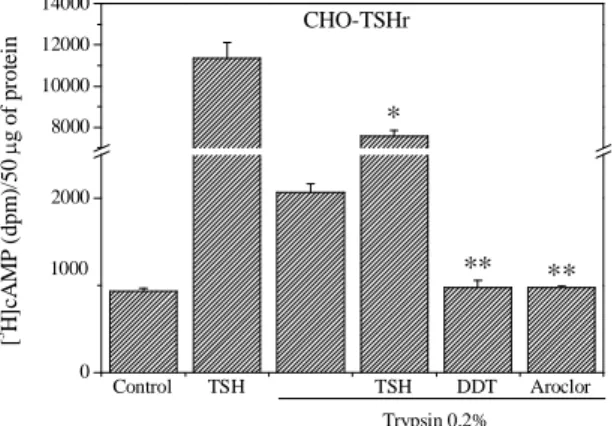
Effect of Aroclor 1254 and DDT in CHO-TSHr cells after light digestion with trypsin. To establish which domain of the TSHr (ECD or TMD) may interact with
DDT and possibly Aroclor 1254 to produce their inhibitory effects, we treated CHO-TSHr cells for 1 min with 0.2% trypsin at +25 °C in the presence of phosphodiesterase inhibitors. Trypsin is expected to increase the TSHr constitutive activity by removing the inhibitory effect of the ECD. In line with data by Van Sande et al., (1996) and Chen et al., (2003), trypsin increased significantly the TSHr constitutive activity, but it did not reduce DDT and Aroclor 1254 inhibitory effects (Fig.3). These data suggest, but do not prove, that DDT and possibly Aroclor 1254 might exert their inhibitory effect on the TMD of the TSHr. Trypsin digestion did not entirely abolish the bTSH stimulatory effect, indicating that a fraction of the ECDs were still functional (Fig.3).
Control TSH TSH DDT Aroclor 0 2000 8000 10000 12000 14000 CHO-TSHr ** ** * 1000 Trypsin 0.2% [ 3 H ]c A M P ( d p m )/ 5 0 µ g o f p ro te in
Fig. 3 Effect of trypsin digestion on the constitutive activity of the TSHr. The TSHr was digested with trypsin 0.2% for 1 min at +25°C in the presence of phosphodiesterase inhibitors. Afterwards, trypsin was rinsed and cells were exposed to bTSH (1 mU/ml for 10 min), Aroclor 1254 or DDT (100 µM) for 2 h. The graphic is the mean ± S.E. of a representative of three experiments, each performed in
quadruplicate. *Significantly different from bTSH alone, and **significantly different from trypsin alone.
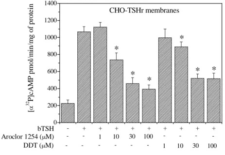
Effect of Aroclor 1254 and DDT on bTSH induced AC activity in CHO-TSHr cell membranes. In view of the fact that there is evidence that DDT might
influence cAMP accumulation in CHO-TSHr by other mechanisms (see Discussion) other than inhibition of TSHr, we tested the efficacy of DDT and Aroclor 1254 to inhibit directly AC on cell membranes.
As shown in Fig.4, both Aroclor 1254 and DDT inhibited the bTSH (1 mU/ml) induced increase in AC activity, beginning with a minimum concentration of 10 µM.
- + + + + + + + + + 0 200 400 600 800 1000 1200 1400
*
CHO-TSHr membranes 100 30 10 1 -DDT (µM) 100 30 10 1 -Aroclor 1254 (µM) bTSH*
*
*
*
*
[ α 3 2 P ]c A M P p m o l/ m in /m g o f p ro te inFig. 4 Effect of Aroclor 1254 and DDT on bTSH (1 mU/ml) induced AC activity in CHO-TSHr cell membranes. Membrane were prepared from confluent CHO-TSHr cells scraped off the plate in a hypotonic solution, containing a cocktail of protease inhibitors. AC activity was measured by monitoring the conversion of [α-32P] ATP to [α-32P] cAMP as described in Materials and Methods. The graphic is the mean ± S.E. of a representative of three experiments, each performed in triplicate. *Significantly different from bTSH alone.
Constitutive activity of the TSHr-trunk mutant in transiently transfected
COS-7 cells. To verify whether TMD of TSHr constitutes the real target for DDT and
in COS-7 cells transiently transfected with a TSHr-trunk. Initially, we characterized the constitutive activity of this truncated receptor in COS-7 cells. Since the constitutive activity of this truncated receptor is very weakly expressed in transiently transfected COS-7 cells, we co-transfected the TSHr-trunk with adenyl cyclase V (ACV) to amplify its effect. In COS-7 cells co-transfected with TSHr-trunk and ACV, and incubated in the presence of phosphodiesterase inhibitors, 2 hour exposure caused cAMP to increase twice as much the level observed in control cells, i.e. not treated with phosphodiesterase inhibitors (Fig.5A). To rule out the possibility that this cAMP increase might be due to the basal activity of ACV, or to the activation of ACV by any constitutive activity of endogenously expressed receptors (Hanke et al., 2001), the experiment was repeated in cells transfected with the ACV alone. Following 2 hour incubation with phosphodiesterase inhibitors, any induced cAMP variation was found to be too low to be significant (insert in Fig.6A). Based on these observations, we can conclude that the amount of cAMP accumulated in COS-7 cells, co-transfected with TSHr-trunk and ACV in the presence of phosphodiesterase inhibitors, is directly correlated with the TSHr constitutive activity.
Effect of Aroclor 1254 and DDT in COS-7 transiently transfected with the TSHr-trunk and ACV or the wild type TSHr and ACV. In COS-7 cells transfected
with the TSH-trunk and ACV, both DDT and Aroclor 1254 were shown to reduce the constitutive activity of the TSH-trunk (Fig.5A). At 30 µM, Aroclor 1254 exerted the strongest inhibition, while at 100 µM its inhibitory effect was consistently less pronounced. By contrast, DDT reached the maximal inhibition at 100 µM. Notice that in COS-7 cells transfected with wild type TSHr and ACV, Aroclor 1254 and DDT exhibited the same inhibitory activity against bTSH (10 mU/ml)-induced cAMP accumulation, even though the effect was much stronger (Fig.5B).
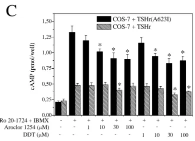
Effect of Aroclor 1254 and DDT in COS-7 cells transiently transfected with the wild type TSHr or the mutant TSHr(A623I). In these experiments we tested the
effect of Aroclor 1254 and DDT on the constitutive activity of the wild type TSHr and the mutant TSHr(A623I) (Parma et al., 1993) transiently transfected in COS-7 cells. In the TSHr(A623I) mutant, the inhibitory effect of the two thyroid disruptors could be
better characterized due to its higher level of constitutive activity. Since ACV was not co-transfected to amplify the signal, cAMP accumulation was determined by the much more sensitive RIA method (see Materials and Methods). As shown in Fig.5C, the constitutive activity of the TSHr(A623I) mutant was inhibited by Aroclor 1254 and DDT from a minimum concentration of 10 µM up to 100 µM. In contrast, the basal activity of the wild type TSHr was inhibited by Aroclor 1254 at only 30 µM and by DDT at 30 and 100 µM. - + + + + + + + + + 0 200 400 600 800 1000 1200 1400 1600 1800
A
COS-7 + TSHr-trunk + ACV
100 30 10 1 -DDT (µM) 100 30 10 1 -Aroclor 1254 (µM) Ro 20-1724 + IBMX * * * * * [ 3 H ]c A M P ( d p m )/ 5 0 µ g o f p ro te in - + + + + + + + + + 0 1000 2000 3000 4000 5000 6000 7000 8000 *
B
* * COS-7 + TSHr + ACV 100 30 10 1 -DDT (µM) 100 30 10 1 -Aroclor 1254 (µM) bTSH * * * * * [ 3 H ]c A M P ( d p m )/ 5 0 µ g o f p ro te in- + + + + + + + + + 0,00 0,25 0,50 0,75 1,00 1,25 1,50 * * * *
C
COS-7 + TSHr(A623I) COS-7 + TSHr 100 30 10 1 -DDT (µM) 100 30 10 1 -Aroclor 1254 (µM) Ro 20-1724 + IBMX * * * * * cA M P ( p m o l/w el l)Fig. 5 Uncompetitiveinverse agonist activity of Aroclor 1254 and DDT for the TSHr-trunk, the wild type TSHr and the TSHr(A623I) mutant expressed in COS-7 cells. COS-7 cells were transiently transfected with the TSHr-trunk (a TSHr truncated by 98% in the ECD) together with the ACV. Cells were incubated in the absence or presence of the phosphodiesterase inhibitors Ro 20-1724 and IBMX for 2 h at +37°C (A). In “B”, COS-7 cells were transiently transfected with the wild type TSHr together with the AC-V. Increasing concentrations of Aroclor 1254 and DDT were tested against bTSH (10 mU/ml). All samples were treated with the phosphodiesterase inhibitors Ro 20-1724 and IBMX. In “C” COS-7 cells were transiently transfected with the wild type TSHr and the mutant TSHr(A632I). As we did not cotransfect ACV to amplify the signal, in this experiment cAMP accumulation was determined using a much more sensitive RIA method (see Materials and Methods). The graphic “A” is the mean ± S.E. of a representative of five experiments, each performed in quadruplicate. *Significantly different from Ro 20-1724 + IBMX. The graphic “B” and “C” are representative of three experiments, each performed in quadruplicate. *Significantly different from bTSH alone (B) and from Ro 20-1724 + IBMX.
Effect of Aroclor 1254 and DDT in COS-7 transiently transfected with ACV alone and in CHO-K1 cells. The effect of Aroclor 1254 and DDT was further tested on
COS-7 cells transfected with ACV alone, in order to see if these two compounds could have any effect other than inhibit the TSHr. To our surprise, both compounds clearly stimulated cAMP accumulation (Fig.6A), although the effect of Aroclor 1254 and DDT in COS-7 cells could be due to inhibition of phosphodiesterases above the level observed in the presence of Ro 20-1724 and IBMX. In order to exclude this possibility, we tested the effect of Aroclor 1254 and DDT in the absence of Ro 20-1724 and IBMX. As shown in the insert of Fig.6A, in the absence of these phosphodiesterase inhibitors
Aroclor 1254 and DDT did not induce any cAMP accumulation over the basal level. In CHO-K1 cells, at variance with COS-7 cells, Aroclor 1254 and DDT did not stimulate cAMP accumulation (Fig.6B). The mechanism of cAMP accumulation, as caused by Aroclor 1254 and DDT, was not further investigated in this study because considered beyond the scope of the present project.
Control 10 30 100 10 30 100 0 200 400 600 800 1000 1200
A
COS-7 + ACV 0 100 200 300 400 500 Ro 20 IBMX DDT Aroclor Basal [ 3H ]c A M P ( d p m ) * * DDT (µM) Aroclor 1254 (µM) * * * [ 3 H ]c A M P ( d p m )/ 5 0 µ g o f p ro te in Control 10 30 100 10 30 100 0 200 400 600 800 1000B
CHO-K1 DDT (µM) Aroclor 1254 (µM) [ 3 H ]c A M P ( d p m )/ 5 0 µ g o f p ro te inFig. 6 Effect of Aroclor 1254 and DDT on COS-7 transfected with adenylyl cyclase V (ACV)
alone (A) and on CHO-K1 cells (B). Increasing concentrations of Aroclor 1254 and DDT were tested in COS-7 transiently transfected with ACV and in CHO-K1 cells. All samples were incubated in the presence of the phosphodiesterase inhibitors Ro 20-1724 and IBMX for 2 h at +37°C. The insert in “A” shows that Aroclor 1254 and DDT do not increase the cAMP level in COS-7 cells in the absence of the phosphodiesterase inhibitors Ro 20-1724 and IBMX. The graphic “A” is the mean ± S.E. of a representative of three experiments, each performed in quadruplicate. The graphic “B” is the mean ± S.E. of a representative of two experiments each, performed in quadruplicate. *Significantly different from Ro 20-1724 + IBMX.
Effect of Aroclor 1254 and DDT on forskolin- or NECA-induced cAMP accumulation in COS-7 transiently transfected with ACV alone or with the adenosine A2a receptor plus ACV, respectively. These experiments were intended to
test the specificity of the inhibitory effect exerted by Aroclor 1254 and DDT on the TSHr. To this purpose we analyzed the effect of the two thyroid disruptors on cAMP accumulation induced by forskolin or NECA in COS-7 transiently transfected with ACV alone or the adenosine A2a receptor plus ACV, respectively. In sharp contrast with
what previously shown in CHO-K1 cells, Aroclor 1254 did not prevent forskolin (1 µM)-induced accumulation of cAMP in COS-7 cells transfected with ACV alone for up to 100 µM of concentration. On the contrary, DDT showed a slight but significant inhibition at 100 µM (Fig.7A). Neither compound inhibited the stimulation of cAMP accumulation as induced by NECA (1 µM) in COS-7 cells co-transfected with the adenosine A2a receptor and ACV (Fig.7A).
Effect of DDT on cAMP accumulation in COS-7 and in CHO-D1 cells. The
specificity of DDT was further tested against other two Gαs coupled receptors, the β2
-adrenergic receptor endogenously expressed in COS-7 cells and the dopamine D1
receptor stably transfected in CHO-K1 cells (CHO-D1). As for A2a, DDT did not inhibit
the isoproterenol (1 µM) and dopamine (10 µM) induced increase in cAMP in COS-7 and CHO-D1 cells, respectively (Fig.7B).
Control Aroclor DDT Control Aroclor DDT 0 500 1000 1500 2000 2500 3000
A
[ 3 H ]c A M P ( d p m )/ 5 0 µ g o f p ro te in 6000 5000 4000 3000 2000 1000 0 COS-7 + A 2a + ACV COS-7 + ACV NECA Forskolin * [ 3 H ]c A M P ( d p m )/ 5 0 µ g o f p ro te in -- -- 10 30 100 -- -- 10 30 100 0 4000 8000 12000 16000 20000B
DDT (µM) [ 3 H ]c A M P ( d p m )/ 5 0 µ g o f p ro te in 5000 4000 3000 2000 1000 0 CHO-D1 COS-7 Dopamine Isoproterenol [ 3 H ]c A M P ( d p m )/ 5 0 µ g o f p ro te inFig. 7 Effect of Aroclor 1254 and DDT on forskolin- and NECA-induced cAMP accumulation upon
ACV and adenosine A2a receptor stimulation (A) and effect of DDT on isoproterenol- and
dopamine-induced cAMP accumulation upon β2-adrenergic and dopamine D1 receptors stimulation (B). COS-7 cells
transiently transfected with ACV or ACV and the adenosine A2a receptor, were stimulated with forskolin
(1 µM) or NECA (1 µM) respectively (A), for 10 min at +25°C in the presence of Aroclor 1254 or DDT at a concentration of 100 µM. β2-adrenergic receptors endogenously expressed in COS-7 cells and
and dopamine (10 µM) respectively (B), for 10 min at +25°C in the presence of different concentration of DDT. All samples were incubated in the presence of the phosphodiesterase inhibitors Ro 20-1724 and IBMX. The graphics are the mean ± S.E. of a representative of three experiments, each performed in quadruplicate. *Significantly different from forskolin alone.
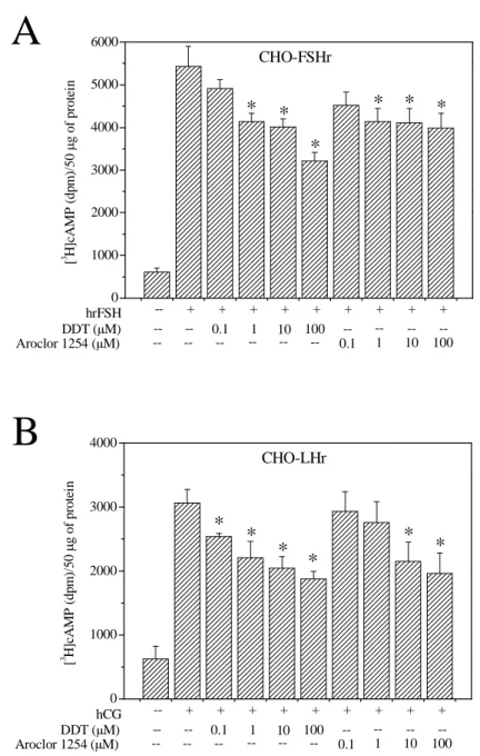
Effect of Aroclor 1254 and DDT on hrFSH and hCG dependent cAMP accumulation in CHO-FSHr and CHO-LH cells. These experiments were performed
to verify whether or not Aroclor 1254 and DDT could exert their inhibitory effect on other members of the glycoprotein hormone receptor family. To this purpose, we tested the inhibitory effect of Aroclor 1254 and DDT on CHO-K1 cells stably transfected with the other two glycoprotein hormone receptors (CHO-FSHr and CHO-LHr). Both Aroclor 1254 and DDT inhibited the hrFSH (100 mU/ml) and hCG (10 mU/ml) induced cAMP accumulation in CHO-FSHr and CHO-LHr cells, respectively. Interestingly, DDT inhibition began at 1 µM concentration in CHO-FSHr cells (Fig.8A) and at 0.1 µM concentration in CHO-LHr cells (Fig.8B). Of the two compounds, DDT at the 100 µM concentration was most efficient by causing a 41% and 39% reduction in cAMP accumulation in CHO-FSHr and CHO-LHr cells, respectively.
-- + + + + + + + + + 0 1000 2000 3000 4000 5000 6000
A
* * CHO-FSHr * * * * 100 10 1 0.1 --100 10 1 0.1 --Aroclor 1254 (µM) DDT (µM) hrFSH [ 3 H ]c A M P ( d p m )/ 5 0 µ g o f p ro te in -- + + + + + + + + + 0 1000 2000 3000 4000 * * CHO-LHrB
* * * * 100 10 1 0.1 --100 10 1 0.1 --Aroclor 1254 (µM) DDT (µM) hCG [ 3 H ]c A M P ( d p m )/ 5 0 µ g o f p ro te inFig. 8 Effect of Aroclor 1254 and DDT on CHO-FSHr (A) and CHO-LHr (B) cells stimulated
with hrFSH or hCG, respectively. Increasing concentrations of Aroclor 1254 and DDT were tested in CHO-FSHr and CHO-LHr cells stimulated with hrFSH (100 mU/ml) or hCG (1 mU/ml). All samples were incubated in the presence of the phosphodiesterase inhibitors Ro 20-1724 and IBMX. The graphics are the mean ± S.E. of a representative of three experiments, each performed in quadruplicate. *Significantly different from hrFSH and hCG alone.
Effects of different DDT analogues on bTSH dependent cAMP accumulation in CHO-TSHr. These experiments were performed to verify whether
DDT inhibitory effects could be attributed to its lipophilicity on the plasma membrane rather than to a direct action on the TSHr. To this purpose, we tested two analogues of DDT for their inhibitory effect on bTSH dependent cAMP accumulation in CHO-TSHr cells: diphenylethylene, a DDT analogues that lacks the Cl substituents and bisphenol A in which the para-Cl atoms are substituted by hydroxyl (OH)-groups. While, diphenylethylene (up to 100 µM) did not inhibit cAMP accumulation induced by bTSH (1 mU/ml), bisphenol A was found to retain in part the inhibitory effect of DDT (Tab.1). Several other structural analogous of DDT were also tested as potential thyroid disruptors (Tab.1). All of these molecules were tested at different concentrations to find out whether some of them could exert an inhibition on the TSH receptor stronger than that exerted by DDT. So far Kanferol, Quercetin and Stilbestrol (Fig.9) proved to be the only compounds capable of being more active at concentrations lower than those employed for DDT. Quercetin could produce a stronger effect than DDT or stilbestrol at a concentration of 10 µM, whereas these compounds had to be used at a concentration of 30 µM to induce a similar inhibitory effect.
Table.1 Percentage of the cAMP accumulation as induced by [1mU/ml] bTSH in CHO-TSHR
calculated by assuming treatment with the bTSH alone equal to 100%. The molecular structure of each compound is depicted on the right lane of the table.
-- -- 10 30 100 -- -- -- -- -- -- -- -- --0 20 40 60 80 100 120
*
*
*
*
*
*
*
100 10 1 _ _ _ _ _ _ _ _ _ _ _ _ _ _ __ _ _ kaempferol [µM] CHO-TSHR _ _ _ 100 0,1 10 100 10 30_ _ __ _ _ _ _ __ _ _ _ Quercetin [µM] Stilbestrol [DDT [µµM]M] Y A x is T it le bTSHFig.9 Effects of DDT, Stilbestrol, Qurcetin and Kaenferol on CHO-TSHr cells stimulated with
bTSH. Increasing concentrations of these compounds were tested in CHO-TSHr cells stimulated with bTSH (1 mU/ml). All samples were treated with the phosphodiesterase inhibitors Ro 20-1724 and IBMX. The graphic is the mean ± S.E. of three representative experiments, each performed in quadruplicate. *Significantly different from bTSH alone. DPE = diphenylethylene.
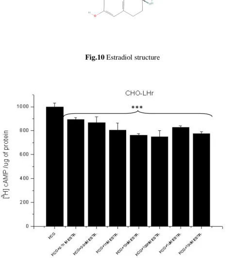
Effects of estradiol on cAMP accumulation in CHO-LHr and CHO-FSH stimulated by their respective agonists. It is recognized that DDT interferes with physiological endocrine processes modulating estrogens receptor activity. The analogy between the chemical structures of DDT and some estrogen compounds has suggested that the target site of DDT on glycoprotein hormone receptors could be a physiological binding site for estrogens. To this end we first tested DDT and Aroclor 1254 on LHr demonstrating that they decrease in a dose dependent way the accumulation of cAMP induced by hCG (Fig.8B). Afterward we tested estradiol (Fig.10) on LHr activity using concentrations that are physiological during oocyte maturation. As shown in Fig. 11 estradiol inhibited the accumulation of cAMP induced by hCG. Interestingly the maximal inhibition occurred when incubation with hCG and estradiol last 30-45 min while it decreased over longer times (Fig.12).
Fig.10 Estradiol structure
Fig. 11 Increasing concentrations of Estradiol (from 1nM to 10uM) in CHO-LHr exposed to
HCG lead to a decrease in cAMP concentration, even though this decrement does not occur in a dose-dependent manner.
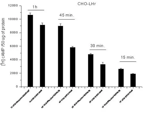
Fig. 12 Data reported on this graph show results from experiments made using different
stimulation times. As it can be clearly seen from the graph, cells exposed to estradiol differ in their ability to inhibit cAMP accumulation in relation to the time of exposure. The highest inhibition of 36% and 39% is obtained at 30 and 45 min of estradiol exposure.
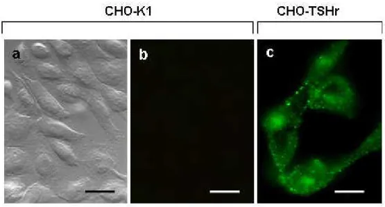
Immunocytochemical detection of TSHr trafficking To provide additional evidence
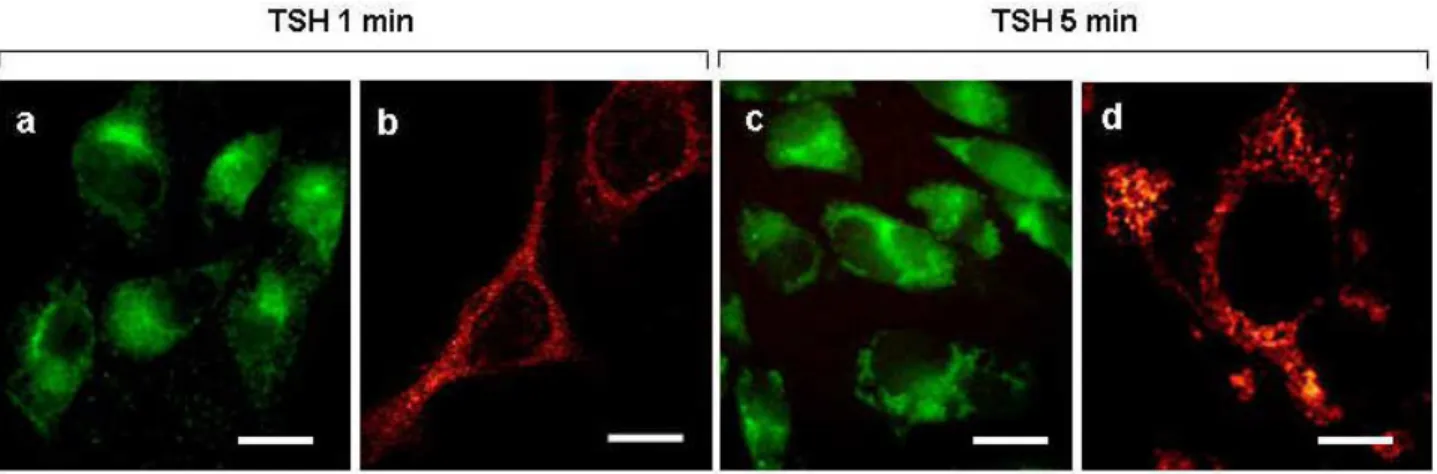
on TSHr trafficking upon exposure to TSH and/or DDT, CHO-TSHr cells were probed with anti-TSH receptor antibodies. Control CHO-K1 cells proved void of any fluorescent staining when exposed to these specific antibodies (see Fig.13a and Fig.13b for transmission). By contrast, CHO-K1 cells transfected with the TSH receptor (CHO-TSHr) became highly fluorescent when exposed to anti-TSH receptor antibodies (Fig.13c) preferentially on the plasma membrane and a number of intracellular spots in all likelyhood corresponding the TSHr constitutive activity (Parnot et al., 2002). Comparison between these two sets of experiments indicate quite clearly the specificity of the TSH receptor immune localization. Essentially similar results were obtained regardless of the monoclonal or polyclonal nature of the antibody employed for this immune localization. Upon exposure to [1mU/ml] TSH for 1 min, CHO TSHr cells exhibited an entirely different labeling pattern, the TSH receptor being displaced from
the plasma membrane to a juxta-nuclear position (Fig.14a). Analysis by confocal fluorescence microscopy confirmed that, under these conditions, the TSH receptor was displaced predominantly in this cell region in the form of spot-like clusters, most likely corresponding to early endosomes (Fig.14b). Prolonging the TSH stimulation for up to 5 minutes exalted both the intensity of fluorescent staining (Fig.14c) and the cytoplasmic spreading of the TSH receptor, as also demonstrated by confocal microscopy (Fig. 14d). Taken together, these observations indicate that TSH exposure causes the TSH receptor to be progressively withdrawn from the plasma membrane and to be and internalized by endocytosis.
Fig. 13 Immune-localization by fluorescent microscopy of anti-TSH receptor antibodies in
CHO-K1 cells (“a” and “b”) and in CHO-TSHr transfected cells (“c”). Cells were cultured on glass cover slips in Dulbecco’s modified Eagle’s medium (DMEM), mildly fixed with aldehydes and eventually exposed to anti-TSH antibodies. Receptor localization was determined by exposure to fluorescein tagged secondary antibodies. Scale bars are 20 µm (“a”, “b”) and 10 µm (“c”), respectively.
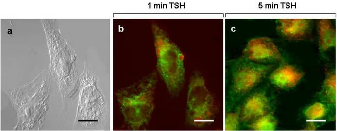
Fig. 14 Immune-localization by fluorescent microscopy of anti-TSH receptor antibodies in
CHO-TSHr transfected cells following exposure for 1 min (“a” and “b”) or 5 min (“c” and “d”) to 10 mU/ml TSH. Cells were cultured in DMEM for 4 days in the presence of G418 (0.6 mg/ml), treated with TSH and immediately afterwards mildly fixed with aldehydes. Cells were eventually treated with anti-TSHr antibodies and the receptor revealed by fluorescein tagged secondary antibodies. “a” and “c” are light microscope images with scale bars of 10 µm. Corresponding confocal microscope images at higher magnifications are shown in “b” with scale bar of 8 µm as well as in “d” with a scale bar of 6.5 µm.
TSH effects on CHO-TSHr cells were further investigated following exposure to lysotracker, a fluorescent acidotropic probe known to label specifically late endosome and lysosomes. As it can be clearly seen in Fig.15b, CHO-TSHr cells exposed for 1 minute to TSH exhibited an intracellular green labelling pattern well distinguishable from the red lysosomal staining (see Fig.15a for the corresponding transmission image). However, within 5 minutes CHO-TSHr cells appeared enriched by a few yellow spots, indicating that some of the endosomal vesicles carrying the internalized TSH receptor may be already acidified or merged with pre-existing lysosomal organelles (Fig.15c). Having previously demonstrated that DDT counteracts TSH action (Rossi et al., 2007), we then wished to investigate how DDT interferes with TSH receptor internalization in CHO-TSHr cells. First, we ascertained that cell viability is not altered when DDT is applied for 2 h up to a concentration of 100 µM, as demonstrated by the fact that a full TSH activity could be completely restored following DDT wash out (data not shown).
Fig. 15 Immune-localization by fluorescent microscopy of anti-TSH receptor antibodies in
CHO-TSHr transfected cells following exposure to 10 mU/ml TSH for 1 min (“b”) or 5 min (“c”) in the presence of 50 nM lysotracker. Pinhole was set at a low value and cells focussed at the major diameter, close to the substrate. Note that 1 min after exposure to TSH, green (anti-TSHr antibodies) and red (lysotracker) spots are still clearly discernible, while some of them have already merged (yellow spots) after 5 minutes of TSH exposure. “a” is the corresponding transmission image. Scale bars are 9 µm.
Figures 16 (“b” and “d”) demonstrate quite clearly that, in the presence of DDT, the
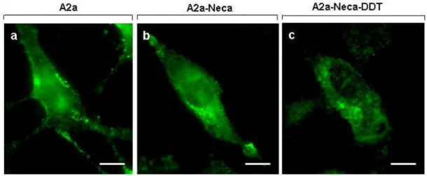
TSH receptor maintained a peripheral location along the plasma membrane, even if stimulated for 1 and 5 minutes with TSH (see Figs. 16a and c for corresponding transmission images). When CHO-TSHr cells were exposed to DDT following pre-labelling with lysotracker, none of the anti-TSHr green fluorescent spots ever merged with any of the red labelled organelles, indicating that endocytic internalization is clearly inhibited (data not shown). To verify that DDT effects on TSHr internalization in CHO transfected cells are specifically exerted onto the receptor itself and do not simply interfere with some general functions compromising cell vitality, the above experiments were repeated on CHO cells transfected with the adenosine A2a receptor. This is also a glycoprotein receptor belonging to the same GPCRs family as the TSH receptor, and it is likewise endowed with a seven-pass transmembrane domain. Fig.17a shows that anti-A2a antibodies identify a number of fluorescent spots along the plasma of CHO-A2a transfected cells. Following stimulation for 1 or 5 minutes with the specific agonist Neca, A2a receptors were seen to be displaced from the plasma
membrane and gradually internalized intracellularly (Fig.17b). However, unlike TSH receptors that were retained along the cell surface upon exposure to DDT, the adenosine A2a receptors were still internalized even if stimulated with Neca in the presence of DDT (Fig.17c). We take these observations as a confirmation of our previous results (Rossi et al., 2007) that DDT inhibits endocytic internalization in CHO cells by interacting with the TSH receptor itself.
Fig.16 Fluorescence microscopy view of DDT effects on CHO-TSHr cells following stimulation by
TSH [1mU/ml]. DDT[100µM] acts by blocking receptor internalization. In presence of 100 µM DDT, the fluorescent signal due to anti-TSHr antibodies is located along the edge of the cell body even following bTSH stimulation, for either 1 or 5 min ("b" and "d"). "a" and "b" are corresponding transmission images. Scale bars are 7 µm.
Fig. 17 Immune-localization by fluorescent microscopy of anti-A2a receptor antibodies in CHO-A2a
transfected cells ("a"), following 1 min exposure to 1 µM Neca ("b") or following 1 min exposure to 1
µM Neca in the presence of 100 µM DDT("c"). Under these conditions, green fluorescent anti-A2a spots are internalized intracellularly following Neca stimulation, regardless of the presence of DDT in the culture medium. Scale bars are 7 µm.
Fluorescent detection of the TSHr bound to the FlAsH chromophore A stretch
of six amino acids containing a tetracysteine motif (TSHr-FlAsH) was introduced into the C-terminal end of the TSH receptor (Fig.18), as specified in Material and Methods.
Initially, the TSHr-FlAsH receptor was transiently transfected in COS-7 cells and tested for its ability to increase the intracellular level of cAMP. Figure 19 shows that TSH administration causes the intracellular level of cAMP to increase in TSHr-FlAsH transfected COS-7 cells in much the same way as in COS-7 cells transfected with the wild type TSH receptor. We took this observation to indicate that insertion of the FlAsH binding motif does not alter normal TSH receptor functioning. Having established this equivalence in COS7 cells, then a stable CHO-TSHr-FlAsH cell line was created, and a number of CHO positive clones selected for their highest cAMP response to TSH stimulation (data not shown). When examined by dynamical confocal microscopy over a period of 35 minutes, CHO-TSHr-FlAsH and CHO-K1 cells appeared to differ in the time course of fluorescent labelling, which was higher in sample than in control cells. However, similar intracellular staining patterns were envisioned in both cell types, suggesting the occurrence of some unspecific binding in CHO cells (data not shown). In