ALMA MATER STUDIORUM - UNIVERSITÀ DI BOLOGNA
SCUOLA DI INGEGNERIA E ARCHITETTURA
DIPARTIMENTO DI INGEGNERIA INDUSTRIALE
CORSO DI LAUREA MAGISTRALE IN INGEGNERIA MECCANICA
TESI DI LAUREA in
LABORATORIO DI MECCANICA DEI TESSUTI BIOLOGICI
IN SILICO METHODS TO EVALUATE
FRACTURE RISK AND BONE MINERAL DENSITY
CHANGES IN PATIENTS UNDERGOING
TOTAL HIP REPLACEMENT
CANDIDATO Andrea Menichetti
RELATORE Chiar.mo Prof. Luca Cristofolini
CORRELATORI Prof. Paolo Gargiulo Prof. Magnús Kjartan Gíslason
Anno Accademico 2015/16 I° Sessione
ii
iii
Abstract
Total Hip Replacement is one of the most successful operations. There are two variants of the prosthesis that differ in the way the implant is anchored to the bone: cemented (fixed by bone cement) and cementless (fixed by press-fitting).
Currently, surgeons do not have any quantitative guideline for choosing between the two typologies, basing the decision just on their experience.
Two of the issues affecting cementless prostheses are the possibility of intra-operative fracture during the press-fitting and the bone resorption after the operation.
Starting from densitometric measurements on CT images of patients that underwent
cementless total hip replacement, two methods were developed: 1) to assess the risk of intra-operative fracture by means of a finite element analysis; 2) to evaluate bone mineral density changes (three-dimensionally around the prosthesis) 1 year after the operation.
A cohort of 5 patients was selected to test both procedures. Each patient was CT-scanned in three different time steps: one before the operation and the other two 24 hours and 1 year post-operatively.
The obtained results confirmed the feasibility of both methods, and allowed to distinguish and quantify the differences between the patients.
The feasibility of both methodologies suggests the possibility to use them in a clinical ambit: 1) knowing the risk of intra-operative fracture may provide clinicians with a guideline for the optimal implant decision-making; 2) knowing bone mineral density changes 1 year post-operatively may be used as a monitoring tool during the follow-up.
Keywords: Total Hip Replacement, Cementless Prosthesis, Intra-operative Fracture Risk; Bone Mineral Density; Finite Element Analysis.
iv
Abstract
La sostituzione totale d’anca è uno degli interventi chirurgici con le più alte percentuali di successo. Esistono due varianti di protesi d’anca che differiscono in base al metodo di ancoraggio all’osso: cementate (fissaggio tramite cemento osseo) e non cementate (fissaggio tramite forzamento). Ad oggi, i chirurghi non hanno indicazioni quantitative di supporto per la scelta fra le due tipologie di impianto, decidendo solo in base alla loro esperienza.
Due delle problematiche che interessano le protesi non cementate sono la possibilità di frattura intra-operatoria durante l’inserimento forzato e il riassorbimento osseo nel periodo di tempo successivo all’intervento.
A partire da rilevazioni densitometriche effettuate su immagini da TC di pazienti sottoposti a protesi d’anca non cementata, sono stati sviluppati due metodi: 1) per la valutazione del rischio di frattura intra-operatorio tramite analisi agli elementi finiti; 2) per la valutazione della variazione di densità minerale ossea (tridimensionalmente attorno alla protesi) dopo un anno dall’operazione.
Un campione di 5 pazienti è stato selezionato per testare le procedure. Ciascuno dei pazienti è stato scansionato tramite TC in tre momenti differenti: una acquisita prima dell’operazione (pre-op), le altre due acquisite 24 ore (post 24h) e 1 anno dopo l’operazione (post 1y). I risultati ottenuti hanno confermato la fattibilità di entrambi i metodi, riuscendo inoltre a distinguere e a quantificare delle differenze fra i vari pazienti.
La fattibilità di entrambe le metodologie suggerisce la loro possibilità di impiego in ambito clinico: 1) conoscere la stima del rischio di frattura intra-operatorio può servire come
strumento di guida per il chirurgo nella scelta dell’impianto protesico ottimale; 2) conoscere la variazione di densità minerale ossea dopo un anno dall’operazione può essere utilizzato come strumento di monitoraggio post-operatorio del paziente.
Parole chiave: Sostituzione Totale d’Anca, Protesi Non Cementata, Rischio di Frattura Intra-operatorio, Densità Minerale Ossea, Analisi agli Elementi Finiti
v
Abbreviations
BMD = Bone Mineral Density CT = Computed Tomography
DEXA = Dual Energy X-ray Absorptiometry
DICOM = Digital Imaging and COmmunications in Medicine EMG = Electromyography
FE = Finite Element
FEA = Finite Element Analysis FEM = Finite Element Method FR = Fracture Risk
FRI = Fracture Risk Index GV = Gray Values GVr = Resulting GV GV1 = GV of Dataset 1 GV2 = GV of Dataset 2 HA = Hydroxyapatite HU = Hounsfield Unit
OECD = Organization for Economic Co-operation and Development PMMA = Polymethylmethacrylate
vi Post 24h = CT-Scan 24 hours after the THA operation Post 1y = CT-Scan 1 year after the THA operation Pre-op = CT-Scan before the THA operation PVE = Partial Volume Effect
ROI = Region of Interest RU = Reykjavik University
STL = Stereo Litography Interface Format THA = Total Hip Arthroplasty
THR = Total Hip Replacement
UHMWPE = Ultra High Molecular Weight Polyethylene WHO = World Health Organization
vii
List of figures
Figure 1.1: Project Timeline ... 2
Figure 1.2 : The structure of a long bone (femur) ... 4
Figure 1.3 : Cortical and Trabecular Bone in proximal femur. [13] ... 5
Figure 1.4: The Haversian System [14] ... 6
Figure 1.5 : Bone Remodeling Phases ... 7
Figure 1.6 : Direction of trabeculae in proximal femur. Red arrows represent daily loads. ... 8
Figure 1.7 : a) Healthy Trabecular Bone vs. b) Osteoporotic Trabecular Bone ... 9
Figure 1.8 : Load direction of body weight ... 10
Figure 1.9 : Anatomy of the Hip Joint [25] ... 11
Figure 1.10 : Movements of Hip Joint [26] ... 12
Figure 1.11 : Statistic on THR, according to [32]... 13
Figure 1.12 : Components of THA. From [34] ... 14
Figure 1.13: Surgical technique to insert cementless stem. Adapted from [46] ... 18
Figure 1.14 : Cemented (a) vs. Non-cemented (b) stems. Retrieved from [48] ... 19
Figure 1.15: Illustration of Helical Tomography delivery ... 21
Figure 1.16 : Scheme of the attenuation principle ... 22
viii
Figure 2.2: Philips' Brilliance 64-slice CT scanner ... 29
Figure 2.3: A) CT-slice of a post-op THR, with artifacts due to the metallic stem. B) The same CT-slice after artifact reduction: no streaks are now present. [10] ... 29
Figure 2.4 : BMD vs. HU relationship ... 30
Figure 2.5 : Steps to create the 3D model ... 32
Figure 2.6 : Convergence Test ... 34
Figure 2.7 : Volume mesh of the femur model ... 34
Figure 2.8 : Materials Histogram ... 36
Figure 2.9 : Material Distribution ... 36
Figure 2.10 : Boundary Conditions ... 37
Figure 2.11 : External loads directions in previous model [53] ... 37
Figure 2.12 : Dynamic model of implant insertion [74] ... 39
Figure 2.13 : Force over time profile of the implant being hit by one hammer blow [74] ... 40
Figure 2.14 : Trend of the force over time on the implant after n hammer blows [74] ... 40
Figure 2.15 : a) Model of the bone-stem conic coupling with forces involved in the system. b) Forces polygon for the equilibrium... 41
Figure 2.16 : Clipped view of the meshed femur ... 43
Figure 2.17 : Lf referring to Zimmer® rasp ... 44
Figure 2.18 : Measurements to calculate perm ... 45
Figure 2.19 : The 7 zones for FR, according to [10] Greater Trochanter zone is evaluated in the lateral side of the bone. ... 46
ix
Figure 2.20 : Different femur positions between Post 24h (a) and Post 1y (b). ... 47
Figure 2.21 : Mimics project before Reslicing ... 48
Figure 2.22 : Mimics project after Reslicing ... 49
Figure 2.23: Femur bone mask (highlighted in purple) ... 50
Figure 2.24: 5 Regions of Interest for BMD, from [79] ... 50
Figure 2.25 : Point-based Image Registration... 51
Figure 2.26 : Landmarks for Registration. Landmark 1 (stem's tip) not visible ... 52
Figure 2.27 : Subtract Fusion Method (in this specific case, Post 24h - Post 1y) ... 53
Figure 2.28 : Steps to evaluate bone loss/gain ... 55
Figure 3.1: Results of FE analysis: Max Principal Elastic Strain Distribution ... 57
Figure 3.2 : Fracture Risk Indexes for different anatomical regions of the proximal femur plotted against patients' age ... 58
Figure 3.3 : HU distribution over the mask of the operated femur’s 3D model. Cancellous and cortical peaks are indicated ... 59
Figure 3.4 : Number of fractured elements against average density of cortical bone ... 60
Figure 3.5 : Number of fractured elements against average density of trabecular bone ... 60
Figure 3.6 : % BMD changes in ROI ... 65
Figure 4.1 : Radiography taken a few days after cementless THR, demonstrating a possible intra-operative fracture. Courtesy of Landspitali – University Hospital of Iceland ... 82
x
List of tables
Table 1.1: Summary of the most-used materials in THR. Adapted from [35] ... 16
Table 2.1 : Subjects' Info ... 28
Table 2.2 : Convergence Test results ... 33
Table 2.3 : Applied Pressure for FRI assessment ... 45
Table 2.4 : Subtraction Fusion Method... 52
Table 3.1: FRI in the 7 areas of investigation ... 58
Table 3.2 : Average BMD [mg/cm3] ... 61
Table 3.3 : Cortical BMD [mg/cm3]... 61
Table 3.4: BMD loss/gain volume fractions ... 66
Table 3.5 : BMD loss/gain volume fractions in ETH protocol ... 67
xi
Contents
Abstract (ENG) ... iii
Abstract (ITA) ... iv
Abbreviations ... v
List of figures ... vii
List of tables ... x
1. Introduction ... 1
1.1 Project Presentation ... 1
1.2 Theoretical Background ... 3
1.2.1 The Bone ... 3
1.2.2 The Hip Joint... 10
1.2.3 Total Hip Replacement ... 12
1.2.4 Basic Principles of Computed Tomography ... 21
1.3 Aim of the Thesis ... 23
1.3.1 Fracture Risk Assessment ... 24
1.3.2 Bone Mineral Density Changes ... 25
2. Materials and Methods ... 26
xii
2.2. Data Acquisition ... 27
2.2.1. Subjects Information ... 27
2.2.2. CT Acquisition ... 28
2.2.3. CT Calibration ... 30
2.3. Fracture Risk Index ... 31
2.3.1. Segmentation... 31
2.3.2. 3D Modeling ... 31
2.3.3. FE Modeling ... 32
2.3.3.1. Material Assignment ... 35
2.3.3.2. Boundary Conditions... 36
2.3.4. Fracture Risk Index Evaluation ... 46
2.4. Bone Mineral Density Changes... 47
2.4.1. Reslicing ... 47
2.4.2. Segmentation... 49
2.4.3. Image Registration and Subtraction ... 51
2.4.4. Bone Mineral Density Gain and Loss Evaluation... 53
3. Results ... 56
3.1. Fracture Risk Index ... 56
3.2. Bone Mineral Density Changes... 61
xiii
3.2.2 BMD changes: volume fractions ... 66
3.2.3. Comparison between two protocols ... 67
4. Discussion... 81
4.1. Fracture Risk ... 81
4.2. Bone Mineral Density Changes... 83
5. Future work and Conclusions ... 86
5.1. Fracture Risk ... 86
5.2. Bone Mineral Density Changes... 87
References ... 88
Appendix A-1 ... 97
The CLS® Spotorno® Stem ... 97
1
1. Introduction
1.1 Project Presentation
Total Hip Replacement (THR) is a widely used and successful orthopedic procedure for the treatment of many crippling diseases that cause advanced damage of hip joints, such as osteoarthritis, femoral neck’s osteonecrosis and femoral neck’s fractures.
THR consists of replacing the articular components (both femoral and acetabular) with artificial ones, in order to relieve patients of pain and re-establish mobility, leading also to a considerable improvement in terms of life quality.
There are two types of THR methodological options, that differ in the way the prosthesis is fixed to the bone, that is to say with or without the use of the bone cement.
In cemented THR, acrylic bone cement ensures the fixation of the implant, while in
cementless THR the primary stability is secured by geometrical interlocking, press-fit forces and friction between bone and implant.
The bone behaves differently depending on which implant typology is used, but there is not an absolute criterion for choosing between cemented and cementless THR, indeed large studies have shown different results in terms of clinical outcome and the issue is still debated [1], [2], [3], [4].
For instance, in the first years following the operation, cases of revision surgery due to periprosthetic fractures are more frequent for cementless implants [2]. However, a revision surgery is much more critical for cemented implants, since residual cemented bone can be removed during cement extraction from the femoral canal.
Moreover, knowing that bone adapts in correlation to the loads upon it (Wolff’s law), the use of the cement reduces the mechanical stress around the prosthesis, leading to a gradual
decrease of the bone mineral density and a consequent raise of the risk of aseptic loosening of the stem [5], [6].
On the other side, the uncemented stem allows a preload of the layer immediately adjacent to the stem and therefore bone is encouraged to grow [7].
Another important consideration is that not every patient is able to withstand the press-fitting operation to insert the cementless stem, since if bone is not strong enough, intra-operative fracture may occur.
2
methodologies, however, presently, there is a lack of a robust, quantitative method to guide physicians to the choice of the proper implant for the patient. In fact, for implant decision making, surgeons usually rely on their experience, just basing on possible bone quality indicators such as age, gender and qualitative assessment of CT images.
This means that generally younger and healthier patients receive cementless implants, while over-65 ones usually get cemented prosthesis.
Nevertheless, it has been clearly shown that other factors, such as bone mineral density and muscle strength play a crucial role in the predisposition to one implant or another. These factors depends not only on age and gender, but also on patient’s lifestyle and genetics [8]. In this context, Landspítali – University Hospital of Iceland and the Reykjavík University started a synergic collaboration to develop a project which aims to establish a subject-specific clinical evaluation score for total hip replacement planning and for post-operative assessment, with the final aims of both improving patient mobility and reducing healthcare costs of
revision surgeries.
This project [9] proposes a novel approach for clinical assessment by collecting unique data. Specific objectives of the project are:
1) to develop monitoring techniques based on gait analysis and electromyography (EMG); 2) to develop assessment methods for bone and muscle density starting from CT scans; 3) to develop and further validate computational processes based on 3D modeling and finite element method (FEM) to estimate mechanical stress acting on femurs and therefore to help selecting the optimal surgical technique;
4) to develop evaluation methods to check bone remodeling short and long-term after THR. Acquisition, elaboration and analysis of the patient’s data follow a standard protocol, whose workflow is resumed in Fig. 1.1. The output from all the elaborations will be correlated to the chosen implant type, to patients anamnesis and rehabilitation results.
Figure 1.1: Project Timeline
A few days before surgery, patients are CT-scanned and undergo a gait assessment. Right after the operation, they undergo another CT-scan. Then, 6 weeks later, patients are called in for gait measurements. 52 weeks post-operatively, they are checked with another CT-scan and gait analysis. Finally collected data are analyzed. [10]
3
The work presented in this paper is included in the frame of the above-mentioned project and specifically concerns two themes: 1) to improve the assessment method, by means of a finite element analysis (FEA), for the femur’s risk of fracture during the press-fitting of the non-cemented stem and 2) to develop a novel procedure to evaluate bone mineral density changes around the prosthesis one year after surgery.
Both assessment methods are based on data from CT-scans acquired in 3 different time steps: one before the operation and the other two 24 hours and 1 year post-operatively.
CT-scans allow to quantify the quality of the patient’s femur in terms of bone mineral density, that is the basic information which both methods are developed with.
1.2 Theoretical Background
1.2.1 The Bone
The bone is a connective tissue whose peculiar feature is to have a mineralized extra-cellular matrix, which gives it stiffness and mechanical strength.
Bone is composed of 65% by inorganic component and 35% by organic matrix, cells and water.
Organic component is principally made of collagen, the rest is non-collagenic proteins. The mineral component mainly consists of hydroxyapatite (HA), namely calcium apatite, Ca10(PO4)6(OH)2, that is present in little crystals (length: 20-40 nm; thickness: 1-1.5 nm)
between collagen fibrils. The organic is the tough and flexible component, while the mineral is the brittle and stiff one.
Bone’s functions are both mechanical and physiological. Mechanical tasks of the bones are to support the body (they form the skeletal system), to protect vital organs, to transfer loads and, in synergy with muscles, to produce motion of the limbs and the entire body. Physiologically speaking, bones are the primary site of hemopoiesis, as formation of blood cells takes place in the bone marrow. Bones are also the major calcium storage of the body, and they play an important role in the regulation (homeostasis) of this fundamental element for several vital processes.
4 Anatomy of the bone
Human adult skeleton counts 206 bones with different shape and dimensions.
For the purpose of this study, just long bones will be considered, since femur belongs to this category.
The anatomy of a long bone is depicted in Fig. 1.2.
Figure 1.2 : The structure of a long bone (femur)
In adults, the long bone is made up of a central hollow part with an approximately cylindrical shape, called diaphysis. The extremities, called epiphysis, are wider, rounded and are covered by a cartilaginous tissue (hyaline cartilage) that reduces friction with adjacent bone segment forming the joint. Intermediate regions between diaphysis and epiphysis are named
methapysis.
The external surface of bone segment, wherever it is not covered by articular cartilage, is called periosteum. It is widely vascularized and has a layer rich of active cells and elastic fibers. It also plays an important role in the remodeling process of the bone. The canal in the middle of long bones is called the medullary canal. The thin layer that surrounds the surface of these canals is called endosteum, which mainly consists of bone-forming cells. [11]
5 Structures of bone tissue
At a microscopical level, adult bone is organized in a modular structure made of adjacent layers, known as lamellae, in which collagen fibrils are oriented in parallel plans. Each lamella has a thickness of about 3-7 μm. Orientation of fibrils in adjacent layers is different, building a plywood-like structure.
Macroscopically, adult human bone is organized into two different architectonical structures: cortical (compact) bone and cancellous (spongy or trabecular) bone. (Fig. 1.3)
Cortical bone, which contributes about 80% of the skeletal mass, has a solid and compact structure, and it constitutes the external part of diaphysial and epiphyseal areas.
Cancellous bone has a porous structure since it is made of a network of little irregularly-shaped rods and plates, named trabeculae; it is mainly present in the epiphyseal regions and hosts red bone marrow – where blood cells are produced – in its interstices.
Cortical bone is heavier, stiffer and stronger, while trabecular one is lighter, less dense and with low mechanical strength properties.
Mechanically, a thin shell of cortical bone internally filled with spongy bone constitute an optimal integrated architecture, since cortical bone has the task to withstand loads, while cortical bone sustains the shell and prevents from buckling. [12]
Figure 1.3 : Cortical and Trabecular Bone in proximal femur. [13]
In cortical bone, a packet of 4-30 concentric lamellae is arranged to surround a central cavity, called haversian canal, that is a longitudinal canal filled with blood and lymphatic vessels
6
and nerves. The haversian canals are interconnected by transverse canals, also called Volkmann canals, which allow the communication with the periosteum and bone marrow. The system made up by lamellae and haversian canal is called osteon or haversian system; it represents the structural unit of cortical bone and its characteristic diameter is 200-250 μm. Externally to the osteon, there is a 1-2 μm thick layer of mineralized matrix, deficient in collagen, called cement line.
Internally to the osteon’s structure, there are little cavities, called lacunae, that host
osteocytes, namely cells trapped during bone growth process. Lacunae are interconnected by a network of little canals, called canaliculi. (Fig. 1.4)
In addition to osteons, lamellae can also organize themselves in other two different structures: circumferential lamellae (continuously surrounding bone’s body) and interstitial lamellae (fragment of residual osteons, filling the gaps between complete osteons). (Fig. 1.4)
Figure 1.4: The Haversian System [14]
Trabecular bone’s structural unit is the trabecular packet, a set of lamellae arranged in thin columns, 50-600 μm thick and circa 1 mm long. Similarly to compact bone, cement lines keep trabecular packets together. [15]
7 Bone modeling and remodeling
Bone is a dynamic tissue that undergoes continual adaption during life to preserve skeletal size, shape, and structural integrity and to regulate mineral homeostasis. Two processes, remodeling and modeling, allow development and maintenance of the skeletal system. Bone modeling is responsible for growth and mechanically induced adaption of bone and requires that the processes of bone formation and bone removal (resorption), although globally coordinated, occur independently at distinct anatomical locations. Bone remodeling is responsible for removal and repair of damaged bone to maintain integrity of the adult skeleton and mineral homeostasis. [16]
Main actors involved in these processes are osteoclasts (poly-nuclear cells assigned to bone lysis, by chemically attacking HA and removing collagen), osteoblasts (mono-nuclear cells assigned to bone formation, by synthesizing and secreting non-mineralized matrix, named osteoid) and osteocytes (the most numerous cells in mature bone, former osteoblasts trapped in the just-created osteoid).
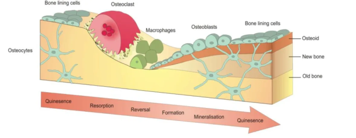
During remodeling process, old bone is resorbed by osteoclasts and replaced with new osteoid. First osteoclasts are activated, and the resorption phase takes approximately 10 days. Following resorption, macrophage-like cells are found at the remodeling site in the reversal phase.
Osteoblast precursors are then recruited, which proliferate and differentiate into mature osteoblasts, before secreting new bone matrix. The matrix then mineralizes to generate new bone and this completes the remodeling process. (Fig. 1.5)
Figure 1.5 : Bone Remodeling Phases
8
It has been found that bone remodeling is not equal for different anatomical sites, even for the same bone segment. Possible reason for this peculiarity is that remodeling process is guided by biomechanical factors. Basically, remodeling tends to maintain physiologically-loaded bone and to reduce bone that is under-loaded. This process involves both cortical and spongy bone: for instance, if bone undergoes loads that are under the physiological level, cortical bone’s thickness diminishes (reducing the resistant section) and trabeculae’s number and dimension decrease, in addition to a reduced mineralization of the matrix. In case of over physiological level loads, bone behaves in the opposite way. It has been proved that bone is able to adapt to mechanical cycling load conditions, both in direction and magnitude. [17], [18]
An emblematic example of this adaptability is provided by the proximal epiphysis of the femur (Fig. 1.6), in which trabeculae of the cancellous bone are aligned following daily loads direction.
It has been also shown that bones are optimized structures. In fact they are able to withstand daily loads and variations of the same with a rather uniform margin of safety and with a minimal mass. [19].
Figure 1.6 : Direction of trabeculae in proximal femur. Red arrows represent daily loads.
Also aging contributes to the variation of bone structure. Basically, aging has a double effect: loss of bone mass and brittleness. The first effect results from the demineralization of bone matrix; this happens earlier and has generally a faster degradation rate for women [20]. The second principal effect of aging on the skeletal system, brittleness, results from deteriorated mechanical integrity of the collagen network, that gives the bone its toughness. This leads the bone to be more susceptible to fracture. As result of aging, for instance, cancellous bone’s
9
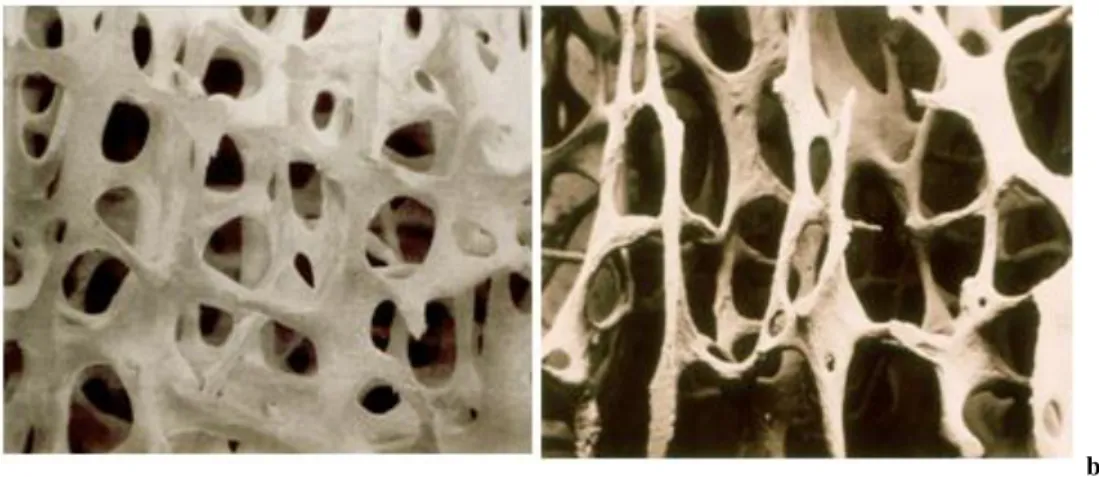
trabeculae become less dense and thinner (Fig. 1.7), while dimensions of cortical bone – both internal and external diameter – raise in order to compensate the loss of mechanical strength by increasing the resistant section. This condition can be classified as osteoporosis. [21], [22]
a) b)
Figure 1.7 : a) Healthy Trabecular Bone vs. b) Osteoporotic Trabecular Bone
Remodeling process is fundamental also for bone – prosthesis interaction.
With regard to THR, since metal implant is much stiffer than the femur, the load transferring through the bone changes, if compared to the preoperative mechanical conditions.
As a consequence, the stem shields the bone – in certain regions – from the stresses it used to be subject. This phenomenon is known as stress shielding. The reduction of the mechanical stimulus to the bone surrounding the stem causes bone resorption, which can consequently compromise stem’s stability and eventually result in implant’s failure. [5] [23] [24] Fig. 1.8 gives a schematic indication on how body weight stress is unloaded in the femoral bone after THR.
10
Figure 1.8 : Load direction of body weight
The stem is stiffer than the bone, this causes stress shielding in the proximal femur, with consequent bone mineral density decrease. Femur remains dense distally to the stem.
1.2.2 The Hip Joint
The hip joint (coxal) is a ball-and-socket synovial type joint that connects the lower limb to the pelvic girdle.
The hip joint consists of an articulation between the head of femur and acetabulum of the pelvis.
The acetabulum is a cup-like depression in the lateral side of the pelvis. The head of femur is hemispherical, and fits completely into the concavity of the acetabulum. Both the acetabulum and head of femur are covered with articular cartilage, acting like a cushion to compressive forces and lubricating the joint.
The hip joint is designed to have a great stability, since it has to withstand body’s weight and transfer it to the lower extremities. This is achieved with the strong muscles and ligaments covering the joint, along with the good fit of the femoral head inside the acetabulum. (Fig. 1.9)
11
a)
b)
Figure 1.9 : Anatomy of the Hip Joint [25]
a) Femural Head and Acetabulum of the hip joint; b) Extracapsular ligaments: they prevent excessive hyperextension (Iliofemoral), excessive abduction and extension (Pubofemoral), excessive extension of the femur (Ischiofemoral).
The movements that can be carried out at the hip joint are flexion/extension,
abduction/adduction and medial/lateral rotation. (Fig. 1.10) The degree to which flexion at the hip can occur depends on whether the knee is flexed, which relaxes the hamstrings, and increases the range of flexion. Extension at the hip joint is limited by the joint capsule, and in particular, the iliofemoral ligament. These structures become taut during extension to limit further movement.
12
Figure 1.10 : Movements of Hip Joint [26]
1.2.3 Total Hip Replacement
THR is the indicated surgical solution to advanced hip joint diseases. Symptoms of hip joint condition counts stiffness, deformity, limb shortening and movement reduction.
Patients seek help from surgery in order to get rid of pain, restore range of motion and improve quality of life.
The most common diseases [27] that can lead to make the choice to undergo total hip arthroplasty (THA) are:
Osteolysis: wear that leads to local loss of bone caused by different articular disorders; Osteoarthritis: degenerative arthritis disease characterized by breakdown of the joint’s
cartilage, leading to friction between ball and socket of the joint that causes severe pain. It is the most common diagnosis between patients undergoing THA; [28] [29]
Avascular Necrosis: lack of blood supply into bone that can progressively lead to joint surface collapse that results in increasing pain;
Rheumatoid Arthritis: chronic inflammatory disorder that involves inflammation in the lining of the joint and may result into cartilage destruction;
Femur’s neck Fracture;
Developmental Dysplasia: femoral head has an abnormal relationship to the acetabulum; Paget’s Disease: metabolic bone disorder that induces an increased and irregular
formation of bone; Tumor.
13
Total hip replacement is one of the most successful and cost-effective interventions in medicine. [28]. Its success is well reported in literature and a survival rate - intended as maintaining the prosthesis in its initial condition - of about 90% is recorded at 15 years follow-up [29] [30] [31].
The most common causes of complications for THA are prosthesis dislocation, aseptic loosening of the stem and/or of the cup due to mechanical wear debris, periprosthetic bone fracture, septic loosening and breakage of the prosthesis (rare) [27] [29]. Most of these complications can directly lead to premature revision surgery, that is to say removal of the failed component and replacement with a new one.
Statistics of Emilia Romagna (Italy) [29] and the rest of the OECD countries [32] show that the number of hip replacements has increased rapidly since 2000. On average, the rate of hip replacements increased by about 35% between 2000 and 2013. (Fig. 1.11 b)
For instance, in Italy [33], number of yearly THR surgeries in 2001 was 45 656, while in 2012 became 62 153.
a)
b)
Figure 1.11 : Statistic on THR, according to [32]
a) Number of THR surgeries in 2013. Iceland counts 185 operations per 100 000 people, Italy 166; b) Trend in hip replacement surgery, for some selected countries, from 2000 to 2013. Average statistics for OECD countries are represented in red.
14
According to data collected between 2000 and 2014 in Emilia Romagna [29], average age at primary THA was 66.7 years; while the largest age group for primary THA was 70-79 years. Male’s average age of patients that underwent hip replacement surgery in 2014 was 66.5 while female’s was 70.2 . However, out of 87 993 patients that underwent primary THA during 2000-2014, 60% were female.
Prosthesis components
THA is the replacement of both articulating surfaces of a degenerated hip joint. In the
conventional approach, the spherical part of the joint is completely replaced. The counterpart of the joint is also replaced by a semi-spherical shell. Artificial hip joints are innovative, high-quality biomedical engineering products. Prostheses are designed to last for at least 20 years, however their lifespan can be limited by wear.
There is a large variety of hip prostheses on the market in terms of material, shape, coating etc. due to the continuous research aiming to reduce the likelihood of post-operation
complications; however, currently, there is a basic modular-design principle common for all of the
implants. (Fig. 1.12)
The damaged hip joint is replaced by two artificial components: the acetabular cup (socket) and the femoral head (ball). The latter is anchored in the femur by the stem, while the former is fixed in the pelvis. The acetabular cup consists of a shell in which a liner is inserted in order to provide the load bearing articulating surface.
15
The modularity of the implant is justified by the fact that different materials with different properties need to be used to meet the different requirements for each component. Modularity can also allow the revision surgery of just the failed component, without replacing all the implant.
In order to take over the physiological function of a hip joint, a prosthesis must have three different compatibility requirements: [35]
- Structural: prosthesis must withstand millions of loading cycles, i.e. it must have an adequate mechanical strength and fatigue strength;
- Tribological: the articulating surfaces must not be compromised by wear, ensuring a correct relative motion of the 2 joint components;
- Biological: stem and shell must integrate well with the bone and resist to body’s corrosive environment. Inevitable debris particles released due to wear must not harm the organism. Hip prostheses can be categorized basing on the utilized materials for the combination femoral head-acetabular cup. The most common combinations are metal-on-polyethylene (MoP), metal-on-metal (MoM) and ceramic-on-ceramic (CoC).
The femoral stem is the component subjected to the highest mechanical stresses and it must provide a uniform transfer of the cycle loads from the prosthesis to the lower limb. Thus, the used material must have high mechanical strength and fatigue resistance, for this reason only metals have been considered to this application so far. Factors like stem length, stem cross-section and neck angle influence stability and therefore must be taken into account when designing it. [36] [37] [38] [39]
The femoral head is coupled to stem’s neck through the latter’s taper junction. Paramount factor is head’s diameter, which plays a fundamental role in the achievable range of motion of the artificial joint, along with its stability. Design criteria for the femoral heads are: i) the minimum surface roughness to get lowest friction and wear rate possible; ii) the maximum outer diameter, to improve stability and range of motion [40]; iii) the mechanical resistance of the material to withstand tensile stresses generated along the taper junction. Metal and ceramic have been used as materials for femoral heads. Ceramic heads, if compared to metal ones, result to have more smoothness (thus less friction coefficient). On the other side there are limitations in manufacturing large diameters, along with higher brittleness.
16
The liner of the acetabular cup is the socket of the hip joint, being the counterpart of the femoral head. It represents the soft component of the hard-soft coupling, therefore it is manufactured in order to have its surface more worn out than femoral head’s one, aiming to maintain relative motion in an acceptable range. The liner can be made of metal (CoCrMo) or ceramic, but it has been usually produced in UHMWPE (ultra high molecular weight
polyethylene), which has allows low friction. However, UHMWPE presents high wear rates, that can be reduced by means of a gamma- or beta-ray irradiation technique (Crosslinked UHMWPE, also known as XLPE). [27] [41]
The acetabular cup’s shell is secured to the pelvis either with bone cement or press-fitting and the fixation technique influences its external surface design: for instance, uncemented
components have porous surface finishing (e.g. with sintered titanium beads) or HA coatings to promote osteointegration. [27] [36]
For MoP and CoC implants, shells must also provide mechanical stability for brittle ceramic or soft UHMWPE liners and is therefore mostly fabricated in pure titanium.
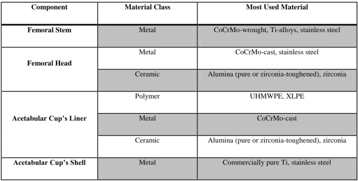
Table 1.1 resumes the most used materials to fabricate THR’s components.
Component Material Class Most Used Material
Femoral Stem Metal CoCrMo-wrought, Ti-alloys, stainless steel
Femoral Head
Metal CoCrMo-cast, stainless steel
Ceramic Alumina (pure or zirconia-toughened), zirconia
Acetabular Cup’s Liner
Polymer UHMWPE, XLPE
Metal CoCrMo-cast
Ceramic Alumina (pure or zirconia-toughened), zirconia
Acetabular Cup’s Shell Metal Commercially pure Ti, stainless steel
17
As already mentioned in § 1.1, the fixation of the implant to the bones characterizes two different categories: cemented and non-cemented prostheses.
For the purpose of this thesis, attention will be put just into femoral stems, knowing that the same fixation techniques can be adopted for acetabular cup’s shells too.
Cemented stem
Historically the first fixation typology (Sir John Charnley, 1961), cemented stems (Fig. 1.14 a) are secured to the femur through an acrylic bone cement. Usually, the material for the cement is Polymethylmethacrylate (PMMA). Actually, bone cement does not bond the stem to bone, but it rather fills the gap between prosthesis and bone.
Regarding the implantation technique, a cavity is created into the femur by reaming trabecular bone from inside the medullary canal. Then a bone plug is inserted down into the canal to keep the bone cement from flowing towards the distal femur. When the stem is inserted, the bone cement is pressurized to flow into the trabecular bone. The interlayer (optimal thickness: at least 2 mm) of bone cement – if stable, homogeneous and complete – assures a uniform
mechanical load transfer from the implant to the bone. [36] [42] [43]
An inhomogeneous distribution of bone cement may result into implant loosening or into increased risk of periprosthetic fractures. Surgeon must also have experience in preparing the bone cement and respect its polymerization time.
Cemented stems must have a smooth surface, since local peaks of stress must be avoided, as they can lead to cracks in the PMMA layer. Furthermore the stem must be stiff enough to avoid mechanical loading of the cement due to elastic deformation of the metal.
Non-cemented stem
In cementless stems (Fig. 1.14 b), used since 1984 [44, 45], a direct press-fit contact between implant and surrounding bone tissue is realized.
The surgical procedure (Fig. 1.13) includes removal (osteotomy) of the neck and the head of the femur. Once osteotomy has been performed, the cavity for stem’s insertion is firstly prepared with an awl and secondly reamed out through a rasp, which has a little smaller dimension than the stem. At this point, a prosthesis of the appropriate size is inserted and driven until it is completely stable. The insertion is carried out by means of hammering blows that need to be carefully controlled in terms of force by the surgeon, since an excessive force, enhanced also by the wedge effect of the stem’s shape, may result in a fracture.
18
Figure 1.13: Surgical technique to insert cementless stem. Adapted from [46]
a) Resection planes: the lesser trochanter is used as a reference; b) Osteotomy of the femoral neck and head; c) Preparation of the cavity in the medullary canal through the awl; c) The cavity is reamed out with the rasp; d) The stem is hammered down to the cavity until it is stable.
Stability of the stem is basically achieved at two stages: [47]
- Primary stability: by hammering the stem into the bone, a geometrical interlocking of the implant is achieved, which guarantees stability in the immediate post-operative period. When the stem is press-fitted, a swelling of the femur’s cross-sectional area is observable, causing assembly strain. The assembly strain consists of a change in the strain
distribution in the bone tissue around the prosthesis. However, if assembly strain is too low, the implant will loosen, if too high bone fracture may occur.
- Secondary stability: The bone around the stem that has been damaged during surgery induces a healing response, according to bone remodeling process, which leads the adjacent trabecular bone to grow onto the stem, providing a solid fixation
(osseointegration). Moreover, preload condition provided by press-fitting is also helpful for bone growing.
Long-term osseointegration is further promoted by having either porous coatings or porous surface finish, that are intended to be filled with newly forming trabecular bone.
19
In the first days post-operatively the patients are prevented from moving, since
osseointegration has not begun yet. Later on, progressively increasing loads are applied to the operated hip to foster osseointegration process.
Figure 1.14 : Cemented (a) vs. Non-cemented (b) stems. Retrieved from [48]
The interface bone-implant is highlighted in the picture. Cemented solution (a) considers the use of bone cement that fills the gap between smooth metal stem and the bone. In cementless prostheses, porous coating encourages
trabecular bone to grow onto it providing osseointegration
Cemented stem vs. non-cemented stem
Large studies have been carried out to test survival rates and clinical outcomes for both implant solutions [1] [2] [3] and a definitive criterion to prefer one typology rather than the other has not been defined yet [10].
Anyway, some differences have been individuated in literature and the most significant will be highlighted here.
For example, advantages of choosing a cemented stem are a faster rehabilitation for the patient (stability of the stem is almost immediate, due to cement) and less thigh pain [49]. These factors contribute to the fact that cemented stems show higher survival rates in the short- and mid-term. [1] [50]. However, issues connected to bone cement can be sometimes detected: for instance, the unreacted PMMA molecules or the temperature peaks due to the exothermal reaction of polymerization may lead to bone necrosis [35], moreover mechanical deterioration of the cement layer is expected with time due to fatigue.
20
Furthermore, stress-shielding in periprosthetic femur is more accentuated for cemented stem [24], also because bone cement acts like a bumper between the metal (stiffer, therefore more loaded) and the bone (less stiff, lower loads), shielding the bone from loads and therefore promoting bone resorption. This phenomenon is likely to implicate stem’s aseptic loosening, that is one of the major reason running to revision surgery.
Revision surgeries, in terms of removing failed stem and replacing it with a new one, are much easier with cementless stem. In fact, by extracting the cement from the femoral cavity, part of the bone in which PMMA had infiltrated can be removed, implicating a further reduction of bone’s quality [51].
Even though cementless implants are more frequently revised because of periprosthetic fracture in the first 2 years post-operatively [2] [52], in long-term period, for younger patients, higher survival rates can be recorded for uncemented stems. [1] [7] [44].
Moreover, porous coating of uncemented stems promotes osseointegration, avoiding stress shielding and depletion of the prosthesis surrounding bone’s quality.
However, during the surgical planning phase, clinicians must check bone’s quality, since a low bone mineral density or a severe osteoporosis may result in an intra-operative fracture: in fact, the bone may not be able to withstand the forces induced by surgeon’s hammer blows. Therefore, cementless stems are generally more appropriate for younger and more active patients, while for older people is more safe to choose the cemented option, even though individual differences can be vast [8] and other factors, such as muscle quality, gender, bone mineral density, stem design, gait patterns, co-morbidities must be taken into account when choosing the optimal prosthesis [53] [54] [55].
The revision rate for infection is similar for both the uncemented THA and the cemented THA with antibiotic cement. [1]
About rehabilitation times, patients undergoing cemented implants get well faster, while for uncemented prostheses recovery period is extended, since secondary stability must be provided with progressive loads, thus activities must be limited for up to 3 months to protect the replaced hip joint.
21
1.2.4 Basic Principles of Computed Tomography
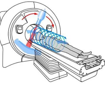
Computed Tomography (CT) is a medical imaging technique, providing slice images of the body, that allows to distinguish different organs and tissues depending on their density. A motorized table moves the patient through a circular opening (gantry) in the CT machine. As the patient passes through the CT imaging system, an X-ray tube generator rotates around the inside of the circular opening. (Fig. 1.15)
The X-ray source produces a beam used to irradiate a section of the patient's body. The radiation that is not absorbed by the tissues is measured by a system of detectors integral with the source: in this way it is possible to record a series of X-ray attenuation profiles of the patient's scanned body.
Many different “snapshots” (angles) are collected during one complete rotation.
Afterwards, the data are sent to a computer, which through complex algorithms is able to reconstruct all of the individual "snapshots" into a cross-sectional image (slice) of the internal organs and tissues for each complete rotation of the X-ray source.
22
Each slice of the body is divided into elementary volume units (voxels). The number of voxels depends on detectors’ number and dimensions.
Knowing the difference between the emitted intensity ( ) and the detected intensity ( ), a CT number is assigned to every voxel. The CT number of a given voxel is the average value of all the linear attenuation coefficients of the beam-crossed tissues considered in the volume and indicates how much energy of the X-ray beam is reduced by passing through these tissues. Attenuation coefficients refer to Lambert-Beer’s law of radiant energy’s attenuation (Equation 1.1):
given as voxels along the path of a ray, as the path length of the ray through -th voxel and as the attenuation coefficient of the material contained within -th voxel. (Fig. 1.16)
Figure 1.16 : Scheme of the attenuation principle
Every tissue has its own linear attenuation coefficient, but since these coefficients can vary due to different CT scan models and different settings on the same scanner, the Hounsfield Unit scale is used to standardize them. [56] [57] [58]
23 The HU is defined as described in Formula 1.2:
where is the average attenuation coefficient of the voxel, while and
( are the linear attenuation coefficient of water and air. Conventionally, at standard pressure and temperature conditions, water is set to 0 HU and air to -1000 HU.
Each voxel with its CT number (i.e. HU) will be represented in a gray values scale.
Since attenuation depends on the density of the scanned volume, different levels of gray lead to distinguish between soft and hard tissues.
Moreover, by calibrating the CT scanner with a phantom it is possible to define the relationship between HU and BMD, as it will be analyzed in § 2.2.3 .
A single voxel may contain of several types of tissues due to the finite spatial resolution of the imaging device. This phenomenon, termed partial volume effect (PVE), complicates the segmentation process. For example, at the interface between a softer and a harder tissue (i.e. outer cortical bone or internal interface between metal stem and bone) the edge may appear as blurred.
1.3 Aim of the Thesis
Measuring bone mineral density in vivo provides quantitative information on bone’s quality, that can be used for several applications. For instance, with regard to THR, bone
densitometry can be used to predict femoral strength [59]; moreover, it provides useful information about bone architecture around implants and its change over time [60] [61]. There are different techniques to measure bone mineral density: plain radiographic absorptiometry, dual energy X-ray absorptiometry (DEXA) and quantitative computed tomography (CT).
Quantitative CT is the most accurate and reproducible method for in vivo evaluation of cortical and cancellous bone density, [62] [63] and is the only technology that allows a three-dimensional volumetric analysis, since it provides a stack of cross-sectional images.
24
However, patients undergo higher radiation dose if compared to DEXA (0.5-1.0 mSv against 0.001-0.1 mSv). [62]
Application of bone densitometry based on CT are multiple: assessment of volumetric BMD is indeed the basis for generating and developing subject-specific FE models both to estimate mechanical structural behavior of the bone-implant system [64] and to predict bone
remodeling and stress shielding around the prosthesis. [63] [65] [66] [67]
In this study, bone densitometry measurements through CT are used for novel subject-specific methods i) to assess the intra-operative risk of fracture (based on FEA) and ii) to evaluate changes in BMD, by comparing post 24h and post 1y data.
1.3.1 Fracture Risk Assessment
The purpose of this part of the study is to develop a novel in silico subject-specific method to evaluate the risk of periprosthetic fracture when the surgeon hammers the cementless stem down to the femoral cavity.
The evaluation of the fracture risk is carried out by generating a 3D model, that is
successively discretized and used for a FE mechanical structural analysis. The 3D model is built thanks to BMD measurements from CT images.
Other aim is to locate which part of the bone is more likely to fail.
This work is based on the previous models [10] [53] [55] developed in the frame of the aforementioned project “Clinical evaluation score for Total Hip Arthroplasty planning and post-operative assessment”.
This method aims to be a development of the previous FR assessment models since it takes into account different external factors in the FE analysis. This means that different boundary conditions, in terms of loads, are here considered in order to answer to the demand of more robustness [53]. In fact, the novelty of this method is taking into account factors like material roughness, tapering degree, cross-sectional area and friction at the bone-implant interface. Moreover, in prior works various failure criteria have been used for FR assessment, such as Von Mises equivalent stress [53]. However, it has been demonstrated that a failure criteria based on maximum principal strains is a suitable candidate for the in vivo
25
By processing 5 patients, this study also wants to test the feasibility and repeatability of the methodology in order to suggest a future use in the clinical evaluation score for pre-operative implant decision making.
1.3.2 Bone Mineral Density Changes
According to the follow-up protocol developed by Landspitali – University Hospital of Iceland and University of Reykjavik, patients undergo 2 CT-scans 24 hours and 1 year after total hip arthroplasty.
Based on BMD measurements by means of these two CTs, this study’s aim is to assess mineral density changes of the femoral bone in 1 year time span after the surgery. Checking how BMD has increased/decreased around the prosthesis is a useful tool to estimate bone’s remodeling.
Differently to DEXA, CT-scan allows to have a three-dimensional measure of the bone mineral density. As suggested in other works too [69], by using the image processing software Mimics, it was possible to exploit this powerful feature and to develop a new protocol for the evaluation of the three-dimensional changes of BMD.
The novelty of this method lies in the opportunity to quantify volumetric bone growth/loss and to visually localize its three-dimensional distribution.
5 patients’ data are processed with this method, in order to investigate the potential of quantitative CT volumetric analysis to quantify bone changes after THR and verify its feasibility and repeatability.
The same 5 patients have been also processed with an alternative method, developed in parallel at ETH – Zürich [70] which uses open source image processing software – instead of Mimics – and a more automated image registration technique. The results have been
compared for a cross-checking validation and to determine advantages and disadvantages of each method.
26
2. Materials and Methods
2.1. Study Workflow
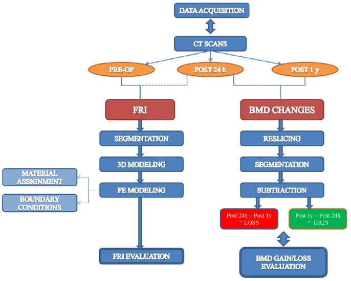
Figure 2.1 shows a scheme representing the overall workflow for both the assessment of the intraoperative FRI and the evaluation of the BMD changes occurring in the periprosthetic area of the femur one year after total hip arthroplasty.
Figure 2.1: Study Workflow
First step is to gather all the CT-scans of the selected patient. For each patient three CT-scan datasets are provided by the University Hospital of Iceland – Landspitali: one of them was carried out pre-operatively, the other two were done 24 hours and 1 year post-operatively. To estimate FRI during the implant hammering operation, pre-op CT-Scan is mainly
27
images, a segmentation is carried out to isolate the interesting areas for the successive creation of the bone’s 3D model.
Successively, the 3D model is discretized with a finite elements mesh. Material properties of the bone are assigned basing on HU values.
Later on, boundary conditions are defined and the strain distribution within the FE model of the femur is calculated in order to assess the FRI.
For the evaluation of BMD changes in the operated proximal femur one year post-operatively, a comparison between post 24h and post 1y CT-scans was made.
Firstly, a reslicing of both the original CT images datasets is done. Next step is to segment the bone, thereafter the two CT images datasets are registered using the subtraction fusion method. It is necessary to make the registration + subtraction operation twice, since BMD gain and loss are evaluated separately: subtracting post 1y dataset from post 24h one leads to assess the BMD increase, while subtracting post 24h from post 1y gives the BMD decrease.
2.2. Data Acquisition
2.2.1. Subjects Information
The two present methods developed to evaluate the intraoperative FRI and the BMD changes were applied to patients that have already joined up the “Clinical evaluation score for Total Hip Arthroplasty planning and post-operative assessment” project, born from the
collaboration between the University of Reykjavik and Landspitali – University Hospital of Iceland. The whole cohort of the project enumerates patients undergoing primary THA
surgery (unilateral arthroplasty), and considers subjects having both cemented and cementless stems.
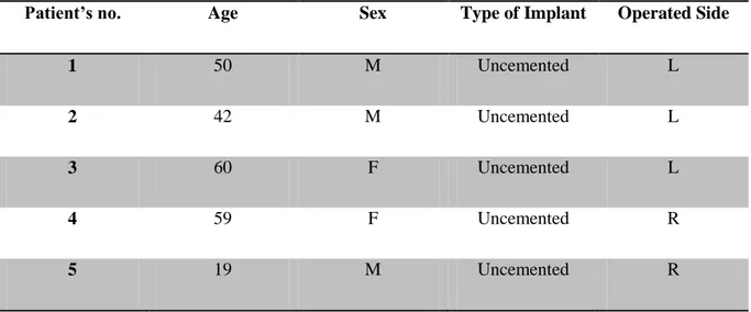
Nevertheless, for the purpose of this study, a total of 5 patients (3 males plus 2 females) having cementless implants has been selected from the whole project cohort. The youngest patient was 19 years old at the time of the first CT, while the oldest one was 60. The average age is 46.0 ± 16.8 years.
28
All the information about patients’ gender, age and operated side are summarized in the Table 2.1.
Patient’s no. Age Sex Type of Implant Operated Side
1 50 M Uncemented L
2 42 M Uncemented L
3 60 F Uncemented L
4 59 F Uncemented R
5 19 M Uncemented R
Table 2.1 : Subjects' Info
2.2.2. CT Acquisition
The patients are scanned with a Philips Brilliance 64 Spiral-CT machine (Fig. 2.2) three times in one year: firstly 1-3 days before surgery and successively 24 hours and 52 weeks after surgery.
The CT scanning region extends from the iliac crest to the middle of the diaphysial femur. Slices thickness is 1 mm, slice increment is 0.5 mm and tube voltage is set to 120 kVp. Every slice has 512x512 pixels; each pixel is represented by a GV belonging to a 12-bit representation scale (4096 different levels of gray). Each GV has a corresponding value in the HU scale, following the relation:
29
Figure 2.2: Philips' Brilliance 64-slice CT scanner
The CT images acquired after the THA are corrupted from metal artifacts that introduce both bright and dark streaks which modify remarkably HU values and therefore bone and muscle density. (Fig. 2.3)
For this reason, post 24h and post 1y CT images are processed in the artifact reduction software Metal Deletion Technique from ReVision Radiology [71]. By means of this
software artifacts are iteratively reduced and the “clean” image reconstructed, thus the BMD assessments explained in this paper can be performed.
Figure 2.3: A) CT-slice of a post-op THR, with artifacts due to the metallic stem. B) The same CT-slice after artifact reduction: no streaks are now present. [10]
30
2.2.3. CT Calibration
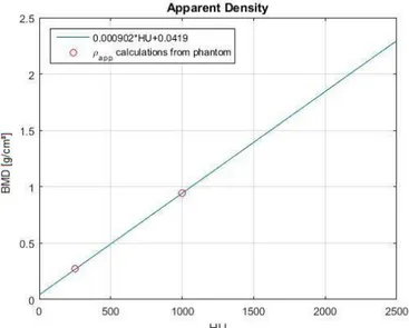
Prior to the study, the CT-Scan device was calibrated [72] using a QUASAR™ Phantom in order to find a mathematical relationship that allows to convert the CT-Scan values (in [HU]) into apparent density, i.e. BMD (in [g/cm³]).
The resulting formula [2.1] is:
Figure 2.4 : BMD vs. HU relationship
31
2.3. Fracture Risk Index
The novel subject-specific method to assess the fracture risk of the periprosthetic area of the proximal femur is described in the following sections. This method was applied to evaluate the FR for all the 5 patients enrolled in this study.
2.3.1. Segmentation
Firstly, Pre-op CT-Scan images (available in DICOM format) are imported into the software Materialise Mimics®.
There, in order to take into account just the part of the image corresponding to the proximal femur, a segmentation is carried out. By using the 3D Live Wire Mimics’ tool, the contour of the bone is traced and all the pixels belonging to the femur are gathered into the same
coloured mask. (Fig. 2.5 a)
2.3.2. 3D Modeling
Once the mask has been created, a three-dimensional model of the pre-op femur is generated. The 3D bone has still got the neck and head: it is necessary to cut them off since they are removed in the first steps of the surgical operation theatre.
To simulate this osteotomy, the 3D model of post-surgery femur (created from post 24h CT-scan) is imported in the pre-op Mimics project.
Using the Reposition tool, the 3D post-op femur is manually dragged and rotated within the pre-op spatial reference system, as long as the 3D post 24h femur overlaps the pre-op one. Once reposition has been performed, the mask of the post 24h bone is obtained from the 3D model, through a tool available in Mimics.
Successively, two Boolean subtractions are carried out. First of them is performed between the just-created masks: by subtracting post 24 h mask from pre-op one, a “scrap mask” is obtained. Secondly, by doing pre-op mask minus scrap mask it is possible to get the mask of the femur ready for the prosthesis insertion. (Fig. 2.5 b and Fig. 2.5 c). Next step is to generate the 3D model from the latter mask.
32
This 3D model is devoid of the neck and the head, and has got the cavity for the stem; moreover, since it has been generated inside the pre-op Mimics file, there are no metal artifacts, that have an adverse effect on proper material assignment for the FE model, which is HU-based, as it will be discussed further.
Additionally, a virtual distal cut is performed orthogonal to the femur’s long axis, about 2 cm below stem’s tip.
Finally, the hollow 3D femur is improved with some refinements, such as Wrapping and Smoothing – tools available in Mimics – to avoid sharp edges (Fig. 2.5 d).
a) b)
c) d)
Figure 2.5 : Steps to create the 3D model
a) Pre-op femur mask ; b) Scrap mask; c) Femur_with_cavity mask; d) 3D model
2.3.3. FE Modeling
The final 3D femur is exported into 3-Matic in order to discretize its volume through a finite element mesh.
First step is to create a surface mesh of triangular elements with automatic Remesh tool, setting the maximum edge length and checking the shape quality to avoid too distorted
elements through mesh-refining tools available in 3-Matic. To do that, a value of 0.3 has been set as the lowest threshold for the shape quality criterion R-in/R-out; moreover, 0.05 has been
33
defined to be the maximum geometrical error, which is the maximum deviation between the part’s surface before and after automatic remeshing operation.
Later on, the volume mesh is generated starting from the surface, using 10-nodes tetrahedral structural solid elements (SOLID187). (Fig. 2.7)
Quality has been checked also for the volume mesh, meaning that before implementing the volume-meshing automatic algorithm, the lower-threshold Aspect Ratio value has been set to 25.
The choice of the optimal elements’ maximum edge length (i.e. the proper mesh density) has been made following the results of a convergence test. Since smaller element’s dimension means larger number of nodes and elements, the average maximum principal elastic strain over a sample area has been calculated to determine the proper mesh density for different maximum edge lengths of the elements. For the convergence test the same boundary conditions as for FR assessment have been set.
The outer lesser trochanter zone is the selected sample area. Table and plot below show the result of the convergence test:
Maximum edge length Number of Elements Number of Nodes Average Max Princ El Strain in Lesser Trochanter Error (compared to previous size result)
8 mm 38916 67745 763 με - 5 mm 55667 89826 1150 με 51% 4 mm 85515 129734 1450 με 26% 3 mm 176372 252111 952 με 34% 2,5 mm 295778 413365 1080 με 13% 2 mm 567491 779603 License not available -
34
Figure 2.6 : Convergence Test
A mesh edge size of 2.5 mm was selected since the difference between results obtained with 2.5 and 3 mm sizes is small if compared to the others; therefore a certain convergence is supposable if increasing the number of elements. Unfortunately, software license has not allowed a further increasing of elements number, and 2.5 mm is the smallest usable size. Moreover, the choice of the smallest size is justified by the fact that the smaller is the element, the lesser number of voxels are counted in a single element, and this allows a more detailed material assignment. In fact, Mimics firstly averages on each element the HU field and then, through mathematical relationships, derives the element Young’s modulus as it will be explained in the next paragraph.
Smaller elements mean higher computational cost in terms of elapsed time for processing, though.
Figure 2.7 : Volume mesh of the femur model
0 200 400 600 800 1000 1200 1400 1600 0 100000 200000 300000 400000 M ax Pi rn ci p al E lasti c Str ai n in Le sser Tr o ch an te r ar e a [ με ] Number of elements
Convergence Test
35
2.3.3.1.
Material Assignment
Once the volume has been discretized, the FE model is imported back to Mimics in order to assign mechanical material properties to the bone, which are substantially based on the average HU of each finite element (see Formula [2.1]).
For simplification, in this method, each finite element has been considered as locally linear-elastic and isotropic.
The inhomogeneity of the bone has been also simplified by defining 50 different materials to be assigned to the bone mesh: this means that elements having similar HU values are “made of” the same material. An apparent density, a Young’s modulus and a Poisson’s ratio are defined for each material.
The apparent density (ρapp) is defined according to the above-mentioned HU-BMD
conversion formula [2.1].
The relationship between ρapp (in [g/cm3]) and Young’s modulus is described by the equation
retrieved from [73]:
[2.2]
Equation 2.2 has been used to both represent trabecular and cortical bone. Finally, the same Poisson’s ratio equals to 0.3 has been set for all the materials.
The mask of the femur – from which the 3D model is generated – may contain pixels with a very low HU: this means that those image elements belong either to soft tissues, bone marrow or air bubbles. For the continuity of the FE model it is necessary to keep also these entities, that are mainly in the inner part of the bone, even though they are not structural elements and they contribute to underestimate the bone strength. Moreover, if these elements have negative HU (in particular < -35 HU), according to formulas [2.1] and [2.2], a negative Young’s modulus would be expected, which is unrealistic.
To overcome this issue, the same “weaker” material is assigned to all of the finite elements having a corresponding apparent density lower than 0.01 g/cm3.
Thus, the fictitious material has been defined to have a ρapp = 0.01 g/cm3, that (according to
[2.2]) leads to E = 7 MPa, which is 2 order of magnitude smaller than the lowest-density bone (255 HU, ρapp = 0.27 g/cm3 and E = 984 MPa).
36
Figure 2.8 and 2.9 shows the distribution of the assigned materials throughout the bone.
Figure 2.8 : Materials Histogram
Yellow/orange elements have the highest HU i.e. the highest ρapp and E
Figure 2.9 : Material Distribution
2.3.3.2.
Boundary Conditions
The three-dimensional FE model of the proximal femur is imported into Ansys Workbench in order to perform the mechanical simulation to assess bone’s behavior consequent to the press-fitting of the non-cemented stem.
Within Ansys Workbench environment, the Finite Element Modeler and the Static Structural modules have been used to carry out the computational simulation.
37
Firstly, a constraint and an external load (boundary conditions) have been defined.
Similarly to previous models [53], a fixed support is determined by selecting a ≈ 2 cm high portion of the most distal part of the femur’s model, right below the stem’s tip (Fig. 2.10 a). This constraint allows an analysis of the strain distribution (as it will be further discussed) just around the periprosthetic area, trying to minimize the bending moment of the femur’s shaft and avoiding any distal displacement.
Figure 2.10 : Boundary Conditions a) Fixed Support; b) Applied Pressure
In the state of the art [9] [53] [55] the external loads are two equals and opposite forces applied in two small areas in the medial and lateral side of the femoral cavity. (Fig.2.11)
38
Thus, this approach means that the same load is applied to all the patient, not considering some factors that can significantly contribute to differentiate the bone’s response to the press-fit, depending on the patient.
This novel method includes factors such as stem design, material roughness, tapering degree, cross-sectional area and friction between bone and stem surfaces, aiming to respond to the lack of robustness of previous methods. All these variables directly influence the external load’s magnitude, which is not a concentrated force anymore, but a uniform pressure, to be applied on the proximal inner surface of the cavity (Fig. 2.10 b).
Theory behind this method considers the bone-stem system modeled as a conic coupling. [74] [75]
Dynamics of the cementless stem being hammered into the femur can be understood starting by considering the collision between two rigid bodies (hammer and stem). Every single hammer blow to the implant can be viewed as two discrete events: in the first one the implant is accelerated due to the impact, in the second one the implant moves within the bone, where it is decelerated until it stops.
During the impact a large force is exerted to the stem over a very short time, this force can be expressed according to Newton’s second law of motion:
having the force F [N], the momentum p [kg m/s], the mass of the implant m [kg] (here
reasonably considered as constant) and the velocity v [m/s]. A large peak force is achieved in a very short time that can be on the order of microseconds. Thus, according to [2.3] the implant is accelerated by the impact.
Now, let’s consider the model (without damping terms) for the dynamics of the implant insertion into a femur, schematized in Fig. 2.12:


![Figure 1.9 : Anatomy of the Hip Joint [25]](https://thumb-eu.123doks.com/thumbv2/123dokorg/7436022.99984/24.892.214.674.102.674/figure-anatomy-hip-joint.webp)
