R E V I E W
Open Access
Proton therapy in the most common
pediatric non-central nervous system
malignancies: an overview of clinical and
dosimetric outcomes
Angela Sardaro, Roberta Carbonara
*, Maria Fonte Petruzzelli, Barbara Turi, Marco Moschetta,
Arnaldo Scardapane and Amato Antonio Stabile Ianora
Abstract
Radiation therapy represents an important approach in the therapeutic management of children and adolescents with malignant tumors and its application with modern techniques– including Proton Beam Therapy (PBT) – is of great interest. In particular, potential radiation-induced injuries and secondary malignancies– also associated to the prolonged life expectancy of patients– are still questions of concern that increase the debate on the usefulness of PBT in pediatric treatments. This paper presents a literary review of current applications of PBT in non-Central Nervous System pediatric tumors (such as retinoblastoma, Hodgkin Lymphoma, Wilms tumor, bone and soft tissues sarcomas). We specifically reported clinical results achieved with PBT and dosimetric comparisons between PBT and the most common photon-therapy techniques. The analysis emphasizes that PBT minimizes radiation doses to healthy growing organs, suggesting for reduced risks of late side-effects and radiation-induced secondary malignancies. Extended follow up and confirms by prospective clinical trials should support the effectiveness and long-term tolerance of PBT in the considered setting.
Keywords: Proton beam therapy, Radiation therapy, Pediatric non-central nervous system malignancies
Introduction
Irradiation of primary or post-operative tumor site rep-resents a fundamental part of the standard therapies for cancer due to the capability of X-rays to damage tumor
cells DNA and induce tumor cells death [1]. Besides
technological advances in photon beam radiotherapy (RT), potential long-term side-effects can affect neuro-cognitive and endocrine functions, as well as body-growth and fertility [2], in oncological pediatric patients. Indeed, it is well known that the use of RT in children and adolescents is particularly challenging [2] because of the increased risk of late toxicities (a serious concern in patients aged under 3 years [2,3]) and secondary malig-nant neoplasms (SMNs) which are related to the higher radiation sensitivity and the increased cell-turnover of developing tissues [2].
Proton Beam Therapy (PBT) is a modality of charged
particle therapy which provides excellent
dose-distributions and an increased dose-sparing of normal tissues due to the absence of an exit-dose and an entrance-dose which is much lower than the target dose [1, 4]. These physical characteristics and the aforemen-tionated dosimetric advantages suggest that PBT could be proposed as an alternative approach to conventional photon RT for the therapeutic management of malignant diseases [5, 6]. Furthermore, besides PBT benefits in normal tissues dose-sparing [3,7–10], early clinical out-comes [4] also emphasized its advantages. Nevertheless, the question of PBT effectiveness and safety in compari-son to modern high-conformal photon techniques is still debated [11, 12] due to the lack of long-term clinical data.
We observed that only selected papers analyzed par-ticular critical issues related to proton treatments and their results were limited. Lodge at al [13]. considered © The Author(s). 2019 Open Access This article is distributed under the terms of the Creative Commons Attribution 4.0
International License (http://creativecommons.org/licenses/by/4.0/), which permits unrestricted use, distribution, and
reproduction in any medium, provided you give appropriate credit to the original author(s) and the source, provide a link to the Creative Commons license, and indicate if changes were made. The Creative Commons Public Domain Dedication waiver (http://creativecommons.org/publicdomain/zero/1.0/) applies to the data made available in this article, unless otherwise stated.
* Correspondence:[email protected]
Interdisciplinary Department of Medicine, Section of Radiology and Radiation Oncology, University of Bari, p.zza Giulio Cesare nr.11, 70124 Bari, Italy
that the use of protons for large tumors located next to critical organs at risk (OARs) was suggested, even if they did not investigated the role of PBT in pediatric patients [5]. Olsen [14] and Brada [15] summarized that PBT did not achieved sufficient evidences supporting its in-creased efficacy compared to other RT. Allen et al. [5] evidenced the benefit of PBT over photon treatments for pediatric Central Nervous System (CNS) tumors and other malignancies, but they underlined that existing data were still limited to provide conclusive recommen-dations for pediatric non-CNS tumors. In the Consensus Report from the Stockholm Pediatric Proton Therapy
Conference published in 2016 [16], expert opinions on
current indications for PBT in pediatrics confirmed that the majority of pediatric cancers which require RT should be treated with PBT. Protons were assumed as a preferred indication for most common pediatric CNS tu-mors, as well as for skull base tumors and retinoblast-oma [16]. Nevertheless, there were different opinions on
Hodgkin lymphoma– curable with lower radiation doses –
and on rhabdomyosarcoma and Ewing sarcoma, for which the relative effectiveness of PBT depends on the tumor location [16].
Worldwide, the number of institutions offering PBT is gradually increasing and in the next years extensive data will provide more information on PBT effectiveness and cost-benefit rates in different scenarios.
We present a literature review of PBT performed for non-CNS pediatric tumors while specifically synthesizing its dosimetric and clinical advantages, which are chan-ging the perspectives in radiation treatments. Whenever possible, we provided a discussion on emerging critical issues. We concluded this review with a summary of on-going trials.
Materials and methods
A PubMed search was carried out using the following Medical Subject Heading (MeSH) terms and arrange-ments: (((particle therapy[MeSH Terms]) OR proton beam therapy[MeSH Terms]) AND radiation therapy[-MeSH Terms]) AND pediatric neoplasms[therapy[-MeSH Terms]). Dosimetric comparison studies between PBT and photon-RT, as well as clinical studies and case series assessing out-comes of PBT in the most common pediatric non-CNS malignancies (retinoblastoma, Hodgkin lymphoma, sarco-mas, Wilms tumor) were included in this review. We con-sidered articles published in English from 2002 to 2018 with the aim to evaluate more recent data. Papers
con-cerning other treatments, other neoplasms,
socio-economic analyses, radiobiological and procedural issues, as well as review articles, editorials, consensus reports, modeling studies, case reports were excluded. Additional references from the retrieved review articles and
consen-sus reports were also considered. Results were
summarized and reported in relation to patients’ popula-tion and study assessment. Furthermore, for an overview of ongoing trials evaluating the application of PBT in the aforesaid setting, we reported an updating of studies cur-rently registered onclinicaltrials.govwebsite.
Results
Among the reviewed papers satisfying the selection cri-teria, 31 articles– mainly reporting retrospective mono-institutional experiences– were included in a qualitative synthesis of dosimetric (Table 1) and clinical (Table 2) PBT outcomes (Fig.1).
Retinoblastoma
Retinoblastoma (RB) represents the most common primary ocular malignancy in childhood and it typically affects chil-dren under 4 years [17]. Patients often present a germ-line mutation of RB1 tumor suppressor gene. RT was used in selected patients to avoid surgical enucleation, even if long-term RT side effects such as conjunctivitis, corneal opacifi-cation, cataract, glaucoma, vitreous hemorrhage, retinop-athy, optic neuropretinop-athy, orbital hypoplasia were observed [17, 47, 48]. Furthermore, although RB is a radiosensitive tumor, the use of RT could increase the risks of radiation-induced SMNs [12, 49], which is greater in children with hereditary RB gene mutation [12,17,50]. For these reasons, RT is currently considered as a salvage-therapy and modern
therapeutic eye-preserving approaches include
crio-ablation, laser, chemotherapy [11, 17, 49]. Various high-conformal RT techniques (fractionated stereotactic RT, intensity-modulated radiotherapy (IMRT), PBT) have been adopted to spare OARs [17, 49] and to reduce radiation
side-effects [17]. PBT represents the most conformal
external-beam RT option currently available for RB [30], since it reduces the integral dose to healthy tissues by de-positing the majority of energy in the“Bragg peak” [17].
Krengli et al. [17], in their study on the optimization of proton-beam arrangements for various intra-ocular tumor locations, concluded that PBT could reduce the risk of radiation-induced SMNs and cosmetic and func-tional side-effects due to its dosimetric benefits (Table
1). In the treatment planning study presented by Lee
et al. [18], (Table 1), PBT achieved the best target-coverage (which clinically might translate into a reduced risk of tumor recurrence) and orbital bone dose-sparing compared to photon-RT techniques.
Long-term tumor control and toxicity outcomes after
PBT were investigated by Mouw et al. [30] (Table 2)
among patients treated for early or locally-advanced dis-ease. A high disease-local control (LC) was observed during a prolonged follow up period both in early and advanced cases, with no patients died for RB or develop-ing metastatic disease; treatment-related ocular side-effects were uncommon, many patients retained useful
Table 1 Summary of literature describing dosimet ric results achieved by PBT and comparison of PBT vs photons Autho r (ye ar) Treat ment pla nning study assessment Number of PBT pediatri c patien ts PB T resul ts Retin oblastoma Kreng li (2005 ) [ 17 ] PBT with different be am arrangem ents/tu mor loc ations; Isod ose comparison, DVH analysi s (for target and O ARs) – Ho mogene ous targ et cove rage, eff ective OARs -sparing. Pot ential red uction of SM Ns and sid e effect s. Lee (2005 ) [ 18 ] PBT vs 3D-C RT, electr ons and IM RT; Isod ose comparison, DVH analysi s (targ et cove rage and mean orbital volume rece iving ≥ 5Gy) 3/8 Su perior target coverag e an d orbital bone dose -sparing Hodg kin lymp homa Ando lino (2011 ) [ 19 ] BS-PT vs 3D-C RT; DVH analysis (brea st parame ters); pai red t-test 10 Si gnificant red uction of dosi metr ic bre ast parame ters Ho ppe (201 2) [ 20 ] INPT vs 3D-CRT and IMRT; Mean heart dose s, me an doses to cardiac subuni ts; Wilc oxon paired t-test 2/13 total IN PT patients (in cluding adults) Re duction of me an heart dose an d mean dose s to all major cardiac subuni ts (p < 0.05) (entire cohort ) Ho ppe (201 2) [ 21 ] INPT vs 3D-CRT and IMRT; 50% red uction in the body V4; me an doses to OARs ; pai red t-tests 1/10 total IN PT patients (in cluding adults) Re duced body V4 (p < 0. 01) and mean doses to OARs (en tire cohort ) Ho ppe (201 4) [ 4 ] a INPT vs 3D-CRT and IMRT; integ ral body dose ; mean dose s to O ARs 5/15 total IN PT patients (in cluding adults) Re duced integ ral dose and mean doses to OARs (en tire cohort ) Kn äusl (201 3) [ 22 ] Treat ment pla nning comparison (dosim etric parame ters and DVHs for targ et an d bre ast, thyroid , lung s, heart, bones ) and SM Ns assessment betw een PET-based RT with 3D-C RT, IMRT and PBT 10 The PE T-based treatme nt planning ens ures dosime tric advantages for O ARs. PBT can furt her im prove these resul ts in te rms of toxicit y risk red uction Soft tissu e sarcoma We ber (2004) [ 23 ] IMPT vs IMRT, dose -escalat ed IMPT; DVH analysis (for targ et and OARs ), inho mogene ity coef ficient, conform ity inde x 5 Si milar le vel of tumor conform ation , im proved homoge neity with min i-beam IMPT , sub stantial reduction of OARs integ ral dose s, dose -escalat ion always possible Rhab domyos arco ma Miralb ell (2002) [ 24 ] PBT, IMPT vs convent ional RT an d IMRT ; mod el-bas ed SMNs risk ass essment 1/2 Re duction of SMN s ris k by a fact or of ≥ 2 Ladra (2014) [ 25 ] PBT vs IMRT; dosi metric parameters for target and OARs; pai red t-tests, Fi sher ’s exa ct test 54 Comp arable target coverag e (p = 0.82) . Re duced mean integ ral dose. Si gnificant spa ring for 26 of 30 OARs (p < 0.05) Ko zak (2009 ) [ 26 ] PBT vs IMRT; dosi metric parameters for target (targ et cova rage and dose-c onform ity) an d OARs two -tailed, Wi lcoxon sign ed-rank test 10 Ac ceptable and comparable target coverag e. Si gnificant sup erior OARs-sp aring, except for ipsilateral cochlea and mas-toid / borderline signific ance for ipsil ateral parotid (p = 0. 05) Cotte r (2011) [ 27 ] PBT vs IMRT; dosi metric parameters for target and OARs Wilc oxon signed -rank test 7 Comp arable target coverag e. Si gnificant red uction in mean OARs dose (p < 0.05 ) and bon e volu me receiving > 3 5 G y
Table 1 Summary of literature describing dosimet ric results achieved by PBT and comparison of PBT vs photons (Co ntinued) Autho r (ye ar) Treat ment pla nning study assessment Number of PBT pediatri c patien ts PB T resul ts Lee (2005 ) [ 18 ] PBT vs 3D-C RT an d IMRT ; Isod ose and dose-volume comparison for targ et an d OARs 3/8 Su perior target coverag e and OARs dose-sparin g (0% of mean ovarian volume rece ived ≥ 2 Gy) Yoc k (2005) [ 28 ] PBT vs 3D-C RT; DVH analysis for OARs (orb ital an d C N S structures) 7 Su perior OARs dose-sparin g Wilm s tumor Hillbra nd (2008 ) [ 29 ] Passively scat tere d/scanned beams PB T vs conve ntiona l RT an d IMRT; DVH analysis (liver and kidney do simetric parame ters); mod el-bas ed SMNs risk ass essment 4/9 Su perior dose-sparing for liv er and kidney (me an liver and kidn ey dose red uced by 40 –60%). Re duced SMNs risk with scan ned beams PBT DVH Dose-volume histogram, SMNs Second malignant neoplasms, BS-PT Breast-sparing proton therapy, INPT Involved-node proton therapy aStudies by Hoppe based on the patients cohort enrolled in an institutional review board-appro ved protocol at the University of Florida Proton Therap y Institute
Table 2 Summary of literature describing clinical ou tcomes of PBT in the most common pediatric non-CNS malignancies Author (year) Method Number of PBT pediatric patients Med FU # mo or y (range) Med Total Dose # CGE or Gy (RBE) (range) Combined treatments Outcomes # Retinoblastoma Sethi (2014) [ 12 ] R/C (protons vs photons) 55/86 6.9 y (1 –24.4) 44.16 Gy (RBE) (40.0 –50.0) Variable ** (chemotherapy) 10y cumulative incidence of in-field SMNs: 0% (vs 14% with photons, p = 0.015) Mouw (2014) a[ 30 ] R 49 (60 eyes) 8 y (1 –24) 44.0 Gy (RBE) (40 –46.8) Variable ** (chemotherapy, cryotherapy/laser) Enucleation-free survival: 81.6% No in-field SMNs Hodgkin lymphoma Hoppe (2014) [ 4 ] P 5/15 (mix A-P patients) 37 mo (26 –55) 15 –25.5 CGE Variable ** (chemotherapy) 3y RFS: 93% (1 relapse among pediatrics) 3y EFS 87% No acute or late grade ≥ 3 toxicities Wray (2016) [ 31 ] R 22 36 mo 21 Gy (RBE; range, 15 –36) including 9 patients treated with a sequential boost due to an incomplete response Variable ** (chemotherapy) 2-year and 3-year OS rates: 94%, 2-year and 3-year PFS rates were both 86%. 3 high-risk pa-tients recurred. No acute or late grade ≥ 3 toxicities Chordoma/Chondro sarcoma Hug (2002) [ 32 ] R 13/29 (mix benign-malignant) 40 mo (13 –92) CH: 73.7 CGE (70 –78.6) CS: 70.0 CGE (69.6 –70.2) Variable ** (surgery; protons-photons) 5y LC*: 60% CH, 100% CS 5y OS*: 60% CH, 100% CS 2% severe late effects Habrand (2008) [ 33 ] R 30 26.5 mo (mean) 68.4 CGE (54.6 –71) (Mean total dose for CS/CH) Variable ** (surgery; protons-photons) 5y OS: 81% CH, 100% CS 5y PFS: 77% CH and 100% CS Grade 2 late toxicity: 7 patients; grade 3: 1 patient Rutz (2007) [ 34 ] R 3/26 (mix A-P patients) 35 mo (13 –73) CH: 72 CGE (59.4 –74.4) Variable ** (surgery; photon RT) 3y OS*: 84% 3y PFS*: 77% Late toxicity: 4 patients Rutz (2008) [ 35 ] R1 0 3 6 m o (8 –77) CH: 74 CGE CS: 66 CGE (63.2 –68) Variable ** (surgery; chemotherapy) LC, OS and FFS: 100% Late toxicity: grade 1 (2 patients), grade 2 (1 patient) Ares (2009) [ 36 ] R 64 (mix A-P patients) 38 mo (mean) (14 –92) CH: 73.5 RBE CS: 68.4 RBE Variable ** 5y LC*: 81% CH and 94% CS 5y DSS*: 81% CH and 100% CS 5y OS*: 62% CH and 91% CS high-grade toxicity: 4 patients Staab (2011) [ 37 ] R 3/40 (mix A-P patients) 43 mo (24 –91) CH: 72.5 Gy (RBE) (mean total dose) (59.4 –75.2) Variable ** (surgery; protons-photons) 5y LC*: 62% 5y DFS*: 57% 5y OS*: 80% (rates were 100% without SS) Rombi (2013) [ 38 ] R 26 46 mo (mean) (4.5 –126.5) CH: 74 RBE (73.8 –75.6) CS: 66 RBE (54.72) Variable ** (surgery) 5y LC*: 81% CH and 80% CS 5y OS*: 89% CH and 75% CS No high-grade late toxicities Soft tissue sarcoma Timmerman (2007) [ 39 ] R 16 (various histologies) 18.6 mo (4.3 – 70.8) 50 CGE (46 –61.2) Variable ** (surgery, chemotherapy) LC: 75% 1y PFS: 81.8% 2y PFS:71.6% 1y OS: 90.9% 2y OS: 69.3% Mild acute toxicity (G3-G4 in bone marrow with concurrent chemotherapy ) Rhabdomyosarcoma
Table 2 Summary of literature describing clinical ou tcomes of PBT in the most common pediatric non-CNS malignancies (Continued) Author (year) Method Number of PBT pediatric patients Med FU # mo or y (range) Med Total Dose # CGE or Gy (RBE) (range) Combined treatments Outcomes # Ladra (2014) [ 25 ] P 54 3.9 y Variable according to tumor site 45 –50.4 Gy (RBE) Variable ** 3y EFS: 69%; 5y EFS: 65% 3y OS: 80%; 5y OS 77% 3y LC: 78%; 5y LC: 78% Late grade 3 toxicity: 3 patients / No SMNs Leiser (2016) [ 40 ] R 83 55.4 mo (0.9 – 126.3) 54 Gy (RBE) (41.4 –64.8) Variable ** (chemotherapy) 5y LC: 78.5% (95% CI, 69.5 –88.5%) 5y OS: 80% (95% CI, 71.8 –90.0%) 5y grade 3 toxicity: 3.6% No grade 4– 5 toxicity SMNs: 1.2% (1/83) Quality of life significantly increased Childs (2012) [ 41 ] R1 7 5 y (2 –10.8) 50.4 Gy (RBE) (50.4 –56.0) Variable ** (chemotherapy, photon RT, surgery) 5y-FFS: 59% (95% CI, 33 –79%), 5y-OS: 64% (95% CI, 37 –82%). 5y-Late effects in 10 patients (58.8%) Cotter (2011) [ 27 ] R 7 27 mo (10 –90) 36 –50.4 CGE Variable ** (surgery, chemotherapy) 71% of patients with no evidence of disease Good treatment tolerance No SMNs Yock (2005) [ 28 ] R 7 6.3 y (3.5 –9.7) 46.6 CGE (40 –55) Variable ** (photon RT, chemotherapy) DFS: 100%, LC: 6/7 patients (85%) Excellent orbital functional outcome Weber (2016) [ 42 ] R 39 Mean 41 mo (9 –106 mo) 54 Gy (RBE) (50.4 –55.8) Neoadjuvant and concomitant chemotherapy 10 patients failed. PFS*: 72% (95% CI, 67 –94%), 5-year OS: 73% (95% CI, 69 –96%). A delay in the initiation of PT (> 13 weeks) was a significant detrimental factor for PFS. 3 (8%) patients had grade 3 toxicity (eye/ear). 5-year grade 3 toxicity free survival*: 95% (95% CI, 94 –96%) Vern-Gross (2016) [ 43 ] R 66 1.5 y 50.4 Gy (RBE) Chemotherapy 2-year LC* and OS*: 88 and 89%. Permanent toxicity affected only 9 pts. (eye, ear, ormonal). Median survival after initial recurrence was 6 months (range: 1– 25) Mizumoto (2018) [ 44 ] R 55 24.5 mo (1.5 – 320) 50.4 GyE (36.0 –60.0) Variable ** (surgery, chemotherapy) 1-and 2-year OS rates were 91.9 and 84.8% 1-and 2-year PFS rates were 81.6 and 72.4% 1-and 2-year LCRs were 95.6 and 93.0% 13 patients recurred Grade > 3 late toxicities were not occurred Ewing sarcoma Rombi (2012) [ 45 ] R 30 38.4 mo (17.4 mo -7.4 years) 54 Gy (RBE) (45 –59.4) Variable ** (surgery; chemotherapy) 3y EFS*, 60% 3y LC*: 86% 3y OS*: 89% Mild/moderate acute skin toxicity 4 hematological SMNs with combined chemotherapy # If not specifically reported, results are referred to entire cohort when mixed population is considered RBE: relative biological effectiveness, CGE: cobalt Gray equivalent; FU Follow-up, A-P Adult-pediatric R Retrospective, C Comparative, P Prospective **variable: different surgery/chemotherap y/RT approaches performed in the patient population LC Local control, OS Overall survival, PFS Progression free survival, RFS Relapse-free survival EFS Event-free survival, FFS Failure-free survival, DSS Disease specific survival, DFS Disease free survival SS Surgical stabilization * actuarial rate CI Confidence interval aStudies by Sethi et al. and Mouw et al. based on the same patients cohort treated with PBT at Massachusetts General Hospital between 1986 and 2012 [ 46 ]
vision in the treated eye and no SMNs were observed
[30]. As focused in the PTOG/PROS/EPTN (Particle
Therapy Co-Operative Group/Pediatric Radiation Oncol-ogy Society/European Particle Therapy Network)
consen-sus statement [11], data by Mouw et Al. suggest that
PBT should be reconsidered for early-stage patients, even if global evidences on the reduction of SMNs-risk using PBT are still low [46]. For this reason, although PBT dosimetric advantages suggest its safety [12,48,50], confirms from further studies with a long-term follow up are necessary.
Hodgkin lymphoma
Hodgkin lymphoma (HL) is a malignancy which usually affects adolescents and young adults [20] with a 5 years-overall survival (OS) rate varying in the range between 85 and 95%, in relation to disease stage and prognostic factors present at diagnosis [19, 51, 52]. Combined-modality treatment regimens for early-stage HL integrate RT for its benefits in loco-regional disease control and overall outcome [53]. In advanced-stages, adjuvant RT is used for patients treated with less-intensive chemother-apy regimens or with incomplete or slow responses to chemotherapy [54,55]. Despite RT advantages in tumor control, an increased risk of treatment-related chronic toxicity in survivors have been observed. In particular, RT on mediastinum combined with anthracyclines in-creases the risk of cardiovascular disease (such as coro-naropathy, valvular diseases, cardiomyopathy, arhythmia, pericarditis) and cardiac death as late sequelae [20, 56]. Also an increased incidence of secondary breast cancer in female survivors has been reported [19, 57, 58]. In
summary, an elevated risk of developing SMNs and car-diovascular diseases have been observed as late side-effects even 30 years after chemo-radiation [21, 56] and at lower RT threshold doses [20].
When both chemotherapy and RT are necessary to treat HL, the risk of toxicity for normal tissues could be limited minimizing treatment field size and reducing
ra-diation doses in combined-modality regimens [20, 55,
59,60]. High-conformal RT techniques– such as IMRT
combined with sophisticated systems for image-guidance or PBT– also allow to reduce the risk of RT-related late effects, with substantial benefits especially in young HL
patients which have high survival rate [20]. Indeed,
IMRT improves OARs dose-sparing in the high-dose re-gion because of its capability to shape the dose distribu-tion around concave structures. However, an important IMRT disadvantage consists in the exposure of OARs to low radiation doses [19].
Andolino et al. [19] (Table1) compared breast-sparing proton therapy (BS-PT) with involved-field 3D-CRT for pediatric female HL patients and concluded that this technique was able to markedly reduce (by 80%) breast dose. Hoppe et al. [4,20,21] enrolled patients (including adults, children and adolescents) with Stage IA-IIIB HL and mediastinal involvement on a prospective study
comparing adjuvant involved-node proton therapy
(INPT) with 3D-CRT and IMRT. They observed [4,21]
that PBT provided the lowest mean dose to heart, lungs and breasts for all patients (Table 1), with an improve-ment in dose-sparing also for esophagus and thyroid.
Authors also reported [20] the reduction of radiation
doses to all major cardiac subunits with PBT, suggesting Fig. 1 Study selection workflow
for a potential limitation of late cardiac toxicities, even if
confirms in long-term follow-up were necessary [20].
Another issue assessed by Hoppe et al. [4] concerns the
early clinical outcomes (Table 2) of performed INPT:
treatments were well-tolerated, with disease outcomes similar to those obtained with conventional photon-RT; nevertheless, a long-term follow-up was considered ne-cessary to evaluate the likely benefit of PBT in reducing the risk of late toxicity [4]. Similar findings were
re-ported by Wray [31] and other authors of the same
re-search group at the University of Florida Health Proton Therapy Institute after the clinical outcomes assessment of 22 HL patients treated with PBT.
To our knowledge, to date, no randomized studies have assessed long-term effectiveness and tolerance of INPT
versus INRT/involved-site (IS) RT – newer photon-RT
approaches with reduced fields/volumes which are becom-ing the new standard of care in radiation treatments for HL in the era of modern imaging [53,61].
To conclude, as reported also by Weber et Al. [11] – who revised the Evidence-based Review on the Use of Proton Therapy in Lymphoma From the Particle Therapy
Coopera-tive Group (PTCOG) Lymphoma Subcommittee[62]– the
dosimetric advantages of PBT are expected to translate into a lower risk of late toxicities and SMNs, establishing the foundation for PBT clinical application and further re-searches to confirm the expected outcomes.
Sarcoma
Chordoma and Chondrosarcoma
Chordoma (CH) and chondrosarcoma (CS) are uncom-mon neoplasms in children with relatively low metastatic potential [37,63]. CH can occur along the axial skeleton, usually at the skull base or near the coccyx [11,37]. CS can involve the pelvis or long bones, with rare presenta-tion at the skull base [11]. Surgery is considered the first-line therapeutic approach [11], however total resec-tion can rarely be achieved because of tumor proximity to critical structures [32, 36]. Also RT planning is lim-ited by OARs tolerance: thus, the delivered RT doses could result in a suboptimal long-term tumor control [32, 64]. Anyhow, because of the low metastatic poten-tial of these tumors, LC, OS and progression free sur-vival (PFS) are very important aspects and adjuvant radiation treatments remain recommended. In this set-ting PBT has established itself as an optimal approach, especially for skull-base CH and CS [36].
Hug et al. [32] (Table 2) analyzed children with mes-enchymal tumors invading the skull base (including CH and CS) which were treated with fractionated proton or combined proton-photon therapy: their data suggested that PBT delivered after a major skull base surgery could offer advantages in tumor control and survival. Also Habrand et al. [33] observed excellent LC and low-grade
late toxicities after high-dose photon-proton therapy performed in a similar setting (Table2).
Rutz et al. [34] evaluated patients with extra-cranial
CH (Table 2) treated with postoperative spot-scanning
PBT1 (performed after a function-preserving surgery)
and observed high OS and PFS rates with acceptable treatment tolerance. In a consecutive study, Rutz et al. [35] (Table 2) reported satisfactory preliminary out-comes of postoperative spot-scanning PBT delivered in combination with or without intensity-modulated proton therapy (IMPT). PBT was well tolerated with late ad-verse events (mild to moderate in degree) reported only in three cases.
The effectiveness of spot-scanning PBT for extra-cranial CH was also evaluated by Staab et al. [37] for adult and pediatric patients (Table 2). In this study, pa-tients with gross residual disease before PBT and no sur-gical stabilization (SS) obtained a 100% LC rate at 5 years; among patients who underwent prior titanium-based SS and reconstruction of the axial skeleton, a sig-nificant reduction (30%) of 5-year LC rate was observed. Authors concluded that PBT achieved high rates of
tumor LC – even for large, unresectable diseases –
which were significantly better in patients without SS. Also Rombi [38] and Ares [36] confirmed excellent out-comes and acceptable late toxicities using fractionated spot-scanning PBT (Table2).
As previously observed in the PTOG/PROS/EPTN consensus [11], since CH and CS are radioresistent tu-mors which require high target doses, PBT could repre-sent an ideal approach to provide target dose-escalation with reduced overall integral dose. The results detailed in our literature review (Table 2) are in line with data
summarized by Weber et Al. [11], who reported the
5-years OS after PBT in the range 68–89%.
Soft tissue sarcoma
RT plays an important role in the multimodal manage-ment of childhood sarcoma [50]. For resectable sarcoma the standard-of-care is surgery followed by adjuvant RT for higher-risk patients, while for unresectable tumors neoadjuvant chemo-RT followed by surgery and
adju-vant chemotherapy is indicated [66]. A preliminary
multidisciplinary evaluation is recommended [66].
Nevertheless, the proximity to dose-limiting OARs (as occurs in head and neck, parameningeal or paraspinal tumor localization) could influence the radiation treat-ment choice. Especially in these cases, PBT could repre-sent a valid alternative RT approach to preserve patients quality of life by reducing OARs doses.
1
Spot-scanning is a PBT delivery method which provide a uniform and complex dose-distribution with the advantage of reduced neutron con-tamination [65]
In Weber’s treatment planning study [23] – which
compared IMRT and IMPT for paraspinal sarcoma– an
increased dose-sparing for OARs and a potential target dose-escalation were reported with PBT.
Timmerman et Al. [39] (Table 2) investigated the
feasibility of spot-scanning PBT in children with soft tis-sue sarcoma (including rhabdomyosarcoma) arise in crit-ical sites and unresectable in the majority of cases: PBT was feasible and well tolerated, with early LC compar-able to outcomes reported with conventional RT.
Rhabdomyosarcoma
Rhabdomyosarcoma (RMS) is the most common soft-tissue sarcoma in childhood which represents a highly-malignant and locally-invasive neoplasm [41]. This tumor commonly occurs in sites within head and neck present-ing also paramenpresent-ingeal locations [67]. A potential reduc-tion of radiareduc-tion-induced SMNs using PBT was suggested by a model-based secondary cancer risk assessment per-formed by Miralbell et al. [24]. PBT has also provided dosimetric and clinical advantages, as highlighted in the following summarized studies (Tables1and2).
Ladra et Al. [25] published the first comparison of
proton vs photon dosimetry from a prospective multi-institutional phase II clinical trial which enrolled 54 chil-dren (the first largest published dosimetric series for RMS); they demonstrated a lower integral dose, an in-creased OARs dose-sparing and satisfactory clinical out-comes with passively-scattered PBT. Leiser et Al. [40] observed minimal late toxicity and improved quality of life in 83 RMS children treated with pencil beam scan-ning PBT combined with chemotherapy. Confirms of the short-term effect and acute tolerability of PBT derive from a recent published experience among 55 RMS pa-tients in Japan [44].
RT and chemotherapy represent the main treatment choices for parameningeal rhabdomyosarcoma (PRMS).
Kozak et al. [26] compared PBT and IMRT plans for
pediatric PRMS: both plans allowed acceptable and com-parable target-coverage, but the higher dose-conformity provided by PBT resulted in a significant improvement of dose-sparing for the majority of the considered OARs. Childs [41] reported tumor LC and survival rates after PBT comparable to those in literature controls. Weber et al. [42] (Table2) reported the clinical outcomes of 39 pediatric PRMS patients treated with PBT and chemo-therapy, evaluated over a median follow-up of 41 months: authors observed encouraging results, in line with the previously reported evidences on the safety and effectiveness of PBT in the considered setting [42].
Cure rates for genitourinary (e.g. bladder/prostate) RMS with a multimodal approach are 70 to 80%, but sig-nificant late side-effects are often observed [27]. Cotter et al. [27] reported comparable target dosimetry between
PBT and IMRT in this setting, but PBT led to a signifi-cant decrease in mean dose to the considered OARs (bladder, testes, femoral heads, growth plates and pelvic bones), suggesting for a reduced risk of late toxicity. Lee et al. [18] compared dose-distributions and dose-volume histograms (DVHs) of 3D-CRT, electron-RT, IMRT and PBT plans for three pediatric disease sites, including pelvic sarcomas: PBT was superior in eliminating any dose to the ovaries and reducing doses to the pelvic bones and verte-brae. Due to these advantages, PBT in pelvic sarcoma could have the potential clinical benefit of preserving reproductive and hormonal functions, as well as body growth. Confirms from extended clinical data are necessary.
Pediatric patients with orbital RMS often receive
com-bined chemotherapy and RT [28]. However, late effects
of photon RT can potentially affect functional and cos-metic results [28]. PBT has shown to provide excellent target distributions with increased OARs
dose-sparing. Indeed, Yock et al. [28] reported PBT
advan-tages in target-coverage and doses to brain, pituitary gland, hypothalamus, temporal lobes and ipsilateral/ contralateral orbital structures as compared to 3D-CRT, even if tumor size and location affected the degree of OARs dose-sparing. Local and distant tumor control with PBT compared favorably to previous published results [28].
Available dosimetric and clinical results of PBT for RMS are promising. As considered also by Weber et Al. [11], PBT could offer an alternative therapeutic approach that should be able to reduce side effects on developing organs.
Ewing’s sarcoma
Ewing’s sarcoma is a rare malignant bone and/or soft tis-sue small blue round cell cancer [11], often occurring in sites that are not easily resected [45]. It is highly respon-sive to RT, which has been widely used [45]. In particu-lar, as summarized by Rombi [45, 50], RT is prescribed in the postoperative setting (for patients with close or positive resection margins and in cases with a poor or slow clinical response to neoadjuvant chemotherapy) or in children with unresectable tumors/higher risk of post-surgical morbidity. Nevertheless, also definitive RT pro-duces side-effects as a result of normal tissues exposure [45] and PBT has been suggested as an alternative treat-ment option to reduce this risk [45,68].
In particular, Rombi et al. [45] reported (Table2) good clinical outcomes and treatment tolerance among 30 children treated with PBT, supporting the premise that protons could be used similarly to photons to achieve analogous disease control rates at comparable doses. Even if available data of PBT application in this setting are still limited to produce significant evidences [46], the main clear advantage of protons is to reduce normal tis-sue doses. The potential reduction of late toxicities and
the possibility of target dose-escalation [11, 68] could represent consequent benefits.
Wilms tumors
In patients affected by Wilms tumor – the most
com-mon childhood renal neoplasm [11]– RT is indicated to improve LC when incomplete resection, higher stage or unfavorable histology occur [11].
Few studies on PBT in this setting are reported. Hill-brand et al. [29] described a substantial reduction in mean liver and kidney dose with PBT compared to
con-ventional RT and IMRT (Table 1). SMNs risk with
scanned beams-PBT was inferior to IMRT and passively-scattered PBT (which is associated to secondary
neu-trons production) (Table 2). Also the PTOG/PROS/
EPTN consensus statement [11] has underlined the
im-provement in OARs dose-sparing with PBT, even if fur-ther evaluations on long-term local failure and patients survival are required.
Ongoing trials
Among the ongoing trials on PBT in pediatric patients registered (clinicaltrials.gov) at the time of this review, fifteen were analyzing outcomes of PBT in the discussed non-CNS malignancies: six were active but not recruit-ing, five were in recruiting status, one was completed and three were terminated.
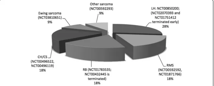
Figure2regroups 11 trials which were evaluating spe-cific pediatric non-CNS tumors.
Six studies were specifically assessing dosimetric issues
(NCT01502150, NCT02070393, NCT00592592,
NCT00850200, NCT00592293, NCT01751412). The
main goal of these prospective study is to obtain infor-mation on PBT effectiveness and toxicity, as well as wider dosimetric data from treatment plans, with the
aim to improve the planning and delivery of PBT for fu-ture approaches in pediatric patients.
Discussion
We present a summary of dosimetric and clinical results achieved with PBT in pediatric patients affected by non-CNS tumors.
At the time of our review, recent authoritative reports [11] have suggested that existing clinical data is still too preliminary to add new directions in clinical practice. Nevertheless, dosimetric advantages of PBT over photon techniques were well-known and knowledge of charged particles’ physical characteristics was consolidated. Bas-ing on the concept that dosimetric data could lead to a prediction of clinical outcomes, we tried to assess if– in
particular cases – PBT dosimetric benefits could
trans-late into clinical gains.
In primis, our research confirmed that PBT improves OARs dose-sparing; additionally, PBT with spot-scanning/ pencil beam scanning and IMPT modalities reduce neu-tron dose-contamination. These are crucial topics that in-crease radiation oncologists’ interest on PBT application in pediatric treatments, especially when radioresistent tu-mors arise next to critical anatomic sites require higher ra-diation doses or a dose-escalation [23,26] (e.g. paraspinal and parameningeal sarcoma).
The overall actual clinical results are confirmed as not-exhaustive to provide high-level evidences in all in-dications [11,46]. Among the retrieved articles, only few studies [12,30] reported long-term clinical data. Despite early tumor control and patients survival rates with PBT resulted high (Table 2), long-term clinical results are mandatory to assess treatment effectiveness and toler-ance in pediatric cohorts.
Costs and difficulties to access to PBT centers (de-pending on the small number of centers able to per-form PBT) are still elevated [69] and could affect the prospect to provide significant evidences on large sample sizes. To overcome these difficulties,
compre-hensive database or wider registries [11] could
sup-port analyses aimed to define the indications for PBT in the multidisciplinary management of pediatric non-CNS tumors.
Careful analyses have also to be performed to
balance PBT cost-effectiveness with requests of
managing potential late effects related to different RT approaches.
Moreover, further studies with appropriate methodolo-gies are required to answer to actual recurring questions, such as: What differences in OARs-sparing and toler-ance profiles can be observed between PBT and modern high-conformal photon techniques supported by sophisti-cated image-guidance systems [5]? Which potential out-comes/effects could be associated to PBT combined with new drugs [11]? Is radiobiology of protons clearly under-stood [6,11,68]? Indeed, one major limitation of previous published works– besides the retrospective nature of their
observations – was the comparison between PBT and
photon-RT with no advanced technologies [11,12]. Mod-ern technologies for image-guidance2and treatment plan-ning (e.g. image-guided IMRT on limited volumes) have improved dose-conformity and OARs dose-sparing even with photon-therapy [11,49]. Nevertheless, clinical results of modern photon-IGRT are still limited. Additionally, in IMRT treatments, developing OARs remain at risk of non-target radiation dose [11] and could be at increased
risk of SMNs as compared to conventional 3D-CRT [2]
due to low radiation doses [1].
In 2018 one of the first treatment planning com-parison between IMPT and highly sophisticated deep-inspiration breath hold Volumetric Modulated Arc
Therapy (VMAT) in adults has been published [71]:
even if VMAT was planned in a very experienced center, IMPT provided higher target coverage and
re-duced mean doses to the considered OARs [71].
These results are promising and could produce impli-cations in pediatric research, inducing radiation on-cologists to further consider PBT application and promote clinical trials.
Conclusion
Since the long life expectancy of patients is a major issue in oncological pediatric treatments, adequate analyses on RT late effects become necessary. PBT has provided
dosimetric advantages for normal tissues as compared to photon-RT, but long-term clinical results and compari-sons with modern photon-RT outcomes are still re-quired. Ongoing and future investigations should clearly define the role of PBT in the multimodal management of the most common pediatric non-CNS tumors. Abbreviations
3D-CRT:Three-Dimensional Conformal Radiotherapy; BS-PT: Breast-sparing
proton therapy; CH: Chordoma; CNS: Central Nervous System; CS: Chondrosarcoma; DVHs: Dose-volume histograms; HL: Hodgkin lymphoma; IMPT: modulated proton therapy; IMRT: Intensity-modulated radiotherapy; INPT: node proton therapy; ISRT: Involved-site radiotherapy; LC: Local control; OARs: Organs at risk; OS: Overall survival; PBT: Proton Beam Therapy; PFS: Progression free survival;
PRMS: Parameningeal rhabdomyosarcoma; PTOG/PROS/EPTN: Particle Therapy Co-Operative Group/Pediatric Radiation Oncology Society/European Particle Therapy Network; RB: Retinoblastoma; RMS: Rhabdomyosarcoma; RT: Radiation therapy (or radiotherapy); SMNs: Secondary malignant neoplasms;
SS: Surgical stabilization; VMAT: Volumetric Modulated Arc Therapy Acknowledgements
“Not applicable”. Authors’ contributions
AS gave substantial contributions to the conception of the study and drafted the work, RC was a major contributor in interpreting the data and writing the manuscript, MFP gave substantial contributions to the acquisition and analysis of data, BT gave substantial contributions to the acquisition and analysis of data, MM gave substantial contributions to the acquisition and analysis of data, AS substantively revised the work, AASI substantively revised the work, All authors read and approved the final manuscript.
Funding None.
Availability of data and materials
All data generated or analysed for this review are included in this published article.
Ethics approval and consent to participate “Not applicable”.
Consent for publication “Not applicable”. Competing interests
The authors declare that they have no competing interests. Received: 17 May 2019 Accepted: 17 December 2019
References
1. Levin W, Kooy H, Loeffler J, DeLaney T. Proton beam therapy. Br J Cancer. 2005;93(8):849–54.https://doi.org/10.1038/sj.bjc.6602754.
2. Greco C, Wolden S. Current status of radiotherapy with proton and light ion beams. Cancer. 2007;109(7):1227–38.
3. Fuss M, Hug EB, Schaefer RA, Nevinny-Stickel M, Miller DW, Slater JM, Slater JD. Proton radiation therapy (PRT) for pediatric optic pathway gliomas: comparison with 3D planned conventional photons and a standard photon technique. Int J Radiat Oncol Biol Phys. 1999;45(5):1117–26.
4. Hoppe BS, Flampouri S, Zaiden R, et al. Involved-node proton therapy in combined modality therapy for hodgkin lymphoma: results of a phase 2 study. Int J Radiat Oncol Biol Phys. 2014;89(5):1053–9.
5. Allen AM, Pawlicki T, Dong L, et al. An evidence based review of proton beam therapy: the report of astros emerging technology committee. Radiother Oncol. 2012;103(1):8–11.
6. Tian X, Liu K, Hou Y, Cheng J, Zhang J. The evolution of proton beam therapy: Current and future status. Mol Clin Oncol. 2018;8(1):15–21.https://
doi.org/10.3892/mco.2017.1499.
2
Nowadays image-guidance is being used in association with current knowledge on optimization/reduction of exposure to imaging doses [70]
7. Archambeau JO, Slater JD, Slater JM, Tangeman R. Role for proton beam irradiation in treatment of pediatric cns malignancies. Int J Radiat Oncol Biol Phys. 1992;22(2):287–94.
8. Palm A, Johansson KA. A review of the impact of photon and proton external beam radiotherapy treatment modalities on the dose distribution in field and out-of-field; implications for the long-term morbidity of cancer survivors. Acta Oncologica. 2007;46(4):462–73.https://doi.org/10.1080/
02841860701218626.
9. Lin R, Hug EB, Schaefer RA, Miller DW, Slater JM, Slater JD. Conformal proton radiation therapy of the posterior fossa: a study comparing protons with three-dimensional planned photons in limiting dose to auditory structures. Int J Radiat Oncol Biol Phys. 2000;48(4):1219–26.
10. Lomax AJ, Bortfeld T, Goitein G, et al. A treeatment planning inter-comparison of proton and intensity modulated photon radiotherapy. Radiother Oncol. 1999;51(3):257–71.
11. Weber DC, Habrand JL, Hoppe BS, et al. Proton therapy for pediatric malignancies: fact, figures and costs. A joint consensus statement from the pediatric subcommittee of PTCOG, PROS and EPTN. Radiother Oncol. 2018; 128:44–55.
12. Sethi RV, Shih HA, Yeap BY, et al. Second nonocular tumors among survivors of retinoblastoma treated with contemporary photon and proton radiotherapy. Cancer. 2014;120(1):126–33.
13. Lodge M, Pijls-Johannesma M, Stirk L, Munro AJ, De Ruysscher D, Jefferson T. A systematic literature review of the clinical and cost-effectiveness of hadron therapy in cancer. Radiother Oncol. 2007;83(2):110–22. 14. Olsen DR, Bruland ØS, Frykholm G, Norderhaug IN. Proton therapy–a
systematic review of clinical effectiveness. Radiother Oncol. 2007;83(2):123–32. 15. Brada M, Pijls-Johannesma M, De Ruysscher D. Proton therapy in clinical
practice: current clinical evidence. J Clin Oncol. 2007;25(8):965–70. 16. Indelicato DJ, Merchant T, Laperriere N, et al. Consensus Report From the
Stockholm Pediatric Proton Therapy Conference. Int J Radiat Oncol Biol Phys. 2016;96(2):387e392.
17. Krengli M, Hug EB, Adams JA, Smith AR, Tarbell NJ, Munzenrider JE. Proton radiation therapy for retinoblastoma: comparison of various intraocular tumor locations and beam arrangements. Int J Radiat Oncol Biol Phys. 2005; 61(2):583–93.
18. Lee CT, Bilton SD, Famiglietti RM, et al. Treatment planning with protons for pediatric retinoblastoma, medulloblastoma, and pelvic sarcoma: how do protons compare with other conformal techniques? Int J Radiat Oncol Biol Phys. 2005;63(2):362–72.
19. Andolino DL, Hoene T, Xiao L, Buchsbaum J, Chang AL. Dosimetric comparison of involved-field three-dimensional conformal photon radio-therapy and breast-sparing proton radio-therapy for the treatment of hodgkins lymphoma in female pediatric patients. Int J Radiat Oncol Biol Phys. 2011; 81(4):e667–71.
20. Hoppe BS, Flampouri S, Su Z, et al. Effective dose reduction to cardiac structures using protons compared with 3DCRT and IMRT in mediastinal hodgkin lymphoma. Int J Radiat Oncol Biol Phys. 2012;84(2):449–55. 21. Hoppe BS, Flampouri S, Su Z, et al. Consolidative involved-node proton
therapy for stage Ia-IIIb mediastinal hodgkin lymphoma: preliminary dosimetric outcomes from a phase ii study. Int J Radiat Oncol Biol Phys. 2012;83(1):260–7.
22. Knäusl B, Fuchs H, Dieckmann K, Georg D. Can particle beam therapy beimproved using helium ions? - a planning study focusing on pediatric patients. Acta Oncol. 2016;55(6):751–9.https://doi.org/10.3109/0284186X.
2015.1125016.
23. Weber DC, Trofimov AV, Delaney TF, Bortfeld T. A treatment planning comparison of intensity modulated photon and proton therapy for paraspinal sarcomas. Int J Radiat Oncol Biol Phys. 2004;58(5):1596–606. 24. Miralbell R, Lomax A, Cella L, Schneider U. Potential reduction of the incidence
of radiation-induced second cancers by using proton beams in the treatment of pediatric tumors. Int J Radiat Oncol Biol Phys. 2002;54(3):824–9.
25. Ladra MM, Edgington SK, Mahajan A, et al. A Dosimetric Comparison of Proton and Intensity Modulated Radiation Therapy in Pediatric Rhabdomyosarcoma Patients Enrolled on a Prospective Phase II Proton Study. Radiother Oncol. 2014;113(1):77–83.https://doi.org/10.1016/j.radonc.
2014.08.033.
26. Kozak KR, Adams J, Krejcarek SJ, Tarbell NJ, Yock TI. A dosimetric comparison of proton and intensity-modulated photon radiotherapy for pediatric parameningeal rhabdomyosarcomas. Int J Radiat Oncol Biol Phys. 2009;74(1):179–86.
27. Cotter SE, Herrup DA, Friedmann A, et al. Proton radiotherapy for pediatric bladder/prostate rhabdomyosarcoma: clinical outcomes and dosimetry compared to intensity-modulated radiation therapy. Int J Radiat Oncol Biol Phys. 2011;81(5):1367–73.
28. Yock T, Schneider R, Friedmann A, Adams J, Fullerton B, Tarbell N. Proton radiotherapy for orbital rhabdomyosarcoma: clinical outcome and a dosimetric comparison with photons. Int J Radiat Oncol Biol Phys. 2005;63(4):1161–8. 29. Hillbrand M, Georg D, Gadner H, Pötter R, Dieckmann K. Abdominal cancer
during early childhood: a dosimetric comparison of proton beams to standard and advanced photon radiotherapy. Radiother Oncol. 2008;89(2):141–9. 30. Mouw KW, Sethi RV, Yeap BY, et al. Proton radiation therapy for the
treatment of retinoblastoma. Int J Radiat Oncol Biol Phys. 2014;90(4):863–9. 31. Wray J, Flampouri S, Slayton W, et al. Proton therapy for pediatric Hodgkin lymphoma. Pediatr Blood Cancer. 2016;63(9):1522–6.https://doi.org/10.1002/
pbc.26044.
32. Hug EB, Sweeney RA, Nurre PM, Holloway KC, Slater JD, Munzen-rider JE. Proton radiotherapy in management of pediatric base of skull tumors. Int J Radiat Oncol Biol Phys. 2002;52(4):1017–24.
33. Habrand JL, Schneider R, Alapetite C, et al. Proton therapy in pediatric skull base and cervical canal low-grade bone malignancies. Int J Radiat Oncol Biol Phys. 2008;71(3):672–5.
34. Rutz HP, Weber DC, Sugahara S, et al. Extracranial chordoma: outcome in patients treated with function-preserving surgery followed by spot-scanning proton beam irradiation. Int J Radiat Oncol Biol Phys. 2007;67(2):512–20. 35. Rutz HP, Weber DC, Goitein G, et al. Postoperative spot-scanning proton
radiation therapy for chordoma and chondrosarcoma in children and adolescents: initial experience at Paul Scherrer Institute. Int J Radiat Oncol Biol Phys. 2008;71(1):220–5.
36. Ares C, Hug EB, Lomax AJ, et al. Effectiveness and safety of spot scanning proton radiation therapy for chordomas and chondrosarcomas of the skull base: first long-term report. Int J Radiat Oncol Biol Phys. 2009;75(4):1111–8. 37. Staab A, Rutz HP, Ares C, et al. Spot-scanning-based proton therapy for
extracranial chordoma. Int J Radiat Oncol Biol Phys. 2011;81(4):e489–96. 38. Rombi B, Ares C, Hug EB, et al. Spot-scanning proton radiation therapy for
pediatric chordoma and chondrosarcoma: clinical outcome of 26 patients treated at Paul Scherrer Institute. Int J Radiat Oncol Biol Phys. 2013;86(3):578–84. 39. Timmermann B, Schuck A, Niggli F, et al. Spot-scanning proton therapy for
malignant soft tissue tumors in childhood: first experiences at the Paul Scherrer Institute. Int J Radiat Oncol Biol Phys. 2007;67(2):497–504. 40. Leiser D, Calaminus G, Malyapa R, et al. Tumour control and quality of life in
children with rhabdomyosarcoma treated with pencil beam scanning proton therapy. Radiother Oncol. 2016;120(1):163–8.
41. Childs SK, Kozak KR, Friedmann AM, et al. Proton radiotherapy for Para-meningeal rhabdomyosarcoma: clinical outcomes and late effects. Int J Radiat Oncol Biol Phys. 2012;82(2):635–42.
42. Weber DC, Ares C, Albertini F, et al. Pencil beam scanning proton therapy for pediatric Parameningeal Rhabdomyosarcomas: clinical outcome of patients treated at the Paul Scherrer Institute. Pediatr Blood Cancer. 2016; 63(10):1731–6.https://doi.org/10.1002/pbc.25864.
43. Vern-Gross TZ, Indelicato DJ, Bradley JA, Rotondo RL. Patterns of failure in pediatric Rhabdomyosarcoma after proton therapy. Int J Radiat Oncol Biol Phys. 2016;96(5):1070–7.https://doi.org/10.1016/j.ijrobp.2016.08.028. 44. Mizumoto M, Murayama S, Akimoto T, et al. Preliminary results of proton
radiotherapy for pediatric rhabdomyosarcoma: a multi-institutional study in Japan. Cancer Med. 2018;7(5):1870–4.https://doi.org/10.1002/cam4.1464. 45. Rombi B, DeLaney TF, MacDonald SM, et al. Proton radiotherapy for
pediatric Ewing’s sarcoma: initial clinical outcomes. Int J Radiat Oncol Biol Phys. 2012;82(3):1142–8.
46. Leroy R, Benahmed N, Hulstaert F, Van Damme N, De Ruysscher D. Proton therapy in children: a systematic review of clinical effectiveness in 15 pediatric cancers. Int J Radiat Oncol Biol Phys. 2016;95(1):267–78. 47. Kaste SC, Chen G, Fontanesi J, Crom DB, Pratt CB. Orbital development in
long-term survivors of retinoblastoma. J Clin Oncol. 1997;15(3):1183–9. 48. Schipper J, Tan KE, van Peperzeel HA. Treatment of retinoblastoma by
precision megavoltage radiation therapy. Radiother Oncol. 1985;3:117–32. 49. Eldebawy E, Parker W, Rahman WA, Freeman CR. Dosimetric study of
current treatment options for radiotherapy in retinoblastoma. Int J Radiat Oncol Biol Phys. 2012;82(3):e501–5.
50. Rombi B, Vennarini S, Vinante L, Ravanelli D, Amichetti M. Proton radiotherapy for pediatric tumors: review of first clinical results. Ital J Pediatr. 2014;40(1):74.
51. Jemal A, Siegel R, Xu J, Ward E. Cancer statistics, 2010. CA Cancer J Clin. 2010;60(5):277–300.
52. Kung FH, Schwartz CL, Ferree CR, et al. A randomized trial comparing chemotherapy with chemoradiotherapy for children and adolescents with stages I, IIa, IIIa1 hodgkin disease: a report from the children’s oncology group. J Pediatr Hematol Oncol. 2006;28(6):362–8.
53. Specht L, Yahalom J, Illidge T, et al. Modern radiation therapy for hodgkin lymphoma: field and dose guidelines from the international lymphoma radiation oncology group (ILROG). Int J Radiat Oncol Biol Phys. 2014;89(4):854–62. 54. Wolden SL, Chen L, Kelly KM, Nachman J, et al. Long-term results of CCG
5942: a randomized comparison of chemotherapy with and without radiotherapy for children with hodgkin’s lymphoma – A report from the children’s oncology group. J Clin Oncol. 2012;30(26):3174.
55. Holtzman A, Flampouri S, Li Z, Mendenhall NP, Hoppe BS. Proton therapy in a pediatric patient with stage III hodgkin lymphoma. Acta Oncol. 2013;52(3):592–4. 56. Oeffinger KC, Mertens AC, Sklar CA, et al. Chronic health conditions in adult
survivors of childhood cancer. N Engl J Med. 2006;355(15):1572–82. 57. Bhatia S, Yasui Y, Robison LL, et al. High risk of subsequent neoplasms
continues with extended follow-up of childhood hodgkins disease: report from the late effects study group. J Clin Oncol. 2003;21(23):4386–94. 58. Dores GM, Metayer C, Curtis RE, et al. Second malignant neoplasms among
long-term survivors of hodgkins disease: a population-based evaluation over 25 years. J Clin Oncol. 2002;20(16):3484–94.
59. Travis LB, Hill DA, Dores GM, et al. Breast cancer following radiotherapy and chemotherapy among young women with hodgkin disease. Jama. 2003; 290(4):465–75.
60. Girinsky T, van der Maazen R, Specht L, et al. Involved-node radiotherapy (INRT) in patients with early hodgkin lymphoma: concepts and guidelines. Radiother Oncol. 2006;79(3):270–7.
61. Eichenauer DA, Aleman BMP, Andrè M, et al. Hodgkin lymphoma: ESMO Clinical Practice Guidelines for diagnosis, treatment and follow-up. Ann Oncol. 2018;29(Supplement 4):iv19–29.
62. Tseng YD, Cutter DJ, Plastaras JP, et al. Evidence-based review on the use of proton therapy in lymphoma from the particle therapy cooperative group (PTCOG) lymphoma subcommittee. Int J Radiation Oncol Biol Phys. 2017;99(4):825e842. 63. Noël G, Feuvret L, Ferrand R, Boisserie G, Mazeron JJ, Habrand JL.
Radiotherapeutic factors in the management of cervical-basal chordomas and chondrosarcomas. Neurosurgery. 2004;55(6):1252–62.
64. Catton C, O’Sullivan B, Bell R, et al. Chordoma: long-term follow-up after radical photon irradiation. Radiother Oncol. 1996;41(1):67–72. 65. Hall EJ. Intensity-modulated radiation therapy, proton, and the risk of
second cancers. Int J Radiat Oncol Biol Phys. 2006;65(1):1–7. 66. Sangkhathat S. Current management of pediatric soft tissue sarcomas.
World J Clin Pediatr. 2015;4(4):94–105.
67. Pastore G, Peris-Bonet R, Carli M, Martínez-García C, de Toledo JS, Steliarova-Foucher E. Childhood soft tissue sarcomas incidence and survival in european children (1978–1997): report from the automated childhood cancer information system project. Eur J Cancer. 2006;42(13):2136–49. 68. Merchant TE. Clinical controversies: proton therapy for pediatric tumors.
Semin Radiat Oncol. 2013;23, Elsevier:97–108.
69. Waddle MR, Sio TT, Van Houten HK, et al. Photon and proton radiation therapy utilization in a population of more than 100 million commercially insured patients. Int J Radiat Oncol Biol Phys. 2017;99(5):1078–82. 70. Hess CB, Thompson HM, Benedict SH, et al. Exposure risks among children
undergoing radiation therapy: considerations in the era of image guided radiation therapy. Int J Radiat Oncol Biol Phys. 2016;94(5):978–92.https://doi.
org/10.1016/j.ijrobp.2015.12.372Epub 2016 Jan 5.
71. Baues C, et al. Proton versus photon deep inspiration breath hold technique in patients with hodgkin lymphoma and mediastinal radiation. Radiat Oncol. 2018;13:122.
Publisher’s Note
Springer Nature remains neutral with regard to jurisdictional claims in published maps and institutional affiliations.