Corresponding author
Giulia Boschi | School of Dentistry, Department of Medical and Surgery Specialties, Radiological Sciences and Public Health, University of Brescia | Piazza Spedali Civili 1, I-25123, Brescia (BS) | Italy
Phone: +39 030383424 | Fax: 030303194 | Email: [email protected] 10.32067/GIE.2020.34.02.11
Società Italiana di Endodonzia. Production and hosting by Ariesdue. This is an open access article under the CC BY-NC-ND license (http://creativecommons.org/licenses/by-nc-nd/4.0/).
Peer review under responsibility of Società Italiana di Endodonzia
Riccardo Tonini1 Giulia Boschi1*
Stefano Alessandro Salgarello1
1School of Dentistry, Department of Medical and Surgery Specialties, Radiological Sciences and Public Health, University of Brescia, Brescia, Italy
ABSTRACT
Aim: External cervical resorption (ECR) assessment and its management planning are
often difficult. This work proposes a standardized workflow for ECR treatment consid-ering the 3D-classification of the lesion by Patel and assesses its clinical applicability to seven preliminary clinical cases.
Summary: ECR cases were detected from medical and dental history, clinical data
and a conventional radiography; a Cone Beam Computed Tomography (CBCT) with a small field of view (FOV) was performed too. A both 2D and 3D-classification was applied on each resorption. After studying better the clinical aspect, especially the accessibility of the lesion, the restorability of the tooth was assessed and the approach was decided between external repair, with or without endodontic treatment, or internal repair. Reparative tissue was removed and the defect was managed through new generation composite resins, mineral trioxide aggregate (MTA) or other cements. A pulp capping was performed in one case and a canal treatment in the others. Clinical and radiographic checks assessed both quality and adequacy of the treatments im-mediately and over time.
Key learning points:
• A standardized workflow could be useful for ECR treatment.
• CBCT is essential for determination of ECR extension and an appropriate treatment
planning.
• ECR management should be conservative, but should also remove the reparative
tissue.
• ECR management could be performed within one or two appointments.
• A follow-up is required to evaluate the treatment over time. The main outcome is
the survival of the element, the secondary outcomes are the absence of resorption progression, no symptoms and healthy periodontal values.
CASE SERIES
Management of seven external cervical
resorptions
KEYWORDS cone-beam computed tomography, conservative treatment, tooth resorption
Received 2020, June 16 Accepted 2020, July 4
Introduction
T
ooth resorption is a condition associated with a process re-sulting in a loss of dentin, ce-mentum and/or bone (1). It is physiologic and desirable in primary teeth as it aids exfoliation of the deciduous tooth and thus facilitates erup-tion of the permanent successor (2), where-as it is pathologic and undesirable in adult teeth as it leads to irreversible damage (3). A preliminary classification of tooth re-sorption considers its localization: it could be an internal or external resorption. Ex-ternal resorption may be further subclas-sified in external inflammatory resorption, external replacement resorption, external cervical resorption (ECR) and external surface resorption (4).ECR is known also as invasive cervical resorption or cervical root resorption (5). It is a relatively uncommon and highly aggressive form of external tooth resorp-tion that starts from the cervical region, below the epithelial attachment, and pro-gressively destroys the tooth structure (4). It is assumed that there must be damage to the periodontal ligament and cementum in combination with a stimulating factor, that can induce and maintain the activity of clastic cells. The histopathogenesis con-sists of three main stages: resorptive (ini-tiation), resorptive (propagation) and re-parative (remodelling), that can occur in parallel in different areas of the same le-sion, while it extends deeper towards the pulp and the middle and apical thirds of the root (3, 6).
Epidemiology shows no difference in sex or age: also young people have been found to be involved (7, 8). Maxillary anterior teeth are usually the most affected (8). Aetiology is rather unclear. Some potential predisposing factors have been already proposed by Heithersay at the end of the last century: orthodontic treatment, dental trauma, internal bleaching, intraoral sur-gical and restorative treatments (9). Mavri-dou investigated also other factors, among which: viruses, systemic diseases, poor oral health, parafunctional habits,
orthog-nathic surgery, music wind instruments, malocclusion, frenulum tension, extraction of a neighboring tooth, eruption disorders and cracks. In the majority of clinical cas-es, more than one potential predisposing factor was identified, indicating that ECR is multifactorial, not idiopathic. Further-more, in 99% of the examined cases, al-most one potential predisposing factor was recognized (7). Nevertheless, a sure causal relationship has not yet been found for any factors and a minority of cases is still with-out a plausible explanation.
Clinical features vary from nothing to a small defect at the gingival margin, which is directly detectable through a visual in-spection, to a pink coronal discoloration of the crown, as a result of soft tissue color shining through the thin tooth tissue over-lying the resorptive cavity, resulting in ultimate cavitation of the enamel. The pro-cess is usually painless because the pulp remains protected by a thin layer of pre-dentin and pre-dentin until late, when a pulpal or periodontal infection supervenes (9, 10). As the resorption is usually without symp-toms through a very long period or even never become symptomatic, the lesion could proceed without anyone becoming aware of it, until the tooth could not be longer saved. So, due to the nature of this lesion, early detection is essential and dif-ficult at the same time. It is very often dis-covered thanks to a routine conventional radiography during a visit as an incidental radiographic finding or, alternatively, be-cause the clinic is rather advanced (5). Until a few years ago, the only way to as-sess the extent of the lesion was the con-ventional X-ray. Here the lesion appears as radiolucency; a radiopaque line demarcates the root canal from the adjacent irregular radiolucency when the latter is close to the canal (11). The first classification by Heithersay was two-dimensional: it cate-gorized ECR according to its extension into the root and its proximity to the root canal, as follows.
Class 1: a small invasive resorptive lesion near the cervical area with shallow pene-tration into dentine.
lesion that has penetrated close to the cor-onal pulp chamber, but shows little or no extension into the radicular dentine. Class 3: a deeper invasion of dentine by resorbing tissue, not only involving the coronal dentine, but also extending at least to the coronal third of the root. Class 4: a large invasive resorptive process that has extended beyond the coronal third of the root canal and may involve almost the entire root (12).
However, the classification above has got limitations, as it does not describe the true nature of ECR (resorptive and reparative) and, above all, it does not read the third dimension; furthermore when there is only a small starting lesion, especially if on the buccal or lingual aspect of the tooth, it may not completely be seen in conven-tional X-ray (13, 3). Therefore the perio-dontal radiography can result in inade-quate assessment or even misdiagnosis and also poor management of root resorp-tion (14).
Fortunately, the Cone Beam Computed Tomography (CBCT) has become available in Endodontics thanks to a reduced field of view (FOV) with a reasonable exposure dosage and good quality, optimizing ex-posure parameters on an individual basis: the principle as low as reasonably achiev-able (ALARA) is therefore followed (15). Several specialist societies published po-sition statements, among which the Euro-pean Society of Endodontology (ESE): they recommended the CBCT to assess and/or manage root resorption, which clinically appears to be potentially amenable to treat-ment (16, 17).
Patel suggested a new three-dimensional classification for ECR based on additional information available from CBCT. It takes into account three parameters thanks to three-dimensional analysis, as follows.
Height (coronal-apical extent) of the lesion:
the lesion
1) is at cement-enamel junction level or coronal to the bone crest (supracrestal); 2) extends into coronal third of the root and apical to the bone crest (subcrestal); 3) extends into mid third of the root; 4) extends into apical third of the root.
Circumferential spread, which is graded
according to the lesion maximum spread within the root:
A) ≤90°
B) >90° to ≤180° C) >180° to ≤270° D) >270°
Proximity to the root canal, which can be
best assessed using axial CBCT views: d) lesion con f i ned to dent i ne; p) probable pulpal involvement (18). The combination of each of the three pa-rameters (e.g. 1Ad) provides to the clini-cian a more accurate determination of the whole extension of ECR and can lead to a more appropriate treatment planning. The same author proposed different strat-egies for the management of ECR: external repair with or without endodontic treat-ment, internal repair, intentional reim-plantation and periodic review until ex-traction. He suggested materials and he tried to connect each strategy with both the Heithersay and the three-dimensional classification, as far as possible (19). Some case reports have already been pub-lished about ECR and its treatment (20-27), but without following a specific workflow. We arranged a preliminary workflow since 2015, then we upgraded it according the ECR classification by Patel (18) and the last ECR review (19). Here we suggest the last version of the workflow for a standardized approach in the anterior as well as in the posterior mouth, as schematized in the diagram of Figure 1. We have also already applied this workflow to new seven cases of ECR, as described below.
We found the cases of ECR thanks to med-ical and dental anamnesis, clinmed-ical data - including probing - and a conventional radiography or directly thanks to conven-tional radiography. We asked the patient about any symptoms, if not directly re-ferred. Then we performed a CBCT with a small FOV, giving a relatively low effec-tive dose and obtaining specific and clear information that made us know the real three-dimensional extension of the lesion. Only in one case we directly chose a larg-er CBCT FOV, as the patient needed also a cysts removal in the right half-jaw, and
in another case an ECR was discovered in a CBCT for an implant plan. Consequently we applied a double classification, both by Heithersay and Patel.
We studied again and better the clinical aspect, especially the accessibility of the lesion, and subsequently we assessed the restorability of the tooth, that was fortu-nately always possible. Then we decided the approach between external repair, with or without endodontic treatment, or inter-nal repair, following instructions and ad-vices in the review by Patel (19). Especial-ly, we tried to remove resorptive tissue when possible and managed the resorptive defect through new generation composite resins, MTA or other cements. We applied a calcium hydroxide cement for pulp cap-ping in one case and used warm gutta-per-cha, endodontic cement and eventually mineral trioxide aggregate (MTA) for the canal treatment in the others.
Clinical and radiographic checks assessed both quality and adequacy of the treat-ments immediately and over time.
Report
#Case1It was the first case in chronological order (year 2015).
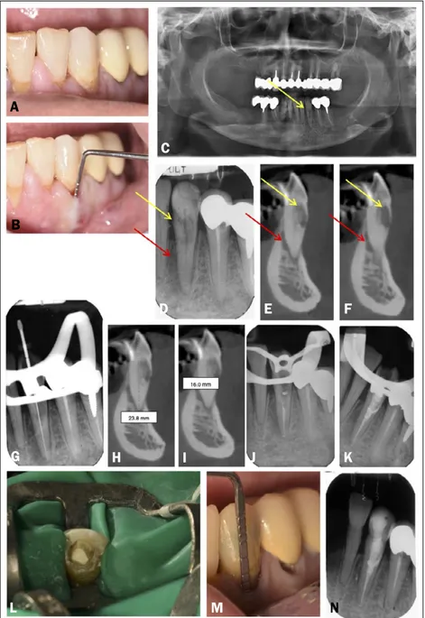
A 17-years-old male presented requiring an orthodontic treatment. At the first vis-it, the oral health seemed good. Any soft oral tissues lesions, any caries or tooth abnormalities were directly detectable; some previous conservative and endodon-tic therapies were present. Periodontal pocket values were between 1 mm up to 3 mm, without bleeding or suppuration on probing. Instead, a malocclusion was as-sessed: therefore we prescribed an pantomography in order to plan the ortho-dontic treatment. Here we noticed a strange Figure 1.
Figure 2 (video 1). ECR case 1. A) Preoperative radiograph of a 3.7 showed ECR (yellow arrow) and preliminary signs of apical periodontitis (red arrow). B) Frontal, C) sagittal and D) axial CBCT slices confirmed the extensive nature of ECR which extends apically and completely around the distal canal (yellow arrows). E) Access cavity preparation revealed the partially reparative nature of ECR. F, G) MTA was located all around the access of the distal canal. (H) Immediate post-fill radiograph. I) Frontal.
orthodontic treatment or any of the other possible predisposing factors and he did not report any symptoms. The clinic aspect of the tooth confirmed to be silent: any pink spots, any gingival irregularities, any strange small cavitations in the crown third part and any deeper periodontal probing values could be detected.
However, we decided to investigate better taking a periodontal radiography with a paralleling technique (Figure 2A): it re-vealed a more widespread radiolucent ar-ea in the distal side of the crown starting from cervical area and extending to the coronal third of the root. Besides, a small suffering area could be seen around the last third of the distal root, whereas it was not detectable in the orthopantomography. According to this further information the resorption was really present and was clas-sified as a class 3 of ECR, following Heither-say classification.
We took a CBCT (Figure 2B-D, M) to un-derstand its real extension. A 3D classifi-cation was not available in 2015, however now we could further classify the lesion as a 3Dp stage following Patel classifica-tion. The CBCT showed also a more im-portant radiolucency into the wave bone around the distal root, therefore the posi-tive answer to vitality test could be due to the mesial part of the teeth. These findings could be explained by CBCT high sensi-tivity: CBCT imaging was demonstrated to have twice the odds of detecting a peria-pical lesion than traditional periaperia-pical radiography (28).
We investigated again the clinical aspects, we decided for the restorability of the ele-ment 3.7, but, because the direct accessi-bility was limited, we concluded that an external repair of the root (the first choice in our workflow for restorable teeth) may have been really difficult. Therefore we chose an internal repair with MTA thanks to its biocompatibility and sealing ability (29), followed by a conventional canal treat-ment with warm gutta-percha.
Therefore, at the first appointment, we ap-plied a stamp technique: we impress the occlusal anatomy and then we performed a classical cavity access (30, 31) with dia-mond burs before and ultrasonic (US) tips radiolucency at the distal cervical area of
his second lower left molar 3.7.
We suspected an ECR. The tooth was a normal responder to both the vitally test (we used the cold test) and the percussion test. The general medical anamnesis was negative; the patient could not recall a his-tory of dental trauma, another previous
A B C
D E F
Figure 2. ECR case 1. J) sagittal, K) axial CBCT slices and L) 3D rendering of two years recall. Comparison between (M) pre-operative and (N) two years recall CBCT.
O) Four years recall periapical X-ray.
later (Figure 2E). Thanks to the micro-scope, we directly saw the reparative tis-sue and we removed it with an endodontic dedicated US tip, also to have access to the coronal third of the canal into the distal root. We located MTA both to the mesial and to the distal aspect of the canal access and we compacted with a micro-brush (Figure 2F). A periodontal X-ray was taken to make us sure that the MTA was correct-ly located (Figure 2G, video 1) and we waited 7 days to allow the complete hard-ening of the material under a temporary restoration. After a week the patient un-derwent the second appointment: we checked the MTA seal, we completed the root canal treatment and performed a di-rect restoration. An immediate post-oper-ative periapical radiography (Figure 2H)
was taken. After 2 years another CBCT was prescribed in order to plan an implant guided surgery of 1.3 and the imaging was extended to the lower jaw, according with the patient: it confirmed a good seal of the previous resorptive lesion and the radiolu-cent periapical area was no longer detecta-ble (Figure 2I-N). Meanwhile, clinical con-trols and periapical X-rays have been taken until 2019 (4 years recall): the tooth has always been clinically asymptomatic, the periapical radiographs confirmed the heal-ing bone process and the absence of new areas of resorption (Figure 2O).
#Case 2
The second case began in 2017.
A 17-years-old female was seen in urgency because of a poorly located strong sponta-neous pain. Her lower left canine 3.3 showed a pink discoloration – therefore we supposed a resorption (11, 10, 3) – and the vitality test led to the following diagnosis: pulpitis (32). Any caries could be detected. A periodontal radiography was taken using a paralleling technique (Figure 3A): it re-vealed a radiolucent lesion with irregular and poorly defined borders in the cervical aspect of tooth extending to the coronal third of the root. Besides, a radiolucency could already be seen into the alveolar bone around the distal aspect of the first third of the root. The diagnosis of ECR was con-firmed. According to this preliminary in-formation the resorption was classified as class 3 following Heithersay classification. The patient confirmed that her general med-ical anamnesis was negative; her main pre-disposing factor was a previous fixed ortho-dontic treatment (9, 7, 33), followed by a post-orthodontic splinting and periodical recalls. She highlighted that she did not experienced any symptoms before, but this finding is not strange as ECR becomes symp-tomatic (if it happens) at its last stages (11). We opened immediately the access cavity in urgency, we put a temporary restoration and we planned a CBCT to further confirm the diagnosis of ECR and assess the true position and extension of the radiolucency. We took a CBCT (Figure 2B-E, video 2) some days later. A 3D classification was not yet available, however now we could classify
J K L
M
Figure 3 (video 2). ECR case 2. A) Preoperative radiograph of a 3.3 showed ECR (yellow arrow) and signs of lateral periodontitis (red arrows). B) Frontal and C, D) axial CBCT slices confirmed both the extensive nature of ECR (yellow arrows) which extends apically the bone crest and a lingual bone lesion (red arrows). E) 3D rendering of the tooth. F) Radiographic working length. G) A gutta-percha cone was temporarily put into the
canal. H) Defect restoration using SuperEBA®Cement and gutta-percha apical seal. I) Schematic view of the fully restored tooth: a fiber post was put into the middle and coronal third of the root. J) One year recall. K) Two years recall.
the lesion as a 2Bp stage following Patel classification. The CBCT showed also an important lingual radiolucency into the wave bone, that broke off the neighboring cortical bone (28).
We reinvestigated the clinical aspects, we
decided for the restorability of the element 3.3 thanks to external repair and endodon-tic treatment. Therefore we removed the temporary restoration and we recorded the working length through a periapical X-ray (Figure 3F) as electronic apex detector did not work correctly because of the perio-dontal tissues interference. We shaped the canal and put a temporary gutta-percha point in it (Figure 3G). We opened a surgi-cal flap and we removed both granuloma-tous tissue that was profusely bleeding and the resorbing tissue thanks to US inserts and operative microscope. Before filling the resorptive defect with SuperEBA® Ce-ment, we controlled the integrity of gut-ta-percha point inserted into the canal to prevent cement from sliding downward. We put SuperEBA® Cement in the defect and closed the flap. Another appointment followed: the apical third of the tooth was filled with endodontic cement and warm gutta-percha (Figure 3H), an hollow fiber post was located because of its favorable mechanical properties (34) and a direct composite restoration was performed. An immediate post-operative periapical radi-ography (Figure 3I) was carried out. Clin-ical and periapClin-ical X-ray has been taken until 2019 - 1 (Figure 3J) and 2 (Figure 3K) years recall: the tooth has been clinically asymptomatic; the periapical radiographs show a healing bone process around the coronal third of the root and no new areas of resorption.
#Case 3
The third case was discovered in November 2018.
A 12-years-old female showed a discolor-ation on her second upper right incisor tooth 1.2 during her fixed orthodontic treatment. We immediately thought that a resorption was happening, as in that case the resorbed structure is replaced by high-ly vascular tissue which could be seen through a thinner enamel, especially in anterior teeth (11, 10, 3). The element was not symptomatic; however endodontic tests were performed: it responded to cold like her neighboring teeth and it was non-ten-der to pressure and percussion.
The general medical anamnesis was
nega-A B C E F G K J D H I
Figure 4 (video 3). ECR case 3. A) Preoperative radiograph of a 1.2 showed ECR (yellow arrow). B, C, D) Sagittal CBCT slices let know the real extension of ECR (yellow arrows) that was mainly palatal and supracrestal. E) 3D rendering of the tooth. F) Immediate post-operative X-ray. G) One year recall.
tive; about her dental history, any other pre-disposing factors could be detected, except for the orthodontic treatment (9, 7, 33). A periodontal radiography was taken using a paralleling technique (Figure 4A): it re-vealed an irregular radiolucency in the me-sial side of the crown just above the bone crest, not extending apically into the radic-ular dentine, but quite deeper through the
coronal dentine, maybe touching the pulp chamber. According to this preliminary information, the lesion was classified as class 2 ECR, following Heithersay classifi-cation.
We took a CBCT (Figure 2B-E) to understand the real extension of the resorption: it was mainly palatal and supracrestal and fortu-nately did not reach the endodontic system. A 3D classification was recently available and we classified the lesion as a 1Bd stage following Patel classification.
We investigated again the clinical aspects, especially the palatal accessibility and we decided for the restorability of the element 1.2. We proposed a direct external repair, trying to maintain tooth vitality.
Therefore we opened a surgical flap and we removed both granulomatous tissue that was profusely bleeding and the re-sorbing tissue thanks to US inserts and operative microscope. The pulp of the chamber was directly observable only under the microscope. So we decided to try a quick direct pulp capping through calcium hydroxide (Dycal, Dentsply Siro-na) (35). Then we performed a direct com-posite restoration followed by many fin-ishing and polfin-ishing steps to remove respectively main irregularities and min-ute scratches (36): we used diamond burs, flexible and semi-flexible abrasive ments (cups, points and wheels), instru-ments coated with abrasives (abrasive disks and strips) and abrasive polishing paste compounds (37). The flap was closed. An immediate post-operative periapical radiography (Figure 4F) was taken. Clin-ical checks with annual periapClin-ical X-ray were planned: the tooth has been clini-cally asymptomatic, has kept its vitality and 1 year recall X-ray (Figure 4G) and probing (video 3) have been satisfactory.
#Case 4
The fourth case was discovered in Novem-ber 2018 too.
A 52-years-old smoker female was referred by her oncologist. She suffered from lung cancer with multiple metastases, mainly localized to the spine, as well as hyperten-sion and panic attacks. She needed to be-gin an intravenous bisphosphonate
thera-A B C
D E
Figure 5 (video 4). ECR case 4. A) Preoperative aspect and B) probing of 3.3. C) Root of 3.3 showed a strange radiolucency in the orthopantomography (yellow arrow). D) Preoperative radiograph of a 1.2 highlighted an ECR (yellow arrow) and a bone lateral radiolucen-cy along the mesial aspect of the root (red arrow). E, F) CBCT sagittal slices showed the exact extension of both the resorptive lesion in the coronal and, partially, middle third of the root (yellow arrows) and the periradicular lesion (red arrows). G, H) Radiographic working length was taken. I) Radiographic resorption level was recorded too. J) The apical third was filled with warm gutta-percha. K) The middle and coronal third were filled with MTA. L) Occlusal view before composite coronal restoration. M) One year probing and N) Two-dimensional X-ray.
ramic multiple-unit fixed dental prosthesis on the upper jaw and two conventional metal-ceramic bridges in the lower jaw. Some caries were evident apically to the metal-ceramic crowns and the loss of tooth-supporting structures around ele-ments 4.2, 4.1 and 3.1 resulted in tooth mobility. There was a generalized loss of clinical attachment, mainly through re-cessions, with no periodontal pocket depths greater than 3 mm, except for the buccal aspect of element 3.3 (Figure 5A), where we recorded the value of 7.5 mm (Figure 5B) with an immediate suppuration on probing. The same element did not re-spond both to the vitality and percussion test. The patient did not report any symp-toms here or elsewhere in the mouth; how-ever, this data was relative as she was tak-ing a high-dose morphine therapy because of her systemic condition.
The patient gave us her latest orthopanto-mography (Figure 5C) that showed some extremely decayed teeth behind the met-al-ceramic crowns, a mandibular cyst be-hind the elements 4.3, 4.2 and 4.1 and 3.1 and a radiolucent area around the root of the element 3.3, as a sign of periodontitis. Besides tooth 3.3 had got a strange radio-lucency also inside the root, just below the cervical area.
A periodontal radiography of 3.3 was im-mediately carried out using a paralleling technique (Figure 5D): it confirmed a mot-tled resorptive lesion into the coronal third of the root, and showed better the bone radiolucency, especially along the distal aspect of the tooth. According to this pre-liminary information, the lesion was clas-sified as a class 3 of ECR, following Heither-say classification.
We supposed the occlusal trauma because of the metal-ceramic opposite prosthesis and the poor oral hygiene as ECR predis-posing factors.
A CBCT scan (Figure 5E, F, video 4) was prescribed to assess the true nature, exten-sion, and position of both the mandibular cist and the radiolucency inside 3.3. The slices confirmed the provisional diagnosis of ECR that had spread at the beginning of the middle third of the root and had reached the pulp; besides the bone radio-py and the oncologist prescribed a previous
dental control to prevent osteonecrosis of the jaws (ONJ). Our dental visit detected a very poor oral hygiene, a single metal-ce-A B C D E F G H I J K L M N
lucency appeared to be in the lingual side above all. We used the new Patel classi-fication: it was a 3Cp lesion. We investi-gated again the clinical aspects, and we initially proposed a direct external repair of the root: we would have begun with an endodontic access without removing the previous distal third class restoration, put a temporary gutta-percha point into the root canal, opened a flap, directly removed the resorptive tissue and re-paired with MTA or SuperEBA® Cement, closed the flap and concluded the canal therapy. We also explained that this ther-apy might have been performed together with 4.3, 4.2, 4.1 and 3.1 extraction and mandibular cyst removal. But the patient refused any unnecessary oral surgery, so we decided for an internal reparation. We opened the endodontic access and we performed a preliminary shaping at the radiographic working length (Figure 5G, H), as the electronic apex detector did not work correctly because of the periodontal tissues interference, as well as in case 2. We performed canal and resorption filling through a sandwich technique, the same proposed in order to deal with root per-foration (38): we closed the apical third (the last 8 mm: working length of 24 mm minus resorption length of 16 mm) (Fig-ure 5H, I) with warm gutta-percha (Fig(Fig-ure 5J), then we used MTA up to the cervical area. The MapSystem (Simit Dental) was very useful. As more than 5 mm of MTA were necessary, we put the material par-tially in a first appointment and partial-ly in a second one (Figure 5K, L), so that the material was able to become complete-ly hard. We ultimate the therapy with a common composite coronal restoration. Subsequently we removed elements 4.3, 4.2, 4.1 and 3.1 because of their advanced mobility, the mandibular cyst and the other teeth with deep caries in the upper jaw. After two months the patient started the intravenous bisphosphonate and we planned almost monthly dental visits. The tooth has always been asymptomat-ic until today and the buccal periodontal probing depth (PPD) has become shorter: it was 5 mm two months later, 4.5 mm three months later, 1 mm seven month
later and 0.5 mm one year later (Figure 5M). At the same time, the two-dimen-sional radiographic aspect became better and no bone lesions could be seen at 1 year recall (Figure 5N).
#Case 5
The fifth case was detected in January 2019.
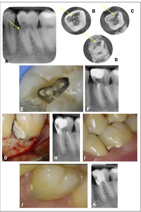
The patient, a 40-years-old male, with good oral health, presented in urgency with the chief complaint of intermittent spontaneous pain related to his lower left side. Any tooth showed directly detectable caries or abnormalities. We performed the cold test on every element and we find that element 3.6 suffered from pulpitis. A periodontal radiography was carried out using a paralleling technique (Figure 6A): it revealed a radiolucent lesion with irregular and poorly defined borders in the cervical mesial aspect of tooth crown extending to the coronal third of the root. An ECR was diagnosed. According to this preliminary information, the resorption was classified as class 3 following Heithersay classification.
The patient general medical history was negative; any ECR predisposing factor could be detected. He highlighted that he did not experienced any symptoms before, until this late stage (11).
We immediately took a CBCT (Figure 6B-D), which confirmed that the resorptive lesion was mainly in the cervical mesial aspect of the teeth and that it had devel-oped deeper and deeper into the dentin until it had reached the pulp chamber. We classified the lesion as a 3Cp stage according with Patel classification. We reinvestigated the clinical aspects, we decided for the restorability of the element 3.6 and we proposed an endo-dontic treatment with internal repair. In the same appointment, we saved a copy of the occlusal anatomy in order to perform a stamp technique (30, 31) for the following coronal restoration, so that it could be quick and precise; then we opened a conservative access cavity and we removed both the pulp tissue and the reparative tissue. We performed a classi-cal canal therapy with warm
gutta-per-Figure 6 (video 5). ECR case 5. A) Preoperative radiograph of a 3.6 showed ECR (yellow arrow) on the mesial aspect of the tooth. B, C, D) Axial CBCT slices let know the real extension of ECR (yellow arrows) that reached the endodontic system. E, F) Internal filling of the lesion with MTA. G, H) External filling of the lesion with SuperEBA®Cement. I) One week clinical control. J) Ten moths clinical and K) radiographic control.
cha, except for the lesion, which we tried to fill with MTA (Figure 6E), but we did not achieve the whole defect reparation. Therefore we put a moistened mi-cro-brush head in order to allow MTA hardening and a temporary coronal res-toration, we took an X-ray in order to
un-derstand how much material was located and exactly where (Figure 6F), and we planned also an external approach. In the following appointment we opened a flap, we located SuperEBA® Cement in the defect (Figure 6G) and closed the flap again. An X-ray confirmed that now the defect was completely filled (Figure 6H). We put the rubber dam and completed the case with direct composite restora-tion, using the stamp technique. 1 week later soft tissues were very good (Figure 6I). A 10 months recall showed a good clinical (Figure 6J) and radiographic heal-ing (Figure 6K): especially, we noticed that interdental papilla was grown; only a small defect was produced on the me-sial aspect of the SuperEBA® Cement filling because of an error during the pro-fessional oral hygiene. The patient has been asymptomatic until today.
All the steps until the last control are better reported in video 5.
#Case 6
The sixth ECR case was found in July 2019.
The patient, a 21-years-old female, present-ed in urgency complaining about a spon-taneous pain in her left lower side. She had also noticed a small localized swelling between teeth in her lower jaw. At the clin-ical control, the patient was caries free and had never had a restoration before. All the teeth were pulp tested using refrigerant spray and electrical pulp test, and all re-sponded within normal limits. Teeth were not tender to percussion and gave a normal sound on percussion, except for element 3.6. On its mesial buccal aspect there was the soft tissue swelling the girl was com-plaining about. Periodontal probing was consistently 3 mm or less and without bleeding or suppuration on probing, but when the periodontal probe reached the mesial buccal site of 3.6 the value became 7 mm with suppuration. We diagnosed a per-iodontal abscess in a healthy mouth. A periodontal radiography was carried out using a paralleling technique (Figure 7A): an irregular radiolucent area was discernible on the mesial aspect of 3.6 crown, especially in the cervical area. An ECR was diagnosed. According to this preliminary informa-A B C D E F G H I J K
Figure 7 (video 6). ECR case 6. A) Preoperative radiograph of a 3.6 showed ECR (yellow arrow) on the
mesial aspect of the tooth. B) Frontal, C) sagittal and D, E, F, G, H, I) axial CBCT slices let know the real extension of ECR (yellow arrows) that reached the
endodon-tic system.J) 3D rendering of the tooth. K) External repair of the defect with a bulk-fill composite. L) Intraoperative radiographic control. M) Postoperative radiographic control. N, O) Conservative restoration. tion, the resorption was classified as class 2 following Heithersay classification. The medical anamnesis was negative; the patient reported no history of trauma, or-thodontic treatment or any other predis-posing factor.
An antibiotic therapy was prescribed and a small FOV CBCT was planned. Seven
days later, the abscess had reduced itself and the patient was asymptomatic, so we performed both the CBCT and the tooth treatment. Sagittal (Figure 7B), frontal (Figure 7C) and axial (Figure 7D-I) slices confirmed that the ECR coronal-apical extension had stopped just above the cor-onal third of the root, but it had developed mainly into the coronal dentine and it had reached the endodontic system, despite we had assessed the pulp vitality. The lesion is an 1Bp stage following Patel clas-sification. The overall extension could be well understood with a 3D rendering im-age (Figure 7J), thanks to the volume ren-dering tool (39).
Considering the 3D rendering image, we performed a further clinical investigation and we proposed to the patient a root ca-nal treatment, as the endodontic system was involved, combined with an external root repair in the same appointment. We performed the first step of the stamp technique, saving the occlusal anatomy (30, 31). A conservative cavity access let us remove the pulp and carry out a pre-flaring. We temporary put a K-file into the buccal mesial canal in order to localize it also from an external view. We opened a surgical flap, we removed the reparative tissue with a bur, we put a gutta-percha point into the canal in order to maintain the patency during the subsequent exca-vation of ECR defect and we performed a composite resin restoration (19). A new generation bulk-fill composite (Beauti-fil-Bulk, Shofu) (Figure 7K) was chosen, as it has got a low polymerization shrink-age and let us reduce the time needed (36, 40). We underwent all the finishing and polishing steps under water: a starting diamond bur delivered a deeper and more aggressive cut; flexible abrasive points and coarse, medium, fine and super fine discs produced a smooth and glossy surface finish (37). Then we closed the flap, we put the rubber dam around the tooth and finished the endodontic treatment with warm gutta-percha (Figure 7L). We used the same bulk-fill composite for direct coronal reconstruction, except for a cap-ping layer of conventional hybrid compos-ite (40). 7 days later, a postoperative
radi-A B C
D E F
G H I J
K L
ographic control was taken (Figure 7M), followed by a further polishing step of coronal restoration with abrasive polish-ing paste compounds, to reduce and smooth any roughness and surface scratch created by finishing instruments (Figure 7N, O). All the steps are shown in video 6. The patient has always been asympto-matic until today and the last mesial buc-cal periodontal probing depth is 3 mm, without bleeding and suppuration on probing.
#Case 7
The seventh case was detected in October 2019.
A 52-year-old male patient in good general health and no oral symptoms had under-gone some extractions some years ago be-cause of carious processes. He presented again requiring an implant treatment. Therefore we performed a new clinical analysis of the mouth without notice any pathological signs and took a CBCT in or-der to plan a guided surgery: here a radio-lucent area was noticed in element 3.7 (Figure 8A-D). It was clearly a 3Bp stage of ECR by Patel classification.
In this case we directly had a definitive diagnosis of ECR, referring to the diagram in Figure 1. A clinical reinvestigation with a periapical X-ray (Figure 8E) followed. A normal response to both cold and percus-sion test was tested on element 3.7. The endodontic diagnosis was normal pulp and normal periapex. The tooth was mesially inclined as 3.6 was lost in youth and nev-er replaced: the 3.6 extraction might have worked as predisposing factor. We pro-posed external debridement and restoration because of a direct mesial accessibility. In the following appointment, a flap was opened and the bulk of reparative tissue was removed with a bur, exposing the pulp tissue, and replaced with SuperEBA® Ce-ment (Figure 8F, G). As the initial debride-ment caused the pulp exposure, an or-thograde endodontic treatment was initi-ated: we removed the pulp and put a tem-porary restoration with metacresylacetate. Subsequently, we performed also an inter-nal reparation with MTA (Figure 8H), a classical canal therapy with warm gut-ta-percha and a composite restoration with a stamp technique (30, 31) (Figure 8I). All the main steps of clinical management are shown in video 7. Controls were planned and the patient has been asymptomatic until today.
Discussion
ECR is a relatively uncommon form of ex-ternal tooth resorption, often undiagnosed or misdiagnosed (4, 5). It invades the tooth from the PDL, apical to the epithelial attach-Figure 8 (video 7).
ECR case 7. A, B) Frontal, C) sagittal and D) axial CBCT slices let diagnose an ECR of 3.7 (yellow arrows). E) Two-dimensional X-ray radiograph of 3.7 showed the resorption (yellow arrow) on the mesial aspect of the tooth. F) External defect restoration using SuperEBA®Cement. G) Two-dimensional X-ray of external defect restoration. H) Internal defect restoration using MTA. I) Postoperative radiographic control.
A B D
I H
C
ment, and develops through both resorp-tive and repararesorp-tive stages (3, 6).
Aetiology is still rather unclear: orthodontic treatment, iatrogenic or accidental dental trauma and poor oral health have been the most frequent predisposing factors detected until today and ECR itself has been proposed to be mainly multifactorial (7).
The lesion could be asymptomatic until later stages, therefore an early diagnosis is difficult and the process is often acciden-tally found with a radiographic control, where it appears as a barely up to a highly discernable radiolucency (11).
Heithersay developed the first classifica-tion system that divides the condiclassifica-tion into 4 classes according to the extent of the lesion in periapical X-rays (12). But the true nature and real extension of an ECR defect cannot be assessed on convention-al radiographs: a three-dimensionconvention-al anconvention-al- anal-ysis is mandatory. CBCT scans clarify the exact location of the entry point of gran-ulation tissue into the root, the dimension of the resorption and the presence of den-tin surrounding the resorption tissue. Besides, it can diagnose also starting le-sions undetectable with periapical X-rays (16, 17). Nowadays a three-dimensional classification by Patel is available: it con-siders height (coronal-apical extent) of the lesion, its circumferential spread and proximity to the root canal and it is easi-ly applicable (18).
Different management approaches have been proposed too, also linked with the three-dimensional classification (19). However, a common standardized work-flow has not been published yet. Therefore we tried to developed it and test its clinical applicability: it start from a preliminary ECR diagnosis, but then a small FOV CBCT is mandatory to confirm it, to study the real extension of the lesion and to choose, with a clinical reinvestigation, the more appropriate treatment planning. When the restorability is assessed, external and in-ternal repair are the most recommended clinical management options.
We used our protocol in 7 consecutive ECR cases (Table 1). Only in case 7 the ECR lesion was directly diagnosed on CBCT imaging, nevertheless the workflow has
been applicable from that step: it has prov-en to be flexible.
Six of the seven cases of ECR were de-tected in the post-lateral lower jaw and only in case 3 an anterior element was affected, in contrast with previous find-ings (8). Both male and female patients presented to our attention: ECR does not seem to be linked to the sex of the patient, as already established (7). These patients were of different ages, even young people: case 3 was diagnosed in a 12-years-old female; which agrees with the new liter-ature data (7). Predisposing factors have not always been found; when verified they were: a previous (case 2) or contex-tual (case 3) orthodontic treatment, oc-clusal trauma and poor oral health (case 4) or a previous surgery in the same oral area - extraction of a neighboring tooth - (case 7) (7). 3 cases were symptomatic on a late stage (case 2, 5, 6): as a matter of fact, these patients were seen in urgency; whereas the other 4 ECR (case 1, 3, 4, 7) were casually found (11).
Everyone accepted a CBCT study which turned out to be useful especially for pulp involvement investigation and management choice (19): fortunately, in all cases the el-ements were maintainable with an external or internal repair, associated with an en-dodontic treatment except for case 3, where an external approach associated with a pulp capping was mandatory, especially considering the age of the patient (35). Three main rules were followed: we tried to remove reparative tissue, as its incom-plete removal is likely to result in the re-currence of ECR (19), we apply the most conservative approach for each case and we perform the management within one or two appointments. Stamp technique succeed in maintaining most of the coronal tissue and allowing a fast and anatomical-ly precise restoration (case 1, 5, 6, 7) (30, 31). Different materials, such as MTA (case 1, 4, 5, 7), SuperEBA® Cement (case 2, 5, 7) and composite (case 3, 7), were already proposed for clinical ECR management because of their sealing ability and biocom-patibility (19) and turned out to be useful in our clinical practice. In cases 5 and 7, a combination of techniques and of materials
Table 1
Overview of the 7 ECR clinical cases
CASE TOOTH PATIENT SEX PATIENT AGE (years old) PREDISPOSING FACTOR(S) MANAGEMENT HEALING FIGURE VIDEO
1 3.7 male 17 not revealed Internal repair with MTA + root canal therapy yes 2 1 2 3.3 female 17 previous orthodontic treatment External repair with SuperEBA® Cement + root canal therapy yes 3 2 3 1.2 female 12 contemporary orthodontic treatment External repair with resin composite + pulp capping yes 4 3 4 3.3 female 52 occlusal trauma and poor oral health + root canal therapy (sandwich Internal repair with MTA
technique) yes 5 4 5 3.6 male 40 not revealed Internal repair with MTA + external repair with SuperEBA® Cement
+ root canal therapy yes 6 5 6 3.6 female 21 not revealed composite + root canal therapyExternal repair with bulk-fill yes 7 6 7 3.7 male 52 same oral area: extraction previous surgery in the
of element 3.6
External repair with SuperEBA® Cement + internal repair with MTA
+ root canal therapy yes 8 7
was even used, in order to fill the irregular form of the lesions.
All patients were extremely motivated to follow-up visits, with both clinical and radiographic checks in order to evaluate the treatment over time: it is an extremely important aspect. The main outcome is the survival of the element, the secondary out-comes are the absence of resorption pro-gression, no symptoms and a healthy per-iodontal depth. All the seven cases de-scribed have reached both the primary and secondary outcomes until today, however their follow-up must continue.
Additional supporting information:
Video Legends
Video 1. ECR case 1 – element 3.7. The main steps of the treatment under operating mi-croscope are here reported: reparative
tis-sue removal, MTA location, irrigation, root canal filling.
Video 2. ECR case 2 – element 3.3. The consecutive axial CBCT slices are compiled as a video.
Video 3. ECR case 3 – element 1.2. 1 year recall probing: a light bleeding on probing matched with healthy periodontal probing values.
Video 4. ECR case 4 – element 3.3. The con-secutive sagittal CBCT slices are compiled as a video.
Video 5. ECR case 5 – element 3.6. The main steps of the treatment under operat-ing microscope are here reported: access cavity, pulp removal, reparative tissue re-moval, irrigation, root canal filling with gutta-percha, internal MTA location, ex-ternal SuperEBA® cement location, 10 months recall.
main steps of the treatment under operat-ing microscope are here reported: occlusal anatomy stamp, access cavity, pulp remov-al, preliminary shaping, surgical flap, re-parative tissue removal, external defect repair with a bulk-fill composite, root ca-nal treatment, adhesive restoration. Video 7. ECR case 7 – element 3.7. The main steps of the treatment under operating mi-croscope are here reported: reparative tissue removal, external SuperEBA® cement loca-tion and linked X-ray, internal MTA localoca-tion, root canal therapy, adhesive restoration.
Conclusions
A correct and predictable management of ECR needs a standardized workflow (in-cluding a CBCT study), a quick and defin-itive conservative approach and a fol-low-up overtime. This case series has succeeded in applying the rules above for the treatment of 7 external cervical root resorptions. However, both long-term fol-low-up visits and other clinical cases are necessary to confirm the real usefulness of the method.
Clinical Relevance
The workflow above can help clinicians to have a correct diagnosis of ECR cases but also in the decision making process regarding their treatment plan.
Conflict of Interest
The authors deny any conflict of interest.
Acknowledgements
The authors certify that there are no spe-cial acknowledgements: we did not receive institutional, private or corporate financial support for the work and there were not contributors to the article other than the authors accredited.
References
1. American Association of Endodontists. AAE glossa-ry of endodontic terms, 10th edn Chicago: American Association of Endodontists; 2020.
2. Harokopakis-Hajishengallis E. Physiologic root re-sorption in primary teeth: molecular and histolog-ical events. J Oral Sci 2007;49:1-12.
3. Patel S, Mavridou AM, Lambrechts P. External cer-vical resorption-part 1: histopathology, distribution and presentation. Int Endod J 2018;51:1205-23. 4. Patel S, Saberi N. The ins and outs of root
resorp-tion. Br Dent J 2018;224:691-9.
5. Patel J, Beddis HP. How to assess and manage ex-ternal cervical resorption. Br Dent J 2019;227:695-701.
6. Patel S, Lambrechts P, Shemesh H, Mavridou AM. European society of endodontology position state-ment: external cervical resorption. Int Endod J 2018;51:1323-26.
7. Mavridou AM, Bergmans L, Barendregt D, Lambre-chts P. Descriptive analysis of factors associated with external cervical resorption. J Endod 2017;43:1602-10.
8. Jeng PY, Lin LD, Chang SH et al. Invasive cervical resorption-distribution, potential predisposing factors, and clinical characteristics. J Endod 2020;46:475-82. 9. Heithersay GS. Invasive cervical resorption: an analysis of potential predisposing factors. Quintes-sence Int 1999;30:83-95.
10. Kandalgaonkar SD, Gharat LA, Tupsakhare SD, Gabhane MH. Invasive cervical resorption: a review. J Int Oral Health 2013;5:124-30.
11. Heithersay GS. Clinical, radiologic, and histopatho-logic features of invasive cervical resorption. Quin-tessence Int 1999;30:27-37.
12. Heithersay GS. Invasive cervical resorption follow-ing trauma. Aust Endod J 1999;25:79-85.
13. Patel K, Mannocci F, Patel S. The assessment and management of external cervical resorption with peri-apical radiographs and cone-beam computed tomog-raphy: a clinical study. J Endod 2016;42:1435-40. 14. Gunst V, Mavridou A, Huybrechts B, et al. External
cervical resorption: an analysis using cone beam and microfocus computed tomography and scanning electron microscopy. Int Endod J 2013;46:877-87. 15. Patel S, Brown J, Pimentel T, et al. Cone beam com-puted tomography in Endodontics - a review of the literature. Int Endod J 2019;52:1138-52.
16. Patel S, Durack C, Abella F et al. European society of endodontology position statement: the use of CBCT in endodontics. Int Endod J 2014;47:502-4. 17. Patel S, Brown J, Semper M, et al. European society
of endodontology position statement: use of cone beam computed tomography in endodontics: euro-pean society of endodontology (ESE) developed by. Int Endod J 2019;52:1675-8.
18. Patel S, Foschi F, Mannocci F, Patel K. External cer-vical resorption: a three-dimensional classification. Int Endod J 2018;51:206-14.
19. Patel S, Foschi F, Condon R, et al. External cervical resorption: part 2 – management. Int Endod J 2018;51:1224-38.
20. Coyle M, Toner M, Barry H. Multiple teeth showing invasive cervical resorption - an entity with little known histologic features. J Oral Pathol Med 2006;35:55-57.
21. Pace R, Giuliani V, Pagavino G. Mineral trioxide ag-gregate in the treatment of external invasive resorp-tion: a case report. Int Endod J 2008;41:258-66. 22. Schwartz RS, Robbins JW, Rindler E. Management
of invasive cervical resorption: observations from three private practices and a report of three cases. J Endod 2010;36:1721-30.
23. Yu VS, Messer HH, Tan KB. Multiple idiopathic cer-vical resorption: case report and discussion of management options. Int Endod J 2011;44:75-85. 24. Patel S, Saberi N. External cervical resorption as-sociated with the use of bisphosphonates: a case series. J Endod 2015;41:742-8.
25. Salzano S, Tirone F. Conservative nonsurgical treat-ment of class 4 invasive cervical resorption: a case series. J Endod 2015;41:1907-12.
26. Shemesh A, Ben Itzhak J, Solomonov M. Minimally invasive treatment of class 4 invasive cervical re-sorption with internal approach: a case series. J Endod 2017;43:1901-08.
27. Alqedairi A. Non-invasive management of invasive cervical resorption associated with periodontal pocket: a case report. World J Clin Cases 2019;7:863-71.
28. Aminoshariae A, Kulild JC, Syed A. Cone-beam com-puted tomography compared with intraoral radio-graphic lesions in endodontic outcome studies: a systematic review. J Endod 2018;44:1626-31. 29. Tawil PZ, Duggan DJ, Galicia JC. Mineral trioxide
aggregate (MTA): its history, composition, and clin-ical applications. Compend Contin Educ Dent 2015;36:247-52.
30. Alshehadat SA, Halim MS, Carmen K, Fung CS. The stamp technique for direct class II composite res-torations: a case series. J Conserv Dent 2016;19:490-3.
31. Murashkin A. Direct posterior composite restorations using stamp technique-conventional and modified: a case series. Int J Dentistry Res 2017;2:3-7 32. Zanini M, Meyer E, Simon S. Pulp Inflammation
Diagnosis from Clinical to Inflammatory Mediators: A Systematic Review. J Endod 2017;43:1033-52. 33. Roscoe MG, Meira JB, Cattaneo PM. Association of
orthodontic force system and root resorption: a systematic review. Am J Orthod Dentofacial Orthop 2015;147:610-26.
34. Bovolato L, Tonini R, Boschi G et al. Novel hollow fiber sandwich composite post system: mechanical characteristics. Minerva Stomatol 2020;69:63-71. 35. Duncan HF, Galler KM, Tomson PL, et al. European
Society of Endodontology position statement: Man-agement of deep caries and the exposed pulp. Int Endod J 2019;52:923-34.
36. Bonfanti C, Barabanti N, Piccinelli G, et al. Micro-biological characterization and effect of resin com-posites in cervical lesions. J Clin Exp Dent 2017;9:e40-e45.
37. Jefferies SR. Abrasive finishing and polishing in restorative dentistry: a state-of-the-art review. Dent Clin North Am. 2007;51:379-97.
38. Tonini R. Root canal repair with the MTA sandwich technology. Dental Tribune 2014;14.
39. Venkatesh E, Elluru SV. Cone beam computed to-mography: basics and applications in dentistry. J Istanb Univ Fac Dent 2017;51:S102–S121. 40. Chesterman J, Jowett A, Gallacher A, Nixon P.
Bulk-fill resin-based composite restorative materials: a review. Br Dent J. 2017;222:337-44.