The authors’ full names, academic de-grees, and affiliations are listed in the Ap-pendix. Address reprint requests to Dr. Sanna-Cherchi at the Division of Ne-phrology, Columbia University, College of Physicians and Surgeons, New York, NY 10032, or at ss2517@ cumc . columbia .-edu, or to Dr. Katsanis at the Center for Disease Modeling, 466A Nanaline Duke Bldg., Box 3709, Duke University Medical Center, Durham, NC 27710, or at katsanis@ cellbio . duke . edu.
Drs. Lopez-Rivera, Liu, Verbitsky, Ander-son, and Capone contributed equally to this article.
This article was published on January 25, 2017, at NEJM.org.
DOI: 10.1056/NEJMoa1609009 Copyright © 2017 Massachusetts Medical Society.
BACKGROUND
The DiGeorge syndrome, the most common of the microdeletion syndromes, affects
multiple organs, including the heart, the nervous system, and the kidney. It is caused
by deletions on chromosome 22q11.2; the genetic driver of the kidney defects is
unknown.
METHODS
We conducted a genomewide search for structural variants in two cohorts: 2080
pa-tients with congenital kidney and urinary tract anomalies and 22,094 controls. We
performed exome and targeted resequencing in samples obtained from 586
addi-tional patients with congenital kidney anomalies. We also carried out funcaddi-tional
studies using zebrafish and mice.
RESULTS
We identified heterozygous deletions of 22q11.2 in 1.1% of the patients with
con-genital kidney anomalies and in 0.01% of population controls (odds ratio, 81.5;
P = 4.5×10
−14). We localized the main drivers of renal disease in the DiGeorge
syn-drome to a 370-kb region containing nine genes. In zebrafish embryos, an induced
loss of function in snap29, aifm3, and crkl resulted in renal defects; the loss of crkl
alone was sufficient to induce defects. Five of 586 patients with congenital urinary
anomalies had newly identified, heterozygous protein-altering variants, including
a premature termination codon, in CRKL. The inactivation of Crkl in the mouse model
induced developmental defects similar to those observed in patients with congenital
urinary anomalies.
CONCLUSIONS
We identified a recurrent 370-kb deletion at the 22q11.2 locus as a driver of kidney
defects in the DiGeorge syndrome and in sporadic congenital kidney and urinary tract
anomalies. Of the nine genes at this locus, SNAP29, AIFM3, and CRKL appear to be
critical to the phenotype, with haploinsufficiency of CRKL emerging as the main
genetic driver. (Funded by the National Institutes of Health and others.)
A B S T R A C T
Genetic Drivers of Kidney Defects
in the DiGeorge Syndrome
E. Lopez-Rivera, Y.P. Liu, M. Verbitsky, B.R. Anderson, V.P. Capone, E.A. Otto,
Z. Yan, A. Mitrotti, J. Martino, N.J. Steers, D.A. Fasel, K. Vukojevic, R. Deng,
S.E. Racedo, Q. Liu, M. Werth, R. Westland, A. Vivante, G.S. Makar, M. Bodria,
M.G. Sampson, C.E. Gillies, V. Vega-Warner, M. Maiorana, D.S. Petrey, B. Honig,
V.J. Lozanovski, R. Salomon, L. Heidet, W. Carpentier, D. Gaillard, A. Carrea,
L. Gesualdo, D. Cusi, C. Izzi, F. Scolari, J.A.E. van Wijk, A. Arapovic, M. Saraga-Babic,
M. Saraga, N. Kunac, A. Samii, D.M. McDonald-McGinn, T.B. Crowley, E.H. Zackai,
D. Drozdz, M. Miklaszewska, M. Tkaczyk, P. Sikora, M. Szczepanska,
M. Mizerska-Wasiak, G. Krzemien, A. Szmigielska, M. Zaniew, J.M. Darlow, P. Puri,
D. Barton, E. Casolari, S.L. Furth, B.A. Warady, Z. Gucev, H. Hakonarson,
H. Flogelova, V. Tasic, A. Latos-Bielenska, A. Materna-Kiryluk, L. Allegri, C.S. Wong,
I.A. Drummond, V. D’Agati, A. Imamoto, J.M. Barasch, F. Hildebrandt, K. Kiryluk,
R.P. Lifton, B.E. Morrow, C. Jeanpierre, V.E. Papaioannou, G.M. Ghiggeri,
A.G. Gharavi, N. Katsanis, and S. Sanna-Cherchi
Original Article
D
eletions on chromosome 22q11.2
are the most common cause of the
Di-George syndrome (Online Mendelian
In-heritance in Man [OMIM] number, 188400) and
the velocardiofacial syndrome (OMIM number,
192430) and constitute the most common
micro-deletion disorder in humans, with an estimated
prevalence of 1 in 2000 to 4000 live births.
1-3The
DiGeorge syndrome is a debilitating,
multisys-temic condition that features (with variable
ex-pressivity) cardiac malformations, velopharyngeal
insufficiency, hypoparathyroidism with
hypocal-cemia, and thymic aplasia with immune
defi-ciency. Additional phenotypes include
neurodevel-opmental defects and urogenital malformations.
4-7The long arm of chromosome 22 contains
multi-ple segmental duplications (low-copy repeats) that
confer a predisposition to genomic
rearrange-ments.
8-10Most frequently, the DiGeorge
syn-drome is caused by a de novo heterozygous
dele-tion of approximately 2.5 mb in length on
chromosome 22q11.2 between low-copy repeats
(LCR22) A and D. Less frequently, the syndrome
is the result of deletions between LCR22 A and B,
between B and D, or between C and D.
5,8,11Congenital kidney and urinary tract
anoma-lies are present in approximately 30% of the
pa-tients with the DiGeorge syndrome.
4,6,12,13Al-though some of the hallmarks of this syndrome
(e.g., heart defects) can be attributed in part to
haploinsufficiency of TBX1,
14-18the identity of the
genes that are responsible for such congenital
kidney and urinary tract anomalies remains
un-known.
M e t h od s
Study Samples
We studied samples obtained from 2666 patients
affected by congenital kidney and urinary tract
anomalies at 26 international centers, along with
additional samples provided by the Chronic
Kid-ney Disease in Children Study (see the Methods
section and Table S1 in the Supplementary
Ap-pendix, available with the full text of this article
at NEJM.org). We performed genomewide
geno-typing for analysis of copy-number variations in
2080 of these samples. Among an additional 586
patients with congenital kidney and urinary tract
anomalies, we performed either whole-exome
se-quencing (in 60 samples) or targeted
next-gener-ation sequencing and Sanger validnext-gener-ation (in 526
samples). All the patients provided written
in-formed consent. The study was approved by the
institutional review board at each site.
(Descrip-tions of the patients, analyses of convolution
de-fects in zebrafish, analysis of tissue localization
in the patients and zebrafish, and the generation
and analysis of a mouse model are provided in the
Methods section in the Supplementary Appendix.)
Genetic Analyses
Using samples obtained from 2080 patients with
congenital kidney and urinary tract anomalies and
22,094 controls, we performed genomewide
geno-typing for analysis of copy-number variation by
means of high-density single-nucleotide
polymor-phism (SNP) microarrays manufactured by
Illu-mina (1820 samples) or Affymetrix (260 samples),
as described previously.
19-21We also performed
whole-exome sequencing on samples obtained
from 60 patients through the Yale Center for
Mendelian Genomics, as described previously.
22-24We performed high-throughput next-generation
sequencing for eight genes in the 370-kb
mini-mal region of overlap for the DiGeorge syndrome
in samples obtained from an additional 526
pa-tients using microfluidic
polymerase-chain-reac-tion capture (Fluidigm) coupled with
next-gener-ation sequencing on an Illumina 2500 HiSeq
system, as described previously.
25,26We subjected
CRKL
coding exons to next-generation
resequenc-ing in samples obtained from 576 unaffected
controls and from 1152 patients affected by IgA
nephropathy but with normal results on renal
ultrasonography. These additional 1728 controls
were matched with the patients according to their
ancestral origin and recruitment site.
R e s u l t s
Patients with 22q11.2 Deletions
In a genomewide search for rare copy-number
variations in a discovery cohort of 1752 patients
with congenital kidney and urinary tract
anoma-lies, we identified deletions at the chromosome
22q11.2 locus in 11 patients (0.6%) and in 3 of
22,094 population controls (0.01%; odds ratio for
patients versus controls, 46.4; P = 9.7×10
−11). An
analysis of breakpoints indicated that all deletions
in the 11 patients overlapped with the common
deletion between LCR22 A and D: 2 patients
had the classic deletion of DNA between A and D,
1 patient had a smaller deletion (bounded by
B and D), and 8 patients had the smallest
dele-tion, between C and D (Table 1 and Fig. 1, and
Table S2 in the Supplementary Appendix).
Of the 11 patients, 9 had renal agenesis or
hypodysplasia, and 2 had an isolated ureteric
phenotype, findings indicating that the 22q11.2
locus between LCR22 C and D is critical for
hu-man nephrogenesis and is possibly specific for
renal agenesis or hypodysplasia (in 9 of 765
pa-tients [1.2%]). In a replication study involving an
additional 328 patients with renal agenesis or
hy-podysplasia, we identified 3 (0.9%) with 22q11.2
deletions, for a total of 14 patients with these
deletions (Table 1 and Fig. 1). Taken together, we
identified deletions at this locus in 12 of 1093
patients (1.1%) with renal agenesis or
hypodyspla-sia, as compared with 3 of 22,094 controls (odds
ratio, 81.5; P = 4.5×10
−14), which implicates
dele-tions at the locus associated with the DiGeorge
syndrome as the second most common genomic
disorder of the kidney and urinary tract after the
17q12 microdeletion associated with the renal
cysts and diabetes syndrome (Table S2 in the
Supplementary Appendix).
19,27Of the 14 patients with the 22q11.2 deletion,
Patients P1, P2, and replication Patient 1 (RP1)
carried the most frequent deletion between LCR22
A and D; in Patient P2, the deletion was
inher-ited from the mother, in whom a clinical
diag-nosis of the DiGeorge syndrome had not been
made. In all the patients, the molecular genetic
diagnosis preceded a clinical diagnosis of the
DiGeorge syndrome (in which some but not all
features of the syndrome were observed) and
had a direct effect on the patient’s treatment. In
patients with deletions between LCR22 B and D
and C and D, additional urinary tract defects
con-sisted of vesicoureteral reflux in 6 patients and
hypospadia in 1 patient. Extrarenal defects were
rare and mild in patients with deletions between
LCR22 B and D and C and D. The deletion
be-tween LCR22 C and D that was identified in
Patient P10 was also observed in a sibling who
was affected by left renal agenesis and an
unde-scended testis.
The analysis of the breakpoints in
copy-num-ber variation that was based on SNP array data
localized the critical region for the phenotype
as-sociated with congenital kidney and urinary tract
anomalies to a locus of approximately 370 kb,
which contains nine genes (Fig. 1, and Table S3
in the Supplementary Appendix). This region
ex-cluded the gene encoding T-box 1 (TBX1), a protein
that is not expressed in the murine embryonic
kidney,
28so Tbx1-null mice have normal early
nephrogenesis (Fig. S2 in the Supplementary
Ap-pendix). Interrogation of the “22q and You”
data-base from the Children’s Hospital of Philadelphia
identified kidney malformations in 2 of 10 patients
with the 22q11.2 deletion between LCR22 C and
D (Table S4 in the Supplementary Appendix).
Finally, we reexamined the three controls with
22q11.2 deletions; one carried the typical
dele-tion between LCR22 A and D, one the deledele-tion
between B and D, and one the deletion between
C and D. We obtained clinical records for
Con-trol C1, who had Parkinson disease, congenital
hypoparathyroidism, and advanced chronic
kid-ney disease (Table S5 in the Supplementary
Ap-pendix). Thus, we found a patient with
undiag-nosed DiGeorge syndrome with renal involvement
among our 22,000 population controls, which
provided further support for the pathogenicity of
the 22q11.2 deletion in patients with congenital
kidney and urinary tract anomalies. After removal
of this patient from the control data set, the
strength of association between 22q11.2 deletions
and renal agenesis or hypodysplasia increased
further (12 of 1093 patients vs. 2 of 22,093
con-trols, P = 8.5×10
−15; odds ratio, 123.7).
Functional Modeling in Zebrafish
The genetic data suggested that dosage
perturba-tion of one or more of the nine genes in the
micro-deletion on 22q11.2 is a driver of congenital kidney
and urinary tract anomalies. We had previously
found that systematic in vivo suppression of
ex-perimentally tractable genes within a deletion
copy-number variant, coupled with quantitative
phenotyping, can determine the contribution of
specific transcripts to disease associated with
copy-number variation in humans.
29-31We first sought to establish a phenotypic
sur-rogate for congenital kidney and urinary tract
anomalies in zebrafish embryos. Previous
stud-ies in mice and humans have shown the critical
role of the gene encoding ret proto-oncogene (RET)
for kidney development and branching
morpho-genesis.
32-35We therefore injected an established
morpholino oligonucleotide (MO) against RET
36into zebrafish that were engineered to enable
visualization of the developing nephron and then
examined the convolution of the pronephros (the
earliest developmental stage in the zebrafish) at
T ab le 1 . Characteristics of 14 Patients with Microdeletions at the 22q11.2 Locus Associated with the DiGeorge Syndrome.* Patient No. Deletion Type† Kidney Phenotype Kidney Location Urinary Tract Phenotype Extrarenal Phenotype Sex Nationality Age at Diagnosis Outcome Discovery cohort P1 A–D Renal hypodysplasia (right), cortical cyst (left) Bilateral None None M French 14 days Normal renal function P2 A–D Renal agenesis Right None Developmental delay M Italian 10 yr Normal renal function P3 B–D Normal renal paren -chyma Left Left ureterovescical junction obstruc -tion, hypospadia Arched palate, mild an -timongoloid slants M Macedonian 3 mo M ild r en al in su ff ic ie n cy P4 C–E Renal agenesis Left Vesicoureteral reflux, megaureter Ventricular septal defect M French 3 mo M ild r en al in su ff ic ie n cy , m ic ro al b u m in u ri a P5 C–D Renal hypodysplasia, cortical cysts Left Left megaureter Undescended testis, epicanthus, chest hemangioma M Czech 1 mo Normal renal function P6 C–D Renal agenesis Bilateral None None M French Prenatal Termination of preg -nancy P7 C–D Reflux nephropathy None Vesicoureteral reflux None F European American 16 yr M ild r en al in su ff ic ie n cy P8 C–D Renal hypodysplasia Left None None F Polish 9 yr Normal renal function P9 C–D Renal hypodysplasia and scarring Bilateral Ureteropelvic junction obstruction, vesico -ureteral reflux, blad -der diverticuli Phimosis M Italian Birth Chronic kidney disease P10 C–D Renal agenesis Left Vesicoureteral reflux Undescended testis M Swiss Birth Normal renal function P11 C–D Renal agenesis Left None None F Italian 21 yr Normal renal function Replication cohort RP1 A–D Renal agenesis Right None None M Polish 8 yr Normal renal function RP2 B–D Renal agenesis Left Vesicoureteral reflux None F Polish 1 yr Normal renal function RP3 B–D Renal hypodysplasia Left Vesicoureteral reflux None M Polish 4 mo Normal renal function * A total of 1752 patients were included in the discovery cohort and 328 patients in the replication cohort. F denotes female, M male, and RP patients in the replication cohort. † According to the megabase coordinates from the Human Genome 19 release, the boundaries for the chromosome 22q11.2 deletions are from 18.9 to 21.5 mb for the region between LCR22 A and D, 20.7 to 21.5 mb for the region between B and D, 21.1 to 21.5 mb for the region between C and D, and 21.0 to 22.1 mb for the region between C and E.
4.5 days after fertilization.
37The injection of 8.0 ng
of a splice-blocking MO, which suppressed
ap-proximately 80% of wild-type message and
in-duced the inclusion of intron 2, followed by
staining of embryos with an antibody against
sodium–potassium ATPase, induced convolution
defects of the proximal pronephros and an
over-all reduction in the length of the tubules (Fig. S3
in the Supplementary Appendix). We captured this
phenotype by measuring the length of the tubule
corrected for the overall length of the embryonic
body axis, thus controlling for possible
develop-mental delay due to the mechanical
manipula-tion of embryos (P<0.05 for all comparisons
between MO knockdown and wild type) (Fig. 2A
and 2B). This phenotype was specific; not only
were we able to rescue this anomaly by
coinjec-tion of 200 pg of human capped RET messenger
RNA (mRNA) (Fig. 2B), but deletions at this locus
that were mediated by CRISPR–Cas9 also
repro-duced this anomaly in a manner
indistinguish-able from the MO, both qualitatively and
quan-titatively (Fig. 2C and 2D). We therefore proceeded
to deploy this assay across all testable genes
within the region of copy-number variation.
First, we used the Basic Local Alignment
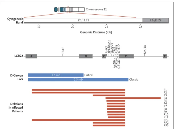
Figure 1. Genomic Organization of Chromosome 22q11.2 and the Deletions Associated with Kidney and Urinary Tract Malformations Identified in This Study.
In approximately 90% of the patients with the DiGeorge syndrome, the congenital disorder is caused by a classic de novo heterozygous deletion of approximately 2.5 mb in length spanning chromosome 22q11.2 low-copy repeats (LCR22) A and D, as shown in blue. Less than 10% of the patients with this syndrome carry the critical 1.5-mb dele-tion between LCR22 A and B. Shown in red are deledele-tions that were identified in 14 patients who were affected by congenital anomalies of the kidney and urinary tract among the 2080 patients who were tested. According to the megabase coordinates for the Human Genome 19 release, the proximal and distal breakpoints for the chromosome 22q11.2 deletions that were identified in the patients are as follows: P1, 18.88 to 21.47 mb; P2, 18.89 to 21.47 mb; P3, 20.73 to 21.46 mb; P4, 21.02 to 22.47 mb; P5, 21.05 to 21.47 mb; P6, 21.06 to 21.47 mb; P7, 21.06 to 21.46 mb; P8, 21.06 to 21.46 mb; P9, 21.07 to 21.46 mb; P10, 21.08 to 21.47 mb; P11, 21.09 to 21.47 mb; Patient 1 from the replication cohort (RP1), 18.88 to 21.46 mb; RP2, 20.74 to 21.46 mb; and RP3, 20.74 to 21.46 mb. The deletion be-tween LCR22 C and D defines the smallest region of overlap for congenital kidney disease among patients with 22q11.2 deletions. Cytogenetic Band Genomic Distance (mb) LCR22 DiGeorge Loci Deletions in Affected Patients 22 21 20 19
TBX1 PI4KA SERPIND1 SNAP29 CRKL THAP7 MAPK1
LZTR1 SLC7A4 P2RX6 AIFM3 Critical Classic P1 P2 P3 P4 P5 P6 P7 P8 P9 P10 P11 RP1 RP2 RP3 Chromosome 22 22q11.21 22q11.22 A B C D E 1.5 mb 2.5 mb
snap29 -MO WT mRNA (150 pg) snap29 -MO (8.0 ng) crkl -M O + WT mRNA (100 pg) crkl -MO (1.5 ng) aifm3 -M O + WT mRNA (150 pg) aifm3 -MO (8.0 ng) ret -M O + WT mRNA (200 pg) ret -MO (8.0 ng) Control Control ret -gRNA +Cas9 crkl -gRNA +Cas9 snap29 -gRNA +Cas9 1.2 1.1 1.0 0.9 0.8 0.7
Relative Pronephros Length
1.2 1.1 1.0 0.9 0.8 0.7
Relative Pronephros Length
* ** *** *** * * * Relative lengt h =
pronephros (a) body axis (b)
C
A
Zebrafish Embryos after CRISPR-Cas9 Injection
MO Knockdown of Zebrafish Embryos Control ret
-MO (8.0 ng) aifm3 -MO (8.0 ng) crkl -MO (1.5 ng) snap29 -MO (8.0 ng) Control ret -gRNA/Cas9 crkl -gRNA/Cas9 snap29 -gRNA/Cas9
D
Relative Length of Pronephros after MO Knockdown Relative Length of Pronephros after CRISPR-Cas9 Injection
B
b
Search Tool (BLAST) algorithm for sequence
searching, in which we detected orthologues for
seven of nine genes. RNA sequencing data
indi-cated that all seven genes were expressed in the
early embryo, between 2 and 4 days after
fertil-ization.
38We therefore designed MOs to knock
down the expression of these genes and injected
them into zebrafish reporter lines in parallel with
the ret–MO as a control. For four of the transcripts
(lztr1, pi4ka, serpind1, and slc7a4) we observed no
differences in convolution complexity or length
of the pronephros between the knockdown
zebra-fish and controls in 26 to 34 embryos, with each
analysis repeated twice with blinded scoring
(Fig. S4 in the Supplementary Appendix). In
con-trast, the suppression of crkl expression or
interrup-tion of splicing of aifm3 and snap29 phenocopied
the pathologic features of RET (Fig. 2A and 2B).
These phenotypes could be rescued for each of the
three genes by coinjection with human mRNA
(Fig. 2B). In addition, deletions of snap29 and crkl
mediated by CRISPR–Cas9 on the day of
fertiliza-tion induced inserfertiliza-tions or delefertiliza-tions in 60 to 80%
of cells within each mutant embryo (Fig. S5 in the
Supplementary Appendix). (The gene aifm3 was
intractable to this method.) Subsequently, the
mu-tant fish fully reproduced the renal disease (Fig. 2C
and 2D). We observed no renal phenotypes when
each human mRNA was injected alone, nor did
we find any other gross morphologic defects in
embryos subjected to either MO knockdown or
overexpression at the studied developmental time
points that might indicate nonspecific toxicity.
Because kidney morphogenesis could be affected
by extrarenal defects (e.g., loss of cardiac output
and collective cell migration of the nephron
in-duced by loss of flow), we analyzed heart
func-tion in both ret and crkl mutants and found no
effect on the morphologic features or rate of the
heart. We also found no evidence of kidney cysts,
which would be expected if cilia-dependent flow
were to be impaired. Analysis of body length as
an indication of global-developmental delay
showed no significant difference between “knocked
down” zebrafish and control zebrafish (Fig. S6
in the Supplementary Appendix). Thus, we
con-cluded that the defects we observed were not due
to the known indirect causes of failed nephron
convolution in zebrafish and support our use of
this assay as a screening technique for intrinsic
kidney defects.
Previous functional dissections of
copy-num-ber variation have revealed a complex genetic
ar-chitecture, in which a single driver may account
for the induction of disease either alone or in cis
epistasis with other genes within the
copy-num-ber variation.
29-31We tested this possibility in
vivo by asking whether the three transcripts in
zebrafish embryos that induce congenital kidney
and urinary tract anomalies could interact
ge-netically. For this purpose, we injected embryos
with subeffective doses of each transcript, with
the requirement that each dose by itself should
induce modest or no disease; we then tested all
possible pairwise combinations. We observed no
genetic interaction between crkl and either aifm3
or snap29. In contrast, cosuppression of aifm3 with
Figure 2 (facing page). Functional Modeling of the DiGeorge Syndrome Terminal Deletion Genes Associated with Kidney and Urinary Tract Malformations.
Panel A shows zebrafish larvae 4.5 days after fertiliza-tion, in which the proximal tubule is folded into a hair-pin structure, displaying proper anterior convolution in noninjected control embryos (staining with anti-body against sodium–potassium ATPase). Knockdown of ret, aifm3, crkl, and snap29 by the injection of 8.0 ng of a splice-blocking morpholino oligonucleotide (MO) against RET resulted in major convolution defects, which are apparent by the failure of the anterior por-tion of the pronephros (the earliest developmental stage in the zebrafish) to progress, along with an over-all reduction in the length of the tubules. Panel B shows the relative length of the pronephros, which was defined as the ratio of the length of the proneph-ros (a) to the length of the body axis (b), in individual larvae (inset). The number of replicate measurements were as follows: control or sham-injected control, 177 in Panel A and 68 in Panel B; MO, 50;
ret-MO+mRNA, 42; aifm3-MO, 38; aifm3-ret-MO+mRNA, 42; crkl-MO, 43; crkl-MO+mRNA, 58; snap29-MO, 48; snap29-MO+mRNA, 39; ret-gRNA+Cas9, 44; crkl-gRNA+Cas9, 31; and snap29-crkl-gRNA+Cas9, 41). Mor-phant phenotypes could be rescued by the coinjection of each respective human messenger RNA (mRNA). In each box-and-whisker plot, the horizontal line rep-resents the median, the top and bottom of the boxes the interquartile range, and the I bars the minimum and maximum values. Panel C shows embryos that have been injected with CRISPR–Cas9 and that are re-producing the convolution defects observed in the morphant embryos. Guide RNA (gRNA) that targeted each respective gene was coinjected with purified Cas9 protein, and the relative length of the proneph-ros was measured in founders, as shown in Panel D. In Panels B and D, a single asterisk indicates P<0.05, two asterisks P<0.01, and three asterisks P<0.001. WT denotes wild type.
snap29
phenocopied the convolution defect of
strong morphants and CRISPR mutants, which
suggested a contributory role to the copy-number
variation pathology. This interaction was specific
and not due to toxicity caused by the presence of
multiple MOs, since it could be rescued by
coin-jection of SNAP29 mRNA (Figs. S7 and S8 in the
Supplementary Appendix).
Whole-Exome and Targeted Sequencing
of CRKL
We asked whether sporadic patients with
con-genital kidney and urinary tract anomalies might
have loss-of-function lesions in any of the nine
genes included in the minimal region of overlap
for the kidney defects of the DiGeorge syndrome.
We first queried exome-sequencing data from
60 patients with renal agenesis or hypodysplasia.
None of the genes showed excess burden of rare
truncating mutations as compared with controls
(Table S6 in the Supplementary Appendix). LZTR1,
P2RX6
, and SLC7A4 have a high frequency of
loss-of-function mutations (defined as premature
ter-mination, splicing, and frameshift mutations), a
prevalence that approaches or exceeds that of
such anomalies in the general population.
Con-versely, SERPIND1, SNAP29, CRKL, and THAP7
carry loss-of-function mutations in no more than
2 of 10,000 persons. Among more than 60,500
publicly available population controls from the
Exome Aggregation Consortium (ExAC) database
(exac.broadinstitute.org), only 1 had a
high-qual-ity loss-of-function variant in CRKL, which ranks
in the top second percentile in the genome for
haploinsufficiency — in other words, there is a
high probability of a detrimental effect on
pheno-type when only one copy of the gene is deleted.
This finding suggests that loss-of-function
vari-ations in CRKL have deleterious effects on
ge-netic fitness (Table S3 in the Supplementary
Appendix).
39We also performed targeted next-generation
resequencing of all 107 coding exons of PI4KA,
SERPIND1
, SNAP29, CRKL, AIFM3, THAP7, P2RX6,
and SLC7A4 in 526 patients with renal agenesis or
hypodysplasia. We identified six loss-of-function
variants in 11 patients: two in SERPIND1, one in
CRKL
, one in AIFM3, and two in P2RX6 (in 7
pa-tients) (Table S7 in the Supplementary Appendix).
Loss-of-function mutations in SERPIND1 have been
associated with a mendelian clotting disease
(hep-arin cofactor II deficiency) that has no known
associations with kidney and urinary tract
devel-opment.
40In contrast, the CRKL truncating
mu-tation, p.Q31*, was found in a patient (P13) with
isolated unilateral renal agenesis and was
pre-dicted to result in haploinsufficiency. We also
identified four additional missense variants that
were absent from the ExAC database, that were
conserved across vertebrates, and that were
pre-dicted to affect protein structure and function
(Table S8 and Figs. S9 and S10 in the
Supple-mentary Appendix).
Whole-exome sequencing of DNA obtained
from P13 did not show pathogenic mutations in
genes that had previously been implicated in
congenital kidney and urinary tract anomalies or
loss-of-function variants in newly plausible
can-didates (Table S9 in the Supplementary
Appen-dix). Finally, because of the formal possibility
that the discovered CRKL variants were
popula-tion-specific polymorphisms, we performed
tar-geted resequencing on samples obtained from
576 additional controls and from 1152 patients
with IgA nephropathy and normal results on
renal ultrasonography who were matched with
our patients according to ethnic background and
recruitment site. All CRKL variants were absent
in the more than 60,500 population controls from
the ExAC database and in the 1728 controls.
Ag-gregating our sequencing data and performing
burden tests between our 586 patients with
con-genital kidney and urinary tract anomalies and
33,352 European controls from ExAC or 1728
ethnically and geographically matched controls
showed significant excess of rare functional CRKL
variants in our patients (P = 3.7×10
−3by Fisher’s
exact test for the comparison with ExAC controls;
odds ratio, 5.2; and P = 4.9×10
−3for the
compari-son with matched controls; odds ratio, 14.8)
(Ta-ble S10 in the Supplementary Appendix).
Expression and Functional Studies of CRKL
We performed mRNA and protein expression
stud-ies in relevant tissues and examined a mouse
model with a Crkl mutation. In humans, CRKL
protein showed mild, diffuse cytoplasmic
ex-pression in both ureteric bud and metanephric
mesenchyme derivatives during the sixth week
of fetal development (Fig. S11A in the
Supple-mentary Appendix). At week 21, CRKL was
de-tected only in proximal tubules and collecting
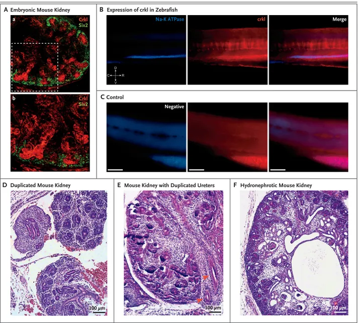
Figure 3. Localization of Crkl in Developing Urinary Tracts in Mice and Zebrafish and Phenotypes of Crkl Knockout Mice.
Panel A shows immunostaining for Crkl in kidney obtained from a transgenic mouse on embryonic day E15.5, in which Six2 has been tagged with enhanced green fluorescent protein (GFP), with specific Crkl staining of the ureteric bud (in red) surrounded by Six2-posi-tive cap mesenchyme cells (in green) (subpanel a). A magnified field shows ureteric-bud branching within condensing metanephric mesenchyme (subpanel b). Panel B shows specific pronephros expression of crkl in zebrafish, as shown by colocalization after staining with antibody against sodium–potassium ATPase. In the orientation symbol, D denotes dorsal, V ventral, C caudal, and R rostral. Panel C shows images of negative controls (i.e., fish treated with fluorophore-conjugated secondary antibodies only). In Panels B and C, the scale bars represent 100 µm. In a mouse model that targets Crkl exon 2, three crosses with transgenic Cre-recombinase mice were creat-ed to effect the deletion of exon 2 in specific compartments: E2a-Cre for global knockout, Six2-Cre in the cap mesenchyme, and Hoxb7 in the structures derived from ureteric buds. Panel D shows tissue from a Six2-Cre mouse in which duplication of the right kidney is ac-companied by an irregular, dysplastic pattern or ureteric-bud branching on embryonic day E15.5. Panel E shows tissue from an E2a-Cre mouse in which a single kidney with duplicated ureters (arrowheads) is accompanied by failure of medullary and renal papillary develop-ment on day E14.5. Panel F shows tissue from a Six2-Cre mouse, in which the kidney is hydronephrotic with dilated pelvis, absence of medullary architecture, and several microcystic glomeruli and tubules on day E15.5.
D
ab
Duplicated Mouse Kidney
C
ControlEmbryonic Mouse Kidney
Crkl Na-K ATPase Negative crkl Merge Six2 Crkl Six2 Expression of crkl in Zebrafish
B
A
E
Mouse Kidney with Duplicated UretersF
Hydronephrotic Mouse Kidneytubules at the apical side of epithelial cells (Fig.
S11B in the Supplementary Appendix). In the
kid-ney of a 1.5-year-old boy, we observed abundant
CRKL expression in the proximal and collecting
tubules at the apical side, along with diffuse
cy-toplasmic signaling in glomerular endothelial
cells, podocytes, Bowman’s capsule, and distal
tubules (Fig. S11C in the Supplementary
Appen-dix). The expression of SNAP29 and AIFM3,
al-though present at very low levels in zebrafish
pronephros (not shown), was seen in the urinary
tract in fetuses and children (Figs. S12 and S13
in the Supplementary Appendix).
In the mouse kidney on embryonic day E15.5,
Crkl showed specific expression in structures
de-rived from the ureteric bud and, occasionally, in
S-shaped bodies and developing proximal tubules
(Fig. 3A, and Fig. S14 in the Supplementary
Ap-pendix). In zebrafish, crkl was specifically
ex-pressed in the pronephros (Fig. 3B and 3C). RNA
studies that were performed with the use of flow
cytometry and cell sorting, along with in situ
hy-bridization, confirmed that crkl was expressed in
the pronephric convoluted tubule and
proneph-ric duct (Figs. S15 and S16 in the Supplementary
Appendix).
Finally, we engineered a mouse model that
targets Crkl exon 2. We generated three different
crosses with transgenic Cre-recombinase mice to
effect the deletion of exon 2 in specific
compart-ments: E2a-Cre for global knockout, Six2-Cre in
the cap mesenchyme, and Hoxb7 in the ureteric
bud–derived structures. We analyzed four litters
(one E2a, one Hoxb7, and two Six2) at embryonic
days E14.5 through E15.5. We observed
develop-mental anomalies in the kidney and urinary tract,
including bilaterally duplicated kidneys,
duplicat-ed ureters, ureteric bud–branching defects,
dys-plastic features, hydronephrosis, microcystic
tu-bules and glomeruli, and tubular and glomerular
capsule dilatation, in eight mice (Fig. 3D, 3E,
and 3F, and Fig. S17 in the Supplementary
Ap-pendix). We observed phenotypes related to
con-genital kidney and urinary tract anomalies in
embryos that were heterozygous and those that
were homozygous for the targeted deletion.
Di s c u s s ion
We determined that deletions in the telomeric
22q11.2 classic region are associated with
spo-radic congenital kidney and urinary tract
anom-alies and renal disease in the DiGeorge syndrome.
Correlations between genotype and phenotype
suggest that these variants are specific for
kid-ney parenchyma defects (i.e., renal agenesis or
hypodysplasia), rather than ureteric and lower
urinary tract disease. However, the presence of
these variants may be an indication of kidney
dis-ease in persons with apparently isolated ureteric
defects, since the two patients with obstructive
uropathy and vesicoureteral reflux whom we
iden-tified in this study showed renal insufficiency. We
observed that the 22q11.2 deletions were present
in 1.1% of our sample of 1093 patients with renal
agenesis or hypodysplasia, which suggests that
such deletions constitute the second most
com-mon structural variant associated with
congeni-tal kidney and urinary tract anomalies after the
17q12 deletion that causes the renal cysts and
dia-betes syndrome, which we identified in 2.2% of
patients with renal agenesis or hypodysplasia from
the same cohort. Our data also support the
hy-pothesis that 22q11.2 microdeletions are medically
actionable variants that confer a predisposition to
renal hypodysplasia and kidney disease.
A review of the literature indicates the
pres-ence of kidney and urinary tract defects in about
one third of the patients with chromosome
22q11.2 deletions spanning LCR22 B and D or C
and D,
5,41a prevalence that is nearly identical to
that of kidney and urinary tract defects among
patients with the DiGeorge syndrome caused by
the typical 22q11.2 deletions spanning LCR22 A
and D.
4,6,42These observations, together with our
data, strongly suggest that the kidney disease
as-sociated with the DiGeorge syndrome is
attribut-able largely to haploinsufficiency of one or more
genes located between LCR22 C and D.
Genetic interaction studies using zebrafish
sug-gested a complex genetic architecture, in which
haploinsufficiency of crkl had a potent detrimental
effect on renal development, whereas knockdown
of its flanking genes, aifm3 and snap29, generated
the phenotype only with cosuppression.
Consis-tent with these data, we found deleterious CRKL
variants, including a premature truncating allele,
in approximately 1% of the patients with sporadic
congenital renal agenesis or hypodysplasia. We
obtained other molecular data in humans, mice,
and zebrafish that supported the role of CRKL in
urinary tract development.
CRKL
encodes an adapter protein that regulates
intracellular signaling transduction from
multi-ple growth factors, including the fibroblast growth
factors,
43which are key regulators of kidney and
urinary tract development.
44,45Inactivation of Crkl
in mice recapitulates some of the phenotypes of
the DiGeorge syndrome, in particular cardiac
mal-formations,
46,47but the kidney phenotype in the
mutant embryos had not hitherto been studied.
We observed that genetic inactivation of Crkl in
the mouse model results in developmental
phe-notypes of the kidney and urinary tract that
re-semble congenital anomalies in the human
uri-nary tract.
We suggest that CRKL mutations sensitize the
genetic background and contribute to the
pene-trance of congenital kidney and urinary tract
anomalies in patients with the DiGeorge
syn-drome. It is possible that other genes within or
outside the locus of the DiGeorge syndrome and
22q11.2 deletions might also be involved. Two of
the genes in the minimal region were refractory
to our studies, and it is possible that the deletion
copy-number variant affects the expression of
genes across the chromosome or elsewhere in the
genome, as has been shown for other
copy-num-ber variants.
48In conclusion, our approach provides support
for the causal role of CRKL in the pathogenesis of
kidney developmental defects. Such defects occur
specifically in the context of the DiGeorge
syn-drome and 22q11.2 deletions and, more broadly,
in sporadic congenital kidney and urinary tract
anomalies.
Supported by grants (1R01DK103184, 1R21DK098531, and UL1 TR000040, to Dr. Sanna-Cherchi; P50DK096415 and P30DK096493, to Dr. Katsanis; 2R01DK080099, to Dr. Gharavi; 3U54DK104309, to Drs. Gharavi and Barasch; P01HD070454, to
Ms. McDonald-McGinn and Dr. Morrow; 4R01GM030518, to Dr. Honig; R37HD033082, to Dr. Papaioannou; and 1R01DK105124, to Dr. Kiryluk) from the National Institutes of Health (NIH); a grant-in-aid (13GRNT14680075, to Dr. Sanna-Cherchi) from the American Heart Association; a grant (RF-2010-2307403, to Drs. Sanna-Cherchi and Ghiggeri) from the Joint Italian Ministry of Health and NIH Young Investigators Finalized Research; a grant (HG006504, to Dr. Lifton) from the National Human Genome Research Institute Centers for Mendelian Genomics; a grant (to Dr. Ghiggeri) from the Fondazione Malattie Renali nel Bambi-no; a grant (AAE07007KSA, to Drs. Salomon and Jeanpierre) from the GIS-Institut des Maladies Rares; and a grant (AOM07129, to Drs. Salomon and Jeanpierre) from the Pro-gramme Hospitalier de la Recherche Clinique Assistance Pub-lique; by the Polish Ministry of Health (to Drs. Materna-Kiryluk and Latos-Bielenska); by the Polish Kidney Genetics Network (POLYGENES), the Polish Registry of Congenital Malformations (PRCM), and the NZOZ Center for Medical Genetics (GENESIS); by grants (to the Chronic Kidney Disease in Children Study) from the National Institute of Diabetes and Digestive and Kid-ney Diseases and the Eunice Kennedy Shriver National Institute of Child Health and Human Development; by grants (U01DK66143, U01DK66174, U01DK082194, U01DK66116, and RO1DK082394) from the National Heart, Lung, and Blood Insti-tute; and by the Paul Marks Scholar Award (to Dr. Sanna-Cher-chi); and a Kolff Postdoc Fellowship Abroad grant (15OKK95, to Dr. Westland) from the Dutch Kidney Foundation.
Disclosure forms provided by the authors are available with the full text of this article at NEJM.org.
We thank the patients and their families for participating in the study; Katarzyna Zachwieja (Dialysis Unit, Jagiellonian Uni-versity Medical College, Krakow, Poland), Daria Tomczyk (De-partment of Pediatrics, Immunology and Nephrology Polish Mother’s Memorial Hospital Research Institute, Lodz, Poland), Tomasz Jarmolinski (Miedzyrzecz Regional Hospital, Depart-ment of Pediatrics, Miedzyrzecz, Poland), Robert Pawluch and Maria Katarzyna Boroszewska-Kornacka (Neonatal and Inten-sive Care Department, Medical University of Warsaw, Poland), Piotr Adamczyk (Department of Pediatrics, School of Medicine with the Division of Dentistry in Zabrze, Medical University of Silesia in Katowice, Poland), and Klaudia Korecka (Department of Pediatric Surgery and Urology, Medical University of Silesia, Upper Silesian Child’s Health Center Katowice, Poland) for re-cruting patients for this study; and Cyrus Zabetian (University of Washington, Seattle) and Haydeh Payami (University of Ala-bama, Birmingham) for sharing clinical and genetic data from the control population.
Appendix
The authors’ full names and academic degrees are as follows: Esther Lopez-Rivera, Ph.D., Yangfan P. Liu, Ph.D., Miguel Verbitsky, Ph.D., Blair R. Anderson, Ph.D., Valentina P. Capone, M.D., Edgar A. Otto, Ph.D., Zhonghai Yan, Ph.D., Adele Mitrotti, M.D., Jeremiah Martino, Ph.D., Nicholas J. Steers, Ph.D., David A. Fasel, B.S., Katarina Vukojevic, M.D., Ph.D., Rong Deng, B.S., Silvia E. Racedo, Ph.D., Qingxue Liu, M.S., Max Werth, Ph.D., Rik Westland, M.D., Ph.D., Asaf Vivante, M.D., Gabriel S. Makar, B.S., Monica Bodria, M.D., Matthew G. Sampson, M.D., Christopher E. Gillies, Ph.D., Virginia Vega-Warner, Ph.D., Mariarosa Maiorana, M.D., Donald S. Petrey, Ph.D., Barry Honig, Ph.D., Vladimir J. Lozanovski, M.D., Ph.D., Rémi Salomon, Ph.D., Laurence Heidet, M.D., Wassila Carpen-tier, Ph.D., Dominique Gaillard, M.S., Alba Carrea, Ph.D., Loreto Gesualdo, M.D., Daniele Cusi, M.D., Claudia Izzi, M.D., Francesco Scolari, M.D., Joanna A.E. van Wijk, M.D., Ph.D., Adela Arapovic, M.D., Mirna Saraga-Babic, Ph.D., Marijan Saraga, M.D., Nenad Ku-nac, Ph.D., Ali Samii, M.D., Donna M. McDonald-McGinn, M.S., Terrence B. Crowley, Ph.D., Elaine H. Zackai, M.D., Dorota Drozdz, M.D., Monika Miklaszewska, M.D., Marcin Tkaczyk, M.D., Przemyslaw Sikora, M.D., Maria Szczepanska, M.D., Malgorzata Mizer-ska-Wasiak, M.D., Grazyna Krzemien, M.D., Agnieszka Szmigielska, M.D., Marcin Zaniew, M.D., John M. Darlow, M.D., Ph.D., Prem Puri, M.D., David Barton, Ph.D., Emilio Casolari, M.D., Susan L. Furth, M.D., Ph.D., Bradley A. Warady, M.D., Zoran Gucev, M.D., Ph.D., Hakon Hakonarson, Ph.D., Hana Flogelova, M.D., Velibor Tasic, M.D., Ph.D., Anna Latos-Bielenska, M.D., Anna Materna-Kiryluk, M.D., Landino Allegri, M.D., Craig S. Wong, M.D., M.P.H., Iain A. Drummond, Ph.D., Vivette D’Agati, M.D., Akira Imamoto, Ph.D., Jonathan M. Barasch, M.D., Ph.D., Friedhelm Hildebrandt, M.D., Krzysztof Kiryluk, M.D., Richard P. Lifton, M.D., Ph.D., Bernice E. Morrow, Ph.D., Cecile Jeanpierre, Ph.D., Virginia E. Papaioannou, Ph.D., Gian Marco Ghiggeri, M.D., Ph.D., Ali G. Gharavi, M.D., Nicholas Katsanis, Ph.D., and Simone Sanna-Cherchi, M.D.
The authors’ affiliations are as follows: the Division of Nephrology (E.L.-R., M.V., V.P.C., Z.Y., A.M., J.M., N.J.S., D.A.F., R.D., M.W., G.S.M., M.B., J.M.B., K.K., A.G.G., S.S.-C.) and the Division of Nephrology in Medicine and Zuckerman Mind Brain Behavior Institute
(B.H.), the Departments of Systems Biology (D.S.P., B.H.), Biochemistry and Molecular Biophysics (B.H.), and Pathology (V.D.), and the Howard Hughes Medical Institute (D.S.P., B.H.), Columbia University, and the Department of Genetics and Development, Columbia University Medical Center (Q.L., V.E.P.), New York, and the Department of Genetics, Albert Einstein College of Medicine, Bronx (S.E.R., B.E.M.) — all in New York; the Center for Human Disease Modeling, Duke University, Durham, NC (Y.P.L., B.R.A., N. Katsanis); the Departments of Internal Medicine–Nephrology (E.A.O.) and Pediatrics–Nephrology (M.G.S., C.E.G., V.V.-W.), University of Michigan School of Medicine, Ann Arbor; the Department of Anatomy, Histology, and Embryology, School of Medicine, University of Split (K.V., M.S.-B.), and the Departments of Pediatrics (A.A., M. Saraga) and Pathology (N. Kunac), University Hospital of Split, Split, Croatia; the Department of Pediatric Nephrology, VU University Medical Center, Amsterdam (R.W., J.A.E.W.); the Department of Medicine, Boston Children’s Hospital (A.V., F.H.), and Harvard Medical School, Boston (A.V., F.H., I.A.D.), and the Nephrology Division, Massachusetts General Hospital, Charlestown (I.A.D.) — all in Massachusetts; the Division of Nephrology, Dialysis, Transplantation, and Laboratory on Pathophysiology of Uremia, Istituto G. Gaslini, Genoa (M.B., A.C., G.M.G.), the Department of Clinical and Experimental Medicine, University of Parma (M.B., M. Maiorana, L.A.), and the Pediatric Surgery Unit, University Hospital of Parma (E.C.), Parma, the Section of Nephrology, Department of Emergency and Organ Transplantation, University of Bari, Bari (L.G.), the Department of Medical Sci-ences, University of Milano, and Institute of Biomedical Technologies, Italian National Institute of Research ITB-CNR, Milan (D.C.), and Dipartimento Ostetrico-Ginecologico e Seconda Divisione di Nefrologia ASST Spedali Civili e Presidio di Montichiari (C.I.) and Cattedra di Nefrologia, Università di Brescia, Seconda Divisione di Nefrologia Azienda Ospedaliera Spedali Civili di Brescia Presidio di Montichiari (F.S.), Brescia — all in Italy; the Department of General and Transplant Surgery, University Hospital of Heidelberg, Ger-many (V.J.L.); the Department of Pediatric Nephrology, Centre de Référence des Maladies Rénales Héréditaires de l’Enfant et de l’Adulte (R.S., L.H., C.J.), INSERM UMR 1163, Laboratory of Hereditary Kidney Diseases (R.S.), Necker–Enfants Malades Hospital, Paris Des-cartes–Sorbonne Paris Cite University, Imagine Institute (R.S.), Sorbonne Universités, UPMC 06, Plateforme Post-génomique de la Pitié–Salpêtrière, UMS 2 Omique, Inserm US029 (W.C.), Paris, and the Department of Genetics, Centre Hospitalier Universitaire de Reims, Unité de Formation et de Recherche de Médecine, Reims (D.G.) — both in France; the Department of Neurology, University of Washington School of Medicine, and Northwest VA Parkinson’s Disease Research, Education and Clinical Centers, Seattle (A. Samii); the Division of Human Genetics, Department of Pediatrics, 22q and You Center, Children’s Hospital of Philadelphia and Perelman School of Medicine at the University of Pennsylvania (D.M.M.-M., T.B.C., E.H.Z., S.L.F.), Division of Nephrology, Children’s Hospital of Philadelphia (S.L.F.), and the Department of Genetics, University of Pennsylvania (H.H.), Philadelphia; the Dialysis Unit, Jagiellonian University Medical College (D.D.), and the Department of Pediatric Nephrology, Jagiellonian University Medical College (M. Miklasze-wska), Krakow, the Department of Pediatrics, Immunology and Nephrology, Polish Mother’s Memorial Hospital Research Institute, Lodz (M.T.), the Department of Pediatric Nephrology Medical University of Lublin, Lublin (P.S.), the Department of Pediatrics, School of Medicine with the Division of Dentistry in Zabrze, Medical University of Silesia, Katowice (M. Szczepanska), the Department of Pe-diatrics and Nephrology, Medical University of Warsaw, Warsaw (M.M.-W., G.K., A. Szmigielska), and Krysiewicza Children’s Hospital (M.Z.) and the Department of Medical Genetics, Poznan University of Medical Sciences, and Center for Medical Genetics GENESIS (A.L.-B., A.M.-K.), Poznań — all in Poland; the Department of Clinical Genetics (J.M.D., D.B.), National Children’s Research Centre (J.M.D., P.P.), and University College Dublin School of Medicine (D.B.), Our Lady’s Children’s Hospital Crumlin, and the National Children’s Hospital Tallaght (P.P.), Dublin, Ireland; the Division of Pediatric Nephrology, Children’s Mercy Hospital, Kansas City, MO (B.A.W.); University Children’s Hospital, Medical Faculty of Skopje, Skopje, Macedonia (Z.G., V.T.); Faculty of Medicine, Palacky Uni-versity, Olomouc, Czech Republic (H.F.); the Division of Pediatric Nephrology, University of New Mexico Children’s Hospital, Albuquer-que (C.S.W.); Ben May Department for Cancer Research, University of Chicago, Chicago (A.I.); and the Department of Genetics, Howard Hughes Medical Institute, and Yale Center for Mendelian Genomics, Yale University, New Haven, CT (R.P.L.).
References
1. Lindsay EA. Chromosomal microdele-tions: dissecting del22q11 syndrome. Nat Rev Genet 2001; 2: 858-68.
2. Devriendt K, Fryns JP, Mortier G, van Thienen MN, Keymolen K. The annual in-cidence of DiGeorge/velocardiofacial syn-drome. J Med Genet 1998; 35: 789-90. 3. McDonald-McGinn DM, Sullivan KE. Chromosome 22q11.2 deletion syndrome (DiGeorge syndrome/velocardiofacial syn-drome). Medicine (Baltimore) 2011; 90: 1-18. 4. Kobrynski LJ, Sullivan KE. Velocar-diofacial syndrome, DiGeorge syndrome: the chromosome 22q11.2 deletion syn-dromes. Lancet 2007; 370: 1443-52. 5. Burnside RD. 22q11.21 Deletion syn-dromes: a review of proximal, central, and distal deletions and their associated features. Cytogenet Genome Res 2015; 146: 89-99.
6. Noël AC, Pelluard F, Delezoide AL, et al. Fetal phenotype associated with the 22q11 deletion. Am J Med Genet A 2014; 164A: 2724-31.
7. Bassett AS, Chow EW, Husted J, et al. Clinical features of 78 adults with 22q11
Deletion Syndrome. Am J Med Genet A 2005; 138: 307-13.
8. Edelmann L, Pandita RK, Spiteri E, et al. A common molecular basis for rear-rangement disorders on chromosome 22q11. Hum Mol Genet 1999; 8: 1157-67. 9. Saitta SC, Harris SE, Gaeth AP, et al. Aberrant interchromosomal exchanges are the predominant cause of the 22q11.2 deletion. Hum Mol Genet 2004; 13: 417-28. 10. Shaikh TH, O’Connor RJ, Pierpont ME, et al. Low copy repeats mediate distal chromosome 22q11.2 deletions: sequence analysis predicts breakpoint mechanisms. Genome Res 2007; 17: 482-91.
11. Shaikh TH, Kurahashi H, Saitta SC, et al. Chromosome 22-specific low copy re-peats and the 22q11.2 deletion syndrome: genomic organization and deletion end-point analysis. Hum Mol Genet 2000; 9: 489-501.
12. Kujat A, Schulz MD, Strenge S, Froster UG. Renal malformations in deletion 22q11.2 patients. Am J Med Genet A 2006; 140: 1601-2.
13. Wu HY, Rusnack SL, Bellah RD, et al.
Genitourinary malformations in chromo-some 22q11.2 deletion. J Urol 2002; 168: 2564-5.
14. Yagi H, Furutani Y, Hamada H, et al. Role of TBX1 in human del22q11.2 syn-drome. Lancet 2003; 362: 1366-73. 15. Paylor R, Glaser B, Mupo A, et al. Tbx1 haploinsufficiency is linked to be-havioral disorders in mice and humans: implications for 22q11 deletion syn-drome. Proc Natl Acad Sci U S A 2006; 103: 7729-34.
16. Jerome LA, Papaioannou VE. Di-George syndrome phenotype in mice mu-tant for the T-box gene, Tbx1. Nat Genet 2001; 27: 286-91.
17. Merscher S, Funke B, Epstein JA, et al. TBX1 is responsible for cardiovascular de-fects in velo-cardio-facial/DiGeorge syn-drome. Cell 2001; 104: 619-29.
18. Lindsay EA, Vitelli F, Su H, et al. Tbx1 haploinsufficieny in the DiGeorge syn-drome region causes aortic arch defects in mice. Nature 2001; 410: 97-101. 19. Sanna-Cherchi S, Kiryluk K, Burgess KE, et al. Copy-number disorders are a
common cause of congenital kidney mal-formations. Am J Hum Genet 2012; 91: 987-97.
20. Verbitsky M, Sanna-Cherchi S, Fasel DA, et al. Genomic imbalances in pediat-ric patients with chronic kidney disease. J Clin Invest 2015; 125: 2171-8.
21. Westland R, Verbitsky M, Vukojevic K, et al. Copy number variation analysis identifies novel CAKUT candidate genes in children with a solitary functioning kidney. Kidney Int 2015; 88: 1402-10. 22. Sanna-Cherchi S, Sampogna RV, Pa-peta N, et al. Mutations in DSTYK and dominant urinary tract malformations. N Engl J Med 2013; 369: 621-9.
23. Westland R, Bodria M, Carrea A, et al. Phenotypic expansion of DGKE-associat-ed diseases. J Am Soc Nephrol 2014; 25: 1408-14.
24. Choi M, Scholl UI, Ji W, et al. Genetic diagnosis by whole exome capture and massively parallel DNA sequencing. Proc Natl Acad Sci U S A 2009; 106: 19096-101. 25. Halbritter J, Diaz K, Chaki M, et al. High-throughput mutation analysis in pa-tients with a nephronophthisis-associated ciliopathy applying multiplexed barcoded array-based PCR amplification and next-generation sequencing. J Med Genet 2012; 49: 756-67.
26. Halbritter J, Porath JD, Diaz KA, et al. Identification of 99 novel mutations in a worldwide cohort of 1,056 patients with a nephronophthisis-related ciliopathy. Hum Genet 2013; 132: 865-84.
27. Mefford HC, Clauin S, Sharp AJ, et al. Recurrent reciprocal genomic rearrange-ments of 17q12 are associated with renal disease, diabetes, and epilepsy. Am J Hum Genet 2007; 81: 1057-69.
28. Chapman DL, Garvey N, Hancock S, et al. Expression of the T-box family genes, Tbx1-Tbx5, during early mouse de-velopment. Dev Dyn 1996; 206: 379-90. 29. Golzio C, Willer J, Talkowski ME, et al. KCTD13 is a major driver of mirrored
neuroanatomical phenotypes of the 16p11.2 copy number variant. Nature 2012; 485: 363-7.
30. Carvalho CM, Vasanth S, Shinawi M, et al. Dosage changes of a segment at 17p13.1 lead to intellectual disability and microcephaly as a result of complex ge-netic interaction of multiple genes. Am J Hum Genet 2014; 95: 565-78.
31. Dauber A, Golzio C, Guenot C, et al. SCRIB and PUF60 are primary drivers of the multisystemic phenotypes of the 8q24.3 copy-number variant. Am J Hum Genet 2013; 93: 798-811.
32. Costantini F, Kopan R. Patterning a complex organ: branching morphogene-sis and nephron segmentation in kidney development. Dev Cell 2010; 18: 698-712. 33. Schuchardt A, D’Agati V, Larsson-Blomberg L, Costantini F, Pachnis V. De-fects in the kidney and enteric nervous system of mice lacking the tyrosine ki-nase receptor Ret. Nature 1994; 367: 380-3. 34. Chatterjee R, Ramos E, Hoffman M, et al. Traditional and targeted exome se-quencing reveals common, rare and novel functional deleterious variants in RET-signaling complex in a cohort of living US patients with urinary tract malforma-tions. Hum Genet 2012; 131: 1725-38. 35. Hwang DY, Dworschak GC, Kohl S, et al. Mutations in 12 known dominant dis-ease-causing genes clarify many congeni-tal anomalies of the kidney and urinary tract. Kidney Int 2014; 85: 1429-33. 36. de Pontual L, Zaghloul NA, Thomas S, et al. Epistasis between RET and BBS mu-tations modulates enteric innervation and causes syndromic Hirschsprung disease. Proc Natl Acad Sci U S A 2009; 106: 13921-6. 37. Vasilyev A, Liu Y, Mudumana S, et al. Collective cell migration drives morpho-genesis of the kidney nephron. PLoS Biol 2009; 7(1): e9.
38. Borck G, Hög F, Dentici ML, et al. BRF1 mutations alter RNA polymerase III-dependent transcription and cause
neuro-developmental anomalies. Genome Res 2015; 25: 155-66.
39. Huang N, Lee I, Marcotte EM, Hurles ME. Characterising and predicting haplo-insufficiency in the human genome. PLoS Genet 2010; 6(10): e1001154.
40. Kondo S, Tokunaga F, Kario K, Mat-suo T, Koide T. Molecular and cellular basis for type I heparin cofactor II defi-ciency (heparin cofactor II Awaji). Blood 1996; 87: 1006-12.
41. Rump P, de Leeuw N, van Essen AJ, et al. Central 22q11.2 deletions. Am J Med Genet A 2014; 164A: 2707-23.
42. Besseau-Ayasse J, Violle-Poirsier C, Bazin A, et al. A French collaborative sur-vey of 272 fetuses with 22q11.2 deletion: ultrasound findings, fetal autopsies and pregnancy outcomes. Prenat Diagn 2014; 34: 424-30.
43. Moon AM, Guris DL, Seo JH, et al. Crkl deficiency disrupts Fgf8 signaling in a mouse model of 22q11 deletion syn-dromes. Dev Cell 2006; 10: 71-80. 44. Bates CM. Role of fibroblast growth factor receptor signaling in kidney devel-opment. Am J Physiol Renal Physiol 2011; 301: F245-F251.
45. Schedl A. Renal abnormalities and their developmental origin. Nat Rev Gen-et 2007; 8: 791-802.
46. Guris DL, Fantes J, Tara D, Druker BJ, Imamoto A. Mice lacking the homologue of the human 22q11.2 gene CRKL pheno-copy neurocristopathies of DiGeorge syn-drome. Nat Genet 2001; 27: 293-8. 47. Racedo SE, McDonald-McGinn DM, Chung JH, et al. Mouse and human CRKL is dosage sensitive for cardiac outflow tract formation. Am J Hum Genet 2015; 96: 235-44.
48. Migliavacca E, Golzio C, Männik K, et al. A potential contributory role for ciliary dysfunction in the 16p11.2 600 kb BP4-BP5 pathology. Am J Hum Genet 2015; 96: 784-96.