UNIVERSITÀ DEGLI STUDI DI SASSARI
SCUOLA DI DOTTORATO IN SCIENZE BIOMOLECOLARI E BIOTECNOLOGICHE
INDIRIZZO BIOCHIMICA E BIOLOGIA MOLECOLARE XXVI CICLO
DIRETTORE: PROF.SSA CLAUDIA CROSIO
LRRK2
– AN IMPORTANT PLAYER IN PARKINSON’S
DISEASE –
ROLE IN VESICLE AND RECEPTOR TRAFFICKING
Dottorando:
Dott.ssa Del Giudice Maria Grazia
Tutor: Dott. Ciro Iaccarino
INDEX
Page
SUMMARY
1
1. INTRODUCTION
5
1.1 PARKINSON DISEASE
6
1.2 DOPAMINE AND DOPAMINE RECEPTORS
9
1.2.1 Dopamine Synthesis and Metabolism
10
1.2.2 Dopamine Receptors trafficking
11
1.3 TREATMENT
14
1.3.1 Drug Therapy14
1.3.2 Surgical Therapy16
1.4 ETIOLOGY16
1.4.1 Environmental Factors17
1.4.2 Genetic Factors19
1.5 LEUCINE RICH REPEAT KINASE 2 (LRRK2)
26
1.5.1 Physio-Pathological role of LRRK2 (PARK8)
26
1.6 LEUCINE RICH REPEAT KINASE 2 AND LEUCINE RICH REPATKINASE 1
2. MAERIALS AND METHODS
48
2.1 Materials
49
2.2 Methods
52
3. RESULTS
61
3.1
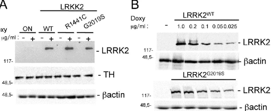
Characterization of PC12 Cells Stably ExpressingDoxycycline-inducible LRRK2 WT or Pathological Mutants
62
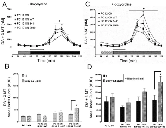
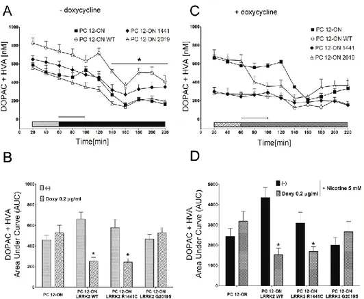
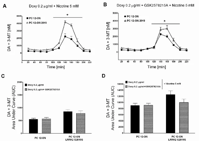
3.2 LRRK2 Influences the Basal and Nicotine-induced secretion of DA in PC12 Cells
64
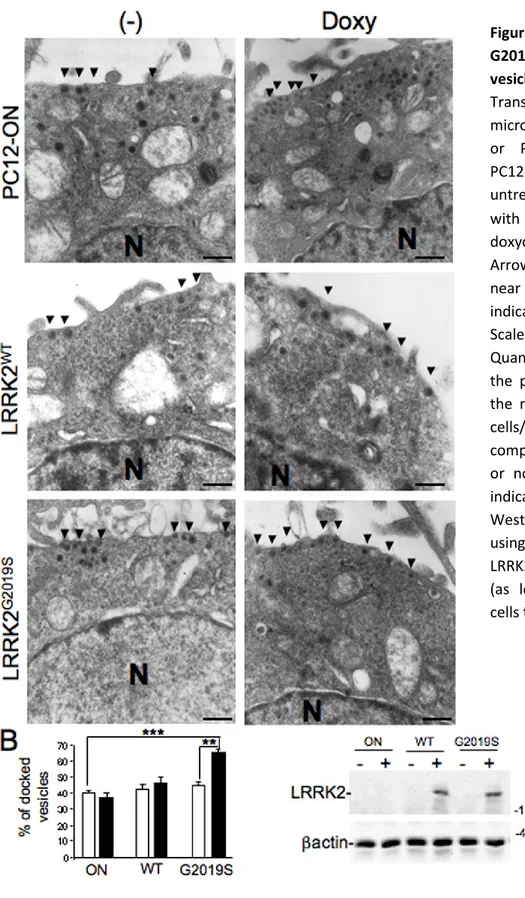
3.3 Vesicle Distribution in PC12 Cells Expressing LRRK2 G2019S Pathological Mutant
69
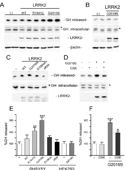
3.4 LRRK2 Increases Growth Hormone Extracellular Level in Neuronal Cells
71
3.5 LRRK2 Affects the Localization of Dopamine Receptor D1 both in Neuronal Cells and Transgenic Mouse Tissues
73
3.6 LRRK1 and LRRK2 functional redundancy in receptors trafficking
76
4. DISCUSSION
87
5. REFERENCES
108
LRRK2 – an important player in Parkinson’s Disease – role in vesicle
and receptor trafficking
SUMMARY
State of the art: PD (Parkinson’s disease) is the second most common neurodegenerative disease affecting 4 million people worldwide. It is characterized by loss of pigmented neurons in the SNpc (substantia nigra pars compacta) and presence of cytoplasmic inclusion bodies (called Lewy bodies). In particular, the loss of dopaminergic neurons is responsible for both depletion of dopamine in the caudate putamen and motor dysfunction.
Although the majority of cases are sporadic, mutations in the leucine-rich repeat kinase 2 (LRRK2) gene (PARK8; OMIM #609007) are linked to late-onset autosomal dominant PD, accounting for up to 6-7% of familial PD cases compatible with dominant inheritance and 1–2% of sporadic PD cases. LRRK2 linked PD patients show clinical and neurochemical indistinguishable phenotype from sporadic affected ones. Up to date, LRRK2 protein is the most significant identified player in PD pathogenesis. Despite its predominance in PD, the physiological function of LRRK2 is not known, and therefore its precise role in the etiology of PD is far from being understood. Recent studies suggest a potential role of LRRK2 in vesicle trafficking. Subcellular fractionation and confocal analysis demonstrate the presence of LRRK2 in microsomal, synaptic vesicle–enriched and synaptosomal cytosolic fractions from animal brain, as well as the mitochondrial outer membrane. Moreover perturbations of LRRK2 expression have been shown to influence neurite extension and vesicle endocytosis. Finally, LRRK1 (Leucine Rich Repeat Kinase 1), the LRRK2 closest homologue, affects the EGF receptor (EGFR) endosmal trafficking. LRRK1 and 2 share protein domain organization and enzymatic activity typology although LRRK1 is not reported to be involved in PD pathogenesis. The assessment of a contingent crosstalk between these two proteins is still a gap in the field.
Objectives and models: My research focuses on studying LRRK2 role in the secretion process, neurotransmitter release and receptors trafficking. In order to study these aspects I used the following models: i) to study dopamine (DA) release, PC12 cells expressing LRRK2 WT or bearing pathological mutations under inducible promoter control. These cells are a
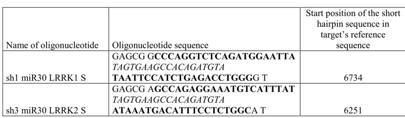
good model to investigate neurotransmitter release mechanisms since they share the same secretion system with dopaminergic neurons; ii) SH-SY5Y or HEK-293 cells transiently co-transfected with murine growth hormone (mGH) expressing construct as a secretion reporter and LRRK2 WT or mutant; iii) HEK-293 or SH-SY5Y, transiently co-transfected with construct coding for Dopamine Receptor D1 (DRD1) and LRRK2 WT or mutant; iv) Striatum and hippocampus from WT or transgenic G2019S LRRK2 mice. Finally, I conducted a functional redundancy study between LRRK2 and its closest homologue LRRK1, in respect to EGFR trafficking. EGFR and dopamine receptors (DAR) are members respectively of tyrosine kinase receptors family and G protein coupled receptors family. For this study I developed a confocal microscopy assay making use of different SH-SY5Y cell lines: LRRK2 Knock-Down (KD) stably expressing LRRK1, LRRK1 KD SH-SY5Y stably expressing GFP-LRRK2, or stable SH-SY5Y co-expressing a KD control construct and GFP-LRRK1 or 2 .
Experimental approach and results: LRRK2 is reported to interact with a broad range of proteins involved in synaptic functions, cytoskeleton dynamics, and vesicular trafficking, moreover previous work done in the laboratories have provided different hints of a potential role of LRRK2 in vesiscle trafficking . However, up to date, the physiological function of LRRK2 is elusive.The main topic of my PhD thesis was to figure out which role has LRRK2 in the vesicle trafficking using the following secretion reporter assays: SH-SY5Y were transiently co-transfected with mGH and WT or mutant LRRK2 both pathological and kinase dead. After 24 hours of protein expression, growth medium and cell lysates were collected and the mGH secretion level was measured in presence of WT or mutant LRRK2. The results demonstrate that expression of WT or pathological mutant LRRK2 determines an increase in basal GH extracellular level, moreover this increase is greater in presence of the pathological G2019S mutation. Furthermore the LRRK2 effect on mGH secretion is absent when a LRRK2 kinase dead mutant is co-transfected. To further elucidate LRRK2 kinase activity contribution in the observed mGH reporter secretion changes, the same assay was performed treating SH-SY5Y cells co-transfected with mGH and G2019S with the LRRK2 kinase inhibitor GSK2578215A. The presence of LRRK2 inhibitor significantly reduces the increase in GH extracellular level due to LRRK2 G2019S expression.
Neurotransmitter release has been studied in collaboration with a pharmacology research group of Department of Clinical and Experimental Medicine - University of Sassari - that has performed all the microdialysis experiments. To control LRRK2 expression in PC12, the Tet-ON system was used. Upon induction with doxycycline to obtain LRRK2 expression, cells were treated with nicotine to induce dopamine release. Direct measuring of dopamine released was performed by microdialysis. Main result of this set of experiments is that the pathological mutant G2019S determines a significant DA extracellular level increase compared to WT in both basal and nicotine induced condition. The same PC12 model was utilized to study vesicle distribution by Electron Microscopy (EM). Resultant images play up a significant increase of the electron-dense vesicles in the proximity of the plasma membrane in presence of G2019S mutant. These vesicles are probably recruited for docking phase of exocytosis process.
Vesicle trafficking regulates different cellular functions, including neurotransmitter or protein release and localization of membrane receptors. The previous results prompted me to analyze the possible effect of LRRK2 on membrane receptor localization using the membrane levels of DRD1 as a read-out. SH-SY5Y cells were transfected with plasmid coding for DRD1 in absence or in presence of LRKK2 G2019S, LRKK2 D1994A (kinase dead mutant), LRRK2 R1441G, or LRRK2 completely lacking the kinase domain (ΔKIN LRRK2). 48 hours after transfection, the sub-cellular distribution of D1DR receptors from cell lysates was analyzed by purification of different cell components. The presence of LRRK2 G2019S or R1441G and, to a lesser extent, LRRK2 D1994A or WT determines a significant increase in the level of membrane-associated DRD1 compared to DRD1 alone, while the effect is completely abolished in presence of the ΔKIN mutant. Interestingly all these results seem to be evident only in neuronal cells since they are not displayed in non-neuronal cell lines such as HEK-293.
To extend the analysis to a more physiological system I analyzed the distribution of two neurotransmitter receptors in the striatum of transgenic LRRK2 G2019S mice compared to non-transgenic ones. In particular, I analyzed the NMDA-NR1 receptor (NR1) and DRD1 distribution in total, membrane or vesicle fractions obtained from the striatum of 5
different animals of the two genotypes. LRRK2 G2019S expression leads to a significant increase of DRD1 in membrane fraction paralleling a significant decrease in vesicle fraction, with no significant differences in total protein extracts between the two genotypes. No significant differences among the different fractions were observed for NR1, clathrin or sec8, a member of the exocyst complex.
In line with these results on vesicle trafficking, I developed a confocal microscopy assay to assess functional redundancy between LRRK2 and LRRK1 in respect to EGFR trafficking. SHSY5Y KD for one of the two proteins but expressing a GFP fusion of the homologue one are treated with EGF-Rhodamine conjugate to track the receptor internalization. GFP-LRRK1 is co-localizing with EGFR in both LRRK2 KD or control conditions. On the opposite, LRRK2 does not co-localize with EGFR in control condition as well as in LRRK1 KD background. Thus LRRK2 seems not able to compensate for LRRK1 function in EGFR internalization process. This result has been validated by two-way ANOVA analysis. At the moment I am developing a similar assay to investigate the role of LRRK1 or 2 on dopamine receptor trafficking.
CHAPTER 1
INTRODUCTION
1.1 PARKINSON DISEASE
Parkinson Disease (PD) is a chronic, neurodegenerative, progressive disease. The main neuropathological features are the extensive and selective loss of neurons in the Substantia Nigra pars compacta (SNpc) and the presence of intraneuronal proteinacious cytoplasmic inclusions, termed “Lewy Bodies” (LBs). The cell bodies of nigrostriatal neurons are in the SNpc, and they project primarily to the putamen (in the striatum). The loss of these neurons, which normally contain conspicuous amounts of neuromelanin, produces the classic gross neuropathological finding of SNpc depigmentation (Figure 1B)(2). Dopamine (DA) depletion in the striatum and neuron loss in SNpc are also responsible of the characteristic motor symptoms of PD: tremor at rest, rigidity, postural instability and bradykinesia. At the onset of symptoms, striatal DA is depleted of ≈80%, and ≈60% of SNpc
dopaminergic neurons have already been lost. Nevertheless, the neurodegeneration is not limited to the SNpc but extends to other encephalic sub regions including noradrenergic neurons in the locus coeruleus and serotoninergic neurons in the nucleus basalis (2).
Incidence of PD is estimated in about 20/100000 cases per year in the population over 50 years, up to 120/100000 new cases per year among the over 70 years old (2). Sometimes, differences in incidence are observed between different ethnic groups probably due to disease etiology namely linked to environmental risk factors exposure or genetic susceptibility. The average age onset is about 60 years, even if 4% of patients display an early disease development (before 50 years). Furthermore, since the clinical features of PD emerge only when a high percentage of SNpc neurons is compromised and the striatal DA levels are strongly reduced, it is plausible that epidemiology studies underestimate the disease incidence.
Parkinson Disease has an heterogeneous nature, there are slow and fast progression forms; relatively simple symptomatology beside clinically complex forms.
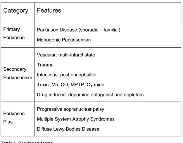
Indeed Parkinson forms are classified into: Primary Parkinson, Parkinson Plus and secondary parkinsonism (see table 1) (3)
Idiopathic PD, also known as Primary Parkinson, is characterized by rigidity, resting tremor, slowness of movement, rigidity and balance loss. This form of pathology has slow progress rate and at present there are no external causes ascribable to the symptomatology.
In life, the diagnosis of PD is made on clinical features, but definite diagnosis requires the identification of both LB and SNpc dopaminergic neuron loss. LBs are not specific for PD, however, and are also found in AD, in a condition called “dementia with LB disease” and as an incidental pathologic finding in people of advanced age at a greater frequency than the prevalence of PD (3). LBs are spherical eosinophilic cytoplasmic protein aggregates composed of numerous proteins, including α-synuclein, parkin, ubiquitin, and neurofilaments, and they are found in dopaminergic neurons in all affected brain regions (4, 5). LBs are more than 15 µm in diameter and have an organized structure containing a dense hyaline core surrounded by a clear halo. Electron microscopy reveals a dense granulovesicular core surrounded by a ring of radiating 8–10 nm fibrils (6).
Category
Features
Primary Parkinson
Parkinson Disease (sporadic – familial) Monogenic Parkinsonism
Secondary Parkinsonism
Vascular: multi-infarct state Trauma
Infectious: post encephalitic Toxin: Mn, CO, MPTP, Cyanide
Drug induced: dopamine antagonist and depletors
Parkinson Plus
Progressive sopranuclear palsy Multiple System Atrophy Syndromes Diffuse Lewy Bodies Disease
Table 1. Parkinson forms.
Another parkinsonism form is “Parkinson Plus”. In this category are grouped some neurodegenerative disorders showing a strict clinical similarity with PD, but characterized by concurrent presence of other neurological aspects (cerebellar, pyramidal, vegetative, cognitive). The most important among the Parkinson Plus manifestations are: Progressive sopranuclear palsy, Multiple System Atrophy Syndrome and the corticobasal degeneration (7).
The last parkinsonism category is Secondary Parkinsonism. These forms display similar characteristics to PD, have known etiology, variable progress and a very low prevalence. Secondary Parkinsonisms can be induced by toxins (Manganese, carbon monoxide, cyanide), drugs (neuroleptics, antiemetics, calcium blockers), or traumatic brain damages (i.e. Pugilistic Parkinson). Drug-induced parkinsonism are marked out by the lack of neuronal loss and are subject to remission after drug assumption interruption. On the contrary, neurotoxins damage to the SNpc is irreversible (8). All Secondary Parkinsonisms
are characterized of a very fast clinical development, lacking or reduced drug response and no Lewy Bodies presence in respect to the Primary Parkinson forms.
For years, diagnostic criteria for idiopathic Parkinson Disease have been developed. Recently Gelb and colleagues have reviewed them highlighting how clinical diagnosis of PD is established on the presence of cardinal motor symptoms in combination with the exclusion of specific uncharacteristic symptoms. Among motor cardinal signs:
- Resting tremor - Rigidity
- Bradykinesia - Asymmetric onset - Postural instability.
Lastly, also the responsiveness to levodopa (L-Dopa) is considered. This is, with sporadic exceptions, a necessary but not exclusive requisite for PD diagnosis, inasmuch is observed also in atypical forms of the disease (9, 10).
1.2 DOPAMINE AND DOPAMINE RECEPTORS
As mentioned before, a progressive demise of the dopaminergic cells in the SNpc is observed in post-mortem brains of PD patients. From SNpc the dopaminergic neurons form synapses mainly with neurons of the striatum (another nucleus of the basal ganglia). This system constitutes the nigrostriatal pathway, which regulates the extrapyramidal system designated to the movement, posture and balance minding control (11).
Dopamine is synthetized by dopaminergic cells in SNpc (pre-synaptic level) (12, 13), thus, in the parkinsonian patient, SNpc cells degeneration dramatically drops dopamine production and dopamine levels in the striatum producing all the motor symptoms which characterize the pathology.
1.2.1 Dopamine synthesis and metabolism
Dopamine, together with adrenaline and noradrenaline, constitutes the catecholamine neurotransmitter class. They are all synthetized starting from tyrosine through a shared pathway including five enzymes: the tyrosine-hydroxylase, an aromatic amino acid decarboxylase; the dopamine β-hydroxylase; the pteridine-reductase and the feniletanolamina-N-methyltransferase. The first enzyme in the pathway, the tyrosine-hydroxylase, converts the amino acid tyrosine to L- dihydroxiphenilalanine (L-DOPA). At the next step of the pathway, the L-DOPA is decarboxylated by the DOPA-decaroxylase obtaining dopamine and CO2. The dopamine β-hydroxylase converts dopamine in norepinephrine.
Once synthesized in the SNpc neurons, dopamine is stored in vesicles and released in the inter-synaptic space, where is free to interact with DA receptors (both at pre and postsynaptic level) and exert its action. Dopamine activity is terminated with a process called “re-uptake” at the presynaptic terminal. In this process dopamine is transported in the presynaptic neuron by the dopamine transporter (DAT), and, is degradated by two enzymes: the monoamine-oxidase (MAO) and the catechyl-O-metyl-transferase (COMT). The MAOs are localized in the external mitochondrial membrane, albeit other MAOs are
also present outside the dopaminergic neuron, respectively MAO A and MAO B; whereas the COMT are extraneuronal enzymes that inactivate catecholamines by methylation of the hydroxyls on the catechol ring (Figure 2).
1.2.2 Dopamine receptors trafficking
Dopamine receptors belong to the large family of heptahelical transmembrane spanning G protein-coupled receptors (GPCRs). Five mammalian dopamine receptor subtypes have been identified and are classified into two major groups, the D1- (D1 and D5) and D2- (D2,
D3, D4) like receptors (14, 15). Although dopamine receptors are similar in structure, receptor subtypes differ by their affinity for dopamine and coupling to downstream effectors, including heterotrimeric guanine nucleotide-binding proteins (G proteins) (14, 16). Dopamine receptor subtypes are expressed differentially throughout the brain. Among the D1-like receptors, D1 receptors are the most abundant, with mRNA transcripts found in the neostriatum, nucleus accumbens, and olfactory tubercle. Lower levels of D1 receptor mRNA are found in the cerebral cortex, hypothalamus, and thalamus (14). Among the D2-like receptors, D2 receptors are the most abundant, with mRNA transcripts found in the neostriatum, nucleus accumbens, and olfactory tubercle, as well as the midbrain, including the substantia nigra and ventral tegmental area (14).
The canonical mechanisms for GPCRs signaling, reviewed in (17) can be extended to dopamine receptors D1 and D2.
Binding of ligands (such as hormones, neurotransmitters or sensory stimuli) induces conformational changes in the transmembrane and intracellular domains of the receptor thereby allowing interactions with heterotrimeric G proteins. Activated GPCRs act a guanine nucleotide exchange factors (GEFs) for the α subunits of heterotrimeric G proteins, catalyzing the release of GDP and the binding of GTP for G protein activation. The activated G protein subunits (α•GTP and βγ) can then associate with downstream effectors (17) including second messengers like adnylate cyclase and ion channels, to modulate various aspects of cellular physiology. A distinguishing feature of D1- and D2-like receptors is their differential coupling to heterotrimeric G proteins. In many cell types D1-like receptors couple to Gαs, which activates adenylate cyclase (52, 53) while D2-like receptors couple to the pertussis toxin-sensitive Gαi or Gαo, which inhibit adenylate cyclase (48, 54)
G protein-mediated signaling by agonist-activated GPCRs can be terminated through GPCR phosphorylation by GPCR kinases (GRKs) and concomitant GPCR association with arrestins, which interact with clathrin and the clathrin adaptor AP2 to drive GPCR internalization into endosomes (see figure 4 from (17)) Finally, G protein activity is terminated by GTP hydrolysis and the re-formation of the G protein heterotrimer. GPCR internalization regulates the functional process of receptor desensitization (17), a process that determines the end of the GPCR signaling.
Following internalization after association with arrestins, GPCRs can be trafficked to lysosomes, where they are ultimately degraded, or to recycling endosomes for recycling back to the cell surface in the functional process of re-sensitization — whereby the cell is re-sensitized for another round of signaling.
GPCR internalization is heavily influenced by two of the canonical families of GPCR-interacting proteins, the GRKs and the arrestins. However, certain other GPCR-GPCR-interacting proteins can also regulate the endocytic trafficking of GPCRs in a more receptor selective manner. For example, the GPCR-associated sorting proteins (GASPs) promotes D2R trafficking to lysosomes following agonist stimulated endocytosis (17).
Dopamine receptor-mediated signaling is a tightly regulated process that is highly dependent on the accessibility of receptors to agonist binding at the cell surface. This availability of functional surface receptors is governed by a strict balance of the various intracellular receptor trafficking pathways that, in association with receptor sensitivity, work in concert to regulate the amplitude of agonist-mediated cellular responsiveness (14). It has been demonstrated that GPCRs are not static within the plasma membrane but can move in the plane of the membrane by the passive process of lateral diffusion (18-20). One of the key factors that govern the dynamics of lateral diffusion of GPCRs is their association with other cellular proteins. The formation of these protein complexes can serve to restrict the movements of the GPCRs, effectively stabilizing the receptors in specific microdomains within the membrane, such as the synapses for example. Movement by lateral diffusion has been reported for several GPCRs including dopamine D1 receptors. In cultured neurons, approximately 65% of D1 receptors are mobile with the remaining receptor anchored within the membrane. This ratio of mobile vs anchored receptors is not fixed, but fluctuating. Scaffolding proteins have a role in this process, either stabilizing DA receptors at the membrane or decreasing their stability.
1.3 TREATMENT
Current PD medication treat symptoms none halt or retard dopaminergic neuron degeneration. The main obstacle to developing neuroprotective therapies is a limited understanding of the key molecular events that provoke neurodegeneration (2).
1.3.1 Drug Therapy
As mentioned above, to date, no defined neuroprotective treatment for PD influencing the natural progressive course of the disease has been established. Therefore current treatments aim at correcting the dopaminergic deficit in the Substantia Nigra either through (i) the substitution of dopamine via its precursor levodopa, or (ii) the prolongation of the synaptic availability of dopamine via inhibitors of enzymes of the dopamine metabolism: COMT (catechyl-O-metyl-transferase) and MAO B (monoamine-oxidase B), or (iii) the activation of functional relevant receptors via dopamine-agonists (21).
Levodopa
Levodopa (L-dopa) is the most effective drug in the treatment of PD and virtually all patients benefit from this treatment (21). L-dopa is the natural parent for dopamine, it enters in the dopaminergic neurons that convert it into dopamine. Direct administration of
dopamine as a drug is not possible as it is not able to pass the hematoencephalic barrier and thus reach the dopaminergic neurons. The L-dopa is orally administered and absorbed in the bowel, and finally enters the systemic circulation. The L-dopa decarboxylase, outside of CNS, catalyzes the transformation of L-dopa into dopamine by removing its carboxylic
group. Since dopamine has no therapeutic effect at peripheral level rather unpleasant side effects, L-dopa is routinely administered in combination with a decarboxylase inhibitor to avoid peripheral L-dopa decarboxylase activity. Once in the brain, L-dopa is converted in dopamine by neuronal L-dopa decarboxylase in the cytoplasm, here is actively transported in the synaptic vesicles. Dopamine storing in vesicles avoids its enzymatic degradation by MAO and it is essential for the following release of the neurotransmitter in the inter-synaptic space upon neuronal stimulation. When the impulse reach the dopaminergic
neurons, the vesicles fuse their membrane with the neuronal plasma membrane releasing dopamine in the synaptic space, where it can reach the receptors on the post-synaptic neurons and complete its therapeutic activity.
Although levodopa represents the gold standard in PD therapy, chronic levodopa treatment is known to result in motor complications (dyskinesia, dystonia) or neuropsychiatric problems in later stage of disease (21).
Dopamine agonists
Dopamine agonists are a class of drugs that share the capacity to directly stimulate dopamine receptors. This class of therapeutics compounds offers some advantages over levodopa therapy as the reduced incidence of levodopa-related adverse effects, no requirement of metabolic conversion and potential neuro-protective benefits. However, the anti-Parkinson efficacy is limited and motor complications are not completely prevented. COMT inhibitors
To date, the entacapone is the only substance available in this therapeutic class and it is administered in association with levodopa as previously described. COMT inhibitors exert their therapeutic effects via inhibition of peripheral levodopa catabolism and therefore
increasing levodopa bioavailability. Thus, COMT inhibition is associated with an increased stability of levodopa plasma levels, suppressing peaks of levodopa concentrations, which are thought to be associated with motor complications (21).
MAO B inhibitors
The most diffused MAO B inhibitor in PD therapy is selegiline; it is a selective inhibitor of the oxidative catabolism of dopamine via irreversible inhibition of the monoamine-oxidase B. Therefore selegiline amplifies the effect of levodopa. In experimental studies selegiline was not only able to improve parkinsonian symptoms but also to exert some neuroprotective effects on dopaminergic neurons. However, clinical studies on the efficacy of this therapeutics have produced controversial results (21).
1.3.2 Surgical therapy
Recent advances in neurosurgery and imaging techniques have allowed the development of novel therapeutic approaches for PD. In general there is a main recently developed approach to the surgical therapy of PD: the deep brain stimulation (21) is a surgical treatment involving the implantation of a medical device called a brain pacemaker, which sends electrical impulses to specific parts of the brain.
1.4 ETIOLOGY
Parkinson Disease has a complex and multifactorial etiology, involving genetic and environmental factors. However the molecular details of the degenerative process are still barely known even though the discovery of PD related genes has shed new light on the cellular mechanisms that might be affected in the PD neurodegeneration.
1.4.1 Environmental Factors
The environmental etiology hypothesis posits that PD-related neurodegeneration results from exposure to some dopaminergic neurotoxins. Theoretically, the progressive neurodegeneration in PD could be produced by chronic neurotoxin exposure or by limited exposure initiating a self-perpetuating cascade of deleterious events (2). An example of how an exogenous toxin can mimic the clinical and pathological features of PD is the MPTP (1-methyl-4-phenyl-1, 2, 3, 6,-tetrahydropyridine or meperidine) intoxication (22). The discovery of MPTP as dopaminergic neurotoxin date in 1982 when young drug users developed a rapidly progressive parkinsonian syndrome due to intravenous use of a street preparation of
1- methyl-4-phenyl-4-propionoxypiperidine (MPPP), an analog of the narcotic Demerol (22) in which MPTP
was present as
contaminant. In
humans and monkeys, MPTP produces an irreversible and severe parkinsonian syndrome characterized by all the PD features, including tremor, rigidity, slowness of movement, postural instability and freezing. In MPTP-intoxicated individuals the beneficial response to levodopa and development of long-term motor complications to medical therapy are virtually identical to those seen in PD patients. Similar to PD, MPTP susceptibility increases with age and is observed the same topologic pattern in the damages to the dopaminergic system (2) (23, 24) In PD, dopaminergic neurons seem to be more sensible to degeneration in respect to other neuronal populations in the brain, these neurons are also more susceptible to MPTP induced degeneration (2). All these clinical
manifestations on MPTP intoxication corroborates the environmental hypothesis of PD etiology and the MPTP-treated monkeys became a good model for PD studies. Some of these studies unraveled the MPTP mechanism of toxicity. After systemic administration, MPTP, which is highly lipophilic, cross the blood-brain barrier. Once in the brain the pro-toxin MPTP is oxidized to 1-methyl-4-phenyl-2, 3, -dihydropyridinium (MPDP+) by MAO-B in the glia, and probably by spontaneous oxidation it becomes MPP+, the active toxic molecule. Thereafter it is released in the extracellular space by an unknown mechanism. Finally is concentrated into dopaminergic neurons through its high affinity for the dopamine transporter (DAT). Inside neurons MPP+ can follow at least three routes: (i) it can bind to the vesicular monoamine transporter-2 (VMAT2 ) (25), which translocate MPP+ into synaptosomal vesicles; (ii) it can be concentrated within the mitochondria (26) and (iii) it can remain in the cytosol to interact with the cytosolic enzymes (27). The toxic mechanism of MPP+ is carried out in the mitochondria where damages electron transport chain (see figure 7). Here MPP+ blocks complex I, which interrupts the transfer of electrons to ubiquinone. This perturbation enhances the production of ROS species and decreases the synthesis of ATP with the final result of apoptosis in the neurons (2, 22). The MPTP toxicity mechanism supports the idea of an involvement of the mitochondria and oxidative stress in PD pathogenesis.
Many epidemiologic studies have been done to clarify if toxin exposure can cause PD. Subject of these studies have been pesticides and insecticides with structure similar to MPP+ as paraquat and rotenone but none of these studies reported unmistakable results. So far, there is no clear evidence that exposure to herbicides or insecticides can cause PD. Other epidemiological studies reported that cigarette smoking and coffee drinking are inversely associated with the risk of PD development. Based on the apparent protective effect of smoking, the therapeutic effect of nicotine has been tested in a few clinical trials, but no improvement of motor symptoms with transdermal nicotine treatment has been documented (28).
Another possibility, which does not fit into a genetic or environmental category, is that an endogenous toxin may be responsible for PD neurodegeneration. Distortions of normal
metabolism might create toxic substances due to environmental exposures or inherited differences in metabolic pathways. One source of endogenous toxins may be the normal metabolism of DA, which generates harmful reactive oxygen species (ROS)(29). Consistent with this hypothesis is the report that patients harboring specific polymorphisms in the gene encoding xenobiotic detoxifying enzyme cytochrome P450 may be at higher risk of developing young onset PD (30). Further, toxic derivatives of isoquinoline have been recovered from PD brains (2, 31).
In addition to toxins, other environmental factors, which can be considered as risk factors rather than causative elements, are lifestyle and habits. For instance, repetitive or simply violent traumas can provoke a parkinson-like progressive syndrome as the one frequently observed among boxers from which the name “Pugilistic Parkinson Syndrome”. Another two risk influencing factors are age and sex, males are indeed more affected in respect to women (in a 1,5:1 ratio). In humans, neuronal and SNpc neuromelanin loss increases around 60 years, coinciding with the average age of PD onset. Since neuromelanin has a protective effect in neurons against free radicals and toxins, the decrease in this pigment may predispose the brain of aged people to Parkinson (32).
Finally, it has been hypothesized an infective etiology: clinical manifestations similar to PD have been observed in patients affected, in the past, by a viral encephalitis propagated in pandemic in 1920 (33, 34).
1.4.2 Genetic Factors
For a long time Parkinson Disease has been considered a sporadic disease, until Leroux in 1880 tracked recurring PD cases in the same family. This observation led to the hypothesis that hereditary factors could increase susceptibility to the pathology.(28). In the last decade several genome wide association studies allowed the identification of numerous mutations in different genes in families with mendelian inheritance parkinsonisms. These findings have been made possible through the analysis of rare familial PD forms characterized by clinical and neuropathological features sometimes different from idiopathic forms. More
2 : Su m m ary o f P AR K 1 -PA R K1 3 e xpressi o n patt ern s ( fo r refer ences, pleas e see t ex t) . Fr o m Bisku p e t al . ( 1 )
recently some susceptibility loci for PD have been identified, but further analysis are required to confirm their association with Parkinson Disease (35, 36).
Up to date, 16 mendelian transmission forms of Parkinson Disease are known. Some of them have an autosomal dominant (AD) inheritance pattern while others have an autosomal recessive (AR) one. Gene products have been associated to 10 out of these 16 forms whereas other causative (PARK 3) or predisponent (PARK 10, PARK 12, PARK 16) chromosomal loci are still waiting to be linked to a precise gene product (37) (See table 2).
Mendelian inheritance Parkinson forms and associated genes
Although genetic PD forms represent only a little percentage of the total cases, studies on cellular or animal models based on the expression familial Parkinson gene have importantly contributed to elucidate disease etiopathology. In this session, the main characteristics of PD associated genes are described, giving particular relevance to those ones that are widely accepted to be conclusively associated with mendelian forms of the disease (35) and have been more extensively studied (PARK1/4, PARK2, PARK5, PARK6, PARK7). For a more detailed presentation of PARK8 locus see paragraph 1.5.
- Alpha-synuclein (SNCA or α-Syn) - PARK1/PARK4
Although Lewy bodies (LB) represent one of the principal landmarks of PD, and their first observation date to 1912, only in 1997 Alpha Synuclein has been identified as the most important component of these protein inclusions (5). In the same year, 1997, the identification of SNCA as the first gene implicated in PD resulted from linkage analysis in the ‘‘Contursi’’ kindred, with the point mutation A53T being associated with the disease in this family. Since then, two other point mutations, A30P and E46K, as well as duplications and triplications of the SNCA gene, have been identified among PD patients. The point mutations and gene rearrangements result in rare autosomal dominant, middle to late onset PD. The loss of dopaminergic neurons and the presence of Lewy bodies, not only in the Substantia Nigra, but also in the Locus Coeruleus, have been observed in the SNCA linked PD (35).
The SNCA gene has six exons, encoding an abundant 140 amino acid cytosolic protein, which is found at presynaptic terminals and thought to be involved in synaptic function (35). Despite intensive studies, the exact role of α-SYN at the synapse remains elusive. There is evidence that the protein plays a role in maintenance of synaptic vesicle pools and activity-dependent dopamine release (1), Larsen et al.(38) have provided evidence that α-SYN might modulate synaptic vesicle priming. Nevertheless α-α-SYN knockout mice have little
The α-synuclein protein is organized in three distinct domains: an N-terminal amphipathic region with conserved repeats (KTKEGV), a central hydrophobic NAC domain (non-amyloid component), and an acidic C-terminal region. All the three point mutations are located in the amphipathic region and are believed to exacerbate the toxic protofibril and fibril formation. Indeed, monomeric mutant a-synuclein has the propensity to form stable β-pleated sheets; the ensuing fibrillogenesis (formation of oligomers, protofibrils, and finally fibrils), results in the generation of pathological inclusions, known as Lewy bodies. Moreover, dopamine has been shown to modulate -synuclein aggregation, perhaps explaining the selective vulnerability of the Substantia Nigra in PD.
Studies in drosophila indicate that Lewy bodies may be a way of detoxifying the cell of damaged -synuclein. The enhanced toxicity of mutant -synuclein may also be explained by the assembly of oligomeric -synuclein into annular structures (39), that integrate into cellular membranes to form pores, thereby affecting membrane permeability. Of note, the overexpression of the wild-type protein is sufficient to cause PD, as duplications or triplications of the wild-type SNCA produce the disease in patients (35) although the precise relationship between aggregation, cellular dysfunction and cell death underlying PD is unknown (1).
- Parkin - PARK2
A year after the discovery of the SNCA gene, Parkin has been associated with PD (35). It is one of the largest genes in the human genome, mapping 1.38 Mb and comprising 12 exons (18). The Parkin gene encodes an ubiquitous 465 amino acid protein (35) organized in domains: an N-terminal ubiquitin like domain, a central linker region and C-terminal RING domain consisting of two RING finger motifs separated by an in between RING domain (19). Parkin functions as an E3 ubiquitin protein ligase similar to other RING finger containing proteins by targeting misfolded proteins to the ubiquitin proteasome pathway for degradation, and the loss of its E3 ligase activity due to mutations lead to autosomal recessive early-onset PD. Parkin functions as a neuroprotective protein in a variety of toxic
insults crucial for dopamine neuron survival (19); it is therefore conceivable that the loss of parkin function may lead to the accumulation of a nonubiquitinated substrate, which is deleterious to the dopaminergic cell, but, due to its nonubiquitinated nature, does not form typical Lewy bodies (20).
Parkin interact with several presynaptic proteins (summarized by Moore (40). α-SYN and the α-SYN-binding synaptic protein (SNCAP) synphilin are two prominent examples. In addition parkin has been shown to modulate the function of a G-protein coupled receptor (GPR37) that interacts with the dopamine transporter DAT (1). The molecular events generated by mutated Parkin that lead to neuronal degeneration seem to be in this order: accumulation of substrates and other proteins, proteasome inhibition and cellular death. Actually, animal models allowed disclosing a possible additional role of Parkin in mitochondrial maintenance and oxidative stress prevention in cooperation with another PD-associated protein, PINK1 (see hereinafter). In particular, PINK1 would be involved in mitochondrial membrane integrity maintenance and could assist Parkin in the removal of damaged proteins.
Finally, oxidant molecules, among which dopamine itself, are able to alter Parkin solubility leading to its aggregation (41).
- Ubiquitin carboxyl-terminal esterase L1, UCHL-1 - PARK5
A mutation in Ubiquitin carboxyl-terminal esterase L1 gene was identified in affected members of one single family of German ancestry. To date, no other bona fide pathogenic mutations of this gene have been found. Whether UCHL1 really is a PD-responsible gene is not yet clear. Interestingly loss of UCHL1 function leads to neurodegeneration in mice (1). UCHL-1 is an abundant brain enzyme; its function might correlate with polymeric ubiquitin recycling and its conversion into monomeric units. Mutations in PARK 5 gene cause enzymatic activity reduction of UCHL-1 with consequent proteasome activity alteration (42).
- PTEN-induced kinase 1, PINK-1 - PARK6
This gene is particularly interesting within the context of the findings linking PD to mitochondrial dysfunction and oxidative stress, as PINK1 is a mitochondrial protein kinase. The PINK1 gene encodes a 581 amino acid ubiquitous protein, consisting of an N-terminal 34 amino acid mitochondrial targeting motif, a conserved serine–threonine kinase domain and a C-terminal auto regulatory domain. The majority of the identified mutations are in the kinase domain, indicating the importance of PINK1 enzymatic activity in PD pathogenesis (35).
A substrate of PINK1 has been recently identified as TNF receptor-associated protein 1 (TRAP1). TRAP1 is a mitochondrial molecular chaperone, also known as heat shock protein 75. Wild-type PINK1 has been shown to phosphorylate TRAP1, moreover this phosphorylation is significantly increased in response to oxidative stress induced by H2O2. Interestingly, the PD-linked PINK1 mutants, G309D or L347P, abolish this kinase activity, although not affecting either the PINK1-TRAP1 interaction or their mitochondrial co-localization. Overexpression of wild-type PINK1 further revealed that cytochrome c release from mitochondria was reduced under oxidative stress conditions. TRAP1 is therefore proposed as being a downstream effector of PINK1: when phosphorylated by PINK1, TRAP1 may indirectly suppress cytochrome c release from mitochondria, protecting against oxidative stress-induced cell death (43). It is therefore plausible to assume that Parkin, PINK1, and TRAP1 are interacting in the same signaling pathway, the defects of which may lead to PD.
PINK1 seems to be involved in another important protection mechanism against oxidative stress, once again acting in concert with another protein, htrA2, which is a serine protease located in the mitochondria. PINK1 phosphorylates htrA2 thus tuning its proteolytic activity which in turn contributes to increased cell resistance to oxidative stress (44). More recently, Wang et al. described a new mechanism trough which PINK1, immobilizing damaged mitochondria, confines cellular damages produced by ROS species to a little region in the cell, putting damaged organelles to quarantine prior to their definitive
removal by autophagosome. Cells are able to maintain their energy balance preventing oxidative stress injuries by fine tuning of trafficking, distribution and clearance of mitochondria. PINK1 acts in the control of this balance intervening on mitochondrial motility. Specifically, it has been demonstrated that PINK1 phosphorylates the protein Miro, which is part of a complex anchoring the kinesin to the mitochondrial surface, thus controlling mitochondria transport alongside the axon (45). In fact phosphorylated Miro is targeted for degradation in a Parkin-dependent pathway. Removal of Miro from the mitochondrion also detaches kinesin from its surface arresting the damaged mitochondria until their complete degradation occurs, likely by autophagosome engulfment. This PINK1-Parkin activity is fundamental for ensuring the removal of damaged organelle preventing the injury diffusion in a larger area in the cell (46, 47). As a consequence, neurons defective for PINK1 and/or Parkin show an impairment in mitochondrial clearance which makes them susceptible to degeneration (42, 48).
- DJ-1 - PARK7
Parkinson Disease form 7 is one of the rare AR parkinsonisms with juvenile onset. Clinical features associated to PARK7 gene are very similar to those linked to PARK2 except for the presence of psychiatric symptoms. This gene, located in chromosome 1 encodes a 189 amino acids protein identified as DJ-1 by Bonifati et al. in 2003. It has ubiquitous expression in a variety of mammalian tissues including brain and it localized into mitochondria (49). The wild type form is present as homodimer located in the cytosol, nucleus and mitochondria, but upon oxidative stress conditions it is relocated in the matrix and in the intermembrane space of the mitochondrion where it exerts a protective activity against ROS. Many lines of evidences suggest that DJ-1 functions as an antioxidant protein. Oxidative stress leads to an acidic shift in the DJ-1 isoelectric point by oxidation of Cys106 which can be converted to cysteine sulfinic acid (Cys-SO2H). Because of its inherent ability to undergo self-oxidation to eliminate H2O2 it may function as a scavenger of reactive oxygen species (ROS). In fact overexpression of wild-type DJ-1 both in cell culture and dopaminergic neurons in vivo protects against wide variety of toxic injury due to oxidative
stress. DJ-1 also functions like a redox-dependent chaperone to inhibit -synuclein aggregation and subsequent cell death. Some familial DJ-1 mutants may have altered abilities to undergo key oxidative modifications, namely, a decreased propensity to undergo oxidation at position 106, and/or an increased susceptibility to undergo oxidative modifications with potentially deleterious effects on DJ-1(49) protective function.
1.5 LEUCINE RICH REPEAT KINASE 2 – (LRRK2)
1.5.1 Physio-pathological Role of LRRK2 (PARK8)
PARK8 locus have been associated for the first time to PD in 2002, by Funayama’s group (50), in a Japanese family presenting a autosomal dominant form of parkinsonism without Lewy Bodies presence. Two years later two other groups independently have identified the gene LRRK2 in the PARK8 locus (51, 52); subsequently many pathological mutations have been identified.
LRRK2 mutations are usually associated to clinical parkinsonism – with typical movement problems of tremor, rigidity, bradykinesia and postural instability. Where autopsies have been performed, prominent loss of melanised dopaminergic neurons in the Substantia
Nigra pars compacta has been noted. As might be expected, movement problems in these
patients respond to L-DOPA treatment. PD cases due to LRRK2 mutation are therefore similar to sporadic PD and distinct from some other types of inherited parkinsonisms. Most, but not all, cases appear to have Lewy bodies, the characteristic pathology of α-synuclein deposition seen in PD and related conditions (53).
LRRK2 : general features and domains
The LRRK2 coding gene is located in chromosome 12 (12.q12), is composed of 51 exons giving rise to a 2527 amino acid protein consisting of 7 functional conserved domains (figure 10)(54, 55) :
An “Armadillo Repeats” domain; An “Ankirin Repeats” domain;
A “Leucine Rich Repeats” (LRR) domain, composed of a 22-28 amino acids motif repeated 13 times;
A Roc domain with GTPase activity; A Carboxy Terminal of Roc (COR) domain
A kinase domain, with homology with MAP-Kinase-Kinase-Kinase (MAPKKK) A C-terminal WD40 domain.
The ”Armadillo Repeats” domain is composed by a repeated 42 amino acids motif organized in 3 α-helixes and was firstly identified in the “Armadillo” protein of Drosophila, which human homologous is β-catenin. The Armadillo domain forms a versatile molecular interaction platform available for different protein interactors (56).
The “Ankirin Domain” consists of seven ankyrin repeats, each forms two antiparallel helices followed by a β-hairpin or loop (Figure 8 - a from Mata et al 2006). The repeats stack together to form a gently curved structure in the ankyrin repeat domain. Ankyrin repeats are found in diverse bacterial and eukaryotic proteins, including cytoskeletal proteins, transcription factors, signaling proteins and cell cycle regulators (57).
Each of the 13 identified LRRs in the leucine rich repeat domain is predicted to form a β-strand followed by an α-helix that line up side-by side to form an arch-like structure (Figure 8 - b from Mata et al. 2006). LRR domains participate in the interactions with different proteins through binding to their extended solvent-accessible surface (58).
The WD40 domain is composed by repeated motifs of 40 amino acids, mainly tryptophan-aspartate. Each repeat contains a four-stranded, antiparallel β-pleated sheet and together these repeats form a circular bladed propeller-like structure. The predicted WD40 domain of LRRK2 comprises seven WD40 repeats (Figure 9 - a modified from Mata et al. 2006). This seven-bladed propeller is thought to form a rigid platform for reversibly interacting with proteins, possibly including those that contain other WD40 domains. The WD40 domains are the most common repeats found in human proteins and normally they coordinate the assembling of multi proteic complexes. They have been identified in functionally diverse proteins including the Gβ subunit of heterotrimeric G proteins, transcriptional regulators, protein phosphatase subunits, RNA processing complexes, cytoskeletal assembly proteins, and proteins involved in vesicle formation and trafficking (58)
Catalytic domains of protein kinases are long 250-300 amino acid residues, containing a small N-terminal lobe and a larger C-terminal lobe connected by a hinge-like region to form a cleft in which Mg2+ -ATP and the protein substrate binds (Figure 9 - b - modified from
Mata et al. 2006). The activation segment is composed of 20–35-residues within the large C-terminal lobe. The majority of protein kinases require phosphorylation of the activation segment for full activity. Upon phosphorylation, the activation segment is believed to adopt an active conformation, enabling substrate access and catalysis to take place (58). The putative GTPase domain of LRRK2 belongs to the ROCO family, in which the predicted GTPase (Roc) is always found in tandem with the COR domain, the function of which is still unknown. This Roc–COR module is conserved throughout evolution, suggesting the functional interdependence of the two domains. The Roc-GTPase domain of the LRRK2 resembles most closely the GTPases of Rab family, which have been implicated in vesicular
trafficking and transport (58) processes by acting as molecular/regulatory switches that cycle between GTP and GDP-bound conformations (59). A functional link is postulated to exist between the intrinsic GTPase activity and downstream kinase activity. Different studies have suggested that LRRK2 kinase activity is dependent on the existence of a functional GTPase domain, whereas GTPase activity can function independently of the kinase domain (59). An additional hypothesis proposes that GTPase activity would be able to regulate the kinase activity and/or the kinase activity would have the ability to regulate the GTPase activity by an auto-phosphorylation mechanism. In support of this latter hypothesis are the various studies that assess auto phosphorylation ability of LRRK2. The kinase domain is indeed not only able to phosphorylate some potential substrates but also, with highest efficacy, the protein itself in correspondence to the ROC domain (see figure 10 from Rideout et al. (55)).
Dimerization of the ROC or ROC-COR domains results in an intermolecular interaction between the kinase domains subsequently allowing an increased kinase activity, thereby structurally explaining the interplay between these enzymatic domains. Dimerization of LRRK2 has been demonstrated by yeast two-hybrid analysis, native gels, size exclusion chromatography and co-immunoprecipitation from mammalian cells (59, 60)
The presence of multiple protein interaction domains (armadillo, ankyrin, LRR and WD40) suggests that LRRK2, in addition to its predicted protein kinase and GTPase activities, might serve as a scaffold for assembly of a multiprotein signaling complex. However, because these domains bind diverse proteins ranging from transcription factors to signaling proteins (58), the physiological LRRK2 interactors and substrates and its function remain unknown.
Recent mRNA expression analysis studies demonstrate the LRRK2 transcript presence in the brain and in various peripheral districts. In rodents, high levels of LRRK2 mRNA have been detected in brain and lymph nodes. LRRK2 is particularly abundant in the dopamine-innervated areas as (53) cortex, striatum, cerebellum, hippocampus and olfactory tubercle; while lower levels are observed in the substantia nigra (55). The subcellular localization of LRRK2 is mainly cytosolic with an important component associated with the membranes,
vesicles and cellular organelles as Golgi, mitochondria, lysosomes and RE, as well as microtubules and cytoskeleton structures (61)
LRRK2 pathological mutations do not alter protein stability and/or localization but some of them increase the kinase activity; they are, in particular, clustered in the central part of the gene in the ROC GTPase domain and the kinase domain (36). An increase in the LRRK2 kinase activity appears to be sufficient to generate neuronal toxicity implicated in PD (54, 59, 62). Studies performed on cell lines demonstrate that LRRK2 has long half-life, and, especially if co-expressed with Parkin, tends to form aggregates which are prone to ubiquitination through a process involving Parkin (63)
More the 75 variants of LRRK2 are described, but not all of them are PD-causative mutations. On the other hand some mutations are associated to PD and they are localized in the enzymatic region of the protein (figure 10 from Rideout et al): The ROC domain is affected by multiple mutations: R1441G, R1441C, R1441H) (52). The effects of these mutations result in an alteration of GTPase activity, but the final effect of these alterations on the kinase activity are quite contrasting. In the ROC domain only a pathological mutation has been described (Y1699C) while in the kinase domain there are two adjacent mutations, G2019S and I2020T. G2019S is the most common pathogenic substitution in the familial forms of PD but is also associated to sporadic forms (36, 64, 65). Various studies performed on cellular models have consistently showed that this substitution leads to an increase in kinase activity up to threefold. The 2019 residue is indeed located in a loop which is closing like a string the catalytic site, the substitution of glycine with serine forces the catalytic site in a constitutively active state leading probably to the increased kinase activity (59, 66).
From the first association between LRRK2 and PD in 2004, many epidemiologic studies have assessed that LRRK2 is responsible of roughly 10% of PD familial forms and importantly also the 4% of the sporadic ones. The glycine to serine substitution in 2019 position of LRRK2 represents the most frequent substitution in the Caucasian population; it is indeed responsible for 1-2% of idiopathic PD and 6% of familial PD; moreover, G2019S mutation is higher in some population of North Africa and in Ashkenazi Jews (13-41% sporadic cases and up to 40% of familial ones)(67).
Concerning Sardinia, a study published in 2006 by Cossu et al. shows that G2019S substitution is present in the highland with a lower frequency in respect to other mediterranean population, while R1441C is very uncommon in Sardinian population (68).
LRRK2 physiologic function and involvement in Parkinson Disease
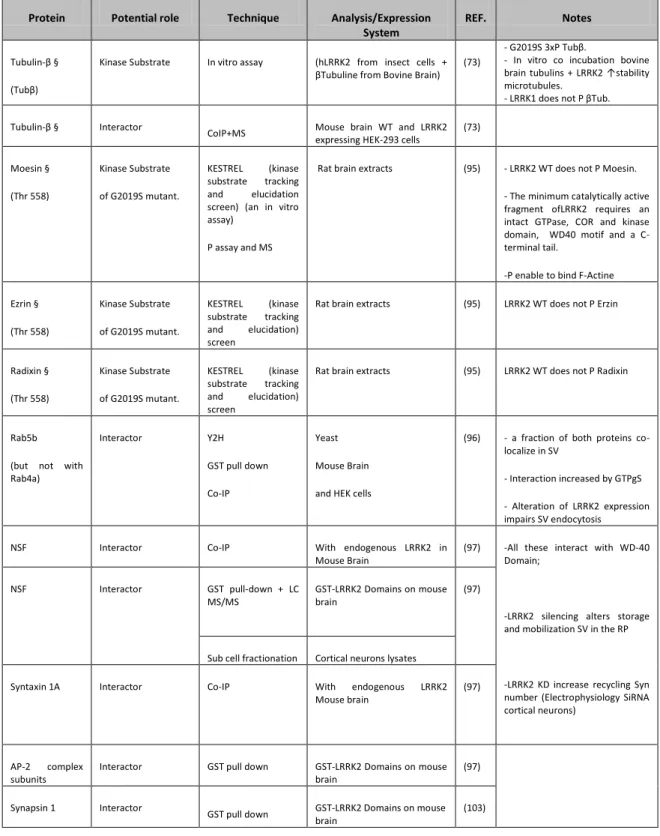
Despite the extensive studies performed, up to date, the physiological role of LRRK2 remains elusive. Among these studies, very indicative of the possible function of LRRK2 are the identifications of potential protein interactors. Through different methodological approaches and various models, a plethora of interactors has been suggested; a summary of those is given in table 3, which link LRRK2 to a broad range of cellular functions. Inter alia, more than one group found that LRRK2 phosphorylates moesin, suggesting that LRRK2 can take part in neurite length maintenance (69, 70). Moesin is a member of ERM (Ezrin/Radixin/Moesin) protein family which main function is to anchor the actin cytoskeleton to the plasma membrane; moesin and radixin are indeed implicated in neurite elongation. In fact, LRRK2 pathological mutant expression causes neurite shortening and neurite branching inhibition (69, 71). In C. elegans, LRK-1, the worm homologue of LRRK2, plays a role in vesicle trafficking between axons and dendrites (72).
Other data support the hypothesis that LRRK2 is involved in cytoskeleton dynamics. Hyper-expressed LRRK2 co-immunoprecipitates with endogenous tubulin in HEK-293 cells. Furthermore, G2019S mutant is able to phosphorylate tubulin more than the WT protein leading to microtubules anomalies that interfere with the normal neuronal functionality (73). Studies using LRRK2 transgenic mice models describe an abnormal Golgi apparatus fragmentation, which integrity is maintained with the help of microtubules, with consequences on Golgi and endoplasmic reticulum balancing in vesicular trafficking control (70). Microtubule stability and integrity are fundamental both for cellular morphology maintenance and for molecules and organelles transport that in neurons must cover longer distances in respect to other cellular types.
Moreover Leucine Rich Repeat Kinase 2 might be involved in translation control, since it has been shown to interact with the 4E-BP1 translation initiation factor.
Furthermore LRRK2 seem to be implicated in apoptosis activation by interaction with FADD proteins and caspase (74, 75). Iaccarino et al. demonstrated that G2019S expression in SH-SY5Y cells causes neuronal death by mitochondrial dependent apoptosis mechanism. This
is carried out by caspase 3 activation depending on Apaf1 which together with pro-caspase 9 and cytochrome c participate in the apoptosome formation (75). In this study ETNA-/- cells had been used as control. These cells are deficient for Apaf1 and are a common cellular model for apoptosis studies in neurodegenerative diseases. When ETNA -/- are transfected with LRRK2G2019S the lacking of Apaf1 prevents caspase 3 activation and the nucleus condensation while cytochrome C is as usual released from the mitochondria. These results point at a pivotal role of the mitochondria in LRRK2 mutant induced cell death and indicate Apaf1 as a fundamental mediator in this pathway. In the same study Iaccarino et al. have analyzed the differential role of LRR and WD40 LRRK2 domains in LRRK2 mutants toxicity showing that the presence of these two domains (LRRKΔLRR or LRRK2ΔWD40 ) are essential for caspase 3 activation and cellular apoptosis.
An interesting study of Greggio et al. demonstrates that PD causing mutants of LRRK2 increase LRRK2 tendency to form cytoplasmic inclusions in cellular models (76) and this property seems to be linked with the kinase activity of the protein (64). Cellular proteic inclusions in neurons of PD patients bearing LRRK2 mutations suggest that one effect of PARK8 locus alterations can be the protein aggregation.
According to different studies there might be a common pathologic mechanism involving LRRK2 and α-Synuclein. The α-Synuclein deposited in Lewy bodies is indeed abundantly phosphorylated on Serine 129. Up to date, LRRK2 is not able to directly phosphorylate α-synuclein although Qing at al. were able to co-immuniprecipitate LRRK2 and α-α-synuclein from Lewy bodies positive tissues and oxidative stressed HEK-293 cells (77). Furthermore recombinant α-synuclein is phosphorylated by some kinases present in the protein lysates from HEK-293 expressing LRRK2 (77, 78). Finally, SH-SY5Y co-transfected with LRRK2 (WT or G2019S) and α-Synuclein show an increase in aggregation level, phosphorylation and release of α-synuclein (79). Taken together these data corroborate a current hypothesis of an important functional connection between LRRK2 and α-Synuclein in triggering neurodegeneration causing PD (80).
Very useful tools to gather clues on the physiopathology of LRRK2 are animal models. In D.
melanogaster, overexpression of mutant LRRK2 causes loss of dopaminergic neurons,
retinal degeneration, motor impairment and shorter life, whereas wild-type protein produces less severe phenotypes (53). In C. elegans models, overexpression of LRRK2 also causes phenotypic changes, including axonal damages in neurons, which seem to involve altered mitochondrial functions (53). Knockout of the nearest homologous gene in Drosophila produces variable effects, with loss of dopamine cells reported in one study, but this was not replicated in an independent laboratory. Knockout of the C. elegans homologue causes changes in axonal polarity and in neurite outgrowth in response to stress (53). Three independent mouse knockouts have been reported (80-82). In all three published studies, the brains of the animals were reported to be grossly normal and there was no loss of dopaminergic neurons in the substantia nigra. Therefore, LRRK2 appears to be dispensable for the survival of neurons under both basal and stressed conditions. Different LRRK2 transgenic rodents have been generated by independent groups. In a few cases the authors describe dopaminergic degeneration in animals expressing pathological mutants (83, 84) while sometimes a reduction in dopamine extracellular content was observed (83, 85, 86). More recently, two groups have reported that transient overexpression of mutant LRRK2 using viral vectors will result in loss of dopaminergic neurons in the substantia nigra of mice (87) or rats (88). Importantly, wild-type protein or kinase-dead versions of LRRK2 had no effect, suggesting that simple overexpression of any similar large protein would not be sufficient to cause neurodegeneration (53). Therefore, this approach has the potential to provide a model that more fully replicates the phenotypes seen in human LRRK2 patients than are not seen in conventional transgenic models. A feature shared by various murine models of PD is an abnormal level of phosphorylated protein Tau induced by G2019S and R1441G LRRK2 mutants. The Tau protein binds tubulin and promotes its assembly in microtubules; when phosphorylated, Tau induces on the contrary, microtubule fragmentation that damages the entire cytoskeleton network (85) (89). Finally, the blockage of Zebrafish LRRK2 protein by morpholinos caused embryonic lethality and severe development defects such as growth retardation and loss of neurons. In addition, the deletion of WD40 domain of zebrafish
LRRK2 revealed Parkinsonism-like phenotypes, including loss of dopaminergic neurons in the diencephalon and locomotion defects. Remarkably, another group failed to reproduce the phenotypic loss of dopaminergic neurons in zebrafish (90)
LRRK2 and vesicle trafficking
Different experimental approaches have demonstrated that LRRK2 is localized throughout the cytoplasm of neuronal perikarya and dendritic processes, where it is associated with vesicular and membranous structures, the microtubule network, mitochondria and other membrane-bound organelles (61, 91). Moreover it has been demonstrated its association with lipid rafts. Although the pathological mutations do not affect the LRRK2 membrane association, this localization is of particular interest since the lipid rafts play important roles in cellular functions such as signal transduction, membrane trafficking and cytoskeletal organization (92). Furthermore, lipid rafts associate with SNARE (soluble N-ethylmaleimide-sensitive fusion protein-attachment protein receptor) proteins and regulate endocytosis and exocytosis (93, 94). Lastly, an extensive analysis of LRRK2 subcellular localization performed by Biskup et al in 2006 either in primary cortical neurons or rodent brains showed that LRRK2 co-localizes consistently to Golgi apparatus and Golgi-associated vesicles, endoplasmic reticulum (ER), lysosomes and mitochondria, and, to a significantly lesser degree, to vesicle markers such as synaptotagmin.
Table 3: Molecular interactors of LRRK2 and co-localizing organelle markers. Abbreviations: MS: Mass Spectrometry; Y2H: Yeast Two Hybrid; SV: Synaptic Vesicle; OE: Over Expression; KD: Knock Down; RP recycling Pool; GNEF: Guanine Nucleotide Exchange Factor; P: Phosphorylation/Phosphate; WB: Western Blot; Co-IP: CoImmunoPrecipitation; ICC: ImmunoCitoChemistry; Mut: mutant/mutation.
Protein Potential role Technique Analysis/Expression
System
REF. Notes
Tubulin-β § (Tubβ)
Kinase Substrate In vitro assay (hLRRK2 from insect cells + βTubuline from Bovine Brain)
(73)
- G2019S 3xP Tubβ.
- In vitro co incubation bovine brain tubulins + LRRK2 ↑stability microtubules.
- LRRK1 does not P βTub. Tubulin-β § Interactor
CoIP+MS Mouse brain WT and LRRK2
expressing HEK-293 cells
(73) Moesin § (Thr 558) Kinase Substrate of G2019S mutant. KESTREL (kinase substrate tracking and elucidation screen) (an in vitro assay)
P assay and MS
Rat brain extracts (95) - LRRK2 WT does not P Moesin. - The minimum catalytically active fragment ofLRRK2 requires an intact GTPase, COR and kinase domain, WD40 motif and a C-terminal tail.
-P enable to bind F-Actine Ezrin § (Thr 558) Kinase Substrate of G2019S mutant. KESTREL (kinase substrate tracking and elucidation) screen
Rat brain extracts (95) LRRK2 WT does not P Erzin
Radixin § (Thr 558) Kinase Substrate of G2019S mutant. KESTREL (kinase substrate tracking and elucidation) screen
Rat brain extracts (95) LRRK2 WT does not P Radixin
Rab5b (but not with Rab4a) Interactor Y2H GST pull down Co-IP Yeast Mouse Brain and HEK cells
(96) - a fraction of both proteins co-localize in SV
- Interaction increased by GTPgS - Alteration of LRRK2 expression impairs SV endocytosis
NSF Interactor Co-IP With endogenous LRRK2 in
Mouse Brain
(97) -All these interact with WD-40 Domain;
-LRRK2 silencing alters storage and mobilization SV in the RP
-LRRK2 KD increase recycling Syn number (Electrophysiology SiRNA cortical neurons) NSF Interactor GST pull-down + LC MS/MS GST-LRRK2 Domains on mouse brain (97)
Sub cell fractionation Cortical neurons lysates
Syntaxin 1A Interactor Co-IP With endogenous LRRK2
Mouse brain
(97)
AP-2 complex subunits
Interactor GST pull down GST-LRRK2 Domains on mouse
brain
(97) Synapsin 1 Interactor
GST pull down GST-LRRK2 Domains on mouse brain