Scuola dottorale in Biologia
Sezione: Scienze Biomolecolari e Cellulari
XXVIII Ciclo
“Optimization and applications of the Micro
Biological Survey method MBS”
Alyexandra Arienzo
Docente guida: Prof. Giovanni Antonini
1
INDEX
Abstract: Ottimizzazione e applicazioni del metodo Micro Biological Survey MBS 4 Abstract: Optimization and applications of the Micro Biological Survey method
MBS 7
1. INTRODUCTION 10
1.1 Conventional and alternative microbiological methods in water analysis 13
1.1.1 Standard methods for drinking water analysis 13
1.1.2 Alternative methods for drinking water analysis 17
1.1.3 Point-of-use tests for drinking water analysis 20
1.2 Conventional and alternative methods for Urinary Tract Infections (UTI)
diagnosis and management 21
1.2.1 Standard methods for UTI diagnosis and management 22
1.2.2 Alternative methods for UTI diagnosis and management 25 1.2.3 Point of Care Tests for UTI diagnosis and management 26
1.3 The Micro Biological Survey Method 27
1.3.1 Micro Biological Survey method biochemical principles 28 1.3.2 Micro Biological Survey method analytical procedure 31
1.3.3 Micro Biological Survey Multireader 31
2
3. MATERIAL AND METHODS 36
3.1 Application of the MBS method for the evaluation of drinking water quality 36
3.1.1 Preliminary study 37
3.1.2 Application of the MBS method on field in the evaluation of the microbiological quality of water in Douala, Cameroon
38
3.2 Application of the MBS method as a Point of Care Test for Urinary Tract
Infections diagnosis and management 40
3.2.1 Preliminary in vitro validation of new MBS reagents for the detection of bacteria in urine and for the evaluation of their susceptibility/resistance to a panel of antibiotics
42
3.2.2 Preliminary clinical study of the developed MBS reagents as a POCT, in
collaboration with the hospital “Azienda Ospedaliera Sant’Andrea” of Rome 44 3.2.3 Optimization of the MBS UBC reagent composition and the operating
procedure for the evaluation of susceptibility/resistance to antibiotics, using the optimized UBC reagent added with antibiotics
46
3.2.4 Study of a new standardized manufacturing process 48
3.2.5 Set up of the optimal detection parameters of the MBS Multireader 49
4. RESULTS AND DISCUSSION 50
4.1 Application of the MBS method for the evaluation of drinking water quality 50
3
4.1.2 Application of the MBS method on field in the evaluation of the microbiological quality of water in Douala, Cameroon
55
4.2 Application of the MBS method as a Point of Care Test for Urinary Tract
Infections diagnosis and management 59
4.2.1 Preliminary in vitro validation of new MBS reagents for the detection of bacteria in urine and for the evaluation of their susceptibility/resistance to a panel of antibiotics
59
4.2.2 Preliminary clinical study of the developed MBS reagents as a POCT, in
collaboration with the hospital “Azienda Ospedaliera Sant’Andrea” of Rome 64 4.2.3 Optimization of the MBS UBC reagent composition and the operating
procedure for the evaluation of susceptibility/resistance to antibiotics, using the optimized UBC reagent added with antibiotics
70
4.2.4 Study of a new standardized manufacturing process 76
4.2.5 Set up of the optimal detection parameters of the MBS Multireader 79
5. CONCLUDING REMARKS 83
6. REFERENCES 85
4
Abstract: Ottimizzazione e applicazioni del metodo Micro Biological Survey MBS
Le analisi microbiologiche tradizionali, impiegate nel controllo della qualità e della sicurezza degli alimenti, nel monitoraggio ambientale e in clinica, sono state sviluppate alla fine del XIX secolo. Nonostante i notevoli progressi tecnologici e l’introduzione nell’arco degli ultimi dieci anni di una innumerevole serie di kit commerciali per la rilevazione e la quantificazione dei microrganismi, i metodi culturali classici sono ancora i più utilizzati, principalmente per la relativa semplicità, tecnica e concettuale, e per la loro approvazione da parte di organismi internazionali (ISO, FDA, USEPA, EU). Tali metodi richiedono tuttavia un notevole investimento in termini di tempo, costi e forza lavoro e presentano numerosi svantaggi, per citarne alcuni: un elevata soggettività nella lettura dei risultati, la possibile interferenza delle matrici biologiche analizzate, e soprattutto l’elevato tempo necessario per ottenere risultati definitivi (da 2 a 7 giorni), dipendente dall’abilità dei microrganismi di moltiplicarsi fino a formare colonie visibili ad occhio nudo. Tali problematiche hanno portato allo sviluppo e al raffinamento di metodi microbiologici alternativi. Tali metodi sono molto più rapidi e agevoli dei rispettivi metodi di riferimento, e portano ad un sostanziale miglioramento riducendo le fasi di preparazione e manipolazione dei campioni, accelerando i tempi di risposta e riducendo i costi. Tali metodi spesso includono qualche forma di automazione e i dati possono venire raccolti elettronicamente riducendo la soggettività d’interpretazione dei risultati e l’errore umano. L’obiettivo finale è quello di garantire risultati più sensibili, accurati, precisi e riproducibili rispetto a quelli ottenuti con le metodiche culturali tradizionali. I metodi microbiologici rapidi alternativi includono generalmente metodi immunologici e molecolari. L’affidabilità variabile, i costi elevati e la necessità di laboratori altamente specializzati di alcuni di questi metodi ne hanno tuttavia finora limitato l’adozione.
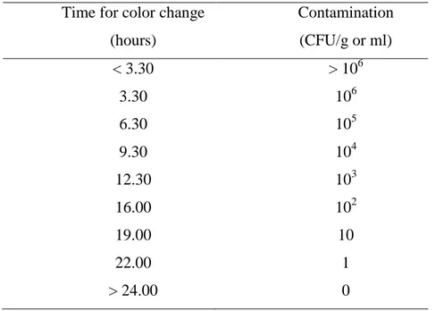
In tale contesto, il metodo Micro Biological Survey (MBS), un metodo alternativo per analisi microbiologiche, è stato sviluppato dall’Università degli Studi “Roma Tre”, con l’obiettivo di fornire risultati affidabili diminuendo i tempi di analisi, facilitando le procedure analitiche e l’interpretazione dei dati, aumentando la sensibilità, limitando i costi e permettendo l’esecuzione di analisi anche in assenza di un laboratorio. Il metodo MBS è un sistema colorimetrico per la rilevazione e quantificazione dei microorganismi eventualmente presenti negli alimenti, nelle acque e nei campioni ambientali. A dispetto dei metodi tradizionali, che misurano la capacità delle cellule di moltiplicarsi fino a formare colonie visibili, il metodo MBS misura l’attività catalitica degli enzimi redox nei pathway metabolici primari batterici permettendo di stabilire una corrispondenza certa tra attività enzimatica misurata e la concentrazione batterica nel campione analizzato. Il tempo
5
necessario per il cambiamento di colore è inversamente proporzionale al logaritmo della concentrazione batterica: come una reazione enzimatica, più elevato è il numero di batteri più veloce è il cambiamento di colore. L’analisi MBS si basa sull’utilizzo di fiale di reazione sterili e pronte all’uso che contengono il reagente specifico per l’analisi da condurre. I campioni possono essere inoculati senza alcun trattamento preliminare. Le analisi possono essere eseguite da personale non specializzato ovunque siano necessarie, senza l’uso di strumentazione particolare a parte un semplice termostato. Questo metodo è stato inizialmente concepito per l’analisi degli alimenti e si è dimostrato un sistema affidabile e rapido per valutare la qualità microbiologica di differenti campioni alimentari. Ulteriori studi hanno dimostrato che la semplicità analitica, la ridotta manualità e l’automazione influiscono positivamente anche sui risultati, che mostrano una maggiore riproducibilità e ripetibilità rispetto ai metodi tradizionali. Il metodo MBS rappresenta una promettente alternativa sia ai metodi tradizionali sia ai metodi alternativi finora sviluppati. Le sue caratteristiche lo rendono utile in molti contesti e sono oggetto di questa tesi. In particolare la mia ricerca è stata finalizzata allo studio di applicazioni alternative del metodo MBS. Il primo obiettivo è stato verificare l’efficacia del metodo MBS come metodo alternativo per l’analisi microbiologica dell’acqua potabile, al fine di sviluppare un nuovo Point of use test per la valutazione della qualità dell’acqua nei Paesi in via di sviluppo. Il secondo obiettivo è stato di esaminare la possibilità di applicare il metodo MBS in ambito clinico ed in particolare per la rilevazione e quantificazione dei batteri nelle urine e per una valutazione preliminare della loro suscettibilità/resistenza agli antibiotici, al fine di sviluppare un nuovo test al Point of care (POCT) per la diagnosi e la gestione delle infezioni delle vie urinarie (IVU). In entrambi i casi la necessità di ottenere risultati accurati e affidabili, in tempi brevi e con una procedura semplice e difficilmente incline ad errori, era una questione centrale per facilitare le analisi e permettere decisioni e azioni correttive rapide.
Nell’ambito dell’analisi dell’acqua potabile ho verificato la possibilità di utilizzare il metodo MBS come metodo alternativo per l’analisi microbiologica dell’acqua dimostrando la sua sensibilità, specificità, linearità e accuratezza. Ulteriori esperimenti sono stati condotti per dimostrare che la determinazione dei coliformi totali in 1 ml di acqua, invece dei 100ml richiesti dalle normative correnti, può essere efficace per dare una valutazione generale della qualità delle acque, specialmente nei Paesi in via di sviluppo, in assenza di strutture e strumentazioni adeguate. Il metodo MBS è stato quindi utilizzato in campo per monitorare la qualità delle acque nella città di Douala in Camerun. Sessantuno siti sono stati selezionati tra 20 quartieri del comune V di Douala. In particolare sono stati presi in considerazione pozzi tradizionali ed incamiciati, utilizzati come fonte di acqua potabile dalla popolazione locale. I dati sono stati collezionati in tre fasi differenti in modo da valutare l’effetto delle variazione meteorologiche inter-annuali sulle caratteristiche delle
6
falde acquifere. Dai risultati ottenuti è stata osservata una netta correlazione tra la tipologia di pozzo e il livello di contaminazione: il 70% dei pozzi tradizionali è risultato almeno una volta contaminato da coliformi con un elevato livello medio di contaminazione; al contrario il 100% dei pozzi incamiciati è risultato esente da contaminazione dimostrando la sicurezza di questa tipologia di fonte e l’importanza del suo utilizzo in zone rurali e non. Il metodo MBS si è dimostrato un metodo valido ed affidabile per valutare la qualità microbiologica delle acque. La semplicità di esecuzione ed interpretazione dei risultati rendono possibile il suo utilizzo anche in aree rurali da personale locale non specializzato e in assenza di un laboratorio microbiologico.
Per quanta riguarda l’applicazione del metodo MBS come test al Point of care per la diagnosi e la gestione delle infezioni delle vie urinarie, la prima fase della ricerca ha riguardato lo sviluppo e validazione preliminare di reattivi MBS specifici per la rilevazione e quantificazione dei batteri nelle urine e una valutazione preliminare della loro suscettibilità/resistenza agli antibiotici. Tali reattivi sono stati testati in uno studio clinico preliminare in collaborazione con l’Azienda ospedaliera Sant’Andrea di Roma che ha evidenziato il grande potenziale del metodo MBS come strumento diagnostico per una rapida e accurata rilevazione dei batteri responsabili di IVU. Uno studio comparativo tra i risultati ottenuti con il metodo MBS e i risultati delle urino colture, eseguite presso il laboratorio microbiologico ospedaliero, è stato condotto utilizzando la Receiver Operating Characteristics (ROC) analysis. I risultati hanno dimostrato che il POCT MBS è stato in grado di rilevare la presenza di una concentrazione significativa di batteri nelle urine e quindi diagnosticare una IVU in solo 5 ore (Area Under the Curve = 0,93). Inoltre il POCT MBS ha dimostrato una maggiore accuratezza (90,2%), sensibilità (91,2%) e specificità (89,9%) rispetto agli stick urinari comunemente utilizzati per un’indicazione presuntiva di IVU. In un tempo relativamente breve rispetto ai metodi tradizionali il metodo MBS è stato in grado di fornire un accurata indicazione di IVU e una valutazione preliminare della suscettibilità/resistenza agli antibiotici degli agenti infettivi, assicurando una pronta diagnosi e guidando la scelta degli antibiotici prima del classico metodo dell’antibiogramma. Successivamente differenti aspetti legati alla specifica composizione del reattivo, alla procedura analitica e al processo di produzione, sono stati ottimizzati al fine di migliorare ulteriormente le prestazioni del metodo e rispondere ai requisiti essenziali necessari per l’introduzione sul mercato.
Nell’insieme i risultati hanno dimostrato che il metodo MBS è una valida alternativa ai metodi microbiologici tradizionali, sia in campo che in laboratorio. I suoi solidi principi lo rendono estremamente flessibile e adattabile a necessità completamente diverse e permettono di ottenere soluzioni semplificate, rapide e affidabili con un impatto significativo nel campo sociale e nella tutela della salute.
7
Abstract: Optimization and applications of the Micro Biological Survey method MBS
Traditional microbiological analyses used in food safety, environmental monitoring and clinical diagnosis, were developed at the end of XIX century. Despite advances in technology and the introduction over the past decade of a plethora of commercial assays and kits for the detection and enumeration of a wide range of microorganisms, traditional culture based methods are still the most used, thanks to their relatively technical and scientific simplicity and their acceptance by regulatory agencies (ISO, FDA, USEPA, EU). These methods are however labor intensive, time consuming and present several shortfalls, such as subjectivity in the interpretation of results, the possible interference of matrices and, above all, the prolonged time (from 2 to 7 days) needed to give definitive results, depending on the ability of the organisms to multiply into visible colonies. These reasons have led to the development and refinement of alternative microbiological methods of analysis. These alternative methods are quicker and easier to perform than the corresponding reference methods; they minimize manipulation, provide results in less time, and reduce costs. They also normally involve some form of automation, and often capture data electronically. In particular alternative microbiological method technologies aim to provide more sensitive, accurate, precise, and reproducible test results when compared with conventional, growth-based methods. Rapid methods generally include immuno-based and DNA-based assays. However the variable reliability, cost, novelty and the need of highly equipped laboratories of some of these methods still limit their adoption.
In this context, an alternative method called Micro Biological Survey (MBS) has been developed by “Roma Tre” University in order to meet the need to provide reliable results thus diminishing the time of analysis, facilitating procedures and interpretation of data, increasing the detection sensitivity, limiting costs and allowing analysis also in the absence of a laboratory. The MBS method is a colorimetric system for detection and selective counting of bacteria in food, water and environmental samples. In contrast with traditional methods that measure the capability of cells to grow in discrete colonies, the MBS method measures the catalytic activity of redox enzymes in the main metabolic pathways of bacteria allowing an unequivocal correlation between enzymatic activity and bacterial concentration in the samples. The time required for a color change is inversely related to the logarithm of bacterial concentration in the sample: like an enzymatic reaction, the greater the number of bacteria, the faster the color change. The MBS analysis is performed in disposable ready to use reaction vials that contain the specific reagent for the analysis to perform. Samples can be inoculated without any preliminary treatment. Analyses can be carried out by
8
untrained personnel and anywhere they are necessary, without the need for any instrumentation other than a thermostatic device. The method was initially conceived for food analysis and it has proven to be a reliable system to evaluate microbiological quality and assess rapidly contamination of different food samples. Further studies have demonstrated that the simple analytical procedure, the reduced labor and automation, positively affect also the analytical performance of the MBS method that displays greater reproducibility and repeatability compared to traditional methods. The MBS method represents a promising and attractive alternative both to traditional and alternative methods until now developed. Its features make it useful in many contexts and are the subject of this thesis. In particular my research was aimed to study alternative applications of the MBS method. The first aim was to verify the effectiveness of the MBS method as an alternative method for microbiological analysis of drinking water, with the purpose to develop a new point of use test to assess water quality in developing countries. The second aim was to investigate the possibility to use the MBS method in clinical settings and in particular for the detection of bacteria in urine and a preliminary evaluation of their susceptibility/resistance to antibiotics, with the final goal to develop a new Point of care test (POCT) for diagnosis and management of Urinary Tract Infections (UTIs). In both cases the need to obtain rapid, accurate, reliable results in a simple, errorless approach, was a central issue to facilitate investigations, allow fast critical decisions and corrective actions.
In the context of the analysis of potable water I investigated the possibility to apply the MBS method as an alternative method for drinking water analysis demonstrating its sensitivity, specificity, linearity and accuracy. Further experiments were carried to demonstrate that the simple evaluation of total coliforms in 1ml of water samples, instead of 100ml as required by law, could be effective to roughly assess water quality, particularly in developing countries, in the absence of specific facilities and instrumentations. I therefore applied the MBS method on field and in particular in the evaluation of the microbiological quality of water in Douala, Cameroon. Sixty one water points were selected within 20 quarters of the Douala V City council. The water points were divided into dug wells and drilled wells and all of them were used as drinking water sources by the resident population. Data were collected in three different phases in order to study the effects of inter-annual meteorological variation of rainfall and temperature on groundwater characteristics. A strict correlation between the well type and the level of contamination was observed: 70% of the examined dug wells resulted at least once contaminated by total coliforms with a high average contamination; on the other hand, 100% of drilled wells resulted non-contaminated, underlining the safeness of these water sources and the importance of their use in both rural and city areas. The MBS method has therefore proven to be a valid and accurate method to frequently evaluate the
9
microbiological quality of many water sources. Its simple procedure and interpretation of results can be suitable for use in rural areas, by local personnel, operating without a microbiological laboratory.
Concerning the use of the MBS method as a Point of care test for diagnosis and management of Urinary Tract Infections the first step was the development and preliminary in vitro validation of new MBS reagents for the detection of bacteria in urine and for the evaluation of their susceptibility/resistance to a panel of antibiotics. These were tested in a preliminary clinical study, performed in collaboration with the “Azienda ospedaliera Sant’Andrea” of Rome, that demonstrated the great potential of the MBS POCT as a diagnostic tool for a rapid and accurate detection of bacteria causing UTIs. A comparative analysis between the results obtained with the MBS method and results of urine cultures, conducted by the hospital laboratory, was performed using the Receiver Operating Characteristics (ROC) analysis. Results demonstrated that the MBS POCT was able to reveal the presence of a significant bacterial load in urine, hence diagnose a clinical UTI, in only 5 hours (Area Under the Curve=0,93). More importantly, the MBS POCT showed much higher accuracy (90.2%), sensitivity (91.2%) and specificity (89,8%) compared to urine dipsticks, widely used for a presumptive diagnosis of UTI. In a relatively short time compared to standard methods, the MBS method was able to give an accurate indication of UTI and a preliminary evaluation of the antibiotic susceptibility of the infecting bacteria, ensuring a prompt diagnosis and guiding the antibiotic choice long before the conventional antibiotic susceptibility test is performed. Different issues linked to the specific composition of the reagent, the operating procedures and the manufacturing process were also optimized in order to further improve the overall performance of the MBS POCT and meet the essential premarketing requirements mandatory for all in vitro diagnostic devices.
Together the results obtained demonstrate that the MBS method is an effective, reliable and precise alternative to traditional microbiological testing, proven in laboratory settings and on field trials. It has also been demonstrated that the solid principles of the MBS method make it extremely flexible and adaptable to completely different needs, allowing simplified, rapid, reliable solutions and achieving health-effective and productive results.
10
1. INTRODUCTION
Detection, quantification and characterization of bacteria are critical issues in many areas, including public health, clinical diagnosis, food safety and environmental monitoring.
Despite advances in technology and the introduction over the past decade of a plethora of commercial assays and kits for the detection and enumeration of a wide range of microorganisms, traditional culture based methods are still the most popular and widely used. Conventional culture methods are the most common category of methods being used by laboratories thanks to their relatively technical and scientific simplicity, the low consumable and equipment costs and their acceptance by regulatory agencies.
Culture test methods used to detect and quantify bacteria are based on the ability of mircoorganisms in a sample to proliferate in or on a specified growth medium, under specified growth conditions. A variety of culture media have been designed to maximize the recovery of different microbial species and are referred by international organizations as the gold standards for detection and quantification of bacteria. Culture dependent methods for enumerating bacterial numbers are known to be biased since bacteria can only be cultivated if their metabolic and physiological requirements can be reproduced in vitro. Microorganisms that will not grow in the selected growth conditions (growth media, temperature) or microorganisms that have lost the ability to reproduce will not be detected. Moreover, although not lacking in sensitivity, these methods can be laborious and depend on the growth of the microorganisms in different culture media (pre-enrichment, selective enrichment, selective plating, identification), which may require several days before results are known [1]. In the past decade, the focus of many researchers has been the study and implementation of improved methods for the isolation, early detection, characterization, and enumeration of microorganisms and their products. In common language, this translates to better methods, automated methods, miniaturized methods, and methods that require less time or expense.
The methods range from simple dip-stick-type tests to very complex automated systems that perform a variety of tests using a variety of techniques. The prices for these methods vary greatly, from a few dollars to hundreds of thousands of dollars. Although these methods are called rapid microbiological methods, many have their roots in other sciences, e.g., chemistry, molecular biology, biochemistry, immunology, immunochemistry, molecular electronics, and computer-aided imaging [2]. These methods can be grouped into three categories: growth-based methods, direct measurement, and cell component analysis. Growth-based methods detect a signal after a short incubation period in liquid or on solid media; examples include detection of CO2 production by
11
colorimetric methods or a change in head space pressure and detection of adenosine triphosphate (ATP) by bioluminescence [3].
Direct measurement methods can detect cell viability without requiring growth of the microorganism. One example of a direct measurement method combines fluorescent labeling and laser scanning cytometry to enumerate organisms. Fluorescence-based methods can also be used to enumerate bacteria. In the food and biotechnology industries, for instance, the automated counting of pure cultures by flow cytometry is well established. However, most bacteria are optically too similar to resolve from each other or from debris using flow cytometry, without artificially modifying the target bacteria using fluorescent labeling techniques such as fluorescent antibodies or fluorescent dyes. Differences in bacterial cell size, coaggregation of bacteria and the presence of different contaminating matrices (e.g. mud, food) can also make meaningful counting difficult, if not problematic, by interference with direct, or fluorescence, microscopy [4].
Rapid enumeration of bacteria can also be achieved by using a variety of molecular approaches. Molecular methods have been developed to increase the rapidity of analysis. They are able to achieve a high degree of sensitivity and specificity without the need for a complex cultivation and additional confirmation steps. These methods offer taxonomic information at different levels, such as classes, genera, species or subspecies. Consequently, some of these methods permit the detection of specific culturable and/or non-culturable bacteria within hours, instead of the days required with the cultivation-based methods [5].
Immunological methods are based on the specific recognition between antibodies and antigens and the high affinity that is characteristic of this recognition reaction. Depending on the taxonomic level of the targeted antigens, immunological methods permit detection of antigens at family, genus, species or serotype levels [6,7].
Most of the nucleic acid methods use molecular hybridization properties, which involve the complementary sequence recognition between a nucleic probe and a nucleic target. A hybridization reaction can be realized between a nucleic DNA probe and a chromosomic DNA sequence (DNA– DNA hybridization) or an rRNA or tRNA sequence (DNA–RNA hybridization). Specificity, here, depends on the phylogenetic degree of conservation of the target within the taxonomic target group. The more frequently used nucleic-acid-based methods are the polymerase chain reaction (PCR) and the in situ hybridization (ISH) methods [8]. PCR is designed to facilitate bacterial identification at any level of specificity: strain, species or genus. In recent years, real-time PCR methods have been developed and described for the rapid detection and identification of several bacterial strains. Real-time PCR is a promising tool for distinguishing specific sequences from a complex mixture of DNA and therefore is useful for determining the presence and quantity of pathogen-specific or other
12
unique sequences within a sample. Real-time PCR facilitates a rapid detection of low amounts of bacterial DNA accelerating therapeutic decisions and enabling an earlier adequate antibiotic treatment [9]. Primers with broad interspecies specificity have been designed to amplify 16S rDNA by PCR and have been used to determine bacterial numbers in complex communities [10].
In situ hybridization techniques use oligonucleotide probes to detect complementary nucleic acids sequences. This method exploits the ability of nucleic acids to anneal to one another in a very specific complementary way to form hybrids. The probes are specific because they are built from, and are complementary to, selected nucleic acids sequences which are unique to a given microorganism, species or group. The probes can target either DNA or RNA molecules. Sequence comparisons make it possible to define targeted regions which are perfectly conserved within different taxonomic levels and consequently specific to these levels, from domains to subspecies. Probes specific to rRNA (mainly 16S and 23S rRNA sequences) have become the standard tools for organism identification. Several oligonucleotide probes are commercially available and the choice of whether to use them or design new, specific ones depends largely on the application [11]. Current work on rRNA in situ hybridization uses fluorescent-labeled nucleotide probes almost exclusively to detect hybridization (FISH). The popularity of the FISH technique is due to its advantages over radioactive labeling, which include sensitivity, speed of visualization of single cells (by means of microscopy or cytometrical devices), stability of the hybridization products, safety, diminished detection time, multiple labels (multiple colors) and ease of use. Depending on the concentration of targeted cells in the sample and to increase resolution, FISH detection can be performed by means of flow or solid-phase cytometry. Flow cytometry enables quantification of the fluorescence intensities for each target-probe hybrid [12].
All analytical methods need to be validated prior to their introduction into routine use [13]. Validation schemes have been established from the major global authorities to assess whether these methods perform at least as well as the corresponding reference methods [14;15,16]. In addition, a European and International Standard, EN ISO 16140, has been developed to provide a common reference protocol for the validation of alternative methods, as well as to determine general principles for their possible subsequent certification [17].
13
1.1 Conventional and alternative microbiological methods in water analysis
Water intended for human consumption is required to be quality tested for a wide variety of contaminants, including potentially harmful microorganisms. While many water quality parameters such as pH, chlorine and turbidity can be measured in near real-time by on-line measurement instrumentation, microbiological testing presents a unique challenge.
1.1.1 Standard methods for drinking water analysis
Various standard and guideline values have been introduced over the years. Many of these have become legally enforceable, while others have been recommended by appropriate bodies and trade associations [18]. Most waterborne diseases are related to fecal pollution of water sources, therefore water microbiological control is largely based on the need to identify indicators of fecal pollution such as coliforms and Escherichia coli (E. coli), but the use of enterococci and Clostridium perfringens is increasing [19]. The two major international bodies, the United States Environmental Protection Agency (USEPA), and the European Union (EU) both include E. coli as a mandatory microbial indicator, and the USEPA regulates for total coliforms, via the Total Coliform Rule. Most drinking water guidelines also refer to the use of total estimates of bacterial numbers in water. This measure is generally called ‘total heterotrophic plate count’ (HPC) or ‘standard plate count bacteria’, and is considered to represent the general cleanliness of drinking water [20;21].
Currently, three primary techniques for the routine detection and enumeration of indicator and opportunistic bacteria currently are used. They are the multiple tube fermentation (MTF), membrane filtration (MF), and plate count techniques. All methods use media designed to select for the growth and identification of specific organisms. Generally, each method also includes inhibitors, restrictive temperatures, and/or limited nutrients to restrict growth of unwanted species [22].
Heterotrophic plate count standard methods. Heterotrophic plate counts (HPC) or total viable counts (TVC) are enumerations of the general population of heterotrophic bacteria present in water supplies. The enumerations may represent bacteria whose natural habitat is the water environment, or those that have originated from soil or vegetation. HPC measurements are used: to indicate the effectiveness of water treatment processes, thus as an indirect indication of pathogen removal; as a measure of numbers of regrowth organisms that may or may not have sanitary significance; to monitor the performance of filtration or disinfection processes. HPC testing may be used in investigating aesthetic quality, and it is used by some authorities as a marker for some of the underlying causes of some aesthetic problems [23].
14
Heterotrophic plate count standard methods provide a standardized means of determining the density of aerobic and facultative anaerobic heterotrophic bacteria in water. Standard methods outline three techniques for determining a standard plate count: pour plate, spread plate, and membrane filtration (MF) [24]. The standard plate count (also called the pour plate method) involves pouring 1 ml of the sample into a plate and pouring an agar solution over the top. The sample is allowed to incubate after the agar has set. This is an effective method for quantifying organisms, and also allows more space for the colonies to grow. The spread plate method differs from the pour plate method in that agar plates are poured and set aside to solidify before inoculation with samples. The spread plate method involves spreading 0.1 ml of the sample over a premade agar plate and allowing it to incubate. This method gives more 3-dimensional colonies than the pour plate method that are much easier to identify and count. The pour plate or spread plate methods are typically used for process water and drinking water [25].
The membrane filtration technique is best suited to test large volumes of low-turbidity, low-count (less than 1 to 10 Colonies Forming Units (CFU) /ml) water. Membrane filtration involves filtering a particular volume of a sample to get a higher concentration of bacteria, then rinsing the filter with a sterile buffer before placing it on an agar plate. This is generally the only method used to test sterile water. It can also be used to test drinking water depending on the application. This method provides good colony morphology, making it a good method for identifying the bacteria present in a sample [26].
In all of these standard test methods there is a significant amount of time and labor required, as well as a high chance of manual error; the processes are highly operator-dependent, and as a result the CFU can vary widely when calculated by different operators and different labs. Also the costs associated with these methods can be high, not only the labor costs but also the cost of materials necessary to conduct the testing. It can take between 48-72 hours to get results, and if the sample is improperly diluted so the results are too high or not enough to count, the sample must be retested. Once testing is completed, the process of counting the number of bacterial colonies is complicated and time-consuming, and it is easy to make mistakes during this process.
Total coliform and Escherichia coli count standard methods. Coliform bacteria belong to the family Enterobacteriaceae but are further defined by functional characteristics rather than systematic genus and species [27]. The World Health Organization has defined coliforms as any rod-shaped, non-spore-forming, gram-negative bacteria capable of growth in the presence of bile salts or other surface-active agents. Continuing, the definition states that coliforms are cytochrome-oxidase negative and able to ferment lactose at either 35 or 37 °C with the production of acid, gas, and aldehyde within 24 to 48 hours [28]. The total coliform group is the most inclusive indicator
15
classification, and contamination indicated by the presence of total coliforms is indicative of inadequate disinfection of drinking water also if it does not always correlate properly with the presence of fecal pollution. Monitoring for total coliforms at the treatment plant and in the distribution and storage system is carried out to provide information on the adequacy of drinking water treatment and on the microbial condition of the distribution system. Their presence in groundwater indicates that it is vulnerable to contamination from the surrounding environment so their detection in the water leaving the well should trigger further actions [28;29]. Escherichia coli is a thermophilic coliform that produces indole from tryptophan, but also defined now as coliform able to produce β-glucuronidase (although taxonomically up to 10% of environmental E. coli may not). The bacterium’s only natural habitat is the large intestine of warm-blooded animals and since E. coli, with some exceptions, generally does not survive well outside of the intestinal tract, its presence in environmental samples, food, or water usually indicates recent fecal contamination or poor sanitation practices in food-processing facilities. Monitoring for E. coli in treated water at the treatment plant and in the distribution system is carried out to provide information on the adequacy of drinking water treatment and on the microbial condition of the distribution system [30].
The US Environmental Protection Agency (EPA) and AFNOR have approved several methods for coliforms and E. coli detection: the multiple-tube fermentation (MTF) technique and the membrane filter (MF) technique [31].
The MTF test uses a specified number of tubes (based on the expected population in the sample) containing a specific medium and sample water. After incubation, each tube is examined for growth of the target organism(s). The number of tubes showing growth are matched to a statistically determined table of numbers to yield the most probable number (MPN) of organisms in the sample. The number of presumptive E. coli is determined by enzymatic testing on samples that give a positive total coliform test [32]. The incorporation of MUGlu into lauryl tryptose broth used as the medium for the multi-tube fermentation (MTF) technique was first proposed for rapid detection and immediate confirmation of E. coli in food and water samples. The presence of methylumbelliferone due to the hydrolysis of MUGlu (positive samples) is detected by exposure to long-wave UV light and visualization of blue-white fluorescence [33] .
Many factors may significantly affect coliform bacteria detection by MTF, especially during the presumptive phase. Interference by high numbers of non-coliform bacteria, as well as the inhibitory nature of the media have been identified as factors contributing to underestimates of coliform abundance. The MTF technique lacks precision in qualitative and quantitative terms. The time required to obtain results is higher than with the membrane filter technique that has replaced the MTF technique in many instances for the systematic examination of drinking water. However, this
16
technique remains useful, especially when the conditions do not allow the use of the membrane filter technique, such as turbid or colored waters. MTF is easy to implement and can be performed by a technician with basic microbiological training, but the method can become very tedious and labor intensive since many dilutions have to be processed for each water sample. However, it is also relatively inexpensive, as it requires unsophisticated laboratory equipment. Nevertheless, this method is extremely time-consuming, requiring 48 h for presumptive results, and necessitates a subculture stage for confirmation which could take up to a further 48 h [34].
The Membrane Filtration (MF) method consists of filtering a water sample on a sterile filter with a 0.45-mm pore size which retains bacteria, incubating this filter on a selective medium and enumerating typical colonies on the filter. If the organisms sought are present, colonies will grow on the membrane filter. Colonies are examined at 10–15X magnification with a stereoscopic microscope and then identified by size, color, shape and sheen. Typical colonies are counted and the number is reported as the number of colonies per 100 ml of sample. The MF technique is fully accepted and approved as a procedure for monitoring drinking water microbial quality in many countries. E. coli is determined from total-coliform-positive samples by the means of specific confirmation media. Different commercial agar media are now available for the detection of E. coli. They include classical agar media modified with specific chromogenic and/or fluorogenic substrates for the detection of β-Dglucuronidase activity. Many media and incubation conditions for the MF method have been tested for optimal recovery of coliforms from water samples [35].
The predominant concern about MF is its inability to recover stressed or injured coliforms. A number of chemical and physical factors involved in drinking water treatment, including disinfection, can cause sub lethal injury to coliform bacteria, resulting in a damaged cell unable to form a colony on a selective medium. Exposure of bacteria to products like chlorine may result in injury and increased sensitivity to bile salts or to the replacement surface-active agents (sodium desoxycholate or Tergitol 7) contained in some selective media. Other authors have suggested that chlorination affects various functions of coliform bacteria activity, such as catalase enzymatic activity. Metabolically active bacteria produce hydrogen peroxide (H2O2), which is usually rapidly
degraded by the catalase. Injured coliforms with reduced catalase activity accumulate toxic hydrogen peroxide, to which they are extremely sensitive [36]. The high number of modified media in use is a reflection of the fact that no universal medium currently exists which allows optimal enumeration of various coliform species originating from different environments and present in a wide variety of physiological states. A significant advantage of the MF technique over the MTF method is that with MF, the examination of larger volumes of water is feasible, which leads to greater sensitivity and reliability. MF also offers a quantitative enumeration comparatively to the
17
semi-quantitative information given by the MTF method. MF is a useful technique for the majority of water quality laboratories as it is a relatively simple method to use. Many samples can be processed in a day with limited laboratory equipment by a technician with basic microbiological training. Nevertheless, since this method is not sufficiently specific, a confirmation stage is needed, which could take a further 24 h after the first incubation period on selective media.
Comprehensively these methods have limitations, such as duration of incubation, antagonistic organisms interference, lack of specificity to the coliform group and a weak level of detection of slow-growing or stressed coliforms. Indeed, depending on the environmental system, only a small portion (0.1–15%) of the total bacterial population can be enumerated by cultivation-based methods. The proportion of non-culturable bacteria may be affected by unfavorable conditions for bacterial growth during culturing or by their entry into viable or active but non-culturable states (VBNC or ABNC) [37].
1.1.2 Alternative methods for drinking water analysis
Conventional microbiological methods display some important limitations linked mainly to the time taken to produce a result and the inability of many methods to recover all of the microorganisms that might be present in a sample. The time taken to produce a result relates to the incubation period required for conventional methods, which rely on agar as a growth medium, or for microorganisms to grow in broth culture. Such methods are relatively slow, and results are only available after an incubation period (somewhere between two to 10 days, depending on the application). A further limitation is culturability and the issue of “viable but non-culturable (VBNC) microorganisms.” Many bacteria, although maintaining metabolic activity, are non-culturable due to their physiology, fastidiousness, or mechanisms for adaptation to the environment.
While some adaptations to cultivation media and cultivation conditions have improved these methods, it has become increasingly evident that conventional cultivation-based detection methods do not suffice to describe all the major bacteriological events that occur during drinking water treatment and distribution.
The new European Drinking Water Directive (EU DWD) defines reference methods for the enumeration of microbiological parameters in drinking water. Member states may use alternative methods in cases where they have demonstrated that these methods produce results that are at least as reliable as those produced by the reference methods [38].
18
Cultivation independent methods for total heterotrophic counts. In the last decade, cultivation independent detection methods, such as adenosine triphosphate (ATP) and flow cytometry total cell count [40;41] have gained foremost interest because these methods are accurate, rapid, quantitative, detect both cultivable and uncultivable microorganisms, and are easy to perform. Using cultivation independent methods, many studies irrefutably demonstrated that the total bacterial cell concentration in water is several orders of magnitudes higher in comparison to heterotrophic plate count (HPC) results [39].
ATP is a parameter that assesses active biomass by quantifying the activated, energy-rich metabolic compounds present in all viable cells. ATP is used as primary energy currency for all bacteria, and therefore it is a parameter suited for the quantification of the active biomass [42]. It has been promoted for its applicability in drinking water and also found a wide application including the analysis of groundwater, assessment of biofilters in water treatment plants, drinking water and biofilms in drinking water distribution systems [43]. An ATP measurement requires 5 minutes per sample, and has additionally the advantages that the equipment is simple to use and relatively sensitive and affordable. However, ATP is not yet widely used in the drinking water industry, due to a lack of knowledge on average ATP concentrations in natural bacteria. Also significant differences can exist between the cellular ATP content of different bacteria cultivated under different conditions or in different physiological states [44].
Flow cytometry (FCM) is a routine method for characterizing cellular biology that has been used since the 1970s. FCM is fast, highly reproducible and it detects all bacteria irrespective of their culturability. It can be used to enumerate the bacteria in a water sample within 20 minutes (inclusive of the required staining time) and it provides a high degree of reproducibility. This method can be used for direct enumeration of the total cell concentrations in water, staining and detection of specific cellular features such as viability, or staining and enumeration of specifically targeted cells with antibodies. A lucrative feature of FCM is that it is fast, accurate and quantitative [45;46;47].
Enzymatic methods for detection and quantification of total coliforms and E. coli. The use of microbial enzyme profiles to detect indicator bacteria is an attractive alternative to classical methods. Enzymatic reactions can be group-, genus- or species-specific, depending on the enzyme targeted. Moreover, reactions are rapid and sensitive. Thus, the possibility of detecting and enumerating coliforms through specific enzymatic activities has been under investigation for many years now. Chromogenic and fluorogenic substrates produce color and fluorescence, respectively, upon cleavage by a specific enzyme. In most tests, the detection of total coliforms is based on the β-galactosidase activity. The enzyme substrates used are chromogenic substrates such as ONPG
(o-19
nitrophenyl-β-D-galactopyranoside), CPRG (chlorophenol red-β-D galactopyranoside), X-GAL (5-bromo-4-chloro-3-indolyl-β-D-galactopyranoside) or cyclohexenoesculetin-β-D-galactoside. Rapid assays for detection of E. coli are based on the hydrolysis of a fluorogenic substrates, 4-methylumbelliferone glucuronide (MUG) by β-glucuronidase, an enzyme found in E. coli. The end product is fluorescent and can be easily detected with a long-wave ultraviolet lamp. Several commercial tests were then developed based on the defined substrate technology: Colilert (IDEXX Laboratories, Portland, ME, USA), Colisure (Millipore corporation, Bedford, MA, USA), Coli- Quick (Hach, Loveland, CO, USA). Most of these are available for a presence/absence response and for enumeration by the multi-tube technique. Numerous and very extensive comparisons between these commercial tests and the classical MTF and MF approaches have been performed to enumerate total coliforms and E. coli in various types of waters [48;49;50]. The main conclusion of these studies was that these tests were effective for the detection of the coliforms from varied source waters, usually as sensitive as the MTF approach for the detection of E. coli and sometimes more sensitive for the detection of total coliforms. A major concern with any assay based on enzyme activity, is the interference that can be caused by the presence of other bacteria. In addition, the use enzyme can be found in numerous organisms. This could lead to the detection of a number of false positive organisms [51;52].
Molecular methods for detection and quantification of total coliforms and E. coli. Over the last 10 years, attempts have been made to use immunological methods for the detection of water quality indicators in drinking water. An ELISA using a monoclonal antibody against the enterobacterial common antigen (ECA), a lipopolysaccharide which is linked within the outer membrane of Enterobacteriaceae, was developed and refined. However, since an epitope can be present in more than one antigenic agent, a rigorous specificity testing of the monoclonal antibodies synthesized with closely and distantly related bacterial strains must precede the routine testing of environmental samples. Most likely due to this problem of cross-reactivity with other bacterial strains, the use of immunological methods for the detection of coliforms and E. coli has not yet been successful [53;54].
The PCR method has often been described for the detection and identification of microorganisms in foods, soils, sediments and waters. Application of this method to the detection of microorganisms in drinking water is, however, recent [55;56]. Since the total coliform group of organisms is a diverse group containing many genera and excluding some that are closely related, the application of PCR technology to the detection of this group of organisms is difficult. Primers to detect total coliforms in water are mainly based on the lacZ gene, encoding the β-galactosidase protein [57]. For the specific detection of E. coli, regulatory region (uidR) and the uidA gene encoding the
β-D-20
glucuronidase (GUD) enzyme are used with success. Many MUG-negative E. coli strains, which include the pathogenic E. coli of serotype O157:H7, were detected after PCR amplification of the uidA gene. Despite its sensitivity, it is difficult to use PCR for the quantification of microorganisms. Two techniques have mainly been developed for DNA quantification by PCR: the most-probable-number PCR and the competitive PCR (addition of a DNA competitor for the PCR stage to calibrate the PCR efficiency). However, these techniques have not been applied up to now for the quantification of coliforms in drinking water. Thus, at present, it does not seem possible to quantify coliform density in drinking water using PCR [58].
1.1.3 Point-of-use tests for drinking water analysis
Microbiological analysis carried out with traditional methods based on bacterial growth, and innovative methods based on genetic and/or enzymatic testing, are expensive or time-consuming and, more importantly, require skilled technicians and sophisticated laboratory equipment and/or expensive reagents. Alternative point-of-use methods using portable, disposable and ready to use devices have been developed in order to meet the need to provide rapid, reliable results, facilitating procedures and interpretation of data, ensuring high analytical performance, limiting costs and allowing analysis in the absence of a laboratory. Their use is crucial in many situations and helps to overcome a great number of economic and logistic restrictions.
A new microbiologic indirect indicator test measuring hydrogen sulfide production using paper strips has been developed for screening bacteriological contamination of drinking water in developing countries. Because hydrogen sulfide is easy and inexpensive to measure, this has been suggested as a new indicator in developing countries. However, hydrogen sulfide can be produced via other mechanisms than bacterial metabolism, and so hydrogen sulfide production is, in effect, measuring an indicator (hydrogen sulfide presence of bacteria) of an indicator (bacteria of human health risk). In addition, there is currently no quantitative test for hydrogen sulfide production, although some are currently being investigated [59].
Presence/Absence (P/A) testing is the simplest direct testing method for microbiological indicators that can be used as a point-of-use test in developing countries. Pre-prepared, disposable, sterile plastic bottles pre-filled with media, such as Hach P/A with MUG broth or IDEXX Colilert, both of which test for total coliforms and E. coli, are commercially available to perform presence/absence analysis. The procedure involves adding a liquid or powdered media to 100 ml of water and incubating for 24-72 hours at 25-35°C. A color change (for total coliform or fecal coliform bacteria) or UV-fluorescence (for E. coli) indicates the presence of bacteria. These tests are certainly simple and applicable also in the presence of minimal equipment. However, at low contamination levels
21
(<10 CFU/100 ml), they can report false negative results, they are not quantitative, and do not allow analytical flexibility.
The only commercially available quantitative on field test is commercialized by IDEXX and can be performed using a sterile, commercial tray with 97 wells of different sizes for simplified MPN testing. To use the tray the sample is mixed with a media and poured into the top of the tray. The tray is sealed using a sealer, and incubated for 24 hours. Yellow and fluorescent wells of each size are counted and compared to a statistical chart which converts the counts to numerical total coliform and E. coli results. This method cannot be used by totally untrained personnel and though the analytical procedure is easy the sensitivity range is small (from 1 max of 2490 CFU/ml). Moreover, Vessels and QuantyTray are quite bulky compared to other microbiological kits and also the incubator and QuantyTray Sealer are bench space consuming.
In all cases (P/A analyses and quantitative analyses) results can be read by an operator only at the end of the analysis (exactly after 24 hours). Due to the need to detect fluorescence for E. coli testing, it is possible to analyze only clear water samples and analyses are not possible for mud containing water, which makes them unsuitable in many settings.
1.2 Conventional and alternative methods for Urinary Tract Infections (UTI) diagnosis and management
Although molecular diagnostic approaches utilizing antigen detection or DNA hybridization and amplification techniques are being applied to the diagnosis of many infections, Urinary Tract Infections (UTI) are still generally diagnosed as they have been for decades. The gold standard for the diagnosis of a urinary tract infection is the detection of the pathogen in the presence of clinical symptoms [60].
The common signs and symptoms of acute uncomplicated lower UTI include: dysuria, frequent and urgent urination, suprapubic pain or tenderness, and possibly hematuria. There is also agreement that the presence of vaginal symptoms (e.g., vaginitis, urethritis) should prompt alternative diagnoses. Clinical manifestations suggestive of pyelonephritis include fever (temperature >38°C), chills, flank pain, costovertebral-angle tenderness, and nausea or vomiting, with or without symptoms of cystitis. Dysuria is also common with urethritis or vaginitis, but cystitis is more likely when symptoms include frequency, urgency, or hematuria; when the onset of symptoms is sudden or severe; and when vaginal irritation and discharge are not present [61;62;63].
22
1.2.1 Standard methods for UTI diagnosis and management
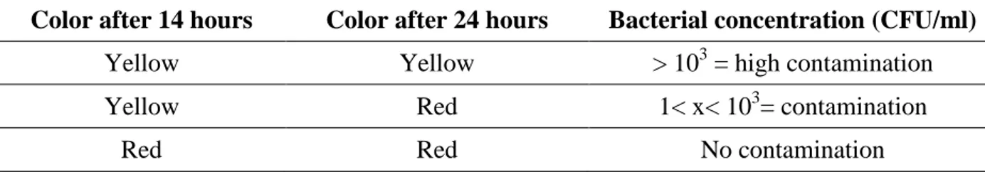
Diagnosis and management of a urinary tract infection is a multistep process which includes the determination of the concentration of pathogens, and the identification of the responsible bacteria, as well as their susceptibility to various antibiotics, the so called antibiogram. The pathogen is detected and identified by urine culture. This also allows an estimate of the level of the bacteriuria. Many laboratories define 105 CFU/ml urine as the threshold. However, this threshold misses many relevant infections. There are therefore other recommendations that recommend the diagnosis of UTI from a count of 103 cfu/ml, depending on the types of bacteria detected [64].
Routine bacterial urine cultures. The laboratory diagnosis of urinary tract infection requires quantitative urine culture on standard agar media not only because it helps to document infection, but also because it is necessary for determination of the identity of the infecting microorganism(s) and for antimicrobial susceptibility testing. This is particularly true because of the increased incidence of antimicrobial resistance. The most commonly used criterion for defining significant bacteriuria is the presence of 105 CFU per milliliter of urine.
Urine cultures have traditionally been performed using sheep blood agar, a nonselective medium, and a selective medium such as MacConkey agar, cysteine lactose electrolyte-deficient (CLED) agar, or eosin methylene blue (EMB) agar. Also chromogenic media have been reported to be an acceptable alternative to traditional media for the isolation of urinary pathogens and offer another option for diagnostic laboratories [65].
Such assays require repeated culturing of a sample and take over 48 hours in order for bacterial colonies to be grown, counted, and exposed to antibiotics using conventional clinical methods. Since the patient cannot remain untreated during this rather prolonged period before definitive diagnosis is obtained, physicians prescribe broad spectrum antibiotics prior to antibiogram results [66].
Non-culture Methods. Key findings pointing toward UTI include pyuria (which can, however, have other causes), hematuria (a finding helpful mainly in excluding alternative diagnoses such as urethritis and vaginitis), and significant bacteriuria.
Assessment for pyuria and bacteriuria is often performed with the use of commercially available dipsticks that test for leukocyte esterase, an enzyme released by leukocytes, and for nitrites, since some bacteria reduce urinary nitrates to nitrites. The dipstick test is most accurate for predicting UTI when the presence of either leukocyte esterase or nitrite is considered a positive result, with a sensitivity of 75% and a specificity of 82%. These tests have low sensitivity, high specificity, low positive-predictive values, and high negative-predictive values. Taken together, the performance characteristics of these tests make them useful as a way to rule out bacteriuria on the basis of a
23
negative test result. Indirect dipstick tests are informative, but less sensitive, than microscopic examination of the urine; however none of these tests establish the diagnosis of UTI [67].
Bacteriuria can be detected microscopically using Gram staining of uncentrifuged urine specimens, Gram staining of centrifuged specimens, or direct observation of bacteria in urine specimens. The performance characteristics of the test are not well-defined. The urine Gram stain test has the important advantage of providing immediate information as to the nature of the infecting bacterium or yeast (rarely infectious agents such as microsporidia) and thereby guiding the physician in selecting empiric antimicrobial therapy. However the Gram stain test has the disadvantage of being too labor intensive for it to be practical for most clinical microbiology laboratories. Moreover it is an insensitive test because it does not distinguish between living and dead bacteria [68].
Standard antibiotic susceptibility testing. Determination of bacterial resistance to antimicrobials is an important part of the management of infections in patients. A limited number of methods for antimicrobial susceptibility testing (AST) of medically important microorganisms have survived the maturation of modern diagnostic clinical microbiology.
In many countries, the disk diffusion method is the most commonly used method in clinical laboratories. This test provides the greatest flexibility and cost-effectiveness; however, the test takes at least 24 h and there are limitations in its accuracy. Another is broth dilution testing, which has attained reference standard status to which all other AST methods are currently compared during development, verification, validation, and clinical trials. Several automated systems are now available that provide rapid antimicrobial susceptibility data. The major limitations of these methods include the requirement for relatively large numbers of viable organisms, complicated pre-analytical processing, limited organism spectrum, pre-analytical variability, time to results, and cost [69].
The disk diffusion method. The disk diffusion method of Kirby and Bauer is simple and practical and has been a well standardized method routinely used. The test is performed by applying a standard bacterial inoculum to the surface of a large (150 mm diameter) Mueller-Hinton agar plate. Up to 12 commercially-prepared, fixed concentration, paper antibiotic disks are placed on the inoculated agar surface. Plates are incubated for 16–24 h at 35°C prior to determination of results. Simultaneous growth of the bacteria and diffusion of the antimicrobial compounds occurs. Growth occurs in the presence of an antimicrobial compound when the bacteria reach a critical mass and can overpower the inhibitory effects of the antimicrobial compound. The zones of growth inhibition around each of the antibiotic disks are measured to the nearest millimeter. The diameter of the zone is related to the susceptibility of the isolate and to the diffusion rate of the drug through the agar medium [70]. The zone diameters of each drug are interpreted using the criteria published by the
24
Clinical and Laboratory Standards Institute (CLSI, formerly the National Committee for Clinical Laboratory Standards or NCCLS). The results of the disk diffusion test are “qualitative,” in that a category of susceptibility (i.e. susceptible, intermediate, or resistant) is derived from the test rather than an minimum inhibitory concentration (MIC). The disk diffusion approach for AST has been standardized primarily for commonly encountered, rapidly growing bacterial pathogens and is applicable to neither anaerobes nor fastidious species that demonstrate marked variability in growth rate from strain to strain. The advantages of the disk method are the test simplicity that does not require any special equipment, the provision of categorical results easily interpreted by all clinicians, and flexibility in selection of disks for testing. It is the least costly of all susceptibility methods. The disadvantages of the disk test are the lack of mechanization or automation of the test [71].
Broth dilution methods. Broth dilution methods may be used to determine the minimum concentrations of antibiotics that are required to inhibit or kill microorganisms. Drugs under study are typically tested at 2-fold doubling (log2) serial dilutions (e.g. 4, 8, 16 μg/ml, and so on), with the
lowest concentration of each antibiotic that inhibits visible growth of organisms designated as the minimum inhibitory concentration (MIC). The concentration ranges tested vary with the antimicrobial agent, the pathogen under study, and the infection site. The broth macrodilution approach is both reliable and well standardized and is of particular utility in research studies and in testing of a single antimicrobial agent for 1 bacterial isolate. The principal disadvantages of the macrodilution method are the tedious, manual task of preparing the antibiotic solutions for each test, the possibility of errors in preparation of the antibiotic solutions, and the relatively large amount of reagents and space required for each test [72].
The miniaturization and mechanization of the test by use of small, disposable, plastic “microdilution” trays has led to its widespread use in both clinical and reference laboratories. Standard trays contain 96 wells, each containing a volume of 0.1 ml that allows approximately 12 antibiotics to be tested in a range of 8 two-fold dilutions in a single tray. Microdilution panels are typically prepared using dispensing instruments that aliquot precise volumes of preweighed and diluted antibiotics in broth into the individual wells of trays from large volume vessels. Following incubation, MICs are determined using a manual or automated viewing device for inspection of each of the panel wells for growth [73].
An example of a commercially available manual broth microdilution (BMD) systems is Sensititre (TREK Diagnostic Systems, Cleveland, OH). Examples of automated BMD platforms include the BD Phoenix (Becton Dickinson, Franklin Lakes, NJ), Microscan (Siemens Healthcare Diagnostics, Deerfield, IL), and Vitek (bioMérieux, Marcy l’Etoile, France)[69].
25
The advantages of the microdilution procedure include the generation of MICs, the reproducibility and convenience of having pre-prepared panels, and the economy of reagents and space that occurs due to the miniaturization of the test. There is also assistance in generating computerized reports if an automated panel reader is used. The main disadvantage of the microdilution method is some inflexibility of drug selections available in standard commercial panels [70].
1.2.2 Alternative methods for UTI diagnosis and management
The standard routine clinical-practice tests currently used to diagnose UTIs regularly misdiagnose infection and antibiotics are often not prescribed when appropriate. Urine cultures followed by classical antibiotic susceptibility testing are not always performed since they delay achievement of results and are laborious and time consuming. This practice has many undesirable consequences, both short term and long term: unsuccessful treatments leading to chronic infections, increased health care costs and increased antibiotic resistance by a growing number of bacterial strains. Given these concerns, it is obvious that a rapid and accurate method of UTI diagnosis and bacterial susceptibility to antibiotics would offer significant health benefits [74].
Alternative methods for UTI diagnosis. Rapid diagnosis methods based on PCR are used to complement conventional culture methods. They have been developed in order to bypass the need for culturing especially with regard to shortening the time to result. Although such PCR assays are fast and very sensitive, they typically require species and strain-specific probes that may or may not be available for a particular organism. Additionally, amplification methods like PCR suffer from contamination problems, complex interpretation of results, as well as high costs [75]. In the diagnosis of UTI, applied real-time PCR methods are presently limited to the detection of single pathogens or the Gram status. A new multiplex real-time PCR test for the detection of 25 common blood stream pathogens (SeptiFastH, Roche Diagnostics GmbH, Penzberg, Germany), introduced recently assures time saving with, however, not very high sensitivity and specificity [76].
Some studies have also hypothesized that bacterial colonization of shed urothelial cells and high levels of urinary ATP (i.e. >50 nmol/l; presumably as a result of a proinflammatory response involving the purinergic system) is a powerful marker of UTI [77].
Another approach is based on the investigation of urine interleukin levels. Active bacterial infection in the lower urinary tract initiates a local inflammatory response including epithelial release of cytokines, acting as neutrophil chemoattractants, acute phase response activators and endogenous pyrogens. In symptomatic UTI, the uroepithelium is triggered to secrete IL-6 and its level to reflect the severity of the infection. Also the investigation of urine interleukin-8 levels was found to be useful for early diagnosis of urinary tract infections [78].