Movement kinematics drive chain selection toward
intention detection
Marco Sorianoa,b, Andrea Cavalloa,b, Alessandro D’Ausilioc,d, Cristina Becchioa,b,1, and Luciano Fadigac,d
aDepartment of Psychology, Università di Torino, 10123 Torino, Italy;bCognition, Motion and Neuroscience Unit, Fondazione Istituto Italiano di Tecnologia, 16152 Genova, Italy;cSection of Human Physiology, Università di Ferrara, 44121 Ferrara, Italy; anddIIT@UniFe Center for Translational Neurophysiology, Fondazione Istituto Italiano di Tecnologia, 44121 Ferrara, Italy
Edited by Peter L. Strick, University of Pittsburgh, Pittsburgh, PA, and approved August 22, 2018 (received for review June 7, 2018) The ability to understand intentions based on another’s
move-ments is crucial for human interaction. This ability has been as-cribed to the so-called motor chaining mechanism: anytime a motor chain is activated (e.g., grasp-to-drink), the observer attrib-utes to the agent the corresponding intention (i.e., to drink) from the first motor act (i.e., the grasp). However, the mechanisms by which a specific chain is selected in the observer remain poorly understood. In the current study, we investigate the possibility that in the absence of discriminative contextual cues, slight kine-matic variations in the observed grasp inform mapping to the most probable chain. Chaining of motor acts predicts that, in a sequential grasping task (e.g., grasp-to-drink), electromyographic (EMG) components that are required for the final act [e.g., the mouth-opening mylohyoid (MH) muscle] show anticipatory activa-tion. To test this prediction, we used MH EMG, transcranial magnetic stimulation (TMS; MH motor-evoked potentials), and predictive models of movement kinematics to measure the level and timing of MH activation during the execution (Experiment 1) and the observation (Experiment 2) of reach-to-grasp actions. We found that MH-related corticobulbar excitability during grasping obser-vation varied as a function of the goal (to drink or to pour) and the kinematics of the observed grasp. These results show that subtle changes in movement kinematics drive the selection of the most probable motor chain, allowing the observer to link an observed act to the agent’s intention.
motor chain selection
|
kinematics|
intention understanding|
transcranial magnetic stimulation|
action observationA
critical aspect of understanding the behavior of another person is recognizing the intent of actions: Is he or she grasping the bottle to pour me a glass of wine or to move the bottle to an ice bucket? It has been proposed that attribution of intentions to actions depends on the same mechanism that governs the motor control of intentional actions (1). During action execution, a high proportion of parietal neurons coding a given motor act (e.g., grasping) discharge differentially depend-ing on the subsequent to-be-executed act (2). Thus, for example, grasp-to-eat neurons show significantly higher firing rates when grasping is followed by bringing a piece of food to the mouth compared with placing the piece of food into a box positioned on the shoulder. This pattern suggests that parietal neurons are dynamically coupled in a way that results in sequential activation—that is, neuronal chaining (2, 3). Anytime an agent has the intention to perform an action (e.g., grasping), depend-ing on the agent’s intention, a specific goal-related neuronal chain is activated (grasp-to-eat).When we observe another person’s actions, a related mecha-nism may operate in the reverse direction. Parietal visuomotor neurons coding grasp-to-eat actions discharge at higher rates when the observed grasp is followed by eating compared with placing (2). Anytime a grasp-to-eat chain is activated, the ob-server can infer the agent’s intention in grasping the object (i.e., to eat). However, the computational principles and neural mechanisms by which a specific chain is selected during action
observation remain poorly understood (3, 4). Given that an ob-server only ever sees a hand grasping an object, how can he or she know whether the observed grasp will be followed by eating or placing?
Since grasping may appear similar across different action chains, some authors argue that chain selection—that is, the process of identifying the most appropriate neuronal chain from among many possible alternatives—is based on context-dependent associations, presumably coded in areas outside the action-observation network (5–9). For example, the presence of a container associated with“placing” may lead to the selection of the grasp-to-place chain. In line with this, the differential response of visuomotor neurons reported during observation of grasp-to-place and grasp-to-eat sequences has been ascribed to contextual cues (2).
A neglected possibility is that slight kinematic variations in the observed grasping action inform mapping to the most probable chain. Initial grasping kinematics, while similar, are not identical. During reach and grasp, the way the hand and digits move to-ward the object is subtly shaped in anticipation of the action intention (10–12), and although it may often be subconsciously, observers are sensitive to subtle variations in movement kine-matics and can use these variations to predict the intention of an observed motor act (13–15). This raises the possibility that, at least in humans, kinematics could drive selection of the most probable chain during action observation.
Here, we describe two experiments designed to test this pos-sibility by studying the level and timing of muscle activation during the execution and observation of grasping sequences.
Significance
Estimation of intentions from the observation of other peo-ple’s actions has been proposed to rely on the same motor chain organization supporting the execution of intentional actions. However, the nature of the mechanism by which a specific neuronal chain is selected among possible alternatives during action observation remains obscure. Our study shows that in absence of discriminative contextual cues, subtle changes in the kinematics of the observed action inform mapping to the most probable chain. These results shed light on the importance of kinematics for the attribution of inten-tions to acinten-tions.
Author contributions: M.S., A.C., C.B., and L.F. designed research; M.S., A.C., and A.D. performed research; A.D. contributed TMS methods; M.S., A.C., and C.B. analyzed data; and M.S., A.C., and C.B. wrote the paper.
The authors declare no conflict of interest. This article is a PNAS Direct Submission.
This open access article is distributed underCreative Commons Attribution-NonCommercial-NoDerivatives License 4.0 (CC BY-NC-ND).
1To whom correspondence should be addressed. Email: [email protected].
This article contains supporting information online atwww.pnas.org/lookup/suppl/doi:10. 1073/pnas.1809825115/-/DCSupplemental.
Chaining of motor acts predicts that, in a sequential grasping task (e.g., grasp-to-drink), electromyographic (EMG) compo-nents that are required for the final act [i.e., the mouth-opening mylohyoid (MH) muscle] show anticipatory activation (16, 17). In Experiment 1, we tested this prediction by measuring MH activity during the execution of grasp-to-drink and grasp-to-pour actions. Having established that MH activity selectively antici-pates the execution of drinking, in Experiment 2, we combined predictive models of movement kinematics with transcranial magnetic stimulation (TMS) to ascertain whether MH excit-ability during action observation, assessed by motor-evoked po-tentials (MEPs), maps onto intention-specific kinematic changes in the observed act (grasp-to-drink vs. grasp-to-pour) in the ab-sence of discriminative contextual cues (Fig. 1).
Results
Experiment 1: Muscle Activation During the Execution of Grasping Sequences.Thirteen volunteers [six females, seven males; mean (M) age, 28 y (range, 23 to 36 y)] took part in the action-execution experiment. Participants were seated on a chair, with their right elbow and wrist resting on a table. They were asked to reach and grasp a bottle positioned on the table with the in-tention to either (i) pour a small glass (height, 8.5 cm; Ø, 5 cm) of water (grasp-to-pour) or (ii) drink water from the bottle (grasp-to-drink). For each participant, a total of 30 trials were administered in two blocks of 15 trials. Trials of the same con-dition were presented within the same block. EMG activity was recorded from the mouth-opening MH muscle and from a con-trol muscle, the extensor carpi radialis (ECR), involved in wrist extension, abduction, and radial deviation.
For each trial, each sample of the EMG signal acquired from reach onset to contact time was recomputed as a ratio of EMG activity atj-th sample of the reach-to-grasp phase divided by the mean of EMG activity in the prereaching phase. EMG ratio was then resampled at intervals of 25% of the normalized movement time to obtain four time bins (i.e., 25%, 50%, 75%, and 100% of movement time). For each participant, for each muscle, the EMG ratio was normalized (z-score) across intention (to pour, to drink) and time bin (25%, 50%, 75%, 100%).
The 2× 4 ANOVA on ECR z-scores showed a main effect of time bin,F(3, 36) = 4.031, P < 0.02, η2p= 0.251. However, none of the post hoc comparisons between time bins survived cor-rection for multiple comparisons (P values ranging from 0.052 to 0.999). The main effect of intention,F(1, 12) = 0.111, P = 0.745,
η2
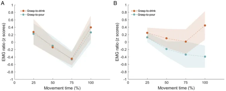
p= 0.009, and the intention-by-time bin interaction, F(3, 36) = 0.334,P = 0.801, η2p= 0.027, were also not significant (Fig. 2A). The 2× 4 ANOVA on MH z-scores showed no main effect of intention,F(1, 12) = 3.682, P = 0.079, η2p= 0.235, and no main effect of time bin,F(3, 36) = 1.271, P = 0.299, η2p = 0.096. A significant intention-by-time bin interaction was present,F(3, 36) = 2.928, P < 0.05, η2
p = 0.196. Post hoc comparisons revealed that at 100% of the movement time (i.e., contact time), MH activity was significantly higher during execution of grasp-to-drink, M = 0.445, SEM = 0.383 (95% CI, −0.389 to 1.279), compared with grasp-to-pour actions, M = −0.394, SEM = 0.295 (95% CI, −1.038 to 0.250), P = 0.019 (Fig. 2B). No statistically significant difference was observed at 25%, 50%, or 75% of movement time (P values ranging from 0.174 to 0.508). Experiment 2: Muscle Activation During the Observation of Grasping Sequences. Having demonstrated that MH activity selectively anticipates the execution of drinking (being significantly higher for grasp-to-drink actions already at contact time), during Ex-periment 2, we assessed whether the same anticipation in MH cortical excitability is seen during observation of grasp-to-drink movements.
Twenty-three new volunteers [12 females, 11 males; mean age, 22 y (range, 19 to 29 y)] took part in the action-observation ex-periment. From the dataset developed by Cavallo et al. (13), we selected 30 representative movies showing the reach-to-grasp phase of grasp-to-pour (n = 15) and grasp-to-drink (n = 15) actions (seeStimuli for more details about movie selection). Only the bottle and the actors’ hand and arm were visible (i.e., all other parts of the body were not shown;Movies S1andS2). Each movie clip was presented at two levels of temporal occlusion (i.e., the movie stopped at 25% or 100% of movement duration). A single TMS pulse was administered at the end of each movie clip via a monophasic stimulator (2002 Magstim) connected to a figure-eight coil (70 mm) positioned over the MH cortical rep-resentation of the left motor cortex. After the TMS pulse was delivered, participants were asked to indicate the intention of the observed act through wrist extension (to signify to drink) or pronation (to signify to pour).
Behavioral results indicated that intentions were categorized above a chance-level accuracy (d′ > 0) at 100% of movement duration,M = 0.31, SEM = 0.13, t22= 2.44, P = 0.023 (95% CI, 0.047 to 0.574), but not at 25% of movement duration, M = −0.03, SEM = 0.08, t22= −0.38, P = 0.708 (95% CI, −0.202 to 0.140). In line with previous findings (13), this demonstrates that kinematic information to discriminate intention is already available at the time of contact with the bottle. Criterionc did not differ from 0 at either of the two time intervals (P values ranging from 0.074 to 0.450), indicating that neither response was favored.
We then conducted a 2 (intention; to pour, to drink)× 2 (time bin; 25%, 100%) repeated-measures ANOVA on normalized MEP areas recorded from MH muscle. The ANOVA yielded no main effect of intention,F(1, 22) = 0.351, P = 0.560, η2p= 0.016, and no main effect of time bin,F(1, 22) = 3.170, P = 0.089, η2p= 0.126. There was, however, a significant intention-by-time bin interaction,F(1, 22) = 6.320, P = 0.020, η2p= 0.223, replicated using a nonparametric permutation-based ANOVA (1,000 per-mutations; empiricalP = 0.013). Post hoc comparisons revealed that at contact with the object (100% time bin), MEP areas were significantly larger during the observation of grasp-to-drink,M = 0.108, SEM= 0.058 (95% CI, −0.012 to 0.228), compared with during the observation of grasp-to-pour actions, M = −0.026, SEM = 0.045 (95% CI, −0.108 to 0.077), P = 0.040 (95% CI, 0.006 to 0.241) (Fig. 3; see also SI Appendix, Fig. S1 for individual MEP areas). No modulation was observed at 25% of movement duration (P = 0.370).
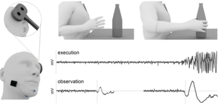
Fig. 1. Schematic illustration of the predicted motor chaining effects. Chaining of motor acts predicts that in a sequential task, EMG components that are required for the final act show anticipatory activation. To test this prediction, in two experiments, we measured the level and timing of the mouth-opening MH activation (Left) during execution and observation of grasp-to-drink actions in the absence of contextual cues (Upper). Hypo-thetical MH EMG records during execution and observation of grasp-to-drink actions are shown (Lower).
NEUROS CIENCE PSYCHOL OGICAL AND COGN ITIVE SC IENCES
Discussion
The ability to understand the intentions of others is of crucial importance for social species such as humans. The way we un-derstand, predict, and attempt to influence another’s actions reflects intention understanding. The precise mechanisms in-volved in the ability of anticipating others’ intentional actions, however, remain obscure.
Coupled with earlier research (11, 13, 15), our results dem-onstrate that in the absence of contextual cues, advanced in-formation pickup from observed movement patterns forms the basis for intention ascription. Most importantly, our findings show that movement kinematics inform the chaining of motor acts during action observation. Subtle changes in the kinematics of the observed action lead to the selection of the most probable action chain.
Significance of Movement Kinematics for Motor Chaining.Debate on the functional role of mirror neurons in an observer’s action understanding has been conditioned by the assumption that the same motor act can be used for several different intentions (7, 18). As Kilner et al. (6) noted, if you see someone in the street raise their hand, they be could hailing a taxi or swatting a wasp— hence, the proposition that any movement-based matching sys-tem will fail to grasp an agent’s intention (19).
But are movement kinematics really substitutable (20)? Studies investigating action observation rarely report on the kinematics of the observed action (14). When they do, the analysis is often confined to a few kinematic landmarks (2, 16, 21). Fogassi et al. (2), for example, report no significant difference in peak wrist velocity and maximal finger aperture between grasp-to-eat and grasp-to-place actions. However, this does not necessarily mean the kinematic profiles of the two grasping actions did not differ across intentions. Two motor acts—for example, grasp-to-drink and grasp-to-pour—may have, on average, similar velocity. How-ever, as shown in Fig. 4, the velocity profile may nevertheless differ across time, providing cues for intention discrimination (11, 13, 14). The possibility that kinematics contributed to chain se-lection in previous studies cannot be ruled out.
The strength of the current design lies in its ability to isolate predictive kinematic information as a potential driver for chain selection. By showing that in the absence of discriminative con-textual cues, activity in MH is greater in anticipation of to-drink
actions compared with to-pour actions, our results demonstrate the significance of kinematics for chain selection.
A New Look at Motor Chaining.The findings reported here chal-lenge two general assumptions underlying the current models of motor chaining. The first assumption is that chain selection is based on context-dependent associations, coded in areas outside the mirror system (22). In the computational model proposed by Chersi et al. (3), this is implemented as an“intention pool”— that is, a pool of spiking neurons in the prefrontal cortex encoding intention based on contextual cues. The model assumes that the activation of this external ensemble initiates the se-quential activation of the appropriate chain. When the cues in the scene are not sufficiently informative, either because they are too few or too many and conflicting to establish the agent’s in-tention, all of the intentions compatible with the given context
Fig. 3. MEP area of MH activation during the observation of grasp-to-drink and grasp-to-pour actions. At contact time (100% of the movement time), MH activity was significantly higher during observation of grasp-to-drink compared with grasp-to-pour actions. Shaded areas depict SEM.
Fig. 2. EMG ratio of ECR (A) and MH (B) during the execution of grasp-to-drink and grasp-to-pour actions. At contact time (100% of movement time), MH activity was significantly higher during execution of grasp-to-drink compared with grasp-to-pour actions. No modulation over time was observed for ECR. Shaded areas depict SEM.
are simultaneously activated and multiple chains are started in parallel (3). Under these conditions, chaining should reveal no anticipation of the agent’s intention. Our findings refute this prediction, showing that in the absence of discriminative con-textual cues, EMG components anticipate the final act.
The second assumption, related to the first, is that selectivity of neurons forming motor chains is independent of the kinematics of the observed act. Contrary to this, our findings show that differ-ential grasping kinematics lead to the selection of the most ap-propriate chain. This supports a model in which the same cortical organization of natural action sequences enabling tailoring to onward action during action execution also supports the predictive linking of kinematic cues to intention during action observation. During the execution of action sequences (e.g., grasping a bottle to drink), motor chaining enables the subtle shaping of the initial grasp in anticipation of the subsequently to-be-performed motor act (to drink). Our results suggest that the same mechanism— chaining of motor acts—is responsible for intention ascription during action observation: Intention-specific kinematic cues drive the selection of the most appropriate chain, allowing an observer to link prospectively the observed act (grasping) to the final act of the sequence (drinking). Evidence that cortical action represen-tations are tuned to movement kinematics has been provided by studies using fMRI (23–25), magnetoencephalography (26), elec-troencephalography (EEG) (27), and TMS (28, 29). The central advance of the present study is the demonstration that the coding of subtle variations in the observed kinematics contributes to ad-vance motor chaining toward intention detection.
Taken together, these results amend the dominant view of chaining as a purely context-based mechanism and suggest that kinematic cues immanent to the observed behavior link together overlapping action segments. Of course, this does not imply that context plays no role. Rather, it suggests that the brain combines contextual information with the stream of incoming kinematic information toward chain selection (30). In keeping with this, Senot et al. (28) found that exposure to incongruent contextual information decreases kinematic modulation, even when the observer is unaware of such incongruence. Future studies simul-taneously manipulating contextual and kinematic information will be necessary to determine how, when, and where this inte-gration takes place and its relationship to conscious perception of
intention. To what extent is conscious perception of intention re-lated to neuronal chaining? This could be examined by asking participants to judge the visibility of the intention of the observed action and then analyzing data according to the visibility judgments made by observers.
Methods
Experiment 1: Muscle Activation During the Execution of Grasping Sequences. Participants. All participants were right-handed, had normal or corrected-to-normal vision, were naïve with respect to the purpose of the experiment, and gave their written informed consent. The experimental procedures were approved by the local ethics committee (Comitato Etico della Provincia di Ferrara) and carried out in accordance with the principles of the Helsinki Declaration (World Medical Association General Assembly, 2008). Stimuli and procedure. Participants were tested individually in a quiet room. They were seated on a height-adjustable chair, with their right elbow and wrist resting on a table (length, 120 cm; width, 80 cm). To ensure a repeatable start position, participants were asked to maintain the right forearm pro-nated, with the right arm oriented in the parasagittal plane passing through the shoulder, and the right hand in a semipronated position, with the tips of the thumb and index finger on a force sensor placed on the working surface. A one-liter glass bottle with about 300 mL of water was positioned over a second force sensor embedded on the table at∼48 cm from participants’ midline. The two force sensors were connected to a 5-V circuit. Variation in resistance of the force sensors led to changes in the output voltage that were read by an analog-to-digital converter connected to Signal software [version 4.08; Cambridge Electronic Design Ltd. (CED)].
Depending on the experimental condition, participants were instructed to start either the grasp-to-pour or the grasp-to-drink movement after hearing a “go” signal from the experimenter. A coexperimenter visually monitored the performance of each trial to ensure participants’ compliance with all requirements. After each trial, the experimenter refilled the bottle to maintain the same weight throughout the experimental session.
Two blocks of 15 trials were administered. The order of block presentation was counterbalanced across participants. Each block was preceded by five practice trials to familiarize participants with the task; these trials were not included in the data analysis. Testing required a session of ∼30 min per participant.
EMG recordings. EMG signals of MH and ECR muscles were acquired using pairs of Ag/AgCl surface electrodes. For the MH recording, electrodes were placed over the right MH muscle, 1 cm lateral to midline, and over the base of the chin. The ECR muscle was recorded with a standard bipolar belly tendon montage. The resulting signals were amplified using MiniWave wireless EMG technology (Cometa Systems), with an input impedance of 20 MΩ and a bandwidth filter between 10 and 500 Hz. All data were sampled at 2 kHz, digitized, and stored for offline analysis using Signal software (version 4.08; CED).
Data analysis. EMG data were filtered (low-pass, 20 Hz) and analyzed offline using a custom MATLAB script (MathWorks). EMG signals of both the MH and ECR muscles were calculated for the reach-to-grasp phase of the movement only, from the reach onset (i.e., the moment the force sensor under the participant’s fingers detected a resistance variation) to the contact time (i.e., the moment the force sensor placed under the bottle detected a resistance variation). The second part of the movement, from the lift of the bottle to the completion of the action sequence, was not considered in the analysis. For each trial, each sample of the EMG signal acquired from reach onset to contact time was recomputed as a ratio of EMG activity at j-th sample of the reach-to-grasp phase divided by the mean of EMG activity in the 200-ms time window preceding the reach onset (i.e., the prereaching phase):
EMG ratioj=
EMGj
μEMG pre−reaching
Values deviating more than±2.5 SD were discarded as outliers. MH and ECR z-scores were analyzed in separate 2× 4 repeated-measures ANOVAs with intention and time bin as within-subjects factors. A significance threshold of P< 0.05 was set for all statistical tests, and a Bonferroni correction applied for post hoc pairwise comparisons.
Experiment 2: Muscle Activation During the Observation of Grasping Sequences.
Participants. Participants who took part in the experiment were right-handed, had normal or corrected-to-normal vision, and had no contraindications for TMS (31). They were naïve to the purpose of the study and provided written informed consent after receiving information about TMS. The experimental Fig. 4. Simulated velocity profiles of exemplar drink and
grasp-to-pour actions. While the two actions have the same mean velocity, their ve-locity profiles differ across time. In particular, the veve-locity of grasp-to-drink actions is higher at 20% and 30% of movement time as compared with grasp-to-pour actions. This pattern reverses between 50% and 60% of movement time. Shaded areas depict SEM.
NEUROS CIENCE PSYCHOL OGICAL AND COGN ITIVE SC IENCES
procedures were approved by the local ethics committee (Comitato Etico della Provincia di Ferrara) and were carried out in accordance with the principles of the revised Helsinki Declaration (World Medical Association General Assembly, 2008). Participants were financially compensated for their time. None of them reported discomfort or adverse effects during TMS. Stimuli. We employed a dataset of 512 actions obtained by Cavallo et al. (13) by recording 17 naïve participants grasping a bottle with the intent to drink or to pour. Apparatus and procedure are described in ref. 13. Briefly, par-ticipants’ right hands were outfitted with 20 lightweight retroreflective hemispheric markers (4 mm in diameter). A near-infrared camera motion-capture system (frame rate, 100 Hz; Vicon System) was used to track the hand kinematics. Kinematic parameters of interest (n= 16, seeSI Appendix, Supplementary Methods) were computed throughout the reach-to-grasp phase of the movement (based on reach onset and contact time) at inter-vals of 10% of the normalized movement time. The second part of the movement, starting from the lift of the bottle, was not considered in the kinematic analysis.
To determine the extent to which grasping movements differed as a function of intention, a linear discriminant analysis was performed. The resulting discriminant function accounted for 100% of variance (function 1= 100% of variance, eigenvalue: 5.279, canonical correlation: 0.917) and signif-icantly differentiated grasp-to-drink and grasp-to-pour movements (P< 0.001). We thus selected the 30 actions (grasp-to-pour, n= 15; grasp-to-drink, n = 15) that minimized the within-intention distance (i.e., the distance from the mean variate score of each intention). This procedure allowed us to identify, for each intention, 15 representative actions. The corresponding movies, filmed from a lateral viewpoint (third-person perspective) using a digital video camera (Sony Handy Cam 3-D; 25 frames per second), were used as stimuli for the current experiment. Movies were edited with Adobe Premiere Pro CS6 (mp4 format, disabled audio, 25 frames per second, resolution 1,280× 800 pixels) so that each movie clip started with the actual reach onset and ended at contact time between the hand and the bottle, with the duration of the movie varying according to the actual duration of the movement (from 840 to 1,640 ms; seeMovies S1andS2), which did not differ between intentions (t28= 0.875, P = 0.389).
EMG and TMS recording. TMS pulses were administered after the localization of the MH cortical representation of the left motor cortex. To localize the MH hot-spot area, the vertex of the cranium was first identified according to the International 10–20 system for EEG electrode placement. The TMS coil was then moved 2 to 4 cm anteriorly and 4 to 6 cm laterally away from the vertex (32–34). Stimulator output was then gradually increased until an MH MEP could be recorded (maximum, 70% of the stimulator output). After the first few MEPs were reliably recognized, the intensity was progressively reduced, the coil was moved around within the area in steps of 1 cm, and the coil orientation rotated to find the position and intensity that maxi-mized stability and amplitude of MEPs. When the hot-spot position was found, it was marked on a bathing cap worn by the participant, and the TMS coil was maintained in a stable position by an articulated arm (Manfrotto). The stimulation intensity was set so that five of five MEPs were detected and clearly discernible from the background EMG activity (stimulation range from 50 to 70% of maximal stimulator output). MEPs were recorded from the right MH muscle using MiniWave wireless EMG technology (Cometa Systems). As in Experiment 1, two Ag/AgCl surface electrodes were placed over the MH muscle (1 cm lateral to midline, and over the base of the chin), and EMG signals were sampled at 2 kHz, fil-tered, and digitized with a data acquisition interface (Micro1401 mk II; CED). Data were displayed and stored for offline analysis using Signal software (version 4.08; CED).
Procedure and task. The experiment was carried out in a dimly illuminated room. Participants sat in a comfortable armchair (dental-chair type) with a fixed headrest in front of a 19-in monitor (resolution, 1,280× 800 pixels; refresh frequency, 75 Hz) at a viewing distance of 50 cm. Before starting the TMS recording, participants were familiarized with the visual stimuli by first watching example movie clips of the reach-to-grasp phase and then the
entire movement for each intention (i.e., four example movie clips were viewed). During the experimental session, each trial began with the ap-pearance of a fixation cross (4,000 ms), followed by the presentation of a movie clip showing either a grasp-to-pour or a grasp-to-drink movement. To ensure movement sequences could be temporally attended (i.e., to allow participants enough time to focus on movement start), 9, 11, or 13 static frames were randomly added at the beginning of each movie. A single TMS pulse was administered at the end of each movie clip. After the TMS pulse was delivered, participants were asked to indicate the intention of the ob-served act through wrist extension (to signify to drink) or pronation (to signify to pour). Participants’ motor response was recorded through a wireless EMG module, equipped with a tri-axis accelerometer (MiniWave; Cometa Systems) positioned over the proximal extremity of the fifth meta-carpal bone of their right hand dorsum. Participants were instructed to re-spond as accurately and quickly as possible. The maximum time allowed to respond was 3,000 ms. A total of 60 movie clips (15 grasp-to-pour and 15 grasp-to-drink) were administered to participants under two levels of tem-poral occlusion: 25% of movement duration (30 clips), 100% of movement duration (30 clips). To avoid participants’ anticipation of TMS delivery, 25% and 100% clips were interspersed with random occlusion trials (60 clips), in which the temporal occlusion occurred at random intervals during reach-to-grasp. To avoid cumulative effects of stimulation, the TMS interpulse in-terval was at least 7,500 ms throughout the experiment (35). The experi-mental trials were preceded by 16 practice trials to familiarize participants with both the task and the TMS pulses (practice pulses). The data for the practice pulses were not included in the main analysis. Stimulus-presentation timing, EMG recording, and TMS triggering, as well as randomization of stimuli were controlled using E-Prime software (version 2.0; Psychology Software Tools Inc.) running on a PC. Testing required a single session of∼60 min per participant.
Data analysis.
Behavioral responses. The proportion of hits (i.e., arbitrarily defined as “to-drink” responses when the observed grasp was to drink) and false alarms (i.e., arbitrarily defined as“to-drink” responses when the observed grasp was to pour) was calculated for each participant for each level of temporal occlusion. From hits and false alarms, we then estimated the criterion c (i.e., the general tendency to respond signal) and sensitivity d′ (i.e., a criterion-independent measure of participants’ performance) (36).
MEPs. Neurophysiological data were analyzed offline using a custom written MATLAB script (MathWorks) and SPSS Statistic software (version 24.0.0.1; SPSS Inc.). For each participant, the area under the rectified MEP curve (MEP area) was calculated within an 8- to 43-ms time window after the TMS pulse. MEP area was chosen instead of peak-to-peak distance because it is more relevant in the context of polyphasic muscle responses (37). To normalize data distribution, MEP area values for each participant were transformed into z-scores, and values deviating more than 2.5 SDs from the grand average of all of the trials were excluded as outliers (3%). To prevent contamination of MEP measurements by background EMG activity, we computed EMG background activity as the area under the rectified EMG signal within a−38- to −3-ms time window before the TMS pulse. Trials characterized by a pre-TMS background exceeding 2.5 times the average were excluded as precontracted trials (<1%) (28). Trials in which MEPs were not identified (2%) were also discarded from further analysis. The remaining normalized MEP data were analyzed in a 2× 2 repeated-measures ANOVA with intention (to pour, to drink) and time bin (25%, 100%) as within-subjects factors. A significance threshold of P< 0.05 was set for all statisti-cal tests, and Bonferroni correction was applied for post hoc pairwise comparisons.
ACKNOWLEDGMENTS. We thank Laura Taverna for her help in figure preparation. The research was funded by the European Research Council under the European Union’s Seventh Framework Programme (FP/2007-2013)/ERC Grant Agreement 312919.
1. Blakemore S-J, Decety J (2001) From the perception of action to the understanding of intention. Nat Rev Neurosci 2:561–567.
2. Fogassi L, et al. (2005) Parietal lobe: From action organization to intention un-derstanding. Science 308:662–667.
3. Chersi F, Ferrari PF, Fogassi L (2011) Neuronal chains for actions in the parietal lobe: A computational model. PLoS One 6:e27652.
4. Grafton ST (2010) The cognitive neuroscience of prehension: Recent developments. Exp Brain Res 204:475–491.
5. Cook R, Bird G, Catmur C, Press C, Heyes C (2014) Mirror neurons: From origin to function. Behav Brain Sci 37:177–192.
6. Kilner JM, Friston KJ, Frith CD (2007) Predictive coding: An account of the mirror neuron system. Cogn Process 8:159–166.
7. Kilner JM (2011) More than one pathway to action understanding. Trends Cogn Sci 15:352–357.
8. Kilner JM, Lemon RN (2013) What we know currently about mirror neurons. Curr Biol 23:R1057–R1062.
9. Press C, Heyes C, Kilner JM (2011) Learning to understand others’ actions. Biol Lett 7: 457–460.
10. Ansuini C, Cavallo A, Bertone C, Becchio C (2014) The visible face of intention: Why kinematics matters. Front Psychol 5:815.
11. Ansuini C, et al. (2015) Predicting object size from hand kinematics: A temporal perspective. PLoS One 10:e0120432.
12. Naish KR, Reader AT, Houston-Price C, Bremner AJ, Holmes NP (2013) To eat or not to eat? Kinematics and muscle activity of reach-to-grasp movements are influenced by the action goal, but observers do not detect these differences. Exp Brain Res 225: 261–275.
13. Cavallo A, Koul A, Ansuini C, Capozzi F, Becchio C (2016) Decoding intentions from movement kinematics. Sci Rep 6:37036.
14. Becchio C, Koul A, Ansuini C, Bertone C, Cavallo A (2018) Seeing mental states: An experimental strategy for measuring the observability of other minds. Phys Life Rev 24:67–80.
15. Ansuini C, Cavallo A, Bertone C, Becchio C (2015) Intentions in the brain: The unveiling of Mister Hyde. Neuroscientist 21:126–135.
16. Cattaneo L, et al. (2007) Impairment of actions chains in autism and its possible role in intention understanding. Proc Natl Acad Sci USA 104:17825–17830.
17. Hamilton AFDC (2013) Reflecting on the mirror neuron system in autism: A systematic review of current theories. Dev Cogn Neurosci 3:91–105.
18. Panasiti MS, Porciello G, Aglioti SM (2017) The bright and the dark sides of motor simulation. Neuropsychologia 105:92–100.
19. Clark A (2016) Surfing Uncertainty (Oxford Univ Press, London).
20. Runeson S (1985) Perceiving people through their movements. Individual Differences in Movement (Springer, Dordrecht, The Netherlands), pp 43–66.
21. Bonini L, et al. (2010) Ventral premotor and inferior parietal cortices make distinct contribution to action organization and intention understanding. Cereb Cortex 20: 1372–1385.
22. Jacob P (2013) How from action-mirroring to intention-ascription? Conscious Cogn 22: 1132–1141.
23. Dayan E, et al. (2007) Neural representations of kinematic laws of motion: Evidence for action-perception coupling. Proc Natl Acad Sci USA 104:20582–20587. 24. Casile A, et al. (2010) Neuronal encoding of human kinematic invariants during action
observation. Cereb Cortex 20:1647–1655.
25. Koul A, et al. (2018) Action observation areas represent intentions from subtle ki-nematic features. Cereb Cortex 28:2647–2654.
26. Press C, Cook J, Blakemore SJ, Kilner J (2011) Dynamic modulation of human motor activity when observing actions. J Neurosci 31:2792–2800.
27. Avanzini P, et al. (2012) The dynamics of sensorimotor cortical oscillations during the observation of hand movements: An EEG study. PLoS One 7:e37534.
28. Senot P, et al. (2011) Effect of weight-related labels on corticospinal excitability during observation of grasping: A TMS study. Exp Brain Res 211:161–167. 29. Agosta S, Battelli L, Casile A (2016) Human movements and abstract motion displays
activate different processes in the observer’s motor system. Neuroimage 130:184–193. 30. Becchio C, Koul A, Ansuini C, Bertone C, Cavallo A (2018) The observability principle and beyond: Reply to comments on“seeing mental states: An experimental strategy for measuring the observability of other minds” by Cristina Becchio et al. Phys Life Rev 24:114–117.
31. Rossi S, Hallett M, Rossini PM, Pascual-Leone A; Safety of TMS Consensus Group (2009) Safety, ethical considerations, and application guidelines for the use of transcranial magnetic stimulation in clinical practice and research. Clin Neurophysiol 120: 2008–2039.
32. Hamdy S, et al. (1996) The cortical topography of human swallowing musculature in health and disease. Nat Med 2:1217–1224.
33. Gallas S, et al. (2007) Mylohyoid motor-evoked potentials relate to swallowing function after chronic stroke dysphagia. Neurogastroenterol Motil 19:453–458. 34. Park E, et al. (2017) Effects of bilateral repetitive transcranial magnetic stimulation on
post-stroke dysphagia. Brain Stimul 10:75–82.
35. Chen R, et al. (1997) Safety of different inter-train intervals for repetitive transcranial magnetic stimulation and recommendations for safe ranges of stimulation parame-ters. Electroencephalogr Clin Neurophysiol 105:415–421.
36. Green DM, Swets JA (1966) Signal Detection Theory and Psychophysics (Wiley, New York).
37. Macdonald DB (2006) Intraoperative motor evoked potential monitoring: Overview and update. J Clin Monit Comput 20:347–377.
NEUROS CIENCE PSYCHOL OGICAL AND COGN ITIVE SC IENCES