Review
The Interplay between Natural Killer Cells and
Human Herpesvirus-6
Eva Eliassen1, Dario Di Luca2, Roberta Rizzo2,* and Isabel Barao3,* 1 HHV-6 Foundation, Santa Barbara, CA 93108, USA; [email protected]
2 Section of Microbiology, Department of Medical Sciences, University of Ferrara, 44121 Ferrara, Italy; [email protected]
3 Department of Microbiology and Immunology, University of Nevada, Reno, NV 89557, USA
* Correspondence: [email protected] (R.R.); [email protected] (I.B.); Tel.: +39-0532-455382 (R.R.); +1-775-343-6750 (I.B.)
Received: 24 October 2017; Accepted: 29 November 2017; Published: 1 December 2017
Abstract: Human Herpesvirus 6 (HHV-6) is a set of two closely related herpes viruses known as HHV-6A and HHV-6B. Both are lymphotropic viruses that establish latency in the host. The ability to evade the immune responses of effector cells is likely a major factor contributing to the development of a persistent HHV-6A/B (collectively termed HHV-6) infection. Natural killer (NK) cells are lymphocytes that, along with neutrophils and monocytes/macrophages, participate in the critical innate immune response during viral infections, but can also mediate the antigen-specific memory responses generally associated with adaptive immunity. NK cells compose the first barrier that viruses must break through to continue replication and dissemination, and a weak NK cell response may predispose an individual to chronic viral infections. Both HHV-6A and HHV-6B can interfere with NK cell-mediated anti-viral responses but the mechanisms by which each of these viruses affect NK cell activity differs. In this review, we will explore the nuanced relationships between the two viruses and NK cells, discussing, in addition, relevant disease associations.
Keywords:NK cells; HHV-6; disease; immune cells; infection
1. Introduction
Human herpesvirus 6 (HHV-6) is a member of the Herpesvirales order, Herpesviridae family, Betaherpesvirinae subfamily, and Roseolovirus genus. HHV-6 is classified into HHV-6A and HHV-6B as two distinct species [1]. The term HHV-6 remains in usage and collectively refers to the two species.
HHV-6 exhibits wide cell tropism in vivo and, as with other herpesviruses, induces a lifelong latent infection in humans (Table1). HHV-6 preferentially replicates in activated CD4+ T lymphocytes [2,3] and uses specific cell receptors permitting virus anchorage to the cell surface: HHV-6A uses CD46, a regulator of complement activation expressed on all nucleated cells, while CD134 (also called OX40), a member of the tumor necrosis factor (TNF) receptor superfamily present only on activated T lymphocytes, functions as a specific entry receptor for HHV-6B [4,5]. In addition to CD4+ T lymphocytes, HHV-6 can infect in vitro CD8+ T lymphocytes (only with HHV-6A), human fibroblasts, natural killer (NK) cells, liver cells, epithelial cells, endothelial cells, astrocytes, oligodendrocytes, and microglial cells [2,6–13]. The host tissue range of HHV-6 in vivo appears to be broader than might be expected from in vitro studies and includes the brain, tonsils, salivary glands, kidneys, liver, lymph nodes, heart, lungs, gastrointestinal tract, and monocytes/macrophages [2,14–16]. The preferential sites for virus latency are suspected to be monocytes/macrphages, bone marrow progenitors and central nervous system (CNS) cells [17–19].
As a key difference with other human herpesviruses, HHV-6 DNA can be integrated into the subtelomeric region of chromosomes in every nucleated cell of the body, including germ cells.
This condition, known as inherited chromosomally integrated HHV-6 (iciHHV-6/ciHHV-6), is present in about 1% of the general population and is vertically transmitted [20].
Table 1.HHV-6A and HHV-6B host-interaction characteristics.
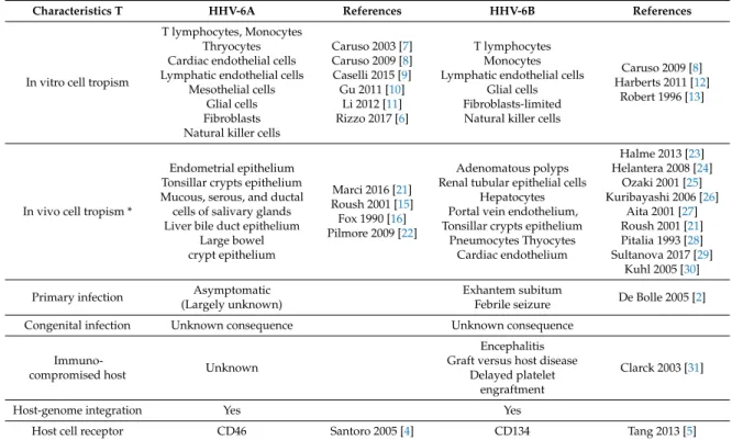
Characteristics T HHV-6A References HHV-6B References
In vitro cell tropism
T lymphocytes, Monocytes Thryocytes Cardiac endothelial cells Lymphatic endothelial cells
Mesothelial cells Glial cells Fibroblasts Natural killer cells
Caruso 2003 [7] Caruso 2009 [8] Caselli 2015 [9] Gu 2011 [10] Li 2012 [11] Rizzo 2017 [6] T lymphocytes Monocytes Lymphatic endothelial cells
Glial cells Fibroblasts-limited Natural killer cells
Caruso 2009 [8] Harberts 2011 [12]
Robert 1996 [13]
In vivo cell tropism *
Endometrial epithelium Tonsillar crypts epithelium Mucous, serous, and ductal cells of salivary glands Liver bile duct epithelium
Large bowel crypt epithelium Marci 2016 [21] Roush 2001 [15] Fox 1990 [16] Pilmore 2009 [22] Adenomatous polyps Renal tubular epithelial cells
Hepatocytes Portal vein endothelium, Tonsillar crypts epithelium
Pneumocytes Thyocytes Cardiac endothelium Halme 2013 [23] Helantera 2008 [24] Ozaki 2001 [25] Kuribayashi 2006 [26] Aita 2001 [27] Roush 2001 [21] Pitalia 1993 [28] Sultanova 2017 [29] Kuhl 2005 [30] Primary infection Asymptomatic
(Largely unknown)
Exhantem subitum
Febrile seizure De Bolle 2005 [2]
Congenital infection Unknown consequence Unknown consequence
Immuno-compromised host Unknown
Encephalitis Graft versus host disease
Delayed platelet engraftment
Clarck 2003 [31]
Host-genome integration Yes Yes
Host cell receptor CD46 Santoro 2005 [4] CD134 Tang 2013 [5]
* In addition to those listed in vitro. HHV = Human herpesvirus. 2. HHV-6 Epidemiology and Infection
HHV-6A and HHV-6B are ubiquitous viruses [2] that are detected in more than 90% of adult populations in developed countries. Primary infection usually occurs very early in life, between 6 months and 2 years of age, following the loss of protective maternal antibodies [32]. Generally, primary HHV-6B infection is accompanied by fever and a rash (exanthema subitum). At an even earlier stage of life, congenital infection following intrauterine transmission or germ line transmission of ciHHV-6 (i.e., congenital ciHHV-6) can occur and has been reported in about 1% of children. It is generally believed that in the majority of countries, primary HHV-6B infection occurs first, and in many cases is associated with clinical symptoms, whereas HHV-6A is acquired later, through asymptomatic infection.
Saliva is assumed to be the main vehicle for virus transmission, as supported by the frequent detection of HHV-6 in saliva and salivary glands. Virus transmission through organ transplantation has been described infrequently, while blood transfusion and breastfeeding have never been reported as the origins of primary infections [33,34].
The clinical symptoms associated with HHV-6 reactivations, which are most commonly reported in transplant recipients, include fever, rash, and transiently decreased numbers of circulating blood cells belonging to the granulocyte/macrophage, erythroid, and megakaryocytic lineages [35–37]. In addition, subacute limbic encephalitis and delayed engraftment are now recognized as typical opportunistic diseases due to HHV-6 reactivation in hematopoietic stem cell transplant (HSCT) recipients [31]. While the effects of chronic HHV-6 infection are yet to be fully understood, the virus has been implicated as a possible trigger for multiple sclerosis (MS) [38,39], chronic fatigue syndrome [40–42], myocarditis and subsequent chronic cardiomyopathy [30,43–45], Hashimoto’s thyroiditis (HT) [46], and, through interference with the normal status of endometrial NK cells, primary idiopathic female infertility [21].
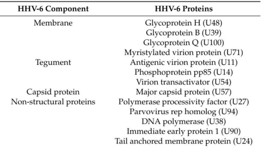
3. HHV-6 Proteins
Roseolovirus genomes are approximately 65 to 90 kb shorter than the 235 kb human cytomegalovirus (HCMV) genome. HHV-6A and HHV-6B are ~90% identical across their genomes, with ~95% of their identity conserved across the herpesvirus core genes. When the viral genome is released into the nucleoplasm, the virus uses the cellular transcription and translation machinery to produce three kinetic classes of viral proteins (immediate early (IE), early (E), and late (L) (Table2).
Table 2.HHV-6 proteins.
HHV-6 Component HHV-6 Proteins
Membrane Glycoprotein H (U48)
Glycoprotein B (U39) Glycoprotein Q (U100) Myristylated virion protein (U71) Tegument Antigenic virion protein (U11)
Phosphoprotein pp85 (U14) Virion transactivator (U54) Capsid protein Major capsid protein (U57) Non-structural proteins Polymerase processivity factor (U27)
Parvovirus rep homolog (U94) DNA polymerase (U38) Immediate early protein 1 (U90) Tail anchored membrane protein (U24)
HHV-6 DNA replication requires seven virally encoded factors. First, the origin binding protein, encoded by the U73 gene, binds to the origin of lytic replication (ori-lyt) and denatures a portion of the circular viral DNA genome [47]. This gap is maintained by the helicase/primase complex, consisting of the U43, U74 and U77 gene products, which also provides RNA primers for the lagging-strand DNA synthesis [48]. The single-stranded DNA in the “replication bubble” is stabilized by the major DNA binding protein, encoded by U41, until second-strand synthesis is catalyzed by the DNA polymerase, pU38 [49], driven by a processivity factor, pU27 [50]. The four proteins encoded by the U79 and U80 genes of HHV-6 are suspected of being involved in DNA replication as well, although their roles are not yet understood [51]. As the new strand grows, the circular replication structure is nicked to form a rolling circle intermediate. Long concatameric strands of progeny DNA are encapsidated by the interaction of cleavage and packaging proteins with specific packaging (pac) signals at the end of the viral genomes. Notably, ori-lyt and pac sequences are different for HHV-6A and HHV-6B [52]. The mature capsids bud out of the nucleus (thereby temporarily acquiring an intermediate membrane devoid of glycoproteins) into the cytoplasm, where they acquire a tegument and a secondary spiked viral envelope at the Golgi complex or at annulate lamellae, where viral glycoproteins accumulate. These are sequentially glycosylated in transport vesicles prior to the release of mature virus particles into the extracellular space by exocytosis. The HHV-6 maturation pathway is different from that of the other herpesviruses in that no viral glycoproteins are detectable in the cell membrane of infected cells [53]. The total time from infection to release of new virions takes approximately 72 h.
Like the other human herpesviruses, HHV-6 is capable of persisting in the host after primary infection, and non-productive infection is characterized by the presence of latency-associated transcripts [54]. The U94 gene product, the expression of which has impaired the migration of endothelial cells [5] and human oligodendrocyte progenitor cells in vitro [55], may enable the establishment and/or maintenance of latent infection [56]. Although HHV-6 has primarily been studied in cases of acute productive infection, as in instances of drug-induced hypersensitivity syndrome/drug reaction with eosinophilia and systemic symptoms (DIHS/DRESS) and reactivation in solid organ and HSCT recipients, the ability of the virus to induce changes in its environment in the absence of strong episodes of replication [57,58] has spurred growing interest in chronic, semi-latent HHV-6 infections.
As HHV-6 is able to infect a great range of cells in vivo, pathogenic effects of chronic infections could have far-reaching consequences.
4. Interactions with the Immune System
HHV-6 is able to both stimulate and modify immune responses [2]. Prior to the emergence of adaptive immunity, HHV-6 stimulates the effectors of innate immunity; an increased secretion of proinflammatory cytokines, such as interleukin-1 beta (IL-1beta), TNF-alpha, and alpha interferon (IFN-alpha), is observed in peripheral blood mononuclear cells, and NK cell activity associated with IL-15 synthesis increases during HHV-6A infection [59,60]. Moreover, HHV-6 has been shown to promote a cytokine profile shift from Th1 to Th2 by upregulating IL-10 and human leukocyte antigen-G (HLA-G) expression [61] while downregulating HLA class I [6] and IL-12 [62,63]. Blocking IL-12 abolishes dendritic cell (DC)-induced IFN-gamma secretion by NK cells (Figure1) [64]. On the other hand, IL-15 upregulation by DC and monocytes/macrophages stimulates NK cell IFN-gamma secretion. These effects are mediated by HHV-6-encoded proteins which act as analogues of cell chemokines and are believed to promote viral growth, viral dissemination, and/or escape from the immune response. Specifically, HHV-6 encodes putative chemokines and chemokine receptors and induces the production of cytokines that are able to modify the immune response.
Viruses 2017, 9, 367 4 of 16
latent HHV-6 infections. As HHV-6 is able to infect a great range of cells in vivo, pathogenic effects of chronic infections could have far-reaching consequences.
4. Interactions with the Immune System
HHV-6 is able to both stimulate and modify immune responses [2]. Prior to the emergence of adaptive immunity, HHV-6 stimulates the effectors of innate immunity; an increased secretion of proinflammatory cytokines, such as interleukin-1 beta (IL-1beta), TNF-alpha, and alpha interferon (IFN-alpha), is observed in peripheral blood mononuclear cells, and NK cell activity associated with IL-15 synthesis increases during HHV-6A infection [59,60]. Moreover, HHV-6 has been shown to promote a cytokine profile shift from Th1 to Th2 by upregulating IL-10 and human leukocyte antigen-G (HLA-antigen-G) expression [61] while downregulating HLA class I [6] and IL-12 [62,63]. Blocking IL-12 abolishes dendritic cell (DC)-induced IFN-gamma secretion by NK cells (Figure 1) [64]. On the other hand, IL-15 upregulation by DC and monocytes/macrophages stimulates NK cell IFN-gamma secretion. These effects are mediated by HHV-6-encoded proteins which act as analogues of cell chemokines and are believed to promote viral growth, viral dissemination, and/or escape from the immune response. Specifically, HHV-6 encodes putative chemokines and chemokine receptors and induces the production of cytokines that are able to modify the immune response.
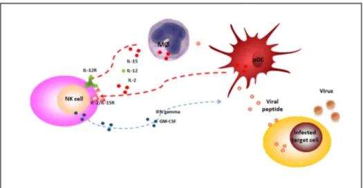
Figure 1. NK cells during viral infections. During viral infections, NK cells are activated by 2,
IL-12 and IL-15, released by plasmacytoid dendritic cells (pDC) and monocytes/macrophages (MØ) (red dotted lines) and secrete potent cytokines, such as interferon gamma (IFN-gamma), that lyse susceptible targets and enhance innate and adaptative immune responses (blue dotted line). NK: natural killer; IL-2/IL-15R: IL-2/IL-15 receptor; IL-12R: IL-12 receptor; IFN-gamma: Interferon gamma; GM-CSF: Granulocyte-macrophage colony-stimulating factor.
5. NK Cells and HHV-6 Infection
5.1. NK Cells
Evidence indicates that HHV-6 alters NK cell activity by affecting their ability to control viral infection. As NK cells are known to represent an important natural defense mechanism in controlling viral infection, evasion of the NK cell response could confer considerable benefits to the virus [65–67].
NK cells, like B and T cells, are a lymphocyte lineage derived from the common lymphoid progenitor (CLP), and like B cells, they are thought to develop primarily in the bone marrow. NK cells also undergo an “education” process during development, during which they acquire the ability to recognize lack of self HLA class I, or “missing-self”, a feature that facilitates their surveillance of Figure 1. NK cells during viral infections. During viral infections, NK cells are activated by IL-2, IL-12 and IL-15, released by plasmacytoid dendritic cells (pDC) and monocytes/macrophages (MØ) (red dotted lines) and secrete potent cytokines, such as interferon gamma (IFN-gamma), that lyse susceptible targets and enhance innate and adaptative immune responses (blue dotted line). NK: natural killer; IL-2/IL-15R: IL-2/IL-15 receptor; IL-12R: IL-12 receptor; IFN-gamma: Interferon gamma; GM-CSF: Granulocyte-macrophage colony-stimulating factor.
5. NK Cells and HHV-6 Infection
5.1. NK Cells
Evidence indicates that HHV-6 alters NK cell activity by affecting their ability to control viral infection. As NK cells are known to represent an important natural defense mechanism in controlling viral infection, evasion of the NK cell response could confer considerable benefits to the virus [65–67].
NK cells, like B and T cells, are a lymphocyte lineage derived from the common lymphoid progenitor (CLP), and like B cells, they are thought to develop primarily in the bone marrow. NK cells also undergo an “education” process during development, during which they acquire the ability to recognize lack of self HLA class I, or “missing-self”, a feature that facilitates their surveillance of target cells that have downregulated HLA class I during infection or malignancy. NK cells rely on both
cytokines and transcription factors to promote and control their development. Cytokine signaling from IL-15 is critical for the development of NK cells and is required throughout their lifetime.
Plentiful studies in the mouse and human demonstrate NK-cell-dependent protective effects during infections with coxsackievirus, human immunodeficiency virus (HIV), hepatitis C virus (HCV), influenza virus, and poxvirus, and herpesviruses [68–70]. Their direct antiviral effects include killing of infected target cells and production of interferon γ (IFN-γ) [70]. The antiviral responses are regulated by a repertoire of germline encoded NK cell receptors (NKRs) recognizing ligands on virus-infected cells and by innate cytokine responses induced during infections.
Inhibitory receptors signal via immunoreceptor tyrosine-based inhibition motifs (ITIMs) located in their cytoplasmic tails. These types of signals predominate under normal conditions, as the majority of target cells express HLA-I molecules. When target cells are transformed or infected, surface expression of HLA-I molecules can be abrogated; the absence of inhibitory NKR ligation allows the NK cell to kill its target (via “missing-self” recognition). In addition to a requirement for release from dominant inhibitory signaling, effective NK cell killing also requires the ligation of activating NKRs. Activating NKRs lack ITIMs, but they associate with membrane-bound adaptor molecules, such as DAP12 (12 kDa transmembrane transduction receptor element) (i.e., Ly49, KIR (killer cell Immunoglobulin-like receptors)), FcεRγ, and CD3ζ (CD16, NKp46), that bear immunoreceptor tyrosine-based activation motifs (ITAMs) [71]. Interestingly, a KIR2DL4 receptor can have both inhibitory and stimulating activity.
5.2. NK Cells Role in HHV Infections
Herpesviruses employ mechanisms used to evade host-specific immunity. HCMV, for instance, downregulates HLA class I expression by using several different proteins in an effort to evade CMV-specific CD8+ T cell responses. HCMV also encodes for several HLA class I homologs that engage inhibitory KIRs to avoid NK cell activation. Additional HCMV proteins serve as ligands for other inhibitory NK cell receptors. One such protein is UL18, which binds to the inhibitory leukocyte immunoglobulin-like receptor 1 (LIR-1) expressed on NK cells, thereby dampening LIR-1+ NK cell function [71,72]. Furthermore, HCMV expresses several proteins that downregulate the expression of activating NKRs and their ligands [73–76].
5.3. NK Cells in HHV-6 Infections
Taking into consideration the fact that other herpesviruses (i.e., HCMV and Epstein–Barr virus) are able to remodel NK cell compartments [77,78], the relationship between HHV-6 and NK cells has emerged as an intriguing field of study. At this time, there are few studies on the consequences of HHV-6 infection on NK cell activity per se, in large part due to the absence of suitable animal models. However, the literature as it stands indicates that the two are strongly interconnected. Upon infection, NK cells act to control the dissemination of the virus, lysing autologous HHV-6-infected PBMC (peripheral blood mononuclear cells) [79]. Accordingly, during the acute febrile phase of HHV-6 infection, there is a significant increase in IFN-α and NK activity when compared to the convalescent phase [59,80]. The heightened NK cell activity is enhanced by the interactions between both IL-15 and CD122 and the beta subunit of the IL-2 receptor. It is known that IL-15 is able to increase the cytotoxicity of NK cells [81], and, in turn, induce the production of the anti-viral factor IFN-γ by CD4+ and NK cells [82].
HHV-6 can also infect NK cells [83]. It has been reported that NK cells rapidly internalize HHV-6 (i.e., within 1 h) but signs of productive infection are only observed if cells are cultured for several weeks prior to infection, a time at which no residual NK cytotoxic activity is observed [83]. Interestingly, infection of NK cells by HHV-6 also leads to de novo expression of CD4, an antigen not expressed by NK cells, thereby predisposing these cells to infection by HIV-1. These results provide evidence that direct infection of NK cells may represent a potential strategy to suppress the natural anti-viral immunity of the host.
In vivo studies revealed an increase in the cytotoxicity and percentages of the CD56brightCD16neg/ dim NK cell subset in patients with Hashimoto’s thyroiditis (HT), a disease in which HHV-6A has been considered as a potential contributor [29,46,84]. These results suggest that the NK cells of patients with HT might be inherently altered to respond to HHV-6A infection with high levels of cytotoxicity, thereby encouraging the development of autoimmunity and furthering the disease. Moreover, the myriad cytokines produced by activated NK cells and direct intercellular interactions can activate dendritic cells, NKT (Natural killer T) cells, B cells, and CD4+ T cells [85], as well as autoreactive T cells [85] that can lead to autoimmune reactions. Indeed, elevated NK cell cytotoxicity has been found in a range of autoimmune disorders, including rheumatoid arthritis [86], autoimmune diabetes [87], and primary biliary cirrhosis [88], where a possible role of viral infection in pathological exacerbation is suggested [89–91].
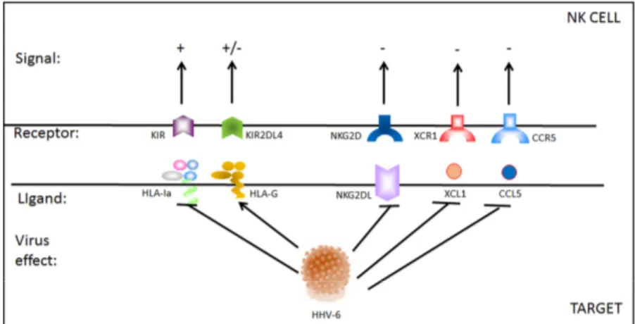
On the other hand, CD56bright NK cells have been found to be helpful in combatting symptoms of autoimmunity, as in some patients with multiple sclerosis (MS) in whom NK cells reduce demyelination through the suppression of T cells [92,93]. This presents a conflicting view of NK cells, in which they are beneficial at times in mitigating an infection or a disease, especially at the onset of an infection, yet at other times, they exacerbate the pathological response, presumably through dysfunctional activity. Both the time point and the functions of the NK cell subset are therefore of key importance in elucidating the course of infection and the development of the disease. Tissue-resident NK cells, for example, possess unique features, such as different receptor repertoires, in comparison to their peripheral blood counterparts, and this variation in cellular characteristics leads to differing downstream effects. Consequently, HHV-6 infection may affect tissue-resident CD56bright NK cells while peripheral blood CD56bright NK cells are not affected in the same manner. In a prime example of this phenomenon, results from a recent study demonstrated a reduction in the CD56brightCD16neg/dim NK cell subset resident in the endometrium of idiopathic infertile women with a concurrent endometrial HHV-6A infection [21]. The uterine NK cells from 43% of women infected with HHV-6A also displayed ex vivo increased activation and degranulation (CD107a expression) towards HHV-6A infected cells, indicating that specific receptor-antigen recognition and signaling (e.g., KIRs-HLA and NKG2D-MICs (MHC class I polypeptide-related sequence), etc.) may be modified by the presence of HHV-6 infection (Figure2). In contrast, peripheral blood NK cell levels were not significantly different among women with HHV-6A and those without the virus.
Viruses 2017, 9, 367 6 of 16
In vivo studies revealed an increase in the cytotoxicity and percentages of the CD56brightCD16neg/dim NK cell subset in patients with Hashimoto’s thyroiditis (HT), a disease in which HHV-6A has been considered as a potential contributor [29,46,84]. These results suggest that the NK cells of patients with HT might be inherently altered to respond to HHV-6A infection with high levels of cytotoxicity, thereby encouraging the development of autoimmunity and furthering the disease. Moreover, the myriad cytokines produced by activated NK cells and direct intercellular interactions can activate dendritic cells, NKT (Natural killer T) cells, B cells, and CD4+ T cells [85], as well as autoreactive T cells [85] that can lead to autoimmune reactions. Indeed, elevated NK cell cytotoxicity has been found in a range of autoimmune disorders, including rheumatoid arthritis [86], autoimmune diabetes [87], and primary biliary cirrhosis [88], where a possible role of viral infection in pathological exacerbation is suggested [89–91].
On the other hand, CD56bright NK cells have been found to be helpful in combatting symptoms of autoimmunity, as in some patients with multiple sclerosis (MS) in whom NK cells reduce demyelination through the suppression of T cells [92,93]. This presents a conflicting view of NK cells, in which they are beneficial at times in mitigating an infection or a disease, especially at the onset of an infection, yet at other times, they exacerbate the pathological response, presumably through dysfunctional activity. Both the time point and the functions of the NK cell subset are therefore of key importance in elucidating the course of infection and the development of the disease. Tissue-resident NK cells, for example, possess unique features, such as different receptor repertoires, in comparison to their peripheral blood counterparts, and this variation in cellular characteristics leads to differing downstream effects. Consequently, HHV-6 infection may affect tissue-resident CD56bright NK cells while peripheral blood CD56bright NK cells are not affected in the same manner. In a prime example of this phenomenon, results from a recent study demonstrated a reduction in the CD56brightCD16neg/dim NK cell subset resident in the endometrium of idiopathic infertile women with a concurrent endometrial HHV-6A infection [21]. The uterine NK cells from 43% of women infected with HHV-6A also displayed ex vivo increased activation and degranulation (CD107a expression) towards HHV-6A infected cells, indicating that specific receptor-antigen recognition and signaling (e.g., KIRs-HLA and NKG2D-MICs (MHC class I polypeptide-related sequence), etc.) may be modified by the presence of HHV-6 infection (Figure 2). In contrast, peripheral blood NK cell levels were not significantly different among women with HHV-6A and those without the virus.
Figure 2. HHV-6 effect on NK cell receptor–ligand interaction. During HHV-6 infection, both NK cells and infected target cells present a modified pattern of receptor–ligand expression, that affect NK cell status. KIR: killer immunoglobulin-like receptors with inhibitory functions; KIR2DL4 with both inhibitory and activating functions; NKG2D activating receptor; XCR1 and CCR5 (C-C chemokine receptor) chemokine receptors. Virus effect: arrow means activated, T bar means inhibited; signal: + means activating signal, +/− means both activating and inhibitory signal, − means inhibitory signal.
Figure 2. HHV-6 effect on NK cell receptor–ligand interaction. During HHV-6 infection, both NK cells and infected target cells present a modified pattern of receptor–ligand expression, that affect NK cell status. KIR: killer immunoglobulin-like receptors with inhibitory functions; KIR2DL4 with both inhibitory and activating functions; NKG2D activating receptor; XCR1 and CCR5 (C-C chemokine receptor) chemokine receptors. Virus effect: arrow means activated, T bar means inhibited; signal: + means activating signal, +/−means both activating and inhibitory signal,−means inhibitory signal.
6. HHV-6 Control of NK Cell Responses
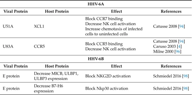
HHV-6 early antigens are involved in regulating NK cell responses (Table3). The expression of the U51A HHV-6A viral receptor in concert with the expression of the HHV-6A ligand U83A affects the binding of ligands to NK activating receptors. U51A binds the ligand XCL1 (Chemokine (C motif) ligand), which normally binds to the CCR7 receptor on CD56brightCD16neg NK cells and the XCR1 receptor on the CD56dimCD16pos NK cells [94]. This may lower the ability of receptor–ligand interactions to activate NK cells, but it is also possible that it promotes chemotaxis of the infected cell to uninfected cells that are secreting the XCL1 and may in turn become infected. Indeed, chemotaxis of infected primary human leukocytes toward the chemokines CCL11 (C-C motif chemokine) and CCL19, which also bind to U51A, has been shown to occur in vitro [94]. These experiments also add to the credence of the notion that the binding of certain ligands to U51A results in temporary protection for the virus by demonstrating that U51A was internalized upon binding to the target ligands. Moreover, under normal circumstances, XCL1 triggers apoptosis in CD4+ T cells when it binds to XCR1, which could be detrimental to the longevity of an HHV-6 infection. Taken together, these responses underline how important it is for HHV-6 to counter the initial immune response.
The viral ligand U83A is produced early in infection and binds CCR5, which blocks binding by CCL5 [94,95]. At the same time, U51A reduces CCL5 expression, resulting in a peak at 24 h post-infection [94] and a subsequently drop. Later in infection, at 6 days and onward, CCL5 is upregulated [58,96]. Early downregulated CCL5 expression might contribute to lower NK cell activation and interruption of the migration of NK cells to HHV-6 infected cells. On the whole, the data indicates that early viral genes seem to lower NK cell activation and allow the virus to spread undetected, but persistent infections may result in activation of NK cells with an altered NK cell response taking the form of atypical chemokine receptor–ligand profiles. In fact, HHV-6B infection decreases the expression of the cellular ligands of several NK receptors (MICB, ULBP1 (UL16 binding protein), ULBP3 and B7-H6) [97].
In combination with its expression of U51A and U83A, HHV-6 suppresses the expression of surface proteins that alert immune cells by triggering two major activating receptors on NK cells: NKG2D and NKp30 [98]. Consequently, the ability of NK cells to counteract HHV-6 reactivation and recruit adaptive immune cells is hindered. Experiments point to the presence of at least two early viral proteins that cause the downregulation of the NKG2D ligands through proteosomal degradation at a post-translational level, as well as a separate mechanism that affects the transcription of B7-H6 mRNA, the cellular ligand of the activating receptor NKp30. These findings are corroborated by the drastically reduced clearing efficiency exhibited by NK cells when they were added to cultures of HHV-6 infected cells after 24 h [99]. Moreover, the results suggest that an inefficient NK cell response may have a significantly detrimental impact on the course of infection and the establishment of a persistent infection.
Notably, both HHV-6A and HHV-6B, although with species-specific differences, induced significant modifications in miRNA expression [6]. miRNAs, small non-coding RNA molecules, are known to play an essential role in fine-tuning host immune homeostasis and responses, as miRNA-mediated regulation of gene expression has a profound impact on immune cell development, function, and response to invading pathogens. HHV-6 infection modifies the expression of specific miRNAs involved in development, maturation and effector functions (i.e., miR-146, miR-155, miR-181, miR-223), and on at least 13 miRNAs with recognized role in inflammation and autoimmunity. Also the expression of transcription factors is significantly modified by HHV-6A/6B infection, with an early increase of ATF3 (Cyclic AMP-dependent transcription factor), JUN and FOXA2 (Forkhead Box A2) by both species, whereas HHV-6A specifically induces a 15-fold decrease of POU2AF1 (POU Class 2 Associating Factor 1), and HHV-6B an increase of FOXO1 (Forkhead box protein O1) and a decrease of ESR1 (Estrogen Receptor 1). Interestingly, ATF3 (Activating Transcription Factor 3), upregulated by both viruses, was reported to regulate negatively NK cell functions in MCMV infected mice, by modulating IFNgamma expression [100]. On the other hand,
although not yet studied in NK cells, several evidences currently associate PPAR-gamma (peroxisome proliferator-activated receptor gamma) to Th lymphocyte differentiation, B lymphocyte effector functions and cytokine expression [101]. POU2AF1, down-modulated by HHV-6A, was recently reported to induce upregulation of host defense genes, including IL-6, in airway epithelium [102]. FOXO1 and ESR1, respectively increased and decreased by HHV-6B, are important regulators of the immune response, being FOXO1 a negative regulator of NK cell maturation and functions [103], whereas ESR1 has been associated to regulation of inflammatory pathways of innate immune cells [104]. JUN is involved in several biological processes as regulators of cell cycle progression, hematopoietic cell differentiation and apoptosis [105–108], and might be studied in detail in the NK cell context.
Table 3.HHV-6A and HHV-6B proteins involved in NK cell activation control.
HHV-6A
Viral Protein Host Protein Effect References
U51A XCL1
Block CCR7 binding Decrease NK cell activation Increase chemotaxis of infected cells to uninfected cells
Catusse 2008 [94]
U83A CCR5 Block CCR5 binding
Decrease NK cell activation
Catusse 2008 [94] Caruso 2003 [4] Milne 2000 [96] HHV-6B
Viral Protein Host Protein Effect References
E protein Decrease MICB, ULBP1,
ULBP3 expression Block NKG2D activation Schmiedel 2016 [98]
E protein Decrease B7-H6
expression Block Nkp30 activation Schmiedel 2016 [98]
7. HHV-6 Infection and NK Cell “Memory”
In recent years, NK cell memory, defined as a quantitatively and qualitatively enhanced immune response upon re-challenge, has gained momentum as a topic of interest. For NK cells, two main types of memory exist: (i) In a fashion similar to T cells and B cells, NK cells can exert immunological memory after encounters with stimuli such as haptens or viruses, resulting in the generation of antigen-specific memory NK cells; (ii) NK cells can “remember” an inflammatory cytokine environment that imprints long-lasting non-antigen-specific NK cell effector function [109].
Using an experimental system in which Ly49Hpos NK cells are adoptively transferred into mice lacking this receptor, robust antigen-driven expansion of these Ly49Hpos cells was observed following murine CMV (MCMV) infection [109,110], and after control of the infection, expanded effector NK cells underwent a contraction phase to establish a long-lived and self-renewing “memory” or “adaptive” pool of antigen-specific cells that could be recovered many months following infection in a variety of peripheral tissues. These memory NK cells display a unique transcriptional signature compared to naïve NK cells [110] and functional attributes commonly associated with memory T cells such as secondary expansion, enhanced effector function ex vivo, and increased protection against virus challenge compared to naïve NK cells from uninfected mice [94]. In addition to evidence supporting NK cell memory formation during MCMV infection, several studies suggest that NK cells contribute in secondary immune responses to other viral infections. NK cells previously exposed to herpes simplex virus 2 (HSV-2) or vaccinia virus infection display enhanced IFN-γ production and protection upon re-challenge in a process that is specific to the priming virus, but independent of the adaptive immune system [111,112]. It is tempting to speculate that different types of memory NK cells might be nurtured by different organ environments.
Distinct patterns of tissue localization have been observed for memory NK cells. Vaccinia virus [113] specific memory NK cells reside in the liver. In influenza VLP (virus like particle)-sensitized mice, memory NK cells isolated from the lungs provided protection against influenza challenge after adoptive transfer [114]. Both splenic and hepatic NK cells from rhesus macaques that were infected with SIV (Simian immunodeficiency virus)-mediated antigen-specific memory responses towards SIV Gag or Env proteins [115]. By contrast, MCMV-specific NK cells are distributed systemically in all organs [110]. Together, these results demonstrate that NK cells, like CD8+ T cells, undergo activation, expansion and contraction in an antigen-specific manner to generate long-lived memory cells in response to viral infection, with a possible tissue specificity. Whether HHV-6-antigen-specific NK cells undergo a similar activation will be an interesting topic for future research. In particular, exploring memory NK responses in the presence of long-term, chronic HHV-6A/B infections could shed light on the disease associations of the viruses.
8. Conclusions
NK cell activity is crucial in mounting an early defense against viral infections and in controlling viral reactivation. However, limited data is available on the effects of HHV-6A and HHV-6B on NK cell activity. While it is obvious that functional NK cell deficiencies allow for the development of robust herpesvirus infections, mainly in individuals with genetic predispositions and immunosuppression, it has also been demonstrated that NK cells with a high activation status are involved in incomplete clearance of HHV-6 in the thyroid, brain and uterus [21,46]. The mechanisms behind this dysfunction and high cytotoxicity might be related to the disruptive effects that HHV-6A and -6B are able to induce directly on NK cells, through epigenetic modulation of the host cell, and indirectly via changes in signaling molecules, perhaps as a result of prolonged semi-latent HHV-6 infection. Several cytokines/chemokines are upregulated by HHV-6 reactivation and may contribute to a range of clinical manifestations, including those seen in transplant recipients [35]. In addition, in vitro studies have demonstrated that HHV-6 can modulate host immunity by several mechanisms, including killing lymphocytes and controlling cytokine/chemokine synthesis [94]. Likewise, viral antigens appear to comprise an important barrier to NK-cell-mediated destruction. Abnormal activation of NK cells might also result from the expression of these proteins, but this aspect of HHV-6/NK cell interaction is poorly understood. Together, the cytokines and chemokines induced by HHV-6 infection, as well as viral proteins themselves, may play important roles in both the activation and suppression of the NK cell response, and consequently, in the course of an HHV-6 infection.
Genetic variations may also influence the development of HHV-6 chronic infections and HHV-6-associated autoimmunity, and may predispose certain individuals to these conditions in part through problems in NK cell functionality [115–119]. In light of this, genetic analysis of patients with chronic HHV-6 infections, such as those reported in cases of HT and idiopathic infertility, could prove useful and may ultimately point to partial underlying NK cell deficiencies [21,46].
NK cells aided by dendritic cells may be the most relevant components of the innate reaction to HHV-6 infection. In addition to their non-specific cytotoxic activity, it is evident that NK cells can take on a novel helper role, in that NK cells may well be exploited for enhancing and rescuing the T-cell response in situations in which the CD4 helper response is affected. In this way, NK cells control the quantity and quality of HHV-6-specific CD8+cytotoxic T lymphocytes, thereby affecting a person’s susceptibility to HHV-6-associated diseases.
Further research is needed to characterize the types of NK cells that are affected by HHV-6 and the range of effects that HHV-6 exerts on NK cells. Future studies may reveal whether suppression of NK cell activity, a particular NK cell receptor repertoire, and/or a disrupted Th1/Th2 ratio is a result of inherent NK cell dysfunction that allows HHV-6 to establish a persistent infection, the NK cell dysfunction is a manifestation of the infection itself, or some combination of these scenarios exists. Consequently, exploration of this topic will help to clarify what kind of role HHV-6 plays in such pathological conditions as HT, female infertility, and even MS, in which NK cell activity
is abnormal. Not only will advances in this field shed light on the mechanisms involved in viral persistence and chronic inflammatory states, but it will also open the door for the development of new immunotherapies to treat patients with HHV-6A- and -6B-associated diseases, many of whom would greatly benefit from advances in treatment options. This is particularly relevant in the setting of chronic HHV-6 infections, for which diagnosis and treatment occurs on a limited scale. Since recent studies support a model in which NK cells display virus-specific expansion to form long-lived memory cells that exhibit specific functional recall responses, a better understanding of the role of NK cells in HHV-6 infection could also be useful in determining whether the NK cell compartment can be harnessed in immunization strategies against HHV-6 where no vaccine or specific cure currently exists. Acknowledgments:This work has been supported by the HHV-6 Foundation; FISM (Fondazione Italia Sclerosi Multipla) grant 2015/R/20; Ricerca Finalizzata GR-2011-02346947; Merck research grant; and Simmaron Research. We thank Kristin Loomis and Daria Bortolotti for the support in paper writing.
Author Contributions: Eva Eliassen, Dario Di Luca: Manuscript writing and critical revision; Roberta Rizzo, Isabel Barao: manuscript conception, design and writing; Eva Eliassen, Dario Di Luca: manuscript writing and critical revision; Roberta Rizzo, Isabel Barao: manuscript conception, design and writing.
Conflicts of Interest:The authors declare no conflict of interest.
References
1. Ablashi, D.; Agut, H.; Alvarez-Lafuente, R.; Clark, D.A.; Dewhurst, S.; di Luca, D.; Flamand, L.; Frenkel, N.; Gallo, R.; Gompels, U.A.; et al. Classification of HHV-6A and HHV-6B as distinct viruses. Arch. Virol. 2014, 159, 863–870. [CrossRef] [PubMed]
2. De Bolle, L.; Naesens, L.; de Clercq, E. Update on human herpesvirus 6 biology, clinical features, and therapy. Clin. Microbiol. Rev. 2005, 18, 217–245. [CrossRef] [PubMed]
3. Braun, D.K.; Dominguez, G.; Pellett, P.E. Human herpesvirus 6. Clin. Microbiol. Rev. 2017, 10, 521–567. 4. Santoro, F.; Kennedy, P.E.; Locatelli, G.; Malnati, M.S.; Berger, E.A.; Lusso, P. CD46 is a cellular receptor for
human herpesvirus 6. Cell 1999, 99, 817–827. [CrossRef]
5. Tang, H.; Serada, S.; Kawabata, A.; Ota, M.; Hayashi, E.; Naka, T.; Yamanishi, K.; Mori, Y. CD134 is a cellular receptor specific for human herpesvirus- 6B entry. Proc. Natl. Acad. Sci. USA 2013, 110, 9096–9099. [CrossRef] [PubMed]
6. Rizzo, R.; Soffritti, I.; D’Accolti, M.; Bortolotti, D.; di Luca, D.; Caselli, E. HHV-6A/6B infection of NK cells modulates the expression of miRNAs and transcription factors potentially associated to impaired NK activity. Front. Microbiol. 2017, 8, 2143. [CrossRef] [PubMed]
7. Caruso, A.; Favilli, F.; Rotola, A.; Comar, M.; Horejsh, D.; Alessandri, G.; Grassi, M.; di Luca, D.; Fiorentini, S. Human herpesvirus-6 modulates RANTES production in primary human endothelial cell cultures. J. Med. Virol. 2003, 70, 451–458. [CrossRef] [PubMed]
8. Caruso, A.; Caselli, E.; Fiorentini, S.; Rotola, A.; Prandini, A.; Garrafa, E.; Saba, E.; Alessandri, G.; Cassai, E.; di Luca, D. U94 of human herpesvirus 6 inhibits in vitro angiogenesis and lymphangiogenesis. Proc. Natl. Acad. Sci. USA 2009, 106, 20446–20451. [CrossRef] [PubMed]
9. Caselli, E.; Campioni, D.; Cavazzini, F.; Gentili, V.; Bortolotti, D.; Cuneo, A.; di Luca, D.; Rizzo, R. Acute human herpesvirus-6A infection of human mesothelial cells modulates HLA molecules. Arch. Virol. 2015, 160, 2141–2149. [CrossRef] [PubMed]
10. Gu, B.; Zhang, G.F.; Li, L.Y.; Zhou, F.; Feng, D.J.; Ding, C.L.; Chi, J.; Zhang, C.; Guo, D.D.; Wang, J.F.; et al. Human herpesvirus 6A induces apoptosis of primary human fetal astrocytes via both caspase-dependent and -independent pathways. Virol. J. 2011, 8, 530. [CrossRef] [PubMed]
11. Li, C.; Goodrich, J.M.; Yang, X. Interferon-gamma (IFN-gamma) regulates production of IL-10 and IL-12 in human herpesvirus-6 (HHV-6)-infected monocyte/macrophage lineage. Clin. Exp. Immunol. 1997, 109, 421–425. [CrossRef] [PubMed]
12. Harberts, E.; Yao, K.; Wohler, J.E.; Maric, D.; Ohayon, J.; Henkin, R.; Jacobson, S. Human herpesvirus-6 entry into the central nervous system through the olfactory pathway. Proc. Natl. Acad. Sci. USA 2011, 108, 13734–13739. [CrossRef] [PubMed]
13. Robert, C.; Aubin, J.T.; Visse, B.; Fillet, A.M.; Huraux, J.M.; Agut, H. Difference in permissiveness of human fibroblast cells to variants A and B of human herpesvirus-6. Res. Virol. 1996, 147, 219–225. [CrossRef] 14. Donati, D.; Akhyani, N.; Fogdell-Hahn, A.; Cermelli, C.; Cassiani-Ingoni, R.; Vortmeyer, A.; Heiss, J.D.;
Cogen, P.; Gaillard, W.D.; Sato, S.; et al. Detection of human herpesvirus-6 in mesial temporal lobe epilepsy surgical brain resections. Neurology 2003, 61, 1405–1411. [CrossRef] [PubMed]
15. Roush, K.S.; Domiati-Saad, R.K.; Margraf, L.R.; Krisher, K.; Scheuermann, R.H.; Rogers, B.B.; Dawson, D.B. Prevalence and cellular reservoir of latent human herpesvirus 6 in tonsillar lymphoid tissue. Am. J. Clin. Pathol. 2001, 116, 648–654. [CrossRef] [PubMed]
16. Fox, J.D.; Briggs, M.; Ward, P.A.; Tedder, R.S. Human herpesvirus 6 in salivary glands. Lancet 1990, 336, 590–593. [CrossRef]
17. Kondo, K.; Kondo, T.; Okuno, T.; Takahashi, M.; Yamanishi, K. Latent human herpesvirus 6 infection of human monocytes/macrophages. J. Gen. Virol. 1991, 72, 1401–1408. [CrossRef] [PubMed]
18. Luppi, M.; Barozzi, P.; Maiorana, A.; Marasca, R.; Torelli, G. Human herpesvirus 6 infection in normal human brain tissue. J. Infect. Dis. 1994, 169, 943–944. [CrossRef] [PubMed]
19. Luppi, M.; Barozzi, P.; Morris, C.; Maiorana, A.; Garber, R.; Bonacorsi, G.; Donelli, A.; Marasca, R.; Tabilio, A.; Torelli, G. Human herpesvirus 6 latently infects early bone marrow progenitors in vivo. J. Virol. 1999, 73, 754–759. [PubMed]
20. Arbuckle, J.H.; Medveczky, P.G. The molecular biology of human herpesvirus-6 latency and telomere integration. Microbes Infect. 2011, 13, 731–741. [CrossRef] [PubMed]
21. Marci, R.; Gentili, V.; Bortolotti, D.; Lo Monte, G.; Caselli, E.; Bolzani, S.; Rotola, A.; di Luca, D.; Rizzo, R. Presence of HHV-6A in endometrial epithelial cells from women with primary unexplained infertility. PLoS ONE 2016, 11, e0158304. [CrossRef] [PubMed]
22. Pilmore, H.; Collins, J.; Dittmer, I.; Williams, L.; Carpenter, L.; Thomas, S.; Croxson, M.; Thomas, M. Fatal human herpesvirus-6 infection after renal transplantation. Transplantation 2009, 88, 762–765. [CrossRef] [PubMed]
23. Halme, L.; Loginov, R.; Arola, J.; Turunen, U.; Lautenschlager, I. HHV-6 antigen and HHV-6 DNA expression in sporadic adenomatous polyps of the colon. Scand. J. Gastroenterol. 2013, 48, 1423–1427. [CrossRef] [PubMed]
24. Helanterä, I.; Loginov, R.; Koskinen, P.; Lautenschlager, I. Demonstration of HHV-6 antigens in biopsies of kidney transplant recipients with cytomegalovirus infection. Transpl. Int. 2008, 21, 980–984. [CrossRef] [PubMed]
25. Ozaki, Y.; Tajiri, H.; Tanaka-Taya, K.; Mushiake, S.; Kimoto, A.; Yamanishi, K.; Okada, S. Frequent detection of the human herpesvirus 6-specific genomes in the livers of children with various liver diseases. J. Clin. Microbiol. 2001, 39, 2173–2177. [CrossRef] [PubMed]
26. Kuribayashi, K.; Matsunaga, T.; Iyama, S.; Takada, K.; Sato, T.; Murase, K.; Fujimi, A.; Takimoto, R.; Kawanishi, J.; Niitsu, Y. Human herpesvirus-6 hepatitis associated with cyclosporine-A encephalitis after bone marrow transplantation for chronic myeloid leukemia. Intern. Med. 2006, 45, 475–478. [CrossRef] [PubMed]
27. Aita, K.; Jin, Y.; Irie, H.; Takahashi, I.; Kobori, K.; Nakasato, Y.; Kodama, H.; Yanagawa, Y.; Yoshikawa, T.; Shiga, J. Are there histopathologic characteristics particular to fulminant hepatic failure caused by human herpesvirus-6 infection? A case report and discussion. Hum. Pathol. 2001, 32, 887–889. [CrossRef] [PubMed] 28. Pitalia, A.K.; Liu-Yin, J.A.; Freemont, A.J.; Morris, D.J.; Fitzmaurice, R.J. Immunohistological detection of human herpes virus 6 in formalin-fixed, paraffin-embedded lung tissues. J. Med. Virol. 1993, 41, 103–107. [CrossRef] [PubMed]
29. Sultanova, A.; Cistjakovs, M.; Gravelsina, S.; Chapenko, S.; Roga, S.; Cunskis, E.; Nora-Krukle, Z.; Groma, V.; Ventina, I.; Murovska, M. Association of active human herpesvirus-6 (HHV-6) infection with autoimmune thyroid gland diseases. Clin. Microbiol. Infect. 2017, 23, 50.e1–50.e5. [CrossRef] [PubMed]
30. Kuhl, U.; Pauschinger, M.; Seeberg, B.; Lassner, D.; Noutsias, M.; Poller, W.; Schultheiss, H.P. Viral persistence in the myocardium is associated with progressive cardiac dysfunction. Circulation 2005, 112, 1965–1970. [CrossRef] [PubMed]
31. Clark, D.A.; Griffiths, P.D. Human herpesvirus 6, relevance of infection in the immunocompromised host. Br. J. Haematol. 2003, 120, 384–395. [CrossRef] [PubMed]
32. Yamanishi, K.; Mori, Y.; Pellett, P.E. Human herpesviruses 6 and 7. In Fields Virology, 6th ed.; Knipe, D.M., Howley, P.M., Cohen, J.I., Griffin, D.E., Lamb, R.A., Martin, M.A., Racaniello, V.R., Roizman, B., Eds.; Lippincott Williams & Wilkins: Philadelphia, PA, USA, 2013; Volume 2, pp. 2058–2079. [CrossRef]
33. Tesini, B.L.; Epstein, L.G.; Caserta, M.T. Clinical impact of primary infection with roseoloviruses. Curr. Opin. Virol. 2014, 9, 91–96. [CrossRef] [PubMed]
34. Ward, K.N.; Gray, J.J.; Efstathiou, S. Brief report: Primary human herpesvirus 6 infection in a patient following liver transplantation from a seropositive donor. J. Med. Virol. 1989, 28, 69–72. [CrossRef] [PubMed] 35. Razonable, R.R. Infections due to human herpesvirus 6 in solid organ transplant recipients. Curr. Opin.
Organ. Transpl. 2010, 15, 671–675. [CrossRef] [PubMed]
36. Razonable, R.R.; Lautenschlager, I. Impact of human herpesvirus 6 in liver transplantation. World J. Hepatol. 2010, 2, 345–353. [CrossRef] [PubMed]
37. Jeulin, H.; Agrinier, N.; Guery, M.; Salmon, A.; Clement, L.; Bordigoni, P.; Venard, V. Human herpesvirus 6 infection after allogeneic stem cell transplantation: Incidence, outcome, and factors associated with HHV-6 reactivation. Transplantation 2013, 95, 1292–1298. [CrossRef] [PubMed]
38. Challoner, P.B.; Smith, K.T.; Parker, J.D.; MacLeod, D.L.; Coulter, S.N.; Rose, T.M.; Schultz, E.R.; Bennett, J.L.; Garber, R.L.; Chang, M.; et al. Plaque-associated expression of human herpesvirus 6 in multiple sclerosis. Proc. Natl. Acad. Sci. USA 1995, 92, 7440–7444. [CrossRef] [PubMed]
39. Leibovitch, E.C.; Jacobson, S. Evidence linking HHV-6 with multiple sclerosis: An update. Curr. Opin. Virol. 2014, 9, 127–133. [CrossRef] [PubMed]
40. Buchwald, D.; Cheney, P.R.; Peterson, D.L.; Henry, B.; Wormsley, S.B.; Geiger, A.; Ablashi, D.V.; Salahuddin, S.Z.; Saxinger, C.; Biddle, R.; et al. A chronic illness characterized by fatigue, neurologic and immunologic disorders, and active human herpesvirus type 6 infection. Ann. Intern. Med. 1992, 116, 103–113. [CrossRef] [PubMed]
41. Patnaik, M.; Komaroff, A.L.; Conley, E.; Ojo-Amaize, E.A.; Peter, J.B. Prevalence of IgM antibodies to human herpesvirus 6 early antigen (p41/38) in patients with chronic fatigue syndrome. J. Infect. Dis. 1995, 172, 1364–1367. [CrossRef] [PubMed]
42. Ablashi, D.V.; Zompetta, C.; Lease, C.; Josephs, S.F.; Balachandra, N.; Komaroff, A.L.; Krueger, G.R.; Henry, B.; Lukau, J.; Salahuddin, S.Z. Human herpesvirus 6 (HHV6) and chronic fatigue syndrome (CFS). Can. Dis. Wkly. Rep. 1991, 17 (Suppl. 1E), 33–40. [PubMed]
43. Mahrholdt, H.; Wagner, A.; Deluigi, C.C.; Kispert, E.; Hager, S.; Meinhardt, G.; Vogelsberg, H.; Fritz, P.; Dippon, J.; Bock, C.T.; et al. Presentation, patterns of myocardial damage, and clinical course of viral myocarditis. Circulation 2006, 114, 1581–1590. [CrossRef] [PubMed]
44. Leveque, N.; Boulagnon, C.; Brasselet, C.; Lesaffre, F.; Boutolleau, D.; Metz, D.; Fornes, P.; Andreoletti, L. A fatal case of human herpesvirus 6 chronic myocarditis in an immunocompetent adult. J. Clin. Virol. 2011, 52, 142–145. [CrossRef] [PubMed]
45. Takatsuka, H.; Wakae, T.; Mori, A.; Okada, M.; Fujimori, Y.; Takemoto, Y.; Okamoto, T.; Kanamaru, A.; Kakishita, E. Endothelial damage caused by cytomegalovirus and human herpesvirus-6. Bone Marrow Transpl. 2003, 31, 475–479. [CrossRef] [PubMed]
46. Caselli, E.; Zatelli, M.C.; Rizzo, R.; Benedetti, S.; Martorelli, D.; Trasforini, G.; Cassai, E.; degli Uberti, E.C.; di Luca, D.; Dolcetti, R. Virologic and immunologic evidence supporting an association between HHV-6 and Hashimoto’s thyroiditis. PLoS Pathog. 2012, 8, e1002951. [CrossRef] [PubMed]
47. Inoue, N.; Pellett, P.E. Human herpesvirus 6B origin-binding protein: DNA-binding domain and consensus binding sequence. J. Virol. 1995, 69, 4619–4627. [PubMed]
48. Nicholas, J. Nucleotide sequence analysis of a 21-kbp region of the genome of human herpesvirus-6 containing homologues of human cytomegalovirus major immediate-early and replication genes. Virology 1994, 204, 738–750. [CrossRef] [PubMed]
49. Teo, I.A.; Griffin, B.E.; Jones, M.D. Characterization of the DNA polymerase gene of human herpesvirus 6. J. Virol. 1991, 65, 4670–4680. [PubMed]
50. Lin, K.; Ricciardi, R.P. The 41-kDa protein of human herpesvirus 6 specifically binds to viral DNA polymerase and greatly increases DNA synthesis. Virology 1998, 250, 210–219. [CrossRef] [PubMed]
51. Taniguchi, T.; Shimamoto, T.; Isegawa, Y.; Kondo, K.; Yamanishi, K. Structure of transcripts and proteins encoded by U79–80 of human herpesvirus 6 and its subcellular localization in infected cells. Virology 2000, 271, 307–320. [CrossRef] [PubMed]
52. Turner, S.; di Luca, D.; Gompels, U. Characterisation of a human herpesvirus 6 variant A “amplicon” and replication modulation by U94-Rep “latency gene”. J. Virol. Methods 2002, 105, 331–341. [CrossRef] 53. Cirone, M.; Campadelli-Fiume, G.; Foa-Tomasi, L.; Torrisi, M.R.; Faggioni, A. Human herpesvirus 6 envelope
glycoproteins B and H-L complex are undetectable on the plasma membrane of infected lymphocytes. AIDS Res. Hum. Retrovir. 1994, 10, 175–179. [CrossRef] [PubMed]
54. Kondo, K.; Shimada, K.; Sashihara, J.; Tanaka-Taya, K.; Yamanishi, K. Identification of human herpesvirus 6 latency-associated transcripts. J. Virol. 2002, 76, 4145–4151. [CrossRef] [PubMed]
55. Campbell, A.; Hogestyn, J.M.; Folts, C.J.; Lopez, B.; Pröschel, C.; Mock, D.; Mayer-Pröschel, M. Expression of the Human Herpesvirus 6A Latency-Associated Transcript U94A Disrupts Human Oligodendrocyte Progenitor Migration. Sci. Rep. 2017, 7, 3978. [CrossRef] [PubMed]
56. Rotola, A.; Ravaioli, T.; Gonelli, A.; Dewhurst, S.; Cassai, E.; di Luca, D. U94 of human herpesvirus 6 is expressed in latently infected peripheral blood mononuclear cells and blocks viral gene expression in transformed lymphocytes in culture. Proc. Natl. Acad. Sci. USA 1998, 95, 13911–13916. [CrossRef] [PubMed] 57. Skuja, S.; Zieda, A.; Ravina, K.; Chapenko, S.; Roga, S.; Teteris, O.; Groma, V.; Murovska, M. Structural and ultrastructural alterations in human olfactory pathways and possible associations with herpesvirus 6 infection. PLoS ONE 2017, 12, e0170071. [CrossRef] [PubMed]
58. Reynaud, J.M.; Jégou, J.F.; Welsch, J.C.; Horvat, B. Human herpesvirus 6A infection in CD46 transgenic mice: Viral persistence in the brain and increased production of proinflammatory chemokines via Toll-like receptor 9. J. Virol. 2014, 88, 5421–5436. [CrossRef] [PubMed]
59. Flamand, L.; Stefanescu, I.; Menezes, J. Human herpesvirus-6 enhances natural killer cell cytotoxicity via IL-15. J. Clin. Investig. 1996, 97, 1373–1381. [CrossRef] [PubMed]
60. Flamand, L.; Gosselin, J.; D’Addario, M.; Hiscott, J.; Ablashi, D.V.; Gallo, R.C.; Menezes, J. Human herpesvirus 6 induces interleukin-1 beta and tumor necrosis factor alpha, but not interleukin-6, in peripheral blood mononuclear cell cultures. J. Virol. 1991, 65, 5105–5110. [PubMed]
61. Saverino, D.; Ghiotto, F.; Merlo, A.; Bruno, S.; Battini, L.; Occhino, M.; Maffei, M.; Tenca, C.; Pileri, S.; Baldi, L.; et al. Specific recognition of the viral protein UL18 by CD85j/LIR-1/ILT2 on CD8+ T cells mediates the non-MHC-restricted lysis of human cytomegalovirus-infected cells. J. Immunol. 2004, 172, 5629–5637. [CrossRef] [PubMed]
62. Smith, A.; Santoro, F.; di Lullo, G.; Dagna, L.; Verani, A.; Lusso, P. Selective suppression of IL-12 production by human herpesvirus 6. Blood 2003, 102, 2877–2884. [CrossRef] [PubMed]
63. Smith, AP.; Paolucci, C.; di Lullo, G.; Burastero, S.E.; Santoro, F.; Lusso, P. Viral replication-independent blockade of dendritic cell maturation and interleukin-12 production by human herpesvirus 6. J. Virol. 2005, 79, 2807–2813. [CrossRef] [PubMed]
64. Ferlazzo, G.; Pack, M.; Thomas, D.; Paludan, C.; Schmid, D.; Strowig, T.; Bougras, G.; Muller, W.A.; Moretta, L.; Münz, C. Distinct roles of IL-12 and IL-15 in human natural killer cell activation by dendritic cells from secondary lymphoid organs. Proc. Natl. Acad. Sci. USA 2004, 101, 16606–16611. [CrossRef] [PubMed] 65. Herberman, R.B.; Ortaldo, J.R. Natural killer cells: Their role in defenses against disease. Science 1981, 214,
24–30. [CrossRef] [PubMed]
66. Trinchieri, G. Biology of natural killer cells. Adv. Immunol. 1989, 47, 187–376. [PubMed]
67. Münz, C.; Chijioke, O. Natural killer cells in herpesvirus infections. F1000Research 2017, 6. [CrossRef] [PubMed]
68. Biron, C.A.; Nguyen, K.B.; Pien, G.C.; Cousens, L.P.; Salazar-Mather, T.P. Natural killer cells in antiviral defense: function and regulation by innate cytokines. Annu. Rev. Immunol. 1999, 17, 189–220. [CrossRef] [PubMed]
69. Orange, J.S.; Ballas, Z.K. Natural killer cells in human health and disease. Clin. Immunol. 2006, 118, 1–10. [CrossRef] [PubMed]
70. Lee, S.H.; Biron, C.A. Here today—Not gone tomorrow: Roles for activating receptors in sustaining NK cells during viral infections. Eur. J. Immunol. 2010, 40, 923–932. [CrossRef] [PubMed]
71. Parham, P. MHC class I molecules and KIRs in human history, health and survival. Nat. Rev. Immunol. 2005, 5, 201–214. [CrossRef] [PubMed]
72. Prod’homme, V.; Griffin, C.; Aicheler, R.J.; Wang, E.C.; McSharry, B.P.; Rickards, C.R.; Stanton, R.J.; Borysiewicz, L.K.; López-Botet, M.; Wilkinson, G.W.; et al. The human cytomegalovirus MHC class I
homolog UL18 inhibits LIR-1+ but activates LIR-1-NK cells. J. Immunol. 2007, 178, 4473–4481. [CrossRef] [PubMed]
73. Loewendorf, A.; Benedict, C.A. Modulation of host innate and adaptive immune defenses by cytomegalovirus: Timing is everything. J. Intern. Med. 2010, 267, 483–501. [CrossRef] [PubMed]
74. Sun, J.C.; Lanier, L.L. The natural selection of herpesviruses and virus-specific NK cell receptors. Viruses 2009, 1, 362–382. [CrossRef] [PubMed]
75. Stern-Ginossar, N.; Elefant, N.; Zimmermann, A.; Wolf, D.G.; Saleh, N.; Biton, M.; Horwitz, E.; Prokocimer, Z.; Prichard, M.; Hahn, G.; et al. Host immune system gene targeting by a viral miRNA. Science 2007, 317, 376–381. [CrossRef] [PubMed]
76. Nachmani, D.; Lankry, D.; Wolf, D.G.; Mandelboim, O. The human cytomegalovirus microRNA miR-UL112 acts synergistically with a cellular microRNA to escape immune elimination. Nat. Immunol. 2010, 11, 806–813. [CrossRef] [PubMed]
77. Gumá, M.; Angulo, A.; Vilches, C.; Gómez-Lozano, N.; Malats, N.; López-Botet, M. Imprint of human cytomegalovirus infection on the NK cell receptor repertoire. Blood 2004, 104, 3664–3671. [CrossRef] [PubMed]
78. Azzi, T.; Lünemann, A.; Murer, A.; Ueda, S.; Béziat, V.; Malmberg, K.J.; Staubli, G.; Gysin, C.; Berger, C.; Münz, C.; et al. Role for early-differentiated natural killer cells in infectious mononucleosis. Blood 2014, 124, 2533–2543. [CrossRef] [PubMed]
79. Malnati, M.S.; Lusso, P.; Ciccone, E.; Moretta, A.; Moretta, L.; Long, E.O. Recognition of virus-infected cells by natural killer cell clones is controlled by polymorphic target cell elements. J. Exp. Med. 1993, 178, 961–969. [CrossRef] [PubMed]
80. Takahashi, K.; Segal, E.; Kondo, T.; Mukai, T.; Moriyama, M.; Takahashi, M.; Yamanishi, K. Interferon and natural killer cell activity in patients with exanthem subitum. Pediatr. Infect. Dis. J. 1992, 11, 369–373. [CrossRef] [PubMed]
81. Szczepanski, M.; Szajnik, M.; Welsh, A.; Foon, K.; Whiteside, T.; Boyiadzis, M. Interleukin-15 enhances natural killer cell cytotoxicity in patients with acute myeloid leukemia by upregulating the activating NK cell receptors. Cancer Immunol. Immunother. 2010, 59, 73–79. [CrossRef] [PubMed]
82. Jayaraman, A.; Jackson, D.J.; Message, S.D.; Pearson, R.M.; Aniscenko, J.; Caramori, G.; Mallia, P.; Papi, A.; Shamji, B.; Edwards, M.; et al. IL-15 complexes induce NK- and T-cell responses independent of type I IFN signaling during rhinovirus infection. Mucosal Immunol. 2014, 7, 1151–1164. [CrossRef] [PubMed]
83. Lusso, P.; Malnati, M.S.; Garzino-Demo, A.; Crowley, R.W.; Long, E.O.; Gallo, R.C. Infection of natural killer cells by human herpesvirus 6. Nature 1993, 362, 458–462. [CrossRef] [PubMed]
84. Rizzo, R.; Zatelli, M.C.; Rotola, A.; Cassai, E.; Degli Uberti, E.; di Luca, D.; Caselli, E. Increase in peripheral CD3-CD56brightCD16- natural killer cells in hashimoto’s thyroiditis associated with HHV-6 infection. Adv. Exp. Med. Biol. 2016, 897, 113–120. [CrossRef] [PubMed]
85. Shi, F.D.; van Kaer, L. Reciprocal regulation between natural killer cells and autoreactive T cells. Nat. Rev. Immunol. 2006, 6, 751–760. [CrossRef] [PubMed]
86. Van Bijnen, S.T.; Cossu, M.; Roeven, M.W.; Jansen, T.L.; Preijers, F.; Spanholtz, J.; Dolstra, H.; Radstake, T.R. Functionally active NKG2A-expressing natural killer cells are elevated in rheumatoid arthritis patients compared to psoriatic arthritis patients and healthy donors. Clin. Exp. Rheumatol. 2015, 33, 795–804. [PubMed]
87. Wang, Y.; Yuan, W.; Guo, H.; Jiang, Y. High frequency of activated NKp46(+) natural killer cells in patients with new diagnosed of latent autoimmune diabetes in adults. Autoimmunity 2015, 48, 267–273. [CrossRef] [PubMed]
88. Jiao, G.; Wang, B. NK cell subtypes as regulators of autoimmune liver disease. Gastroenterol. Res. Pract. 2016, 2016. [CrossRef] [PubMed]
89. Costenbader, K.H.; Karlson, E.W. Epstein–Barr virus and rheumatoid arthritis: Is there a link? Arthritis Res. Ther. 2006, 8, 204. [CrossRef] [PubMed]
90. Seewaldt, S.; Thomas, H.E.; Ejrnaes, M.; Christen, U.; Wolfe, T.; Rodrigo, E.; Coon, B.; Michelsen, B.; Kay, T.W.; von Herrath, M.G. Virus-induced autoimmune diabetes: Most beta-cells die through inflammatory cytokines and not perforin from autoreactive (anti-viral) cytotoxic T-lymphocytes. Diabetes 2000, 49, 1801–1809. [CrossRef] [PubMed]
91. Floreani, A.; Baragiotta, A.; Leone, M.G.; Baldo, V.; Naccarato, R. Primary biliary cirrhosis and hepatitis C virus infection. Am. J. Gastroenterol. 2003, 98, 2757–2762. [CrossRef] [PubMed]
92. Bielekova, B.; Catalfamo, M.; Reichert-Scrivner, S.; Packer, A.; Cerna, M.; Waldmann, T.A.; McFarland, H.; Henkart, P.A.; Martin, R. Regulatory CD56bright natural killer cells mediate immunomodulatory effects of IL-2Rα-targeted therapy (daclizumab) in multiple sclerosis. Proc. Natl. Acad. Sci. USA 2006, 103, 5941–5946. [CrossRef] [PubMed]
93. Laroni, A.; Armentani, E.; Kerlero de Rosbo, N.; Ivaldi, F.; Marcenaro, E.; Sivori, S.; Gandhi, R.; Weiner, H.L.; Moretta, A.; Mancardi, G.L.; et al. Dysregulation of regulatory CD56(bright) NK cells/T cells interactions in multiple sclerosis. J. Autoimmun. 2016, 72. [CrossRef] [PubMed]
94. Catusse, J.; Spinks, J.; Mattick, C.; Dyer, A.; Laing, K.; Fitzsimons, C.; Smit, M.J.; Gompels, U.A. Immunomodulation by herpesvirus U51A chemokine receptor via CCL5 and FOG-2 down-regulation plus XCR1 and CCR7 mimicry in human leukocytes. Eur. J. Immunol. 2008, 38, 763–777. [CrossRef] [PubMed] 95. Milne, R.S.; Mattick, C.; Nicholson, L.; Devaraj, P.; Alcami, A.; Gompels, U.A. RANTES binding and down-regulation by a novel human herpesvirus-6 beta chemokine receptor. J. Immunol. 2000, 164, 2396–2404. [CrossRef] [PubMed]
96. Grivel, J.C.; Ito, Y.; Fagà, G.; Santoro, F.; Shaheen, F.; Malnati, M.S.; Fitzgerald, W.; Lusso, P.; Margolis, L. Suppression of CCR5- but not CXCR4-tropic HIV-1 in lymphoid tissue by human herpesvirus 6. Nat. Med. 2001, 7, 1232–1235. [CrossRef] [PubMed]
97. Gosselin, J.; TomoIu, A.; Gallo, R.C.; Flamand, L. Interleukin-15 as an activator of natural killer cell-mediated antiviral response. Blood 1999, 94, 4210–4219. [PubMed]
98. Schmiedel, D.; Tai, J.; Levi-Schaffer, F.; Dovrat, S.; Mandelboim, O. Human herpesvirus 6B downregulates expression of activating ligands during lytic infection to escape elimination by natural killer cells. J. Virol. 2016, 90, 9608–9617. [CrossRef] [PubMed]
99. Cerwenka, A.; Lanier, L. Natural killer cell memory in infection, inflammation and cancer. Nat. Rev. Immunol. 2016, 16, 112–123. [CrossRef] [PubMed]
100. Rosenberger, C.M.; Clark, A.E.; Treuting, P.M.; Johnson, C.D.; Aderem, A. ATF3 regulates MCMV infection in mice by modulating IFN-gamma expression in natural killer cells. Proc. Natl. Acad. Sci. USA 2008, 105, 2544–2549. [CrossRef] [PubMed]
101. Da Rocha Junior, L.F.; Dantas, A.T.; Duarte, A.L.; de Melo Rego, M.J.; Pitta Ida, R.; Pitta, M.G. PPARγ agonists in adaptive immunity: What do immune disorders and their models have to tell us? PPAR Res. 2013, 2013. [CrossRef] [PubMed]
102. Zhou, H.; Brekman, A.; Zuo, W.L.; Ou, X.; Shaykhiev, R.; Agosto-Perez, F.J.; Wang, R.; Walters, M.S.; Salit, J.; Strulovici-Barel, Y.; et al. POU2AF1 functions in the human airway epithelium to regulate expression of host defense genes. J. Immunol. 2016, 196, 3159–3167. [CrossRef] [PubMed]
103. Deng, Y.; Kerdiles, Y.; Chu, J.; Yuan, S.; Wang, Y.; Chen, X.; Mao, H.; Zhang, L.; Zhang, J.; Hughes, T.; et al. Transcription factor Foxo1 is a negative regulator of natural killer cell maturation and function. Immunity 2015, 42, 457–470. [CrossRef] [PubMed]
104. Kovats, S. Estrogen receptors regulate innate immune cells and signaling pathways. Cell Immunol. 2015, 294, 63–69. [CrossRef] [PubMed]
105. Foletta, V.C.; Segal, D.H.; Cohen, D.R. Transcriptional regulation in the immune system: All roads lead to AP-1. J. Leukoc. Biol. 1998, 63, 139–152. [PubMed]
106. Wisdom, R.; Johnson, R.S.; Moore, C. c-Jun regulates cell cycle progression and apoptosis by distinct mechanisms. EMBO J. 1999, 8, 188–197. [CrossRef] [PubMed]
107. Ponti, C.; Gibellini, D.; Boin, F.; Melloni, E.; Manzoli, F.A.; Cocco, L.; Zauli, G.; Vitale, M. Role of CREB transcription factor in c-fos activation in natural killer cells. Eur. J. Immunol. 2002, 32, 3358–3365. [CrossRef] 108. Healy, S.; Khan, P.; Davie, J.R. Immediate early response genes and cell transformation. Pharmacol. Ther.
2013, 137, 64–77. [CrossRef] [PubMed]
109. Sun, J.C.; Beilke, J.N.; Lanier, L.L. Adaptive immune features of natural killer cells. Nature 2009, 457, 557–561. [CrossRef] [PubMed]
110. Sun, J.C.; Beilke, J.N.; Lanier, L.L. Immune memory redefined: Characterizing the longevity of natural killer cells. Immunol. Rev. 2010, 236, 83–94. [CrossRef] [PubMed]
111. Abdul-Careem, M.F.; Lee, A.J.; Pek, E.A.; Gill, N.; Gillgrass, A.E.; Chew, M.V.; Reid, S.; Ashkar, A.A. Genital HSV-2 infection induces short-term NK cell memory. PLoS ONE 2012, 7, e32821. [CrossRef] [PubMed] 112. Gillard, G.O.; Bivas-Benita, M.; Hovav, A.H.; Grandpre, L.E.; Panas, M.W.; Seaman, M.S.; Haynes, B.F.;
Letvin, N.L. Thy1+ NK cells from vaccinia virusprimed mice confer protection against vaccinia virus challenge in the absence of adaptive lymphocytes. PLoS Pathog. 2011, 7, e1002141. [CrossRef]
113. Paust, S.; Gill, H.S.; Wang, B.Z.; Flynn, M.P.; Moseman, E.A.; Senman, B.; Szczepanik, M.; Telenti, A.; Askenase, P.W.; Compans, R.W.; et al. Critical role for the chemokine receptor CXCR6 in NK cell-mediated antigen-specific memory of haptens and viruses. Nat. Immunol. 2010, 11, 1127–1135. [CrossRef] [PubMed] 114. Reeves, R.K.; Li, H.; Jost, S.; Blass, E.; Li, H.; Schafer, J.L.; Varner, V.; Manickam, C.; Eslamizar, L.; Altfeld, M.;
et al. Antigen-specific NK cell memory in rhesus macaques. Nat. Immunol. 2015, 16, 927–932. [CrossRef] [PubMed]
115. Rizzo, R.; Bortolotti, D.; Fainardi, E.; Gentili, V.; Bolzani, S.; Baldi, E.; Casetta, I.; Granieri, E.; Rotola, A.; Furlan, R.; et al. KIR2DL2 inhibitory pathway enhances Th17 cytokine secretion by NK cells in response to herpesvirus infection in multiple sclerosis patients. J. Neuroimmunol. 2016, 294, 1–5. [CrossRef] [PubMed] 116. Borghi, A.; D’Accolti, M.; Rizzo, R.; Virgili, A.; di Luca, D.; Corazza, M.; Caselli, E. High prevalence of
specific KIR types in patients with HHV-8 positive cutaneous vascular lesions: A possible predisposing factor? Arch. Dermatol. Res. 2016, 308, 373–377. [CrossRef] [PubMed]
117. Ben Fredj, N.; Rizzo, R.; Bortolotti, D.; Nefzi, F.; Chebel, S.; Rotola, A.; Frih-Ayed, M.; di Luca, D.; Aouni, M. Evaluation of the implication of KIR2DL2 receptor in multiple sclerosis and herpesvirus susceptibility. J. Neuroimmunol. 2014, 271, 30–35. [CrossRef] [PubMed]
118. Caselli, E.; Rizzo, R.; Ingianni, A.; Contini, P.; Pompei, R.; di Luca, D. High prevalence of HHV8 infection and specific killer cell immunoglobulin-like receptors allotypes in Sardinian patients with type 2 diabetes mellitus. J. Med. Virol. 2014, 86, 1745–1751. [CrossRef] [PubMed]
119. Rizzo, R.; Gentili, V.; Casetta, I.; Caselli, E.; de Gennaro, R.; Granieri, E.; Cassai, E.; di Luca, D.; Rotola, A. Altered natural killer cells’ response to herpes virus infection in multiple sclerosis involves KIR2DL2expression. J. Neuroimmunol. 2012, 251, 55–64. [CrossRef] [PubMed]
© 2017 by the authors. Licensee MDPI, Basel, Switzerland. This article is an open access article distributed under the terms and conditions of the Creative Commons Attribution (CC BY) license (http://creativecommons.org/licenses/by/4.0/).