€ 98,00
www.piccin.it
ISBN 978-88-299-2941-2
9 7 8 8 8 2 9 9 2 9 4 1 2
0222890
Anatomy of the Eye
and
Human Visual System
Anatomy of the Eye
and
Human V
1
General Features
Human orbits are a pair of symmetric cavities lying beside the root of the nose (Fig. 1.1). Th e two orbits are separated from each other by the nasal cavities and the ethmoidal sinuses. Th ey lie over the maxillary sinuses, below the anterior cranial fossa, and are medial to the temporal and middle cranial fossae. Each orbit has the shape of a quadrangular pyramid lying horizontally, with the base facing forward, laterally and slightly downward, and the apex directed backward and medially. Th e base of the orbit is known as the anterior orbital opening and the apex corresponds to the optic fora-men. Orbit comparison with a pyramid, frequently used for descriptive purposes, falls short for two main reasons. Firstly, as the fl oor is shorter than the other walls, frontal sections show a quadrilateral profi le only in the anterior orbit whereas more posterior sections assume a triangular shape (Fig. 1.2); secondly, the maximum diameter of the orbit is not at its base but about 1 cm behind it. For these reasons, the shape of the orbit could be also compared to a pear with the stalk in the optic canal. Nevertheless, we will stick to the quadrangular pyramid as the shape of reference, describing one by one its constitutive elements: the four walls (superior, inferior, lateral and medial), the four angles, the apex and the base with its margins.
Th e medial wall of the orbit lies along a sagittal plane whereas the lateral wall di-verges from the former by a 45° angle. Variations of this angle are common and are related to the individual variability of the head conformation. Th e longitudinal axis of the orbit, 44 to 50 mm long, is oriented forward, laterally and downward and makes a 22-23° angle with the sagittal plane (Fig. 1.3).
Th e size of the orbit, particularly with regard to its depth, greatly varies according to individuals, sex, race and age. Its average volume is about 30 mL, while the average volume of the eyeball is 7.5 mL. Overall, human orbit is outlined by seven bones: frontal, ethmoid, sphenoid, lacrimal, maxillary, zygomatic and palatine bones (Fig. 1.4). Th e orbit walls are traversed by several canals/openings through which vessels and nerves enter and leave the orbital cavity (Table 1.1, see p. 17).
Eugenio Bertelli
IOF Inferior Orbital Fissure IR Inferior Rectus
LP Levator Palpebrae superioris LR Lateral Rectus
MR Medial Rectus muscle SOF Superior Orbital Fissure
List of Abbreviations
the fi ssure anteriorly in about 60% of cases. Th rough the IOF the orbit joins the infraorbital fossa and, at the posterior end of the fi ssure, the pterygopalatine fossa. As the posterior extremity of the IOF is also located just under the posterior part of the superior orbital fi ssure (SOF), it roughly represents the converging point of the openings connecting the orbit with three important and very diff erent anatomic regions: the infraorbital fossa, the pterygopalatine fossa and the middle cranial fossa. Th e IOF, almost completely closed by the periorbita, transmits the infraorbital nerve and artery, the zygomatic nerve, often an anastomotic vein connecting the in-ferior ophthalmic vein with the pterygoid plexus (see Chapter 3) and, possibly, some parasympathetic branches to the lacrimal gland coming from the sphenopalatine ganglion (see Chapter 3).
Superolateral Angle
In analogy with the inferolateral angle, also the posterior part of the superolat-eral angle is occupied by an elongated opening. Th is is the superior orbital fi ssure (SOF) which represents the largest communication between the orbit and the mid-dle cranial fossa. It is about 22 mm long and its anterior end lies 30-40 mm behind the orbital margin. Th e fi ssure, though greatly varying in shape and size among in-dividuals (Fig. 1.13), roughly has the shape of a tennis racket with a larger posterior portion, lying inferomedially, and a superolaterally located thin forward-directed elongated portion (the handle of the racket). Th e inferior root of the lesser wing of the sphenoid (optic strut) separates the posterior end of the fi ssure from the optic foramen. Th e inferior and superior edges of the fi ssure are bounded by the great-er and lessgreat-er wings of the sphenoid respectively. Along the infgreat-erior margin, at the border between the larger inferomedial and the thinner superolateral portions, the spina musculi recti lateralis is often present, a small variably-shaped bony spine (Fig. 1.13) which receives the insertion of the common tendinous ring, the fi brous ring that gives origin to the four recti (see Chapter 4); in particular, a part of the LR takes origin from the spina recti lateralis. Th e common tendinous ring, also known as annulus of Zinn, partially overlaps with the SOF, crossing its posterior and larger portion twice: the fi rst time ascending vertically from the spina recti lateralis to the superior edge of the fi ssure, the second time bridging the two edges of the fi ssure almost horizontally just over the posterior end of the fi ssure. In this way the annu-lus of Zinn divides the fi ssure into intraconal and extraconal spaces: the intraconal space, oculomotor foramen, is comprised within the fi brous ring and is crossed by the nasociliary and the abducens nerves, the superior and inferior divisions of the oculomotor nerve, the sympathetic and long roots of the ciliary ganglion, and some-times the deep recurrent ophthalmic artery (see Chapter 3); the extraconal spaces are just under and above the common tendinous ring (Fig. 1.14). Th e space under the fi brous ring is very small and occasionally traversed by the inferior ophthalmic vein. Th e following elements can be found in the space over the annulus of Zinn: the superior ophthalmic vein leaving the orbit, the trochlear, frontal and lacrimal nerves entering it, and frequently two arterial anastomoses crossing it: the menin-goophthalmic artery and the anastomosis between the recurrent meningeal branch of the lacrimal artery and the sphenoidal branch of the middle meningeal artery (see Chapter 3). In addition, in about 10% of orbits the superfi cial recurrent ophthalmic artery ( marginal tentorial artery) leaves the orbit through the SOF. In less than 1% of cases (0.74%) a bony bridge is present at the inferomedial extremity of the SOF. Th is bridge forms a large foramen, referred to as Warwick’s foramen, which con-nects the orbital cavity with the middle cranial fossa (Fig. 1.15).
Spina
musculi recti Spina
musculi recti Superior
orbital fissure orbital fissureSuperior
Superior orbital fissure Superior orbital fissure Orbitomeningeal foramen
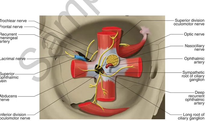
Fig. 1.13 Size and shape variations of the superior orbital fi ssure and spina musculi recti lateralis. Anterior views of the right (A, C) and left orbits (B, D). C D Trochlear nerve Frontal nerve Recurrent meningeal artery Ophthalmic artery Optic nerve Lacrimal nerve Nasociliary nerve Superior ophthalmic vein Abducens nerve Inferior division oculomotor nerve Deep recurrent ophthalmic artery Long root of ciliary ganglion Sympathetic root of ciliary ganglion Superior division oculomotor nerve
Fig. 1.14 Common tendinous ring and superior orbital fi ssure. Vessels and nerves passing through the superior orbital fi ssure.
19
The Eyelids
General Morphology
Th e eyelids are two mobile fi bromuscular folds covered externally by skin and internally by the conjunctiva. Placed in front of the anterior opening of the orbit, they appear convex due to the underlying eyeball bulging forward.
Th e superior eyelid merges upward with the eyebrow at the superior orbito-palpebral sulcus. Th e inferior eyelid is continuous downward with the skin of the zygomatic region; the shallow inferior orbitopalpebral sulcus separates the inferior eyelid from the lid-cheek segment of the midcheek region. It begins close to the medial canthus and runs laterally curving upward to fade in proximity of the lateral canthus (Fig. 2.1). Further downward two additional folds, the shallow nasojugal and palpebromalar folds, can be seen extending into the midcheek region and marking the inferior border of the lid-cheek. Th e former begins close to the medial canthus and runs laterally and downward to end below the inferior margin of the orbital opening, the latter courses downwards and medially from below the lateral canthus toward the inferior part of the nasojugal fold (Fig. 2.2).
With their opposed free edges (i.e. lid margins), the superior and inferior eyelids outline the palpebral fi ssure, a 10-12 mm high and 28-30 mm long opening whose ends are known as medial and lateral canthi (or angles) (Fig. 2.1). Th rough the palpebral fi ssure, depending on the degree of opening, variable portions of cornea and sclera can be seen. On average the upper lid margin is a couple of millimeters lower in adults than in children where it lies just at the level of the upper limbus; its peak is just medial to a vertical line passing through the center of the pupil. Th e lower lid margin is tangential to the inferior limbus and its lowest point lies laterally to the vertical line passing through the pupil. Th e interpalpebral distance is on aver-age 10-12 mm wide. Th is value can be divided into the mean refl ex distance (MRD)
Lacrimal Glands
and Ducts
Eugenio Bertelli, Claudio Nicoletti
CALT Conjunctiva-Associated Lymphoid Tissue
CK Cytokeratin
EALT Eye-Associated Lymphoid Tissue EGF Epidermal Growth Factor
LA Levator Aponeurosis LDALT Lacrimal Drainage-Associated
Lymphoid Tissue
LP Levator Palpebrae Superioris
LPL Lateral Palpebral Ligament M cells Membranous Epithelial Cells MALT Mucosa-Associated Lymphoid Tissue MPL Medial Palpebral Ligament
MRD Mean Refl ex Distance OrOc Orbicularis Oculi OS Orbital Septum SPM Superior Palpebral Muscle VIP Vasoactive Intestinal Peptide
List of Abbreviations
55
Arteries
Th e arterial blood supply of the orbit derives mainly from the ophthalmic ar-tery (OA) (Fig. 3.1). In addition, the orbit receives minor contributions from the infraorbital artery, middle meningeal artery (MMA) and anterior deep tempo-ral artery, branches of the internal maxillary artery, and from the angular artery, terminal branch of the facial artery.
Ophthalmic Artery
Th e OA is the fi rst important branch of the internal carotid artery. It has an av-erage diameter ranging from 1.5 to 1.9 mm. Th e OA takes origin from the internal carotid artery in about 96%-98% of cases. Th e site of origin usually coincides with the emergence of the internal carotid artery from the cavernous sinus. In 4% to 8% of cases, however, it arises earlier when the carotid artery is still in the cavernous sinus. In exceptional cases, the OA stems from a higher position, namely from the supraclinoid portion of the internal carotid artery. In a very limited number of indi-viduals (1% to 3% of cases) the OA arises from the MMA, whereas in 2% to 4% of cases its origin is double; in this instance, the thinner OA usually comes from the in-ternal carotid artery whereas the larger one stems from the MMA (Fig. 3.2). Hence, in 3.4% to 6.6% of individuals (depending on the statistical surveys) the territory supplied by the OA falls entirely, or in large part, into the area of distribution of the external carotid artery. A double OA can also be the result of two arteries arising from the internal carotid artery, one from the intracavernous portion and the other from the supraclinoid area. In extremely rare cases, the OA has been reported as stemming from the middle cerebral artery, the anterior cerebral artery, the posterior communicating artery or even the basilar artery.
For descriptive purposes, the course of the OA is divided into intracranial, in-tracanalicular and intraorbital parts. In its short intracranial course (about 4 mm), the OA runs in the subdural space, between the internal carotid artery, located inferiorly, and the lateral half of the optic nerve (ON) that runs superiorly. At its entrance into
Eugenio Bertelli
CRA Central Retinal Artery E-W Edinger-Westphal
E-Wcp E-W nucleus with centrally projecting cells E-Wpg E-W nucleus with preganglionic cells IOV Inferior Ophthalmic Vein
LA Lacrimal Artery
MMA Middle Meningeal Artery OA Ophthalmic Artery ON Optic Nerve PCA Posterior Ciliary Artery SOV Superior Ophthalmic Vein List of Abbreviations
87
Extraocular Muscles
General Features
Th e orbit contains six striated extraocular muscles that serve to ocular move-ments. Based on their general arrangement, they are divided into four recti muscles and two oblique muscles. Recti muscles are called Medial Rectus (MR), Lateral Rectus (LR), Superior Rectus (SR), and Inferior Rectus (IR). Oblique muscles are called Inferior Oblique (IO) and Superior Oblique (SO). In addition, the Levator Palpebrae superioris (LP), though not involved in ocular movements, is usually described among extraocular muscles due to its intraorbital location, com-mon embryologic development and innervation (Fig. 4.1). Ocular movements take place around the centre of rotation, which is very close to the geometric centre of the eye. Th ey can be described as movements of rotation on the three fundamen-tal axes. A rotation on the vertical axis is called abduction, if the direction of the eye is turned toward the temporal side, or adduction, if the direction of the eye is turned toward the nasal side. A rotation on the transverse axis is called depression (or infraduction), if the centre of the cornea moves downward, or elevation (or supraduction) if it turns upward. A rotation on the sagittal axis is called intorsion (or medial rotation) if the twelve o’clock of the cornea moves nasally or extorsion (or lateral rotation) if the twelve o’clock of the cornea moves temporally (Fig. 4.2). Even though the eyes rotate frequently on non-fundamental axes, each movement can be easily described as a combination of elementary rotations.
Based on its orientation, the contraction of each muscle induces a main move-ment around a fundamove-mental axis, which is called primary action. Th e rotations on the remaining axes are called secondary and tertiary actions (Table 4.1). Th e primary action (main movement) of the IR, for instance, is to depress the eye; this action being more eff ective when the eye is abducted. When the eye looks ahead, the IR also acts inducing adduction (secondary action) and extorsion (tertiary action). Both secondary and tertiary actions become more pronounced as the eye becomes
and Intraorbital
Connective Tissue
Eugenio Bertelli
IO Inferior Oblique muscle IR Inferior Rectus muscle LA Levator Aponeurosis
LP Levator Palpebrae superioris muscle LR Lateral Rectus muscle
MIF Multiply Innervated Fiber
MR Medial Rectus muscle SIF Singly Innervated Fiber SO Superior Oblique muscle SPM Superior Palpebral Muscle SR Superior Rectus muscle List of Abbreviations
General Features
Th e eyeball can be compared with an almost spherical shell housing a nucleus of transparent substances (Fig. 5.1). Th e shell, in turn, is f ormed by three concen-tric membranes, or tunicae (coats), that are structurally and functionally diff erent. Th e outer membrane is the fi brous coat (see Chapter 6), consisting of a larger and opaque posterior portion, the sclera, and a transparent less extensive anterior por-tion, the cornea.
Th e uvea (see Chapter 7) is the intermediate membrane and it is a vascular lay-er. Its posterior 2/3 are in contact with the sclera and are known as the choroid. Forward, though still in contact with the sclera, the uvea has a diff erent and more complex structure, due to the presence of the ciliary muscle. Along with the inner membrane, this part of the uvea is called the ciliary body. Th e iris is the
anterior-of the Eyeball
Eugenio Bertelli
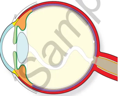
Fig. 5.1 Schematic arrangement of the eye. The eye is formed by a shell of three concentric coats: the outer coat is the fi brous coat and is formed by the sclera (red ) and the cornea (light blue ). The tran-sition area between sclera and cornea is the limbus (yellow ). The second tunica of the eye is the uvea and consists of three segments: choroid (purple ), ciliary body (orange ), and iris (light green ). The innermost coat is the retina which is made of a light sensitive part (brown ) and a blind part (dark green
) which provides an inner covering to the ciliary body and to the iris. The eye shell contains the lens (light sky blue ), just behind the iris, the humor aqueous (white), in front of the iris and around the lens and the
vitreous (beige ), behind the lens. 111
The fibrous tunica ( fibrous coat) is the outer layer of the eyeball. Its consistency gives a minor contribution to the stiffness of the eye which is mainly due to the pos-itive inner pressure. The cornea, the anterior sixth of the fibrous coat, is transparent. The posterior 5/6, the sclera, are opaque and white. The 1.5-2 mm wide transition zone between the cornea and the sclera is referred to as the corneoscleral limbus (Fig. 6.1). On the inner side, the corneoscleral limbus houses a series of structures that are involved in the reabsorption of the aqueous humor from the anterior cham-ber.
Cornea, Sclera and
Corneoscleral Limbus
Eugenio Bertelli, Claudio Traversi, Cosimo Mazzotta, Giulia Borsari
CGRP Calcitonine Gene-Related Peptide CK Cytokeratin
FGF Fibroblast Growth Factor JT Juxtacanalicular Tissue NPY Neuroactive Peptide Y PDGF Platelet Derived Growth Factor SC Schlemm’s Canal
SP Substance P
TEM Transmission Electron Microscopy TGFβ Transforming Growth Factor Β TM Trabecular Meshwork
VAChT Vesicular Acetylcholine Transporter VIP Vasoactive Intestinal Peptide List of Abbreviations
Fig. 6.1 The outer tunica of the eye is the fi brous coat. The po-sterior part is the sclera (red ) whereas its anterior part is the cornea (light blue ). The transition between sclera and cornea is the corneoscleral limbus (yellow ).
121
Sample page
General Features
The vascular tunica of the eyeball, the uvea, is interposed between the nervous and the fibrous coats. The latter, on the other hand, is strictly apposed to the uvea only with its scleral segment. The name uvea derives from the grape-like look that the tunica acquires when isolated from the sclera.
The uvea consists of three portions, each in continuity with the other, with dif-ferent structure and function. From behind forward the uvea comprises the cho-roid, the ciliary body and the iris (Fig. 7.1).
Choroid
The choroid represents the posterior 2/3 of the vascular coat (Fig. 7.1) and, extending forward from the optic nerve to the ora serrata (OS), it shares the same limits of the light-sensitive part of the retina (see Chapter 8). It is mainly formed by vessels that guarantee the metabolic exchange between blood and the outer layers of the retina. In human beings, 90% of oxygen required by the retina comes from the choroid circulation and 90% of all the oxygen delivered to the retina is used by pho-toreceptors. To accomplish such demanding task despite the presence of barriers like Bruch’s membrane (see p. 162-163) and the retinal pigmented epithelium, a steep gradient of oxygen tension between the choroid and the retina has to be kept con-stant. This is achieved by a very high level of blood flow, possibly the highest in the body per unit of tissue weight. It has been estimated that such flow, close to 1200 ml/min/100g, is about 10 folds higher than in the brain. Thanks to this substantial blood flow, oxygen tension in the choroid is maintained high with the arterial/ venous difference of only 3% versus 38% for the retinal circulation. The latter one
Ciliary Body and Iris
Eugenio Bertelli, Paolo Toti
CC Choriocapillaris
CGRP Calcitonin Gene-Related Peptide CK Cytokeratin
CNPE Ciliary Nonpigmented Epithelium CPE Ciliary Pigmented Epithelium JT Juxtacanalicular Tissue nNOS Neuronal Nitric Oxide Synthase NO Nitric Oxide
NPY Neuroactive Peptide Y
NVSMC Non-Vascular Smooth Muscle Cell
OS Ora Serrata
PDGF Platelet Derived Growth Factor SMC Smooth Muscle Cell
SP Substance P
TEM Transmission Electron Microscopy TGFβ Transforming Growth Factor β TM Trabecular Meshwork
VAchT Vesicular Acetylcholine Transporter VIP Vasoactive Intestinal Peptide
List of Abbreviations
157
Sample page
187
General Features
The retina develops from the primitive optic cup (see Chapter 14). Due to the same embryologic origin, the epithelial lining of the ciliary body and the iris epitheli-um should also be considered part of the retina. Indeed, in many textbooks a division is frequently proposed between the light-sensitive and the blind parts of the retina, the latter being that portion of the retina anterior to the ora serrata and corresponding to the ciliary and iris epithelia. Nowadays, it is customary to consider the retina only the portion extending behind the ora serrata (Fig. 8.1). It should be noted, however, that such approach is not justified, either from an embryologic or anatomic viewpoint, since the continuity between these two portions is maintained even when their devel-opment is complete. On the other hand, as the retina, in a clinical context, is usually identified exclusively with its light-sensitive portion, from now on this chapter will conform with this view, the blind portion of the retina having been already dealt with in the paragraphs dedicated to the iris and the ciliary body (see Chapter 7).
The retina is a delicate and thin layer of nervous tissue. The retina anterior bor-der, beyond which it continues with the ciliary epithelium, is marked by the ora serrata, whereas its posterior limit corresponds to the edge of the optic disc, though its nerve fiber layer (see p. 241-242) extends into the optic disc where it forms the optic nerve. The re tina is made up by two concentrically arranged main layers: the pigmented epithelium, externally placed and developed from the primitive optic cup outer layer, and the sensory layer, characterized by a highly complex nervous structure and derived from the optic cup inner layer.
Eugenio Bertelli, Ivanela Kondova, Jan A.M. Langermans
BB S-Cone Bipolar = Blue Bipolar DB Diffuse Bipolar
FMB Flat Midget Bipolar GABA Gamma-Aminobutyric Acid GB Giant Bipolar
GP Glycogen Phosphorylase IMB Invaginating Midget Bipolar ipG Intrinsically photosensitive Ganglion IRBP Interphotoreceptor Retinoid-Binding
Protein L-Cone Long wavelength Cone LGN Lateral Geniculate Nucleus
M-Cone Medium wavelength Cone Me-Cone Melanopsin Cone MG Midget Ganglion PG Parasol Ganglion PKC Protein Kinase C RB Rod Bipolar
RHAMM Receptor for Hyaluronan Mediate Motility
S Stratum/Strata
SBG Small Bistratifi ed Ganglion S-Cone Short wavelength Cone SMG Smooth Monostratifi ed Ganglion
List of Abbreviations
Even their morphology changes according to eccentricity. In peripheral retina, H1 cells have long and scarcely ramified dendrites; overall the dendritic tree measures 160 μm in diameter and makes contact with about 50 pedicles. In contrast, in the fovea H1 cell dendrites are shorter and highly branched, covering an area that measures 16 μm in diameter and contacting only 6-7 pedicles (Fig. 8.27). In the fovea the receptive field of H1 cells coincides with the size of the dendritic tree, whereas in the periphery it is considerable larger (309 μm at 11 mm of eccentric-ity). This difference could be due to a different degree of electric coupling among adjacent cells; in other words H1 cells of the peripheral retina could be more ex-tensively joined through gap junctions.
In contrast to the axon-bearing H1 cells, H2 cells have smaller nuclei and are axonless. They have a local broad dendritic arborization and one or two axon-like longer dendrites (100-300 μm in length) lacking terminal ramifications. H2 cell dendrites receive inputs from all cones but they converge mainly on S-cone pedicles (Fig. 8.27).
On the whole, therefore, dendrites of horizontal cells, either H1 or H2, are post-synaptic to cone pedicles, whereas the axons of H1 cells are postpost-synaptic exclusively to rod spherules. Studies carried out on monkey retinas show that each pedicle is in contact with 3-5 H1 and 3-5 H2 horizontal cells. As we have already mentioned (see p. 209) horizontal cells are provided with AMPA ionotropic (coupled to ion channels) glutamate receptors. The pattern of glutamate receptors is slightly differ-ent according to the horizontal cell type: both H1 and H2 cells are provided with AMPA GluR2-4 receptors on the dendrites, taking part to the tetrads and on the desmosome-like junctions. In addition, however, H1 cells also have KA GluR6/7 receptors on desmosome-like junctions. In absence of luminous stimulation, gluta-mate binding to receptors keeps cationic channels open and maintains horizontal cells in a state of partial depolarization. By decreasing glutamate release, luminous stimulation on photoreceptors induces horizontal cell hyperpolarization. This re-sponse is achieved on H1 cells by stimulations with green or red light, whereas H2 cells show the same response with luminous stimulations with green, red or blue light. Hence, no selectivity has been found for L- or M-cone inputs in H1 cells and even less selectivity has been found for H2 cells, as they hyperpolarize in the same way by stimulating all types of cones.
Horizontal cells have not a center-surround receptive field organization (the re-sponse has always the same sign, whichever area of the receptive filed is stimulated); in addition, even though H1 and H2 cells show different connectivity with cones (H2 cells preferentially contact S-cones, H1 cells tend to skip S-cones), their re-sponse to luminous stimulation is not color opponent.
Horizontal cells are thought to produce and release GABA. The nearest cognate receptors are located on cone pedicles and on bipolar cell dendrites. So far, it is not clear how horizontal cells achieve GABA secretion as well as the exact influence that, at this level, GABA exerts on the transmission of visual information. Nevertheless, even though the precise synaptic mechanisms are not known, the prevalent opinion is that, on the whole, horizontal cells function is to shape the antagonistic periphery of cones and bipolar cell receptive fields. As RB cells (see p. 217) do not possess a center-surround receptive field structure, the role played by H1 horizontal cells in the tetrads of the spherules is possibly different.
Bipolar Cells
Retinal bipolar cells form a heterogeneous population of about 35,000,000 glu-tamatergic neurons. They are 2nd order sensory conducting neurons that transmit vi-sual information from photoreceptors to ganglion and amacrine cells. The cell body
with one or two nucleoli, a well-developed Golgi complex located near the origin of the primary dendrite, a prominent rough endoplasmic reticulum, mitochondria and free ribosomes. Dendrites have a section diameter as large as 0.1-0.2 μm at the fovea and they contain microtubules, numerous slender mitochondria and a few vesicles. The axon, provided with microtubules, vesicles and very rare mitochondria, is sur-rounded by Müller cells cytoplasmic processes up to the inner plexiform layer where it forms its telodendron. The telodendron ends into synaptic terminals containing large mitochondria and synaptic vesicles mainly gathered around synaptic ribbons. Synaptic ribbons in bipolar cells are smaller, usually lacking the arciform density,
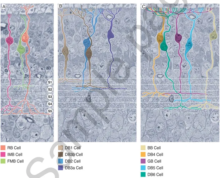
RB Cell IMB Cell FMB Cell DB1 Cell DB3b Cell DB2 Cell DB3a Cell BB Cell DB4 Cell GB Cell DB5 Cell DB6 Cell S1 S2 S3 S4 S5
Fig. 8.27 Retina. Morphology and level of axon terminal ramifi cations of the different types of bipolar cells. A) Rod bipolar (RB) cells (oran-ge) collect inputs from rod spherules and ramify in S5; invaginating midget bipolar (IMB) cells (lilac) gather inputs from single pedicles and ramify their axons in S4-S5; fl at midget bipolar (FMB) cells (light green) collect inputs from single pedicles and their axons end in S1-S2. B) OFF-center diffuse bipolar (DBa ) cells. DB1 cells (light brown) receive inputs from all pedicles within their dendritic tree. They end distributing their axon terminals in S1; DB2 cells (blue) collect inputs from all pedicles within their dendritic tree. Their axon terminals are located in S1-S2; DB3a cells (dark blue) gather visual information mainly from M- and L-cones. Their axons distribute within a narrow stripe of S2; DB3b cells (dark brown) collets inputs from cone pedicles and also from a few spherules. Their axonal distribution overlaps that of DB3a cells. C) ON-center diffuse bipolar (DBb) cells, giant bipolar cells (GB) and S-cone bipolar cells (BB). DB4 cells have a dendritic tree which collects inputs mainly from M- and L-cones. Their axon terminals distribute within S3 (orange); DB5 cells (light blue) collect inputs exclusively from M- and L-cones. Their axon radiates within a narrow stripe of S4; DB6 cells (turquois) have a broad dendritic tree which gathers inputs from all types of cones. Their axon distributes to S5; GB cells (purple) have a large and sparse dendritic tree which collects inputs only from half pedicles. Their axon ends within S3; BB cells (beige) collects inputs from 2-3 S-cones. Their axon arborizes in S5.
A B C
to which they are postsynaptic. Based on this criterion, rod bipolar cells are distin-guished from cone bipolar cells. Whereas the former type of bipolar cells makes up a homogeneous population, the latter type is further classified in at least three major subpopulations (Fig. 8.28): i) midget bipolar cells, which may be split into either ON-center or OFF-center subtypes; ii) diffuse bipolar cells, divided into three ON-center and four OFF-center subtypes; iii) S-cone bipolar cells. In addition, giant bipolar cells have been recently described, but data on this variety are still poor and we will deal with it only briefly.
Rod Bipolar Cells
Rod bipolar cells (RB) are about 20% of all bipolar cells. RB cells are not pres-ent in the fovea and they appear only at 1 mm of eccpres-entricity from the foveal cpres-enter. On average, RB cell body is 10 μm large and it is located in the outer half of the inner nuclear layer, sometimes among the perikaria of horizontal cells on the border of the outer plexiform layer. The cell body gives origin to a robust dendrite that enters the outer plexiform layer ramifying repeatedly and eventually dividing into a series of pointed terminal branchlets, whose number varies as a function of retinal eccentricity. In the area centralis, the dendritic field of RB cells covers a circular surface that measures about 20 μm in diameter, whereas in the peripheral retina this diameter doubles. Each terminal branchlet occupies the central position of a tetrad housed in the deep invagination of a spherule (see p. 205-206). In this way, it is believed that one RB cell makes contact with 30-35 spherules in the area centralis and with 45-50 in the peripheral retina; there are, however, different estimations and according to some investigators the number of rod spherules contacted by each RB cell could be as high as 80-120. RB cell dendrites are provided with APB-sensitive mGluR6 metabotropic glutamate receptors. When engaged by the ligand, these re-ceptors activate a second messenger that closes cationic channels. Without luminous stimulation, when glutamate release from rods saturates the receptors, cationic chan-nels are closed and RB cells are in a hyperpolarized state. The decrease in glutamate release evoked by a luminous stimulation desaturates the receptors. Consequently, cationic channels open allowing an influx of positive ions that depolarizes RB cells. From a functional viewpoint, therefore, synapses between spherules and RB cells are sign-inverting synapses; in addition, all RB cells are ON bipolar cells as they depo-larize to increments of light.
The axon of RB cells dives into the sublamina b of the inner plexiform layer (see p. 232-235) without giving off collaterals. Once reached S5, the deepest stratum of the inner plexiform layer (see p. 228), it gives origin to numerous telodendria that resolve into few large terminals housing synaptic ribbons and glutamate-containing synaptic vesicles (Fig. 8.28). The postsynaptic elements of RB cell ribbon synapses are almost always two dendrites of amacrine cells, one belonging to an AII cell and one to an A17 cell (see p. 223-225). It has been estimated that only in 0.3% of cases one or both postsynaptic elements derive from ganglion cells. Regardless of the identity of their constitutive elements, the postsynaptic complexes formed by two dendritic processes are known as diads. Rarely, it is possible to observe a single postsynaptic element ( monad) that invariably belongs to an amacrine cell. RB cell axonal terminals also act as postsynaptic elements of conventional synapses with amacrine cells. The synaptic output/input ratio in RB cell axons is 0.5. RB cells express some proteins, like protein kinase C (PKC)α, calcium-binding protein-5, Islet-1 transcription factor, and protein Goα, that have been proposed as molecular markers. Unfortunately, these proteins are not exclusive of RB cells and they are even expressed in one type of diffuse bipolar cells.
RB cell receptive field seems to be an exception among bipolar cells. Whereas
The dioptric media of the eye include the tear film, the cornea, the aqueous humor, the lens and the vitreous. The tear film and the cornea have been previ-ously dealt with in Chapters 2 and 6 respectively. This chapter is dedicated to the following items: aqueous humor, lens and vitreous. The ciliary zonule is described together with the lens.
Aqueous Humor
The aqueous humor is a perfectly transparent fluid that occupies the two chambers of the eye. It has a specific weight very close to water (1.006) and it is slightly acidic (pH = 7.21) compared to blood pH. Its refraction index is 1.333. The aqueous humor is produced by the ciliary processes in the posterior chamber (see Chapter 7), with a circadian rhythm (2.61 μl/min by day, 1.08 μl/min by night). It is reabsorbed, mainly through the drainage angle, from the anterior chamber and, in a minor proportion, through the uveoscleral pathway (see Chapter 6). The aqueous humor is an electro-lytic solution containing several organic molecules, such as growth factors, cytokines and many other proteins. These substances meet the metabolic needs of the avascular tissues present in the anterior segment of the eye and they may even play a role in the regulation of intraocular pressure (about 20-25 mm/Hg).
Some ions in the aqueous humor have concentrations similar to those encoun-tered in plasma (Na+, K+, Mg++), whereas the concentration of other ions is quite different; Cl–, for instance, is more concentrated in the aqueous humor (131 μmol/ ml) than in plasma (107 μmol/ml), whereas HCO3– ion concentration is higher in plasma than in the aqueous humor (26 μmol/ml vs. 22 μmol/ml). Other significant differences concern lactic acid concentration, which, in the aqueous humor, is more than double (4.5 μmol/ml) compared to plasma (1.9 μmol/ml), and ascorbic acid concentration, which is 25 folds higher (1.06 μmol/ml) than in plasma (0.04 μmol/ ml). In contrast, glucose concentration is higher in plasma (5.9 μmol/ml) than in the aqueous humor (2.8 μmol/ml) and only traces of free amino acids are present
Ciliary Zonule, Vitreous
Eugenio Bertelli
MAGP Microfi bril Associated Glycoprotein
List of Abbreviations
249
Sample page
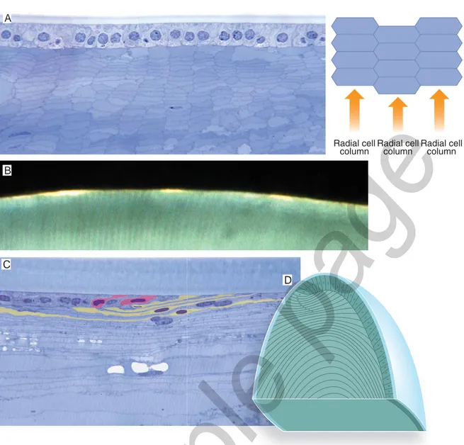
whereas the youngest fibers form arches with a slight outward concavity, those a little older are almost straight and those further older make arches with the concav-ity directed towards the lens axis. Such concavconcav-ity results the more pronounced the longer and older the fibers are (Fig. 9.7).
So far we have described what happens to cells belonging to a single meridional row of the transitional zone. Now, let us see what occurs to the cells that are generat-ed at the same time in all the meridional rows of the transitional zone. These cells lie one beside the other and they will become a single generation of lens fibers to which each radial cell column contributes with a single element. Each generation of elon-gating fibers form a growth shell, a sort of incomplete sheath extending from the anterior to the posterior face of the lens and straddling the equator. Growth shells are not static structures but, with the elongation of their fibers, they gradually ex-pand along the lens faces toward the poles. Their completion is achieved only when they have entirely wrapped the deeper layers. In other words, successive generations of elongating fibers form concentric growth shells that are the more complete the older and the deeper they are (Fig. 9.8).
Radial cell
column Radial cellcolumn Radial cell
column
Fig. 9.7 Lens fi bers. A) Histologic feature of lens fi bers and their general arrangement. Frontal section of the lens. Lens fi bers show a hexagonal profi le and are piled one over the other forming radial cell columns. B) Incident light on the surface of the lens shows the most superfi cial fi bers of the meridionally oriented radial cell columns. Macaca Mulatta.C) Elongating fi bers in the transition zone. Meridional section of the lens. Cells of the anterior epithelium, on the left of the fi gure, moving posteriorly (towards the right of the fi gure) progres-sively get taller and taller and incline the major axis so that they appear as lying down beneath the anterior epithelium. Some cells have been highlighted to better appreciate their progressive growth. Macaca Mulatta. D) Schematic drawing of the lens anterior epithelium and fi bers sectioned along a meridional plane. The arrangement of the elongating fi bers nuclei draws a sort of arch, the nuclear bow.
B
C
D
269
General Features
The visual pathway consists of a series of specialized nervous formations which gather, transfer and integrate visual information. The visual pathway, therefore, be-gins with photoreceptor cells and ends in the brain cortex.
This chapter deals with the intraocular part of the visual pathway, which is formed by the intraretinal circuits that preside over the first level of integration of the visual information. In the retina, photoreceptor stimulation is spatially and temporally integrated and the graded variations of membrane potential of photoreceptor cells are coded into trains of action potentials that leave the eyeball via the optic nerve.
The fibers of the optic nerve are, for the most part, axons of retinal ganglion cells. As we have already noted (see Chapter 8), the retina is characterized by several populations of ganglion neurons, each one consisting of cells similar in shape and synaptic connectivity. Each population of ganglion cells is distributed throughout the retina to form a regular mosaic. Every ganglion element represents a single tile of the mosaic. In doing so, the dendritic tree of each population of ganglion cells covers the entire retinal surface. The diverse connections and kinetics of response to the luminous stimulations define each population of ganglion cells as a pathway through which a particular aspect of the visual information leaves the eyeball and is transmitted centrally. Though our understanding is still largely incomplete, it is evident that a number of parallel pathways, likely equal to the number of ganglion cell populations, take origin from the retina and project to extraocular nervous tar-gets. Not all these pathways have been characterized. The three best known are: the midget ganglion (MG) cell pathway, also referred to as parvocellular pathway, since it projects to the parvocellular layers of the lateral geniculate nucleus (LGN); the parasol ganglion (PG) cell pathway, also known as magnocellular pathway, since it targets the magnocellular layers of the LGN; the small bistratified ganglion (SBG) cell pathway, also referred to as the koniocellular pathway, since it ends in the koniocellular layers of the LGN (see Chapter 11). The shaping of these pathways begins already at the level of photoreceptors, rods and cones, which operate
differ-Intraretinal Circuitry
Eugenio Bertelli
BB S-Cone bipolar = Blue Bipolar DB Diffuse Bipolar
DBa OFF-Center Diffuse bipolar DBb ON-Center Diffuse bipolar
ipG Intrinsically photosensitive Ganglion L-Cone Long wavelength Cone
LGN Lateral Geniculate Nucleus
M-Cone Medium wavelength Cone MG Midget Ganglion
PG Parasol Ganglion RB Rod Bipolar
SBG Small Bistratifi ed Ganglion S-Cone Short wavelength Cone List of Abbreviations
279 Visual information, partially integrated within the eye by intraretinal circuitry
(see Chapter 10), leave the eye via the optic nerve. Though building up within the eyeball, the optic nerve is mainly located outside the globe. The two optic nerves represent the first portion of the extraocular visual pathways. The extraocular visual pathway of each side includes the following nervous structures (Fig. 11.1):
1) optic nerve (ON) 2) optic chiasma (OC) 3) optic tract (OT)
4) lateral geniculate nucleus (LGN) 5) geniculocalcarine tract
6) primary visual area (striate cortex, visual area 1) (V1) 7) extrastriate visual areas (association visual areas) 8) pulvinar
Eugenio Bertelli, Paolo Toti
CO Cytochrome Oxydase
hMT+ (V5) Human Middle Temporal area (Visual area 5)
hV4 human Visual area 4 IPS IntraParietal Sulcus LGN Lateral Geniculate Nucleus LO Lateral Occipital complex NOT Nucleus of the Optic Tract OC Optic Chiasma
ON Optic Nerve OT Optic Tract PA Anterior Pulvinar
PHC Parahyppocampal Cortical area PI Inferior Pulvinar
PIc central nucleus of the Inferior Pulvinar PIcl central lateral nucleus of the Inferior
Pulvinar
PIcm central medial nucleus of the Inferior Pulvinar
PIm medial nucleus of the Inferior Pulvinar
Pip posterior nucleus of the Inferior Pulvinar PL Lateral Pulvinar
PLdm dorsomedial nucleus of the Lateral Pulvinar
PLvl ventrolateral nucleus of the Lateral Pulvinar
PM Medial Pulvinar TO TemporoOccipital area
V1 Primary Visual area (striate cortex, visual area 1)
V2 Visual area 2 V2d dorsal Visual area 2 V2v ventral Visual area 2 V3 Visual area 3 V3A Visual area 3A V3B Visual area 3B V3d dorsal Visual area 3 V3v ventral Visual area 3 V7 Visual area 7 VO Ventral Occipital cortex
List of Abbreviations
323
General Features
Eye movements, promoted by extraocular muscle contraction, are under the di-rect control of three nuclei of gray matter in the brainstem: the oculomotor (III) nucleus, the trochlear (IV) nucleus and the abducens (VI) nucleus (see Chapter
and Palpebromotor System
Eugenio Bertelli
AOS Accessory Optic System CCN Central Caudal Nucleus CEF Cingulate Eye Field
CGRP Calcitonin Gene-Related Peptide: cMRF Central Mesencephalic Reticular
Formation
dLVN Dorsal part of the LVN (Deiter’s nucleus)
EBNs Excitatory Burst Neurons E-W nucleus Edinger-Westpahl nucleus E-Wpc E-W nucleus with centrally
projecting cells E-Wpg E-W nucleus with preganglionic cells FEF Frontal Eye Field
hMT+ Middle Temporal area IBNs Inhibitory Burst Neurons III nucleus Oculomotor nucleus INC Interstitial Nucleus of Cajal IO Inferior Oblique
IR Inferior Rectus IV nucleus Trochlear nucleus IVN Inferior Vestibular Nucleus LGN Lateral Geniculate Nucleus LLBNs Long-Lead Burst Neurons LP Levator Palpebrae LR Lateral Rectus MIF Multiply Innervated Fiber
mMVN Magnocellular component of the MVN MR Medial Rectus
MVN Medial Vestibular Nucleus
NDP Nucleus Dorsalis Paragigantocellularis NOT Nucleus of the Optic Tract
NPC Nucleus of the Posterior Commissure NPH Nucleus Prepositus Hypoglossi NRG Nucleus Reticularis Gigantocellularis
NRI Nucleus Raphe Interpositus NRPC Nucleus Reticularis Pontis Caudalis NRPO Nucleus Reticularis Pontis Oralis NRTP Nucleus Reticularis Tegmenti Pontis OKNM Optokinetic Refl exive Movement OPNs Omnipause Neurons PEF Parietal Eye Field PFEF Prefrontal Eye Field PMT Paramedian Tract pMVN Parvocellular component of the MVN PON Pretectal Olivary Nucleus PPRF Paramedian Pontine Reticular
Formation
RIMLF Rostral Interstitial nucleus of the Medial Longitudinal Fasciculus SC Superior Colliculus SEF Supplementary Eye Field SIF Singly Innervated Fiber SLBNs Short-Lead Burst Neurons SNc Substantia Nigra pars compacta SNl Substantia Nigra pars lateralis SNr Substantia Nigra pars reticulata SOA Supraoculomotor Area SP Substance P SPEM Smooth Pursuit Eye Movement SR Superior Rectus SVN Superior Vestibular Nucleus V1 Primary Visual area VI nucleus Abducens nucleus VII nucleus Facial nucleus
vLVN Ventral part of the Lateral Vestibular Nucleus VOR Vestibulo-Ocular Refl ex VORM Vestibulo-Ocular Refl exive
Movement
List of Abbreviations
415
General Features
The orbit is the small region of the human body that houses the eyeball. It is also densely populated by muscular, nervous and vascular structures. Indeed, it is the high number of vascular and nervous elements enclosed in such a small space that makes the anatomo-topographic study of the orbit an issue of great interest for surgeons. The anterior border of the orbital region is marked by the orbital septum. The structures lying in front of the orbital septum are pertinent to the eyelids (see Chapter 2) and will not be considered in this chapter. The structures located behind the septum, though in some cases extending into the eyelids, belong to the orbital region and will be dealt with in the following paragraphs.
A frontal section passing through the mid-orbit shows two main compartments (Fig. 13.1): one is central, the intramuscular cone (see Chapter 4), and is arranged along the longitudinal axis of the orbit. As it is outlined by the four recti this space is also referred to as the muscular pyramid. The second compartment, extraconal space, surrounds the former one and intervenes between the intramuscular cone and the orbit walls. Both compartments are filled with an adipose atmosphere that forms the adipose body of the orbit.
The intramuscular cone has the shape of a truncated cone with the lesser base corresponding to the annulus of Zinn facing backward and medially. The for-ward-directed greater base is concave as it embraces the posterior hemisphere of the eyeball. A transverse plane passing through the medial and lateral recti helps to make a further division of the extraconal space into two areas located respectively above and under the muscular pyramid (Fig. 13.1).
Intramuscular Cone
As we have already stated, the intramuscular cone is the space outlined by the four recti. At the level of the eyeball, the sheaths of the four recti are bridged togeth-er by the inttogeth-ermuscular membrane. Furthtogeth-er backward, behind the posttogeth-erior pole of
of the Orbit
Eugenio Bertelli ON Optic Nerve OA Ophthalmic Artery List of AbbreviationsSample page
425
Early Development
By 16 days of gestation, on the dorsum of the developing embryo, the ectoderm has produced a central thickening, the neural plate or neuroectoderm. Two days later the neural plate shows a sagittal depression, the neural groove, encompassed between two parallel neural folds (Fig. 14.1). By the end of the third week, the neural folds raise and converge together. In doing so, their tops come into contact and fuse together starting roughly from their midpoint. The fusion of the neural folds, extend-ing caudally and rostrally, transforms the neural groove into the neural tube (Fig. 14.1). The extremities of the neural tube, the neuropores, eventually close. By 20 days of gestation (2 mm embryo), in proximity of the rostral end of the neural groove, the inner side of the neural folds develops a couple of small lateral grooves ( optic sulci) which represent the very first visible event marking the onset of eye development (Fig. 14.2). The sulci, deepening laterally, are then called optic pits and they correspond to the optic evaginations that can be seen on the outer surface of the neural tube. When the rostral neuropore closes (24 days of gestation, 3 mm embryo), the optic evagina-tions expand and form the optic vesicles, a pair of hollow outgrowths bulging from the lateral walls of the neural tube almost in contact with the surface ectoderm (Fig. 14.2). The cavity of the optic vesicles remains in continuity with the cavity of the fore-brain vesicle. By 27 days of gestation (4 mm embryo) the surface ectoderm opposite to the optic vesicles grows thicker forming the lens placodes. The lens placodes and the optic vesicles maintain close relationships, as they are separated only by a very narrow space (Fig. 14.2). It is thought that starting from this moment the development of both the lens placodes and the optic vesicles is coordinated by the secretion of growth factors, particularly fibroblast growth factors (FGF)s. The lens placodes invaginate and form the lens vesicles (Fig. 14.3). The formation of the lens vesicles is marked on the surface of the embryo by two small depressions, one for each side, referred to as the lens pits. At first, each lens vesicle remains attached to the surface ectoderm through a narrow peduncle, the lens stalk, then it detaches (33rd day of gestation). Simultane-ously to the formation of the lens vesicles, the optic vesicles grow laterally remaining connected with the forebrain through a constricted portion, the optic stalk. The optic
Development
Eugenio Bertelli
FGF Fibroblast Growth Factor PAX Paired Box
SIX Sine oculis homeobox
MAF Muscoloaponeurotic Fibrosarcoma Oncogene
PITX Paired-like omeodomain
FOXC1 Forkhead box C RPC Retinal Progenitor Cell SOX SRY homeobox-like M-cones Medium wavelength cones L-cones Long wavelength cones S-cones Short wavelength cones List of Abbreviations