First-trimester detection of abnormally invasive placenta in
high-risk women: systematic review and meta-analysis
F. D’ANTONIO
1, I. E. TIMOR-TRITSCH
2, J. PALACIOS-JARAQUEMADA
3, A. MONTEAGUDO
2,
D. BUCA
4, F. FORLANI
5, G. MINNECI
5, F. FOTI
5, L. MANZOLI
6, M. LIBERATI
4, G. ACHARYA
7and G. CAL`I
51Women’s Health and Perinatology Research Group, Department of Clinical Medicine, Faculty of Health Sciences, UiT-The Arctic
University of Norway, Tromsø, Norway;2Department of Obstetrics and Gynaecology, Division of Maternal-Fetal Medicine, New York
University School of Medicine, New York, NY, USA;3Centre for Medical Education and Clinical Research (CEMIC), University Hospital,
Buenos Aires, Argentina;4Department of Obstetrics and Gynaecology, University of Chieti, Chieti, Italy;5Department of Obstetrics and
Gynaecology, Arnas Civico Hospital, Palermo, Italy;6Department of Medical Sciences, University of Ferrara, Ferrara, Italy;7Department of
Clinical Science, Intervention and Technology, Karolinska Institute, Stockholm, Sweden
K E Y W O R D S: abnormally invasive placenta; first-trimester diagnosis; ultrasound
ABSTRACT
Objectives The primary aim of this systematic review
was to ascertain whether ultrasound signs suggestive of abnormally invasive placenta (AIP) are present in the first trimester of pregnancy. Secondary aims were to ascertain the strength of association and the predictive accuracy of such signs in detecting AIP in the first trimester.
Methods An electronic search of MEDLINE, EMBASE,
CINAHL and Cochrane databases (2000–2016) was performed. Only studies reporting on first-trimester diag-nosis of AIP that was subsequently confirmed in the third trimester either during operative delivery or by pathological examination were included. Meta-analysis of proportions, random-effects meta-analysis and hierar-chical summary receiver–operating characteristics curve analysis were used to analyze the data.
Results Seven studies, involving 551 pregnancies at high
risk of AIP, were included. At least one ultrasound sign suggestive of AIP was detected in 91.4% (95% CI, 85.8–95.7%) of cases with confirmed AIP. The most common ultrasound feature in the first trimester of pregnancy was low implantation of the gestational sac close to a previous uterine scar, which was observed in 82.4% (95% CI, 46.6–99.8%) of cases. Anechoic spaces within the placental mass (lacunae) were observed in 46.0% (95% CI, 10.9–83.7%) and a reduced myometrial thickness in 66.8% (95% CI, 45.2–85.2%) of cases affected by AIP. Pregnancies with a low implantation of the gestational sac had a significantly higher risk of AIP (odds ratio, 19.6 (95% CI, 6.7–57.3)), with a sensitivity
Correspondence to: Dr F. D’Antonio, Department of Clinical Medicine, Faculty of Health Sciences, UiT-The Arctic University of Norway,
Hansine Hansens veg 18, 9019 Tromsø, Norway (e-mail: [email protected])
Accepted: 7 August 2017
and specificity of 44.4% (95% CI, 21.5–69.2%) and 93.4% (95% CI, 90.5–95.7%), respectively.
Conclusions Ultrasound signs of AIP can be present
during the first trimester of pregnancy, even before 11 weeks’ gestation. Low anterior implantation of the placenta/gestational sac close to or within the scar was the most commonly seen early ultrasound sign suggestive of AIP, although its individual predictive accuracy was not high. Copyright© 2017 ISUOG. Published by John
Wiley & Sons Ltd.
INTRODUCTION
Abnormally invasive placenta (AIP) encompasses a spectrum of conditions characterized by an abnormal adherence of the placenta to the implantation site1–4.
AIP is associated with the occurrence of several major maternal complications such as severe hemorrhage, need for blood transfusion, peripartum hysterectomy, intra-and postoperative complications intra-and an increased risk of adverse perinatal outcome1. Accurate prenatal diagnosis of AIP is desirable because it has been shown to reduce the burden of maternal and fetal morbidity associated with the condition, especially by allowing implementation of preplanned management strategies5–13.
The underlying mechanisms leading to AIP are not yet completely understood, but it is known that defective development of the decidua basalis constitutes the anatomical prerequisite for the occurrence of AIP14–16.
Recently, several studies have shown that Cesarean scar pregnancy (CSP), a condition in which the gestational
sac implants on, or in close proximity to, a previous Cesarean section (CS) scar, represents the precursor of AIP, although it is not yet entirely certain whether all subtypes of AIP share this pathophysiology17.
Although prenatal diagnosis of AIP is commonly achieved during the second or third trimester of pregnancy, there are reports suggesting that the signs of AIP are already present in early pregnancy18–20.
Furthermore, the recently proposed association between CSP and AIP suggests that invasion of the uterine scar by trophoblastic tissue may start early in pregnancy, thus being theoretically detectable during the first-trimester scan17.
The primary aim of this systematic review was to ascertain whether ultrasound signs suggestive of AIP are present at the first-trimester scan (i.e. before 14 weeks’ gestation); the secondary aims were to ascertain the strength of association and the predictive accuracy of such signs in detecting AIP in the first trimester.
METHODS
This review was performed according to an a-priori designed protocol recommended for systematic reviews and meta-analyses21–23. MEDLINE, EMBASE, CINAHL
and The Cochrane Library, including The Cochrane Database of Systematic Reviews (CDSR), Database of Abstracts of Reviews of Effects (DARE) and The Cochrane Central Register of Controlled Trials (CENTRAL), were searched electronically on 23rd February 2017,
utilizing combinations of the relevant medical subject heading (MeSH) terms, keywords, and word variants for ‘abnormally invasive placenta’, ‘morbidly adherent placenta’ and ‘ultrasound’ (Table S1). The search and selection criteria were restricted to the English language. Reference lists of relevant articles and reviews were hand-searched for additional reports. PRISMA and STARD guidelines were followed24,25 and the study was
registered with the PROSPERO database (Registration number: CRD42017060513).
Studies were assessed according to the following cri-teria: population, prenatal diagnosis of AIP during the first trimester of pregnancy and study design. AIP was defined based on clinical observation of abnormal placen-tal adherence with evidence of gross placenplacen-tal invasion at the time of surgery and/or histopathological diagnosis of trophoblast invasion through the myometrium with the absence of normal decidua at the basal plate.
Only studies reporting the first-trimester diagnosis of AIP confirmed in the third trimester of pregnancy either at surgery or by pathological examination were included in the analysis. Studies reporting exclusively the prenatal diagnosis of AIP after first- or second-trimester termination of pregnancy and those including only cases of CSP were excluded on the basis that these may represent the severe end of the spectrum of invasive placental disorders.
Prospective and retrospective cohorts, case–control studies and case series were analyzed. Opinions and
studies carried out only in the second and/or third trimester were excluded. Case reports were also excluded to avoid publication bias. Studies published before 2000 were excluded, as we considered that advances in prenatal imaging techniques and improvements in the diagnosis and definition of AIP make these less relevant.
First-trimester ultrasound signs of AIP explored in the present systematic review were: location of the gestational sac within the lower part of the uterus in the isthmic region in proximity to the scar of a previous CS, presence of intraplacental lacunae, reduced myometrial thickness between the placenta/gestational sac and the bladder and abnormal uterus–bladder interface16,17,20.
The gestational age at which the ultrasound scan was performed was also recorded.
Two reviewers (F.D., D.B.) independently extracted data. Inconsistencies were discussed between the review-ers and consensus reached. For those articles in which targeted information was not reported but the method-ology was such that the information might have been recorded initially, the authors were contacted requesting the data. Histopathological findings and/or surgical notes were used as the gold standard for confirmation of the condition.
The quality of the studies was assessed using the revised tool for the quality assessment of diagnostic accuracy studies (QUADAS-2)26. Each item was scored a ‘yes’, ‘no’ or, if there was insufficient information to make an accurate judgment, ‘unclear’.
Funnel plots displaying the outcome rates from individual studies vs their precision (1/standard error) were carried out with an exploratory aim. Tests for funnel-plot asymmetry were not used when the total number of publications included for each outcome was less than 10; in such cases, the power of the test is too low to distinguish chance from real asymmetry27.
First, we explored the prevalence of the different ultrasound signs suggestive of AIP at the first-trimester scan; for the purpose of this analysis, meta-analysis of proportions was used to assess the data. Then, we explored the strength of association between the different ultrasound signs reported in the published literature, and the occurrence of any type of AIP; we planned to use random-effects meta-analysis to compute a summary odds ratio (OR) of the likelihood of each ultrasound sign in fetuses with or without AIP26,28,29. A sensitivity analysis
was performed in order to evaluate the occurrence of each explored ultrasound sign in cases scanned before 11 weeks’ gestation.
Finally, we evaluated the diagnostic accuracy of first-trimester ultrasound compared with intraopera-tive/histopathological diagnosis. We computed summary estimates of sensitivity, specificity, positive and nega-tive likelihood ratios (LR+ and LR–) and diagnos-tic OR (DOR) for the overall predictive accuracy of first-trimester ultrasound in detecting AIP using the hierarchical summary receiver–operating characteristics (HSROC) model30–34. Rutter and Gatsonis HSROC
of sensitivity and specificity to define a summary ROC curve, and its hierarchical modeling strategy can be used for comparisons of test accuracy when there is variability in threshold between studies30–33.
StatsDirect statistical software 2013 (StatsDirect Ltd, Altrincham, Cheshire, UK) was used to analyze the data.
RESULTS
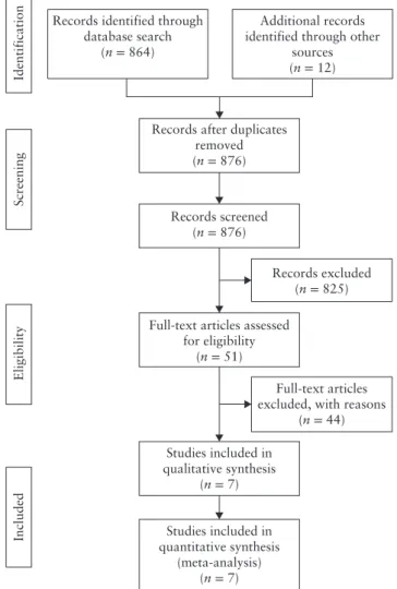
A total of 876 articles were identified. After screening the abstracts, 51 full-text articles were assessed with respect to their eligibility for inclusion (Table S2) and seven studies were included in the systematic review (Figure 1, Table 1)35–41. These seven studies included 551
pregnancies at high risk for AIP, of which 117 (21.2% (95% CI, 17.9–24.9%)) had AIP. The occurrence of placenta accreta, placenta increta and placenta percreta was 45.3% (95% CI, 36.7–54.3%), 15.4% (95% CI, 10.0–23.0%) and 24.8% (95% CI, 17.9–33.3%), respectively.
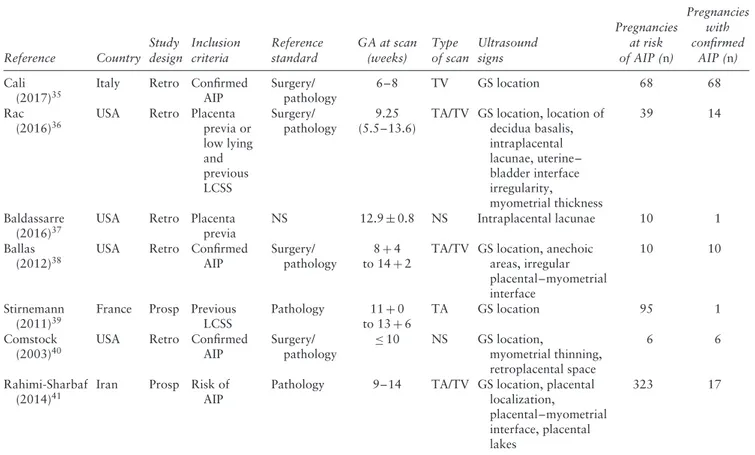
General characteristics of the studies included in this systematic review are reported in Table 1. Most of
Records excluded (n = 825) Records identified through
database search (n = 864)
Additional records identified through other
sources (n = 12)
Full-text articles excluded, with reasons
(n = 44)
Identification
Included
Screening
Eligibility
Full-text articles assessed for eligibility (n = 51) Studies included in qualitative synthesis (n = 7) Studies included in quantitative synthesis (meta-analysis) (n = 7) Records after duplicates
removed (n = 876)
Records screened (n = 876)
Figure 1 Flowchart summarizing selection of studies on
first-trimester diagnosis of abnormally invasive placenta, subsequently confirmed in third trimester, that were included in systematic review and meta-analysis.
the included studies were retrospective series including only cases of AIP confirmed either at surgery or by histopathology, thus not allowing an objective assessment of the specificity of ultrasound in ruling out AIP at the time of the first-trimester scan. Similarly, evaluation of the diagnostic accuracy in terms of specificity could not be assessed in view of the lack of false-positive and true-negative cases for the majority of the ultrasound signs assessed.
Quality assessment based on QUADAS-2 guidelines is shown in Figure 2. Some of the included studies had a high or unclear risk of bias regarding patient selection and the index test, especially because of heterogeneity in the gestational age at scan and the definition of individual ultrasound signs, while there was an overall low risk of bias regarding the reference standard. However, it should be taken into account that such tests have a low statistical power when the overall number of publications is less than 10, as in the present review29.
At least one ultrasound sign suggestive of AIP was detected during the first-trimester scan in 91.4% (95% CI, 85.8–95.7%) of cases with confirmed AIP at delivery. The most common ultrasound feature in the first trimester of pregnancy was a low implantation of the gestational sac close to a previous uterine scar, which was observed in 82.4% (95% CI, 46.6–99.8%) of cases, while anechoic spaces within the placental mass (lacunae) were observed in 46.0% (95% CI, 10.9–83.7%) (Table 2, Figures S1 and S2).
Myometrial thickness was assessed in two studies; Rac
et al.36reported several cut-offs of myometrial thickness, although the ones which showed the optimal combination of sensitivity and specificity were 5 and 6 mm, while in the study by Comstock et al.40 the authors compared the anterior with the posterior myometrium, and defined reduced myometrial thickness as anterior myometrium thinner than posterior myometrium. Overall, a reduced myometrium thickness was present in 66.8% (95% CI, 45.2–85.2%) of cases of AIP scanned in the first trimester. Finally, two studies reported the assessment of an abnormal uterus–bladder interface in the first trimester of pregnancy and reported an overall prevalence of this sign in 51.8% (95% CI, 0.2–100.0%) of cases affected by AIP (Figure S2)36,38.
When considering only cases scanned before 11 weeks’ gestation, the presence of at least one ultrasound sign suggestive of AIP was present in 95.1% (95% CI, 75.3–99.5%) of cases, with a low implantation of the gestational sac within the CS scar being visible in all cases affected by AIP. However, it was not possible to perform a comprehensive pooled assessment of the prevalence of the other ultrasound signs explored in this review because of the very small number of included cases.
A pooled risk assessment between different ultrasound signs and AIP could be performed for only low implantation of the gestational sac and the presence of intraplacental lacunae. Two studies explored the strength of association between low implantation of the gestational sac and the occurrence of AIP in a population at risk for
Table 1 General characteristics of seven studies reporting on first-trimester diagnosis of abnormally invasive placenta (AIP) that was
subsequently confirmed in third trimester, included in systematic review and meta-analysis
Reference Country Study design Inclusion criteria Reference standard GA at scan (weeks) Type of scan Ultrasound signs Pregnancies at risk of AIP (n) Pregnancies with confirmed AIP (n) Cali (2017)35
Italy Retro Confirmed
AIP Surgery/ pathology 6–8 TV GS location 68 68 Rac (2016)36
USA Retro Placenta
previa or low lying and previous LCSS Surgery/ pathology 9.25 (5.5–13.6)
TA/TV GS location, location of decidua basalis, intraplacental lacunae, uterine– bladder interface irregularity, myometrial thickness 39 14 Baldassarre (2016)37
USA Retro Placenta
previa
NS 12.9± 0.8 NS Intraplacental lacunae 10 1
Ballas (2012)38
USA Retro Confirmed
AIP
Surgery/ pathology
8+ 4 to 14+ 2
TA/TV GS location, anechoic areas, irregular placental–myometrial interface 10 10 Stirnemann (2011)39
France Prosp Previous LCSS Pathology 11+ 0 to 13+ 6 TA GS location 95 1 Comstock (2003)40
USA Retro Confirmed
AIP Surgery/ pathology ≤ 10 NS GS location, myometrial thinning, retroplacental space 6 6 Rahimi-Sharbaf (2014)41
Iran Prosp Risk of
AIP
Pathology 9–14 TA/TV GS location, placental
localization,
placental–myometrial interface, placental lakes
323 17
Only first author of each study is given. GA, gestational age; GS, gestational sac; LCSS, low Cesarean section scar; NS, not stated; Prosp, prospective; Retro, retrospective; TA, transabdominal; TV, transvaginal.
0 20 40 60 80 100
Patient selection Index test Reference standard Flow and timing
(a) (b)
Proportion of studies with low, high or unclear risk of bias (%)
QUADAS-2 domain
0 20 40 60 80 100
Proportion of studies with low, high or unclear concerns regarding applicability (%) Figure 2 Quality assessment for risk of bias (a) and concerns regarding applicability (b) of seven studies included in systematic review and
meta-analysis according to QUADAS-2. , low risk; , high risk; , unclear risk.
these anomalies, such as women with a previous CS39,41;
the study by Stirnemann et al.39included women scanned
between 11 and 14 weeks, while that by Rahimi-Sharbaf
et al.41 included those assessed between 9 and 14 weeks. Overall, cases with a low implantation of the gestational sac had a significantly higher risk of AIP, with an OR of 19.6 (95% CI, 6.7–57.3; I2, 0%). Once translated into
predictive accuracy, a low implantation of the gestational
sac had a sensitivity of 44.4% (95% CI, 21.5–69.2%), specificity of 93.4% (95% CI, 90.5–95.7%), LR+ of 7.5 (95% CI, 3.8–14.9), LR– of 0.6 (95% CI, 0.4–0.9) and DOR of 11.0 (95% CI, 4.0–30.3) for the detection of AIP. The presence of intraplacental lacunae in the first trimester of pregnancy did not carry an increased risk of AIP (OR, 1.03 (95% CI, 0.2–4.8)); likewise, the diagnostic accuracy was poor, with a sensitivity of 33.3%
Table 2 Raw and pooled proportions of prevalence of first-trimester ultrasound signs in women with abnormally invasive placenta Ultrasound sign Studies (n) Pregnancies (n/N) Raw proportion (95% CI) (%) I2(%) Pooled proportion (95% CI) (%)
At least one sign 7 100/117 85.47 (77.8–91.3) 88.4 91.42 (85.8–95.7)
Low implantation of gestational sac 5 90/102 88.24 (80.4–93.8) 89.7 82.42 (46.6–99.8)
Placental lacunae 3 12/25 48.00 (27.8–68.7) 71.0 46.03 (10.9–83.7)
Reduced myometrial thickness 2 13/19 68.42 (43.4–87.4) 0 66.79 (45.2–85.2)
Abnormal uterus–bladder interface 2 11/24 45.83 (25.6–67.2) 93.2 51.84 (0.2–100.0)
Bladder
Bladder
Bladder
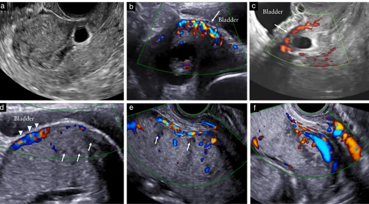
Figure 3 Ultrasound criteria for diagnosis of Cesarean scar (CS) pregnancy and abnormally invasive placenta (AIP) during first trimester of
pregnancy. (a) Transvaginal scan at 5+ 5 weeks’ gestation showing low implantation of gestational sac embedded eccentrically in lower uterine segment and implanted in location of prior CS. (b,c) Assessment with color and power Doppler reveals rich vascular pattern in area between CS and placenta; myometrium beneath placental mass is irregular and scarcely visible at some points (arrows); no intraplacental lacunae can be detected. (d,e) Transabdominal scan at 14 weeks; abnormal location of gestational sac is more difficult to appreciate with advancing gestation; myometrium beneath placenta is not entirely visible in some areas and there is increased subplacental vascularity (arrowheads). Intraplacental lacunae can be seen at this stage as hypoechoic spaces within the parenchyma (arrows). (f) Transabdominal scan at 17 weeks showing classic second-trimester signs of AIP, such as intraplacental lacunae, abnormal uterus–bladder interface with increased vascularity and absence of retroplacental clear space.
(95% CI, 11.8–61.6%), specificity of 67.5% (95% CI, 50.9–81.4%), LR+ of 1.4 (95% CI, 0.5–3.6), LR– of 0.9 (95% CI, 0.6–1.3) and DOR of 1.0 (95% CI, 0.7–1.6).
DISCUSSION Main findings
The findings of this systematic review show that ultrasound signs suggestive of AIP can be detected from the first trimester of pregnancy. At least one ultrasound sign suggestive of AIP was detected in 91.4% of all cases and in 95.1% of those scanned before 11 weeks. Low anterior implantation of the placenta/gestational sac close to or within the scar was the most common
early ultrasound sign associated with AIP, although its individual predictive accuracy was not high.
Strengths and limitations of the study
The very small number of cases in each included study represents the major limitation of this systematic review. In such cases, estimation of the variances of the random effects is subject to a high level of uncertainty, and caution is required when interpreting the results.
Heterogeneity in the inclusion criteria among the different studies and their retrospective design is another major limitation of this systematic review. The majority of the studies included exclusively cases with surgically or histologically confirmed AIP, thus making it impossible to extrapolate any information regarding the specificity of
9w2d 14w2d
17w0d 16w2d
15w2d
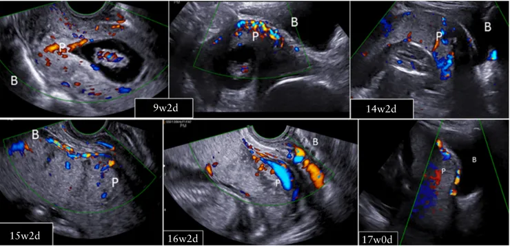
Figure 4 Development of abnormally invasive placenta from Cesarean scar pregnancy. B, bladder; P, placenta. Timeline given in weeks (w)
and days (d).
first-trimester ultrasound in ruling out AIP. Furthermore, the included studies differ with respect to the gestational age at assessment, type of scan and population analyzed. This review included mainly women at risk for AIP; however, such risk assessment differed among the included studies and was ascertained at the time of the second- or third-trimester scan (i.e. women with placenta previa and/or previous CS), thus it is not clinically applicable to women in the first trimester of pregnancy. AIP is still a relatively rare anomaly and the large majority of women with a CS scar would not have AIP, thus questioning the need for such first-trimester assessment. Finally, because of the small number of cases, it was not possible to perform sub-analysis according to the severity of placental invasion.
Implications for clinical practice and research
Prenatal diagnosis of AIP is usually accomplished during the second and/or third trimester of pregnancy, although there is no consensus yet on the optimal gestational age at scan to detect AIP and the number and type of imaging criteria which should be adopted in order to improve the overall diagnostic accuracy of ultrasound18,19,40.
This systematic review showed that AIP can be detected as early as in the first trimester of pregnancy and that early ultrasound signs of AIP are those of a CSP, such as low implantation of the gestational sac, which is classically defined as being less than 5 cm from the external cervical os, although a precise definition of low implantation was not provided in many of the included studies42.
The recently proposed association between CSP and AIP poses the dilemma of how women with a prenatal diagnosis of CSP should be counseled, and whether termination of pregnancy should be the only therapeutic
option offered to these women17. Several studies aimed
at stratifying the risk of AIP in women with a previous CSP have been published recently35,43–45. Kaelin Agten
et al.43showed that CSP implanted ‘on the scar’, defined as a placenta implanted partially or fully on top of a well-healed scar, had a substantially better outcome than did cases in which the CSP implanted into the niche of a deficient or dehiscent scar. Myometrial thickness below 2 mm on first-trimester ultrasound was associated with morbidly adherent placenta at delivery43. Cali et al.35,44
showed that the relationship between the gestational sac of the CSP, previous Cesarean scar and anterior uterine wall thickness can be used to predict not only the evolution of the CSP towards the most severe types of AIP, but also the clinical outcome of these women. Despite this, identification of CSPs that will have successful pregnancy outcome or are amenable to treatment without serious complications remains a challenge (Figures 3 and 4).
The first-trimester diagnosis of AIP has been reported rarely and there is still no consensus on which imaging sign should be sought in order to diagnose AIP early in pregnancy. In the collective authors’ experience, low anterior implantation of the placenta/gestational sac close to or within the scar, a reduced myometrial thickness and abnormal vascularity at the uterus–bladder interface are the most common early ultrasound signs suggestive of AIP, although there is no consensus yet on how to define such signs.
Several cut-offs of myometrial thickness have been reported to be associated with AIP in the first trimester. In the study of Rac et al.36, myometrial thickness≤ 6 mm
measured in the sagittal plane showed the optimal combination of sensitivity and specificity, although the authors did not stratify their analysis according to the depth of placental invasion. However, whether routine
assessment of myometrial thickness in women with a prior CS and low implantation of the gestational sac improves the diagnostic accuracy of first-trimester ultrasound requires confirmation in large prospective studies.
Placental lacunae are among the most commonly detected ultrasound signs in pregnancies with AIP diagnosed in the second and third trimesters18. However,
in the present systematic review, which includes only first-trimester pregnancies, the prevalence of lacunae was about 46%, with substantial heterogeneity between the included studies. Furthermore, the risk of AIP in women presenting with lacunae was not higher than in those not showing this sign on ultrasound. Placental lacunae may be less common, or difficult to identify, in the early first trimester.
Prenatal diagnosis of AIP has been shown to be improved when a multiparametric prediction model, including ultrasound, maternal and pregnancy charac-teristics, is applied to women at risk46. In this scenario,
integrating findings on the first- with the second-and third-trimester scans, together with maternal second-and pregnancy characteristics, might theoretically improve the diagnostic accuracy of ultrasound in detecting the presence and severity of AIP.
In conclusion, ultrasound signs of AIP can be present during the first trimester of pregnancy. Further studies directed at prospectively evaluating women at risk for AIP, such as those with a prior CS, from the first trimester of pregnancy are needed in order to ascertain whether findings on first-trimester ultrasound may help in stratifying such risk and whether they should be combined with those of the second- and third-trimester scans in order to improve the overall diagnostic accuracy of ultrasound in identifying AIP.
ACKNOWLEDGMENT
We would like to thank Dr N. Zosmer for additional information provided.
REFERENCES
1. Belfort MA. Placenta accreta. Am J Obstet Gynecol 2010; 203: 430–439. 2. Oyelese Y, Smulian JC. Placenta previa, placenta accreta, and vasa previa. Obstet
Gynecol 2006; 107: 927–941.
3. Miller DA, Chollet JA, Goodwin TM. Clinical risk factors for placenta previa–placenta accreta. Am J Obstet Gynecol 1997; 177: 210–214.
4. Wu S, Kocherginsky M, Hibbard JU. Abnormal placentation: twenty-year analysis.
Am J Obstet Gynecol 2005; 192: 1458–1461.
5. Tikkanen M, Paavonen J, Loukovaara M, Stefanovic V. Antenatal diagnosis of placenta accreta leads to reduced blood loss. Acta Obstet Gynecol Scand 2011; 90: 1140–1146.
6. Silver RM, Fox KA, Barton JR, Abuhamad AZ, Simhan H, Huls CK, Belfort MA, Wright JD. Center of excellence for placenta accreta. Am J Obstet Gynecol 2015;
212: 561–568.
7. Shamshirsaz AA, Fox KA, Erfani H, Clark SL, Salmanian B, Baker BW, Coburn M, Shamshirsaz AA, Bateni ZH, Espinoza J, Nassr AA, Popek EJ, Hui SK, Teruya J, Tung CS, Jones JA, Rac M, Dildy GA, Belfort MA. Multidisciplinary team learning in the management of the morbidly adherent placenta: outcome improvements over time. Am J Obstet Gynecol 2017; 216: 612.e1–5.
8. Shamshirsaz AA, Fox KA, Salmanian B, Diaz-Arrastia CR, Lee W, Baker BW, Ballas J, Chen Q, Van Veen TR, Javadian P, Sangi-Haghpeykar H, Zacharias N, Welty S, Cassady CI, Moaddab A, Popek EJ, Hui SK, Teruya J, Bandi V, Coburn M, Cunningham T, Martin SR, Belfort MA. Maternal morbidity in patients with morbidly adherent placenta treated with and without a standardized multidisciplinary approach. Am J Obstet Gynecol 2015; 212: 218.e1–9.
9. Fox KA, Shamshirsaz AA, Carusi D, Secord AA, Lee P, Turan OM, Huls C, Abuhamad A, Simhan H, Barton J, Wright J, Silver R, Belfort MA. Conservative management of morbidly adherent placenta: expert review. Am J Obstet Gynecol 2015; 213: 755–760.
10. Flood KM, Said S, Geary M, Robson M, Fitzpatrick C, Malone FD. Changing trends in peripartum hysterectomy over the last 4 decades. Am J Obstet Gynecol 2009; 200: 632.e1–6.
11. Angstmann T, Gard G, Harrington T, Ward E, Thomson A, Giles W. Surgical management of placenta accreta: a cohort series and suggested approach. Am J
Obstet Gynecol 2010; 202: 38.e1–9.
12. Ballas J, Hull AD, Saenz C, Warshak CR, Roberts AC, Resnik RR, Moore TR, Ramos GA. Preoperative intravascular balloon catheters and surgical outcomes in pregnancies complicated by placenta accreta: a management paradox. Am J Obstet
Gynecol 2012; 207: 216.e1–5.
13. Bowman ZS, Manuck TA, Eller AG, Simons M, Silver RM. Risk factors for unscheduled delivery in patients with placenta accreta. Am J Obstet Gynecol 2014;
210: 241.e1–6.
14. Jauniaux E, Jurkovic D. Placenta accreta: pathogenesis of a 20th century iatrogenic uterine disease. Placenta 2012; 33: 244–251.
15. Timor-Tritsch IE, Monteagudo A. Unforeseen consequences of the increasing rate of cesarean deliveries: early placenta accreta and cesarean scar pregnancy. A review.
Am J Obstet Gynecol 2012; 207: 14–29.
16. Timor-Tritsch IE, Monteagudo A, Cali G, Vintzileos A, Viscarello R, Al-Khan A, Zamudio S, Mayberry P, Cordoba MM, Dar P. Cesarean scar pregnancy is a precursor of morbidly adherent placenta. Ultrasound Obstet Gynecol 2014; 44: 346–353.
17. D’Antonio F, Iacovella C, Bhide A. Prenatal identification of invasive placentation using ultrasound: systematic review and meta-analysis. Ultrasound Obstet Gynecol 2013; 42: 509–517.
18. D’Antonio F, Iacovella C, Palacios-Jaraquemada J, Bruno CH, Manzoli L, Bhide A. Prenatal identification of invasive placentation using magnetic resonance imaging: systematic review and meta-analysis. Ultrasound Obstet Gynecol 2014;
44: 8–16.
19. Palacios-Jaraquemada JM, Bruno CH, Mart´ın E. MRI in the diagnosis and surgical management of abnormal placentation. Acta Obstet Gynecol Scand 2013; 92: 392–397.
20. Timor-Tritsch IE, Monteagudo A, Cali G, Palacios-Jaraquemada JM, Maymon R, Arslan AA, Patil N, Popiolek D, Mittal KR. Cesarean scar pregnancy and early placenta accreta share common histology. Ultrasound Obstet Gynecol 2014; 43: 383–395.
21. Henderson LK, Craig JC, Willis NS, Tovey D, Webster AC. How to write a Cochrane systematic review. Nephrology (Carlton) 2010; 15: 617–624.
22. NHS Centre for Reviews and Dissemination. Systematic reviews: CRD’s guidance
for undertaking reviews in health care. University of York: York, UK, 2009.
23. Leeflang MM, Deeks JJ, Gatsonis C, Bossuyt PM; Cochrane Diagnostic Test Accuracy Working Group. Systematic reviews of diagnostic test accuracy. Ann Intern Med 2008; 149: 889–897.
24. Prisma statement. http://www.prisma-statement.org/ [Accessed 10 March 2017]. 25. Bossuyt PM, Reitsma JB, Bruns DE, Gatsonis CA, Glasziou PP, Irwig LM, Lijmer JG,
Moher D, Rennie D, de Vet HC; Standards for Reporting of Diagnostic Accuracy. Towards complete and accurate reporting of studies of diagnostic accuracy: the STARD Initiative. Clin Chem 2003; 49: 1–6.
26. Whiting PF, Rutjes AW, Westwood ME, Mallett S, Deeks JJ, Reitsma JB, Leeflang MM, Sterne JA, Bossuyt PM; QUADAS-2 Group. QUADAS-2: a revised tool for the quality assessment of diagnostic accuracy studies. Ann Intern Med 2011; 155: 529–536.
27. Egger M, Davey Smith G, Schneider M, Minder C. Bias in meta-analysis detected by a simple, graphical test. BMJ 1997; 315: 629–634.
28. Hunter JP, Saratzis A, Sutton AJ, Boucher RH, Sayers RD, Bown MJ. In meta-analyses of proportion studies, funnel plots were found to be an inaccurate method of assessing publication bias. J Clin Epidemiol 2014; 67: 897–903.
29. Higgins JPT, Green S (eds). Cochrane Handbook for Systematic Reviews of Interventions Version 5.0.2 [updated September 2009]. The Cochrane Collaboration, 2009. at www.cochrane-handbook.org. [Accessed 3 December 2016].
30. Rutter CM, Gatsonis CA. A hierarchical regression approach to meta-analysis of diagnostic test accuracy evaluations. Stat Med 2001; 20: 2865–2884.
31. Harbord RM, Whiting P. Metandi: meta-analysis of diagnostic accuracy using hierarchical logistic regression. Stata J 2009; 9: 211–229.
32. Cochrane Handbook for Systematic Reviews of Diagnostic Test Accuracy, Chapter 10; http://srdta.cochrane.org/handbook-dta-reviews.
33. Glas AS, Lijmer JG, Prins MH, Bonsel GJ, Bossuyt PM. The diagnostic odds ratio: a single indicator of test performance. J Clin Epidemiol 2003; 56: 1129–1135. 34. Zamora J, Abraira V, Muriel A, Khan K, Coomarasamy A. Meta-DiSc: a software
for meta-analysis of test accuracy data. BMC Med Res Methodol 2006; 6: 31. 35. Cali G, Forlani F, Timor-Tritsch IE, Palacios-Jaraquemada J, Minneci G, D’Antonio
F. Natural history of Cesarean scar pregnancy on prenatal ultrasound: the crossover sign. Ultrasound Obstet Gynecol 2017; 50: 100–104.
36. Rac MW, Moschos E, Wells CE, McIntire DD, Dashe JS, Twickler DM. Sonographic findings of morbidly adherent placenta in the first trimester. J Ultrasound Med 2016;
35: 263–269.
37. Baldassarre RL, Gabe M, Pretorius DH, Ramos GA, Romine LE, Hull AD, Ballas J, Pettit KE. Placental sonolucencies in the first trimester: incidence and clinical significance. Ultrasound Q 2016; 32: 43–46.
38. Ballas J, Pretorius D, Hull AD, Resnik R, Ramos GA. Identifying sonographic markers for placenta accreta in the first trimester. J Ultrasound Med 2012; 31: 1835–1841.
39. Stirnemann JJ, Mousty E, Chalouhi G, Salomon LJ, Bernard JP, Ville Y. Screening for placenta accreta at 11–14 weeks of gestation. Am J Obstet Gynecol 2011; 205: 547.e1–6.
40. Comstock CH, Lee W, Vettraino IM, Bronsteen RA. The early sonographic appearance of placenta accreta. J Ultrasound Med 2003; 22: 19–23.
41. Rahimi-Sharbaf F, Jamal A, Mesdaghinia E, Abedzadeh-Kalahroudi M, Niroomanesh S, Atoof F. Ultrasound detection of placenta accreta in the first trimester of pregnancy.
Iran J Reprod Med 2014; 12: 421–426.
42. Moschos E, Wells CE, Twickler DM. Biometric sonographic findings of abnormally adherent trophoblastic implantations on cesarean delivery scars. J Ultrasound Med 2014; 33: 475–481.
43. Kaelin Agten A, Cali G, Monteagudo A, Oviedo J, Ramos J, Timor-Tritsch I. The clinical outcome of cesarean scar pregnancies implanted ‘‘on the scar’’ versus ‘‘in the niche’’. Am J Obstet Gynecol 2017; 216: 510.e1–6.
44. Cal`ı G, Forlani F, Minneci G, Foti F, Di Liberto S, Familiari A, Scambia G, D’Antonio F. First-trimester prediction of surgical outcome in abnormally invasive placenta using the cross-over sign. Ultrasound Obstet Gynecol 2018; 51: 184–188.
45. Timor-Tritsch IE, Monteagudo A, Cali G, El Refaey H, Kaelin Agten A, Arslan AA. Easy sonographic differential diagnosis between intrauterine pregnancy and cesarean delivery scar pregnancy in the early first trimester. Am J Obstet Gynecol 2016; 215: 225.e1–7.
46. Rac MW, Dashe JS, Wells CE, Moschos E, McIntire DD, Twickler DM. Ultrasound predictors of placental invasion: the Placenta Accreta Index. Am J Obstet Gynecol 2015; 212: 343.e1–7.
SUPPORTING INFORMATION ON THE INTERNET
The following supporting information may be found in the online version of this article:
Figure S1 Pooled proportion for prevalence of at least one first-trimester ultrasound sign suggestive of
abnormally invasive placenta in women with placental invasion confirmed in the third trimester.
Figure S2 Pooled proportion for prevalence of different first-trimester ultrasound signs suggestive of
abnormally invasive placenta in women with placental invasion confirmed in the third trimester.
Table S1 Combinations of Medical Subject Heading (MeSH) terms, keywords and word variants used for
electronic search of EMBASE, MEDLINE, CINHAL and The Cochrane Library