mRNA was significantly higher in the Nell-1Ytreated group than in the control group, whereas no differences in the expression of aggrecanase or IL-1 mRNA were found between the experimental and the control group. This finding is also in accord with an earlier study in vitro15and suggests that in vivo Nell-1 also has a potent ability to increase type II collagen and aggrecan within the ECM to protect the articular cartilage against destruction in TMJOA.
In conclusion, our results showed that intra-articular injection of Nell-1 can protect the articular cartilage from degeneration by stimulating synthesis of type II collagen and aggrecan in TMJOA models induced by partial perforation of the articular disk, which suggests that intra-articular injection of Nell-1 into the TMJ may be a good alternative for the treatment of cartilage degeneration in OA.
REFERENCES
1. Ong TK, Franklin CD. A clinical and histopathological study of osteoarthrosis of the temporomandibular joint.
Br J Oral Maxillofac Surg 1996;34:186Y192
2. Cascone P, Fonzi Dagger L, Aboh IV. Hyaluronic acid’s biomechanical stabilization function in the temporomandibular joint. J Craniofac Surg 2002;13:751Y754
3. Herb K, Cho S, Stiles MA. Temporomandibular joint pain and dysfunction. Curr Pain Headache Rep 2006;10:408Y414
4. Leonardi R, Michelotti A, Farella M, et al. Fibronectin upregulation in human temporomandibular joint disks with internal derangement. J Craniofac Surg 2004;15:678Y683
5. Dijkgraaf LC, de Bont LG, Boering G, et al. The structure, biochemistry, and metabolism of osteoarthritic cartilage: a review of the literature. J Oral Maxillofac Surg 1995;53:1182Y1192
6. Ting K, Vastardis H, Mulliken JB, et al. Human NELL-1 expressed in unilateral coronal synostosis. J Bone Miner Res 1999;14:80Y89 7. Watanabe TK, Kataqiri T, Suzuki M, et al. Cloning and characterization
of two novel human cDNAs (NELL1 and NELL2) encoding proteins with six EGF-like repeats. Genomics 1996;38:273Y276
8. Zhang X, Carpenter D, Bokui N, et al. Overexpression of Nell-1, a craniosynostosis-associated gene, induces apoptosis in osteoblasts during craniofacial development. J Bone Miner Res
2003;18:2126Y2134
9. Zhang X, Kuroda S, Carpenter D, et al. Craniosynostosis in transgenic mice overexpressing Nell-1. J Clin Invest 2002;110:861Y870 10. Kuroda S, Tanizawa K. Involvement of epidermal growth factor-like
domain of NELL proteins in the novel protein-protein interaction with protein kinase C. Biochem Biophys Res Commun
1999;265:752Y757
11. Kuroda S, Oyasu M, Kawakami M, et al. Biochemical characterization and expression analysis of neural thrombospondin-1Ylike proteins NELL1 and NELL2. Biochem Biophys Res Commun 1999;265:79Y86 12. Bornstein P, McKinney CE, LaMarca ME, et al. Metaxin, a gene
contiguous to both thrombospondin 3 and glucocerebrosidase, is required for embryonic development in the mouse: implications for Gaucher disease. Proc Natl Acad Sci U S A 1995;92:4547Y4551 13. Desai J, Shannon ME, Johnson MD, et al. Nell1-deficient mice have
reduced expression of extracellular matrix proteins causing cranial and vertebral defects. Hum Mol Genet 2006;15:1329Y1341 14. Cowan CM, Cheng S, Ting K, et al. Nell-1 induced bone formation
within the distracted intermaxillary suture. Bone 2006;38:48Y58 15. Lee M, Siu RK, Ting K, et al. Effect of Nell-1 delivery on chondrocyte
proliferation and cartilaginous extracellular matrix deposition. Tissue Eng Part A 2010;16:1791Y1800
16. Zhu S, Zhang B, Man C, et al. NEL-like molecule-1Ymodified bone marrow mesenchymal stem cells/poly lactic-co-glycolic acid composite improves repair of large osteochondral defects in mandibular condyle. Osteoarthritis Cartilage 2011;19:743Y750
17. Man C, Zhu S, Zhang B, et al. Protection of articular cartilage from degeneration by injection of transforming growth factor-beta in temporomandibular joint osteoarthritis. Oral Surg Oral Med Oral Pathol Oral Radiol Endod 2009;108:335Y340
18. Swift JQ, Roszkowski MT, Alton T, et al. Effect of intra-articular versus systemic anti-inflammatory drugs in a rabbit model of
temporomandibular joint inflammation. J Oral Maxillofac Surg 1998;56:1288Y1296
19. Lutfi AM, Kosel K. Effects of intra-articularly administered corticosteroids and salicylates on the surface structure of articular cartilage. J Anat 1978;127:393Y402
20. Lang TC, Zimny ML, Vijayagopal P. Experimental
temporomandibular joint disc perforation in the rabbit: a gross morphologic, biochemical, and ultrastructural analysis. J Oral Maxillofac Surg 1993;51:1115Y1128
21. Zhang X, Cowan CM, Jiang X, et al. Nell-1 induces acrania-like cranioskeletal deformities during mouse embryonic development. Lab Invest 2006;86:633Y644
22. Scho¨n S, Huep G, Prante C, et al. Mutational and functional analyses of xylosyltransferases and their implication in osteoarthritis. Osteoarthritis Cartilage 2006;14:442Y448
23. Billinghurst RC, Dahlberg L, Ionescu M, et al. Enhanced cleavage of type II collagen by collagenases in osteoarthritic articular cartilage. J Clin Invest 1997;99:1534Y1545
24. Plaas A, Osborn B, Yoshihara Y, et al. Aggrecanolysis in human osteoarthritis: confocal localization and biochemical characterization of ADAMTS5-hyaluronan complexes in articular cartilages. Osteoarthritis Cartilage 2007;15:719Y734
25. Kubota E, Imamura H, Kubota T, et al. Interleukin 1 beta and stromelysin (MMP3) activity of synovial fluid as possible markers of osteoarthritis in the temporomandibular joint. J Oral Maxillofac Surg 1997;55:20Y27
26. Hollander AP, Heathfield TF, Webber C, et al. Increased damage to type II collagen in osteoarthritic cartilage detected by a new immunoassay. J Clin Invest 1994;93:1722Y1732
Surgical Management of
Posttraumatic Intraorbital
Hematoma
Matteo Brucoli, MD, Francesco Arcuri, MD, Mariangela Giarda, MD, Rodolfo Benech, Arnaldo Benech, MD
Abstract: Retrobulbar hematoma is a rare condition but represents a diagnostic and therapeutic emergency. It occurs in between 0.3% and 3.5% of facial traumas and can be caused by direct or indirect injury of the orbit; they can be classified into intraorbital and sub-periosteal hematoma. We describe 4 different cases of posttraumatic retrobulbar hematoma treated at the Unit of Maxillofacial Surgery of the Novara Major Hospital between January 2005 and December 2009, each different from the others for morphologic aspects, and we
From the Department of Maxillo-Facial Surgery, Azienda Ospedaliera Maggiore della Carita`, University of Piemonte Orientale BAmedeo Avogadro,[ Novara, Italy.
Received May 23, 2011.
Accepted for publication August 19, 2011.
Address correspondence and reprint request to Francesco Arcuri, MD, S.C.D.U. di Chirurgia Maxillo-Facciale, Ospedale Maggiore della Carita`, Corso Mazzini 18, 28100 Novara, Italy; E-mail: [email protected] The authors report no conflicts of interest.
Copyright* 2012 by Mutaz B. Habal, MD ISSN: 1049-2275
DOI: 10.1097/SCS.0b013e3182418cc9
Brief Clinical Studies The Journal of Craniofacial Surgery
&
Volume 23, Number 1, January 2012e58
* 2012 Mutaz B. Habal, MDdiscuss its diagnosis and management. Surgery decompression of the orbit is recommended when visual deficit arises and when there is no response to pharmacologic therapy. Several techniques for orbital decompression have been proposed. The lateral canthotomy and/or the inferior cantholysis are the 2 techniques most practiced. Anterior-chamber paracentesis is effective, but it is rarely indicated for frequent complications such as cataract formation, herniation of the iris, infection, and trauma to the canal of Schlemm. Other pro-cedures including transantral ethmoidectomy, transantral sphenoi-dectomy, and transfrontal craniotomy are described.
Key Words: Retrobulbar hematoma, orbital decompression
R
etrobulbar hematoma is a rare condition but represents a diag-nostic and therapeutic emergency. It occurs in between 0.3% and 3.5% of facial traumas and can be caused by direct or indirect injury of the orbit; they can be classified into intraorbital and subperiosteal hematoma.1,2The pathophysiology of retrobulbar hemorrhage that leads to blindness still remains unclear. The currently accepted theory states that bleeding into a nonyielding space, the orbit, causes an increased intraorbital pressure, which begins a vicious circle? leading to an irreversible injury if not promptly recognized and treated.3,4
Bleeding into a nonyielding space, the orbit, causes an increased pressure that impairs intraorbital structures such as (1) ocular bulb, (2) optic nerve, and (3) blood vessels. Scientific literature describes the association between retrobulbar hematoma and intracranial extradural/subdural hematoma.5Y7
Initially, the increase in intraorbital pressure displaces the globe anteriorly, which is limited by the eyelid apparatus, the orbital septum, and the optic nerve itself. Therefore, the increased intraor-bital pressure represents a compartment syndrome with venous compression. The consequent venous stasis contributes to increasing the intraorbital pressure, which damages the arterial system with compression of retinal artery, decreasing perfusion pressure, and subsequently ischemic injury.8Y10
We describe 4 different cases of posttraumatic retrobulbar he-matoma treated at the Unit of Maxillofacial Surgery of the Novara Major Hospital between January 2005 and December 2009, each different from the others for morphologic aspects, and we discuss its diagnosis and management (Tables 1 and 2).
MATERIALS AND METHODS
Patient 1
Patient 1 was a 65-year-old man with facial trauma after accidental fall. On physical examination, the patient showed right eyelid
he-matoma, right chemosis, and a deep laceration of the right upper eyelid. The pupils were equal in size. Extraocular muscle motility, light-eyelid reflexes, and visual acuity were present. Computed to-mography (CT) showed right orbital roof and lateral wall fracture. No associated lesions were present.
Twenty-four hours after the trauma, the patient experienced major decline in visual acuity of the right eye accompanied by right occipital headache. He underwent eye examination that reported loss of vision of the right eye and the absence of direct photomotor reflex.
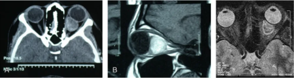
Therapy was initiated with intravenously administered dexa-methasone 1 mg/kg every 6 hours. He underwent magnetic reso-nance imaging (MRI) of the head and neck that highlighted the presence of subperiosteal intraorbital hematoma in the superior compartment, which displaced the superior rectus muscle inferiorly (Figs. 1A, B). There was no evidence of signs or symptoms of intracranial extradural hematoma.
The patient underwent emergency surgery through a 2-cm supraciliary incision and the evacuation of the hematoma by blunt dissection. Intravenous steroid therapy was maintained for 3 days. The patient showed a progressive improvement of visual acuity. The dose of dexamethasone was reduced to 0.5 mg/kg every 6 hours and continued for another 2 days. Oral therapy was then set with pred-nisone 25 mg twice daily for 3 days, later reduced to 5 mg twice daily. After 10 days of hospitalization, the patient was discharged with a good functional recovery. Ophthalmologic and maxillofacial monitoring was planned once a week for the first month and then monthly for the next 6 months. The patient showed no signs and symptoms of recurrent retrobulbar hematoma, showing visual function improvement with some residual impairment.
Patient 2
Patient 2 was a 25-year-old woman with craniofacial trauma after a car accident. Head and neck examination revealed left eyelid
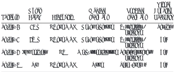
TABLE 2. Clinical Characteristics of the Patients Considered in This Study
Patients Onset Delay Diagnosis Medical Treatment Surgical Treatment Visual Function Recovery Patient 1 24 h CT and MRI Dexamethasone Supraciliary,
drainage
Partial Patient 2 32 h CT and MRI Dexamethasone Supraciliary,
drainage
Yes Patient 3 Immediately CT Methylprednisolone Paralateronasal,
drainage
Yes Patient 4 No CT and MRI None Percutaneous Yes
TABLE 1. Demographic Characteristics of the Patients Considered in This Study
Patients Age,
y Sex
Associated
Lesions Type of Fracture
Location of Hematoma Patient 1 65 Male No Orbitomaxillozygomatic Extraconal, superior
compartment Patient 2 25 Female No No Intraconal Patient 3 28 Male Yes Orbitomaxillozygomatic Intraconal Patient 4 10 Male No None Extraconal, superior
compartment FIGURE 1. A and B, Coronal MRI scan showing intraorbital hematoma in the superior compartment displacing inferiorly the superior rectus muscle.
The Journal of Craniofacial Surgery
&
Volume 23, Number 1, January 2012 Brief Clinical Studies* 2012 Mutaz B. Habal, MD
e59
hematoma, left exophthalmos, and decreased extraocular muscle motility. The direct photomotor reflex and the visual acuity were progressively deteriorated. Computed tomography and MRI showed left retrobulbar hematoma (Figs. 2AYC) without concomitant max-illofacial fracture. Chemotherapy was started with intravenously administered dexamethasone 4 mg every 12 hours for 2 days; the patient underwent immediate decompressive surgery.
Through a left supraciliary medial incision, the medial wall and the roof were approached, the intraconal compartment was exposed by blunt dissection, and the hematoma was evacuated. Intravenous chemotherapy was continued for 3 days after surgery. Three days after surgery, the eye examination showed improvement in ocular motility, and the vision was preserved. The dose of dexamethasone was reduced and continued for another 3 days. Oral therapy was then set with prednisone for 2 weeks.
The patient was discharged with ophthalmologic and maxillo-facial checkups weekly. The patient had a complete recovery of visual function without recurrent retrobulbar hematoma.
Patient 3
Patient 3 was a 28-year-old man with facial trauma after a work accident. Clinical examination showed a bruised right eyelid with a massive hematoma, a supraciliary lacerated wound, exophthal-mos, and chemosis. The extrinsic ocular motility was limited, and the direct photomotor reflex was absent; the affected eye presented no vision. There were concomitant pneumothorax, hepatic con-tusion, and hip fracture. Computed tomography ruled out the presence of orbital wall fracture and anterior displacement of right eyeball by hyperdense retrobulbar adipose tissue of hemorrhagic nature.
The patient underwent emergency surgery. Through a 1-cm paralateronasal incision, the blood collection was drained with a blunt cannula, and careful hemostasis was achieved. Intravenous therapy with methylprednisolone 5.4 mg/kg every hour for 24 hours was set and then prednisone 1 mg/kg per day for 11 days.
Two days after surgery, CT scan documented the reduction of the retrobulbar hyperdensity. Eye examination showed clinical im-provement with recovery of the visual acuity and the extraocular movements. At 4 days after trauma, the patient was discharged with ophthalmologic and maxillofacial checkups weekly. The pa-tient showed no signs and symptoms of recurrent retrobulbar hematoma.
Patient 4
Patient 4 was a 10-year-old boy with craniofacial trauma after a car accident. Clinical examination showed swelling and bruising of the left eyelid with the absence of any sensory and motor deficits. Computed tomography of the orbits showed intraorbital hematoma above the superior rectus muscle (Fig. 3A).
After 6 hours, MRI of the orbits was performed, and it con-firmed the presence of subperiosteal spindle-shaped fluid collec-tion between the roof of the orbit and the superior rectus muscle (Fig. 3B). He underwent emergency surgery through a percuta-neous drainage of the hematoma with a blunt cannula.
Steroid therapy was not set because the patient had no evidence of visual impairment. At the third day, postoperative CT scan showed the reduction of the subperiosteal blood collection. After 4 days of hospitalization, the patient was discharged with ophthal-mologic and maxillofacial assessment every week for the first month and then monthly for the next 4 months. Three months after surgery, MRI showed complete ‘‘restitutio ad integrum’’ of the superior or-bital compartment.
DISCUSSION
Retrobulbar hematoma is a rare complication of blunt periorbital trauma. Hayreh et al11 demonstrated that, in case of increased intraorbital pressure, the vessels anterior to the lamina cribrosa sclerae were first compressed, followed by the peripapillary cho-roidal vessels, the retrolaminar vessels of the optic nerve, and finally the retinal central artery. Moreover, they demonstrated that the ir-reversible retinal injury requires 3 hours or more to set up.
Huang et al12 injected autologous blood into the retrobulbar space in 24 rabbits to evaluate the effects of retrobulbar hemorrhage. Intraorbital injection of blood immediately produced lid edema, lid ecchymosis, chemosis, mydriasis, and proptosis. Subsequently light-eyelid reflexes disappeared. The vision was preserved because the intraorbital pressure was not maintained long enough to produce irreversible optic nerve or retinal damage.
Rahn13stated that intraorbital pressure has to exceed the mean arterial pressure by 60 to 70 mm Hg to result in central artery occlusion. It is known that retinal circulation is usually protected by various compensatory mechanisms, so that the blood flow in the retina is adequately maintained even if the entire system is under stress.
FIGURE 2. AYC, Computed tomography and MRI scans showing left retrobulbar intraconal hematoma.
FIGURE 3. A and B, Computed tomography and MRI scans of the orbits showing the presence of subperiosteal spindle-shaped fluid collection between the roof of the orbit and the superior rectus muscle.
Brief Clinical Studies The Journal of Craniofacial Surgery
&
Volume 23, Number 1, January 2012e60
* 2012 Mutaz B. Habal, MDThe neural tissues are vulnerable to ischemic injury, so that a transient interruption of the blood supply to the retina can cause neural cells to be unresponsive to light stimuli.14The optical nerve damage, caused by expensive lesions into the retrobulbar space, is caused by 3 different mechanisms: neurovascular compression, optic nerve stretch, and ischemia.15
It is usually self-limiting because the hemorrhage into the retrobulbar space is drained into the paranasal sinus through fractures of orbital walls. Nevertheless, in the absence of fractures or in the presence of linear fractures, the drainage could be absent with arising of retrobulbar pressure and irreversible damage of vision system.2
The clinical suspicion of retrobulbar hematoma must be ade-quately investigated and promptly treated to avoid the onset of ir-reversible and devastating complications.6,16Onset of retrobulbar hematoma can be several days after traumatic episode; therefore, the patient should be observed carefully to individuate the presence of chemosis, proptosis, painful exophthalmos, decreased visual acuity, and extraocular muscle motility.1Moreover, the clinical presentation can be variable, and it can be confused with other conditions such as traumatic superior orbital fissure syndrome and carotid-cavernous sinus fistula.
Prevention of devastating sequelae relies on a prompt diagnosis. Magnetic resonance imaging and CT proved to be tightly comple-mentary in the diagnostic assessment, determining (1) orbital wall fractures, (2) extension of any hemorrhagic lesions, and (3) asso-ciated intracranial hematomas.16,17
Magnetic resonance imaging has several advantages including (1) lack of harmful ionizing radiation, (2) multiplanar imaging, and (3) high soft tissue contrast resolution.7It is the modality of choice for characterization of subperiosteal hematomas, which represent the rarest subtype of hemorrhagic intraorbital lesions.5
Treatment of this condition should be addressed as soon as signs of retrobulbar hemorrhage appear, avoiding irreversible events. Despite conservative measures including immediate eye massage to reduce intraorbital pressure, bed rest, and elevation of the head, prompt pharmacologic therapy should be started. Pharmacologic therapy includes administration of mannitol, acetazolamide, and high doses of corticosteroids.
Surgery decompression of the orbit is recommended when visual deficit arises and when there is no response to pharmacologic therapy. Several techniques for orbital decompression have been proposed. The lateral canthotomy and/or the inferior cantholysis are the 2 techniques most practiced.
Under local anesthetic, the lateral canthotomy is performed by placing an artery clip between the upper and lower lids and then transecting the lateral canthus. The inferior cantholysis detaches the inferior crus of the lateral tendon, leading to a complete mobile lower eyelid.
Anterior-chamber paracentesis is effective, but it is rarely indicated for frequent complications such as cataract forma-tion, herniation of the iris, infecforma-tion, and trauma to the canal of Schlemm.
More extended approaches to the internal orbit with a more complex procedure and a higher morbidity are used in the case of extensive retrobulbar hemorrhage and when it is necessary to manage complex fractures or optic nerve decompression. These procedures include transantral ethmoidectomy, transantral sphenoi-dectomy, and transfrontal craniotomy.18,19
Carbonic anhydrase inhibitors such as acetazolamide 500 mg, intravenously administered hydrocortisone 100 mg, and a rapid in-fusion of 20% mannitol are frequently used. The action of mannitol is immediate, whereas that of acetazolamide is delayed. Steroids reduce inflammation and stabilize cell membranes against ischemic damage. Other nonsurgical treatment methods suggested include the
use of timolol maleate eye drops, 0.25% topical solution, which decreases the production of aqueous humor.
The necessity of a rapid diagnosis and management of a retro-bulbar hemorrhage has been described in this article. A combined medical and surgical management is the most appropriate treatment option for this condition; this maxillofacial emergency has a variable clinical presentation, and the onset can be delayed. Visual acuity must be monitored in all patients affected by orbital trauma, and CT or MRI assessment should be performed in any suspected case to permit a prompt diagnosis and management, avoiding permanent blindness.
REFERENCES
1. Naja A, Chellaoui A, Ibahioin K, et al. He´matome sub-pe´rioste´ de l’orbite associe´ a` une lame d’hematome extradural sous-frontalVa` propos d’un cas. Neurochirurgie 2003;48:101Y103
2. Bailey WK, Paul C, Evans LS. Diagnosis and treatment of retrobulbar haemorrage. J Oral Maxillofac Surg
1993;51:780Y781
3. Walsh FB, Hoyte WF. Clinical Neuro-ophthalmology. Vol 3 (ed 3). Baltimore, MD: Williams & Wilkins, 1969:2380
4. Hislop WS, Dutton GN, Duglas PS. Treatment of retrobulbar haemorrage in accident and emergency departments. Br J Oral Maxillofac Surg 1996;34:289Y292
5. Kline LB, Morawetz RB, Swaid SN. Indirect injury of the optic nerve. Neurosurgery 1984;14:756
6. Guirgis MF, Segal WA, Lueder GT Subperiosteal orbital hemorrhage as initial manifestation of Christmas disease (factor IX deficiency). Am J Ophthalmol 2002;133:584Y585
7. Brucoli M, Stecco A, Iaquinta C, et al. Diagnosis and treatment of the orbit post-traumatic subperiosteal haemorrhage in a child associated to a subdural intracranial haemorrhage: a case report. J Craniofac Surg 2005;16:407Y410
8. Katz B, Herschler J, Brick DC. Orbital haemorrhage and prolonged blindness: a treatable posterior optic neuropathy. Br J Ophthalmol 1983;67:549Y553
9. Volpe N, Lessell S, Kline L. Traumatic optic neuropathy: diagnosis and management. Int Ophthalmol Clin 1991;31:142Y156
10. Fry HJH. Reversibile visual loss after proptosis from retrobulbar hemorrhage. Plast Reconstr Surg 1969;44:480
11. Hayreh SS, Kolder HE, Weingeist TA. Central retinal artery occlusion and retinal tolerance time. Ophthalmology 1980;87:74Y78
12. Huang TT, Horwitz B, Lewis SR. Retrobulbar haemorrhage. Plast Reconstr Surg 1977;59:39Y44
13. Rahn H. The pressure-volume diagram of the thorax and lung. Am J Physiol 1946;146:161Y178
14. Hayreh SS. Pathogenesis of occlusion of the central retinal vessels. Am J Ophthalmol 1971;72:998
15. Gellrich NC, Schramm A, Rustemeyer J, et al. Quantification of the neurodegenerative impact on the visual system following sudden retrobulbar expanding lesions: an experimental model. J Craniomaxillofac Surg 2002;30:230Y236
16. Rosdeutscher JD, Stadelmann WK. Diagnosis and treatment of retrobulbar haematoma resulting from blunt periorbital trauma. Ann Plast Surg 1998;41:618Y622
17. Wolter GR, Leenhouts JA, Coulthard SW. Clinical picture and management of subperiosteal haematoma of the orbit. J Pediatr Ophthalmol 1976;13:136Y138
18. Sacks SH, Lawson W, Edelstein D, et al. Surgical treatment of blindness secondary to intraorbital hemorrhage. Arch Otolaryngol Head Neck Surg 1988;114:801Y803
19. Sofferman RA. Sphenoethmoid approach to the optic nerve. Laryngoscope 1981;91:184
The Journal of Craniofacial Surgery
&
Volume 23, Number 1, January 2012 Brief Clinical Studies* 2012 Mutaz B. Habal, MD