Nella revisione della memoria
e della dimensione del destino,
tra il vuoto del presente
e il peso di sogni perduti,
lungo un equilibrio
di respiro lento della notte
di contorni sterili di ombre
e di silenzio del tempo,
cadere è stato facile;
sperimentando
il piacere di precipitare
e il bisogno di risalire;
ma camminare sempre
verso orizzonti più lontani
della mente,
dove la forma
del pensiero
diventava libera…
UNIVERSITÀ DEGLI STUDI DI FOGGIA
FACOLTÀ DI MEDICINA E CHIRURGIA
DIPARTIMENTO DI SCIENZE BIOMEDICHE
____________________________________________________________________
Tesi di Dottorato in
SCIENZE E TECNOLOGIE BIOMEDICHE
XXIV ciclo
Host-probiotic interaction: in vitro analyses
DOCENTI TUTOR:
Dott.ssa Daniela FIOCCO Prof.ssa Anna GALLONE
COORDINATORE DEL DOTTORATO:
Prof. Maurizio MARGAGLIONE
DOTTORANDO:
Pasquale BOVE
____________________________________________________________________
Anni Accademici 2008-2011
INDEX
ABSTRACT
1. INTRODUCTION……….1
1.1. Probiotics………1
1.1.1. History and definition of probiotics………....1
1.2. Microorganisms used as probiotics………..3
1.3. Requisites and mechanisms of probiosis………...8
1.3.1. Beneficial effects of probiotics: clinical studies………...8
1.3.2. Safety………...11
1.3.3. Tolerance to oro-gastrointestinal conditions……….12
1.3.4. Adhesion to host epithelial cells and pathogen displacement………...13
1.4. Probiotic action and interaction with host cells……….16
1.4.1. Molecular interplay between probiotics and host cell………..17
1.4.1.1. Host receptors and signal cascades……….17
1.4.2. Relevance of the bacterial cell surface features in the interaction with the host………....20
1.4.3. Host cell response - Modulation of innate defense mechanisms………..21
1.5. Methodologies to study probiotics………...…....25
1.5.1. Oro-gastrointestinal tract simulators………...25
1.5.2. Methodologies to study bacterial adherence to the intestinal mucosa……....27
1.6. Lactic Acid Bacteria………...28
1.6.1. Lactobacillus plantarum: a model organism to study LAB and probiotics….29 1.7. Stress response in bacteria………...30
1.7.1. Heat shock proteins………...30
1.7.2. Genetic regulation of bacterial heat shock proteins………..32
1.7.3. sHSPs in Lactobacillus plantarum WCFS1………....33
1.7.4. The FtsH protein of Lactobacillus plantarum WCFS1……….34
1.8. Phenotypical features of L. plantarum WCFS1 mutant strains………....35
2. AIMS OF THE RESEACH………...…...37
3. MATERIALS AND METHODS……….…...…..38
3.1. MATERIALS……….…...38
3.1.2. Animal cells……….….38
3.1.3. Culture media……….….38
3.1.4. Food matrices………...39
3.1.5. Antibiotics………...39
3.1.6. Buffers and Solutions………...40
3.1.7. Drugs and Supplements………...41
3.1.8. OGI enzymes………...41
3.1.9. Enzymes and kits for nucleic acids manipulation and analysis…………...41
3.1.10. Oligonucleotides………...………42
3.2. METHODS………....45
3.2.1. Bacteria………..…………...45
3.2.1.1. Bacterial culture conditions……….45
3.2.1.2. Analysis of bacterial stress tolerance by minispot plating and CFUs count...45
3.2.1.3. Biofilm assay………...…...46
3.2.1.4. Microbial adhesion to solvents method (MATS)………...46
3.2.1.5. Scanning electron microscopy (SEM) analysis……….……..47
3.2.1.6. Oro-gastrointestinal transit assay………...47
3.2.2. Molecular cloning procedure……….…...50
3.2.2.1. L. plantarum WCFS1 genomic DNA extraction………..…...50
3.2.2.2. Polymerase chain reaction (PCR)……….…...50
3.2.2.3. Purification of PCR products………...51
3.2.2.4. Restriction enzymes……….……...…..51
3.2.2.5. Dephosphorylation………...……....….51
3.2.2.6. Ligation………...…...52
3.2.2.7. E. coli DH10B Ca2+-competent cells and transformation procedure…...52
3.2.2.8. E. coli DH10B transformation by heat shock………...…..53
3.2.2.9. Screening of trasformants and recombinant clones by colony PCR…...53
3.2.2.10. Plasmid purification and DNA sequencing………....53
3.2.2.11. Electroporation of L. plantarum WCFS1……….…...54
3.2.2.12. Gene knockout of L. plantarum WCFS1 shsp genes………..…....55
3.2.2.13. Disruption of L. plantarum WCFS1 ftsH gene………..…...58
3.2.3.1. THP-1 cell culture and ELISA assay……….….………58
3.2.3.2. Caco-2 cell culture and adhesion test………..59
3.2.3.3. Polarization of Caco-2 cells………..………60
3.2.3.4. Caco-2 cell stimulation assay………...60
3.2.3.5. RNA extraction from bacteria and animal cells…...……….…...61
3.2.3.6. cDNA synthesis………...62
3.2.3.7. Quantitative Real Time PCR………...62
3.2.3.8. Statistics……….63
4. RESULTS………..……...64
4.1. Generation of L. plantarum WCFS1 mutant strains……….………….64
4.1.1. Strategies to delete L. plantarum WCFS1 hsp18.5 and hsp19.3 genes ...…….64
4.1.2. Disruption of the ftsH gene……….…..…...66
4.2. Phenotypic analyses of L. plantarum mutant strains………...….…….67
4.2.1. ftsH gene deletion affects growth of L. plantarum WCFS1……...……...67
4.2.2. Morphological and physico-chemical surface properties of L. plantarum ΔftsH and other mutant strains………...68
4.2.3. Transcript profile of genes associated to probiosis………...71
4.3. Development of an oro-gastrointestinal tract simulator………...72
4.3.1. Survival during the transit through the in vitro OGI tract model………...72
4.3.2. Matrix effect on the viability of L. plantarum WCFS1 wild type during the transit in the oro-gastrointestinal tract model……….….76
4.3.3. Molecular response of the bacteria to the stress conditions of the OGI tract simulator………...79
4.4. Interaction with the host cells………...…….…….85
4.4.1. Differentiation of Caco-2 cell monolayers………..………..85
4.4.2. Adhesion of bacterial cells to human intestinal epithelial cells……...…...86
4.4.3. Effect of beta-glucans addition on bacterial adhesion to Caco-2 cells..…….87
4.4.4. Expression of immune-related genes in Caco-2 cells upon interaction with cells from different L. plantarum strains………...…...87
4.4.5. Modulation of TNF-α production in host immune cells…………....……...92
5. DISCUSSION………...………...94
5.2. Surface properties of the ΔftsH mutant and comparison with the other L.
plantarum strains OGI tract simulator……….…………..…95
5.3. OGI tract simulator……….………...96
5.4. Interaction with the host………..……..…....102
6. CONCLUDING REMARKS………..………107
7. REFERENCES………..……..108
8. PUBLICATIONS AND CONFERENCES………..….…127
ABSTRACT
Functional foods can positively influence functions of the body, by improving the health or reducing the risk of disease. Some functional foods contain ‘probiotics’, defined as ‘live microorganisms which when administered in adequate amounts confer a health benefit on the host’. The development and use of in vitro and in vivo protocols to assess the probiotic efficacy of microorganisms are highly encouraged by FAO and WHO.
In this thesis, the probiotic potential of the lactic acid bacterium Lactobacillus plantarum, wild type and derivative mutant strains, was investigated. The distinctive cell surface features exhibited by stress gene mutants prompted us to produce, by gene knockout, other L. plantarum defective strains and led us to investigate whether these characteristics could affect host-microbe interaction. The bacterial survival of L. plantarum strains and commercial probiotics was evaluated by designing an in vitro system simulating the transit along the human oro-gastrointestinal tract. Different carrier matrices were assayed in relation to possible prebiotic effects. The bacterial molecular response to such stresses was monitored by analysing the expression of stress, adhesion and probiosis genes. Interaction with the host was studied in vitro by i) assessing bacterial adhesive ability to gut epithelial cells; ii) investigating anti-inflammatory properties and induction of innate immunity genes in human host cells.
L. plantarum strains were resistant to the combined stress at the various steps of the simulated oro-gastrointestinal tract. Major decreases in viability were observed mainly under drastic acidic conditions (pH ≤ 2.0) of the gastric compartment. Abiotic stresses associated to the intestinal environment (small intestine) poorly affected bacterial vitality. The protective effect of vehicle matrices correlated with composition and bacterial nutritional needs. A relationship was found between bacterial survival and stress gene pattern. All strains significantly adhered to human intestinal epithelial cells, with the ΔctsR L. plantarum mutant exhibiting the highest adhesion. Colonization ability was improved by addition of prebiotics. Supernatants from all strains of L. plantarum reduced proinflammatory cytokine secretion by activated human immune cells. Induction of immune-related genes resulted generally higher upon incubation with heat-inactivated bacteria, rather than with live ones. For specific genes, a differential transcriptional pattern was observed upon stimulation with the different L. plantarum strains, pointing to a possible role of the knocked out bacterial
genes in modulation of host cells response. Particularly, cells from Δhsp18.55 and ΔftsH mutants strongly triggered immune defence genes.
This study highlights the relevance of the microbial genetic background in host-probiotic interaction and might contribute to: i) define selection criteria and/or conditions for probiotic screening and delivery; ii) identify candidate bacterial genes and/or molecules involved in probiosis, so to tailor probiotics for specific clinical applications.
1
1. INTRODUCTION
1.1. Probiotics.
Humans live in close association with a large number of microorganisms occurring on the skin, in the mouth and all along the gastrointestinal (GI) mucosa. The highest concentration of commensal microorganisms is found in the GI tract, which has more than 400 m2 of surface area. The GI tract harbours a rich and complex microbiota of more than 500 different bacterial species, some of which play important health functions on the host, including immune system stimulation, protection from invading bacteria and viruses, and support to digestion of nutrients (Mcfaralane and Mcfarlane, 1997; O’Hara and Shanahan, 2006; Neish, 2009). The normal gut flora, which is essential for human homeostasis, is rapidly acquired after birth and remains relatively stable throughout the life. While the intestinal microbiota is developing, its interaction with the host results in the evolution of a unique and distinct intestinal immune system. The great challenge for the host mucosal immune system is to discriminate between pathogens and benign organisms by stimulating protective immunity without excessive inflammatory response that may disrupt the integrity of the GI mucosa (Mc Ghee et al, 1999)
The use of antibiotics, immunosuppressive therapy and other treatments, may profoundly alter the composition on the commensal GI microbiota. Therefore, the dietary supplementation of beneficial bacterial species may be a very attractive therapeutic alternative to re-establish the microbial equilibrium and prevent disease (Vanderhoof and Young, 1998). In this regard, the helpful bacteria are the so-called ‘probiotics’. According to the World Health Organization (WHO) and the Food and Agriculture Organization of the United Nations (FAO), ‘probiotics’ are live microorganisms that confer a health benefit on the host (FAO/WHO, 2002). Whereas, the related term ‘prebiotic’ indicates non-digestible food ingredients that improve host health by stimulating growth and activity of beneficial components of the gut microflora (Gibson and Roberfroid, 1995).
1.1.1. History and definition of probiotics.
The Russian scientist and Nobel laureate Eli Metchnikoff, was the first to conceive a positive role of certain bacteria in the human body. At the beginning of the 20th century, he suggested that it might be possible to replace harmful microbes with useful ones. He believed that the aging process was due to toxins such as phenols, indols and ammonia in the large intestine, produced by proteolytic microbes such as Clostridia. He noted that milk
2 fermented with lactic acid bacteria inhibited the growth of the proteolytic bacteria because of the low pH produced by lactose fermentation. Metchnikoff also observed that some rural peoples in Europe, who used to drink milk fermented by lactic acid bacteria, had a relatively long life. He then introduced the use of fermented sour milk, using a bacterial species that he later called ‘Bulgarian bacillus’ (Vaughan, 1965).
The French pediatrician Henry Tissier first isolated a Bifidobacterium. He isolated it from a breast-fed infant and called it ‘Bacillus bifidus communis’ (later renamed Bifidobacterium bifidum). Tissier concluded that this species was predominant in the microflora of breast-fed infants and recommended it for feeding babies suffering from diarrhea (Tissier, 1900).
In 1917, the German professor Alfred Nissle isolated the bacterium Escherichia coli from the feces of a World War I soldier who did not develop enterocolitis during a severe outbreak of shigellosis. He successfully used this strain to treat intestinal diseases such as shigellosis and salmonellosis (Nissle, 1918). At that time antibiotics were not discovered yet. The probiotic E. coli Nissle 1917 is still in use today and recent studies have demonstrated its direct interaction with the host adaptive immune system (Molin, 2001).
In 1920, professor Leo F. Rettger showed that ‘Bulgarian Bacillus’, later known as Lactobacillus delbruekii subsp. bulgaricus, could not live in the human intestine. So, at this time, Metchinikoff’s theory was disputed and the idea of fermented food died out (Cheplin and Rettger, 1920).
Werner Kollath first introduced the term ‘probiotics’. In 1953, he wrote about probiotics as being in contrast to harmful antibiotics, and defined ‘Probiotika’ those ‘active substances that are essential for a healthy development of life’. Rosalie Lilly and Daniel Still well coined the term in 1965. They defined it as ‘a substance produced by a microorganism stimulating the growth of another microorganism’. That is the opposite of antibiotic (Lilly and Stillwell, 1965). In 1974, R. B. Parker gave a different definition: those ‘organisms and substances which contribute to intestinal balance’ are probiotics (Parker, 1974). Roy Fuller, in 1989, defined as probiotic ‘a live microbial food supplement which beneficially affects the animal host by improving its intestinal microbial balance’ (Fuller, 1989).
Over the years, experts have long argued on how to define probiotics. WHO and FAO have recently developed a widely accepted definition: ‘live microorganisms which, when administered in adequate amounts, confer a health benefit on the host’ (FAO/WHO, 2002).
3
1.2. Microorganisms used as probiotics.
Most probiotic microorganisms are Gram-positive bacteria belonging to the genera of Lactobacillus and Bifidobacterium. However, even Lactococcus, Streptococcus, and Enterococcus genera, as well as some non-pathogenic strains of Escherichia, and certain yeast strains are currently used as probiotics (Table 1.1) (Ouwehand et al, 2002).
Genus Species Strain Health benefits
Lactobacillus acidophilus casei johnsonii plantarum rhamnosus La5 Shirota La1 299v GG
- Reduced antibiotic associated diarrhoea
- Shortening of rotavirus diarrhoea
- Reduced recurrence of superficial bladder cancer
- Immune modulation
- Improved oral vaccination
- Reduced colonisation by Helicobacter pylori
- Relief of irritable bowel syndrome - Reduction of LDL-cholesterol
- Shortening of rotavirus diarrhoea - Immune modulation
- Relief of inflammatory bowel disease - Treatment and prevention of allergy
Bifidobacterium longum lactis
BB536
Bb12
- Reduction of incidence of influenza
- Treatment of allergy
- Shortening of rotavirus diarrhoea - Reduced incidence of travellers diarrhoea - Improved oral vaccination
Escherichia coli Nissle 1917 - Fewer relapses of inflammatory bowel disease
Enterococcus faecium SF68 - Fewer relapses of inflammatory bowel disease
4 Lactic Acid Bacteria (LAB), a heterogeneous group of Gram-positive, lactate-producing bacteria, are commonly used in the formulation of functional probiotic foods (Ljungh and Wadström, 2006; Schroeter and Klaenhammer, 2009; Bron and Kleerebezem, 2011). LAB have been traditionally employed for the preparation of fermented foods (milk, meat, vegetables and beverages); moreover, several species are natural inhabitants of the human oro-gastrointestinal (OGI) tract and vagina.
In the following sections, some of the major bacterial species used as probiotics will be briefly described.
Lactobacillus plantarum. Lactobacillus plantarum (Figure 1.1) is a Gram-positive,
aero-tolerant LAB that produces both isomers (D and L) of lactic acid. L. plantarum is extremely widespread as it inhabits foods of plant or animal origin, but also soil and the mammalian gut. It is used for the production and preservation of fermented foods obtained from different raw materials (mostly of plant origin) (Table 1.2), in which it is either present as a contaminant or added as a starter to carry out fermentations. L. plantarum contributes to specific organoleptic and nutritional properties of the final product (Kleerebezem et al, 2003). L. plantarum is among the most common lactobacilli occurring on the human oral and intestinal mucosa (Molin, 2001; de Vries et al, 2006).
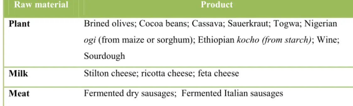
5 Table 1.2. Foods containing L. plantarum (adapted from de Vries et al, 2006).
Diverse L. plantarum strains have been ascribed healthy properties; because of its natural occurrence and history of safe use in food product, L. plantarum is present in a variety of currently marketed probiotic foods (Table 1.3). A well known and broadly used probiotic strain is L. plantarum 299v which was originally isolated from the human intestinal mucosa. Several reports, including human clinical studies, document the potential beneficial effects of such a strain (Molin, 2001; de Vries et al, 2006).
The complete genome of L. plantarum WCFS1, a single colony of L. plantarum NCIMB 8826 (National Collection of Industrial and Marine Bacteria, Aberdeen, UK), isolated from human saliva, was sequenced and annotated in 2003 (Kleerebezem et al, 2003). The availability of such data has prompted the genetic and molecular dissection of this species, also in relation to its probiotic behavior.
Table 1.3. Marketed probiotic food products containing L. plantarum (adapted from de Vries et al, 2006).
Raw material Product
Plant Brined olives; Cocoa beans; Cassava; Sauerkraut; Togwa; Nigerian
ogi (from maize or sorghum); Ethiopian kocho (from starch); Wine;
Sourdough
Milk Stilton cheese; ricotta cheese; feta cheese
Meat Fermented dry sausages; Fermented Italian sausages
Product name Formulation
IFlora Acidophilus Formula Probiotic Eleven
Plantadophilus FloraFood Living Vitamine C caps
Udo’s Choise Super Detox System
Capsule
Proviva Fruit drink
Lactovitale Drink
6
Lactobacillus acidophilus. Lactobacillus acidophilus (Figure 1.2) (Latin name meaning:
acid-loving milk-bacterium) is a homo-fermentative LAB (fermenting sugars into lactic acid), which grows readily at low pH (below 5.0) and has an optimum growth temperature of 37°C. L. acidophilus occurs naturally in the animal and human GI tract, mouth, and vagina. Strains of L. acidophilus are commercially used in many dairy products, such as yoghurt, sometimes together with Streptococcus salivarius subsp. thermophilus and Lactobacillus delbrueckii subsp. bulgaricus (Ashraf and Shah, 2011).
Figure 1.2. Scanning electron microscopy image of L. acidophilus.
L. acidophilus La-14 is a putative probiotic strain usable in therapeutic approaches for humans (Todorov et al, 2011). L. acidophilus La-14 showed ability to produce bacteriocins against Lysteria monocytogens ScottA and was resistant to drugs used in common antibiotic therapies. Therefore its potential use would be appropriate in parallel to pharmacological therapies.
L. acidophilus LA-5 strain has been attributed documented probiotic properties and is extensively used for the preparation of commercial functional foods, especially those containing milk-derived matrices (Chr. Hansen). A recent study revealed a reduction in salivary Mutans Streptococci and Lactobacilli levels in children, after consumption of ‘probiotic ice-cream’ prepared with L. acidophilus LA-5 in association to Bifidobacterium animalis subsp. lactis BB-12 (Singh et al, 2011).
7
Bifidobacterium. Bifidobacterium (Figure 1.3) is a motile, spore-forming,
non-gas-producing, Gram-positive, anaerobic, catalase-negative bacterium with a high GC content. Bifidobacteria cells look like irregular V- or Y-shaped rods. The actual reason for the irregular shape of Bifidobacteria is not yet clearly understood. However, studies have revealed that in vitro growth media can induce the typical bifid shape (Lee and O’Sullivan, 2010). Bifidobacteria produce water-soluble vitamins in the large intestine, including many of the B group. Moreover, Bifidobacteria restore the constipation in elderly people (Mayo et al, 2008).
Figure 1.3. Scanning electron microscopy image of Bifidobacterium.
B. animalis subsp. lactis BB-12 is marketed as probiotic. Such strain was given to preterm infants in a double-blind, placebo controlled, randomized clinical study. Feces from infants supplemented with B. lactis BB-12 showed lower viable counts of Enterobacteriaceae subsp. and Clostridia subsp., than the placebo group. However, B. lactis BB-12 supplementation did not reduce the gut colonization by antibiotic-resistant strains (Mohan et al, 2006).
8
1.3. Requisites and mechanisms of probiosis
.In order to be defined probiotics, microorganisms have to fulfill specific requisites of (Table 1.4). These characteristics include documented clinical efficacy, safety for human consumption, ability to reach, survive and colonize, at least transiently, the human gut, where probiotics exert their beneficial effects (Owehand et al, 2002).
Table 1.4. Main requisites of probiotic microorganisms and related advantages.
1.3.1. Beneficial effects of probiotics: clinical studies.
The rationale of probiotic therapies is to correct and/or prevent imbalances of the indigenous microbiota and gut barrier dysfunctions (Isolauri, 2001; Owehand et al, 2002) (Table 1.5).
Table 1.5. Potential clinical targets of probiotic therapy (adapted from Isolauri et al, 2004). Requisite of probiosis Benefit
Documented clinical effects True health benefits Safety No heatlh risk for consumer Tolerance to gastric acidity, bile
salts and pancreatic enzymes
Survival of passage through the intestinal tract Adhesion to intestinal mucosa Balancing of intestinal microbiota; strengthening of
epithelial barrier; immune modulation Human origin Species specific interactions with the host;
non-pathogenic;
Good technological properties Strain stability; resistance to storage and food processing conditions Effect Mechanism Nutritional management of acute diarrhoea Nutritional management of allergic disease
Reducing the risk of infectious disease Reducing the risk of allergic/inflammatory disease
Reduction in the duration of rotavirus shedding, normalization of gut permeability and microbiota Degradation/structural modification of enteral antigens, normalization of the properties of aberrant indigenous microbiota and of gut barrier functions, local and systemic inflammatory response, increased expression of mucin
Increase in IgA-secreting cells against rotavirus, induced expression of mucins
Promotion of gut barrier functions, anti-inflammatory potential, regulation of the secretion of inflammatory mediators, and promotion of development of the immune system
9 Specific probiotic strains are known to i) normalize altered gut microecology and intestinal permeability; ii) attenuate mucosal hypersensitivity and inflammatory reactions; iii) stimulate non-specific host resistance to microbial pathogens and favour their eradication (Isolauri et al, 2004).
Well-controlled clinical and nutritional studies are necessary to demonstrate the claimed health effects of probiotics. So far, probiotic interventions have been proven to be effective in varied pathologic conditions, such as necrotizing enterocolitis, antibiotic-associated diarrhoea, Helicobacter pylori infections, inflammatory bowel disease, cancer and surgical infections (Reid et al, 2003).
Necrotizing enterocolitis. Necrotizing enterocolitis (NE) is a devastating intestinal
disorder affecting preterm infants. It is a mortal disease characterized by abdominal distension, bilious vomiting, bloody diarrhoea, lethargy, apnoea, and bradycardia. Preterm infants who survive have intestinal obstruction and multi-organ failure (Caplan and Jilling, 2000).
Low weight preterm infants, delivered by Caesarean section, are often breast fed only after several days from birth. In addition, the normal process by which microorganisms such as Lactobacillus species are ingested via vaginal birth and propagated by mother's milk does not take place in these infants. Therefore, these infants are exposed to various pathogenic microbes (Clostridium, Escherichia, Salmonella, Shigella, Campylobacter, Pseudomonas, Streptococcus, Enterococcus, Staphylococcus and coagulase negative Staphylococcus) which colonize the intestine and increase the risk of NE. Furthermore, pre-term infants, given formula feeding, have fewer Lactobacillus and Bifidobacterium species in their stool compared to controls. These findings suggest a correlation between NE and Lactobacillus species. A human trial with live L. acidophilus and B. infantis given to newborn resulted in 60% reduction in NE (Gewolb et al, 1999; Hoyos, 1999).
Antibiotic associated diarrhoea. Probiotics have preventive as well as curative effects
on several types of diarrhoea of different etiologies. The dietary supplementation of probiotics bacteria (e.g., L. rhamnosus GG, E. coli strain Nissle 1917, Enterococcus faecium SF 68) and yeasts (Sacchromyces boulardii) alleviated symptoms of diarrhoea (de Vriese and Marteau, 2007).
10 Antibiotic associated diarrhoea (AAD) still affects hospitalized patients, although new antibiotics with a broad spectrum of activity and fewer side effects have been developed. The complications of AAD include electrolyte imbalance, dehydration, pseudomembrane colitis and toxic megacolon. A clinical study confirms the efficacy of probiotics in the prevention and treatment of AAD with S. boulardii (D'Souza et al, 2002).
Helicobacter pylori infections. H. pylori is a major cause of chronic gastritis and peptic
ulcer and a risk factor for gastric malignancies. Antibiotics for H. pylori eradication are 90% effective, but they are expensive and cause side effects and resistance. Probiotic-based approaches to treat H. pylori consequences have been performed. The studies revealed that probiotics had an in vitro inhibitory effect on H. pylori and reduced H. pylori associated gastric inflammation in animals; moreover, probiotic treatment reduced H. pylori therapy associated side effects (Lesbros-Pantoflickova, 2007).
Inflammatory bowel disease. Inflammatory bowel disease (IBD) includes ulcerative
colitis and Crohn's disease, representing chronic inflammations of the GI tract. Both clinical and experimental observations associate IBD to i) an imbalance in the composition of the intestinal microbiota, with relative predominance of aggressive bacteria and relative paucity of protective bacteria, and to ii) over-stimulation of proinflammatory immunological mechanisms. Preliminary studies suggest a positive response to probiotic interventions in IBD patients. The probiotic mixture VSL#3 provided a support to patients with intestinal mucosa depleted of protective bacteria (Gionchetti et al, 2000; Mitsuyama et al, 2002).
Cancer. In intestinal tumors, Latobacilli prevent or delay the tumor development by
metabolizing and/or binding to mutagenic compounds and suppressing the growth of bacteria which convert pro-carcinogens into carcinogens. Moreover, Lactobacilli reduce the levels of β-glucoronidase and other carcinogens (Ling et al, 1994). Recurrences of urinary bladder cancers decreased following internal instillation of probiotics such as L. casei Shirota, but this finding needs further confirmation (Aso et al, 1995).
Surgical infections. Before the advent of antiseptics and antibiotics, fermented milk was
used for healing wounds and fighting infections. Recent studies show the application of probiotics for treating and preventing surgical infections. L. fermentum RC-14 inhibits Staphylococcus aureus infection and bacterial adherence to surgical implants. One week
11 supplementation of the probiotic strain L. plantarum 299 with oat fibres reduced episodes of infection and pancreatic abscesses (Gan et al, 2002; Olah et al, 2002). Such clinical studies remind us that the potential use of probiotics is not necessarily restricted to heal gut associated dysfunctions, but has broader perspectives of applications.
1.3.2. Safety.
Because probiotics are supplemented as live microorganisms, they may cause infection to the host. Lactobacilli and Bifidobacteria are simply considered safe in reason of their taxonomic position, and for their long traditional use in food preparation. The human origin of the bacterial isolate and/or its natural occurrence in the OGI tract represent a further guarantee of safety for human consumption. In fact, the first human feeding trial shall also assess the safety of probiotic species.
Systemic infections have been rarely reported with Bifidobacterium, although many cases of sepsis with L. rhamnosus GG or L. casei have been reported (Adlerberth et al, 1991). Episodes of sepsis occur mainly in immune-compromised individuals or infants. But the conclusion, based on different reports, is that the risk of infection with probiotic Lactobacilli or Bifidobacteria is similar to infection with commensal strains, and that consumption of such products presents a negligible threat to consumers, including immune-compromised hosts (Ouwehand and Vesterlund, 2003). However, in order to establish safety guidelines for probiotic organisms, FAO and WHO recommend to characterize probiotic strains with a series of essential tests to assess antibiotic resistance pattern, metabolic activities, toxin production, hemolytic activities, infectivity in immune-compromised animal models, side-effects in humans, and adverse outcome in consumers. In 2002, FAO/WHO developed Operating Standards establishing guidelines for all companies producing probiotic products (FAO/WHO, 2002; Reid, 2005).
These guidelines include:
- guidelines for the use of probiotics;
- phase I, II and III of clinical trials to prove health benefits;
- good manufacturing practice and production of high quality products; - studies to identify mechanism of action in vivo;
- informative labelling;
12 - expansion of proven strains to benefit the oral cavity, nasopharynx, respiratory tract, stomach, vagina, bladder and skin as well as for cancer, allergies and recovery from surgery or injury.
1.3.3. Tolerance to oro-gastrointestinal conditions.
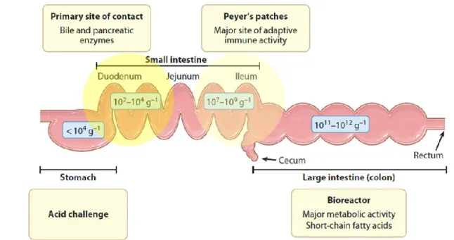
Resistance to the extreme conditions of the oro-gastrointestinal (OGI) tract, including highly acidic gastric juices and pancreatic bile salt secretions, is an essential criterion for the selection of orally delivered (food-borne) probiotics. The viability of probiotics is extremely important in order to guarantee high bacterial loads into the main site of action (e.g., the intestine) and their optimal functionality (Figure 1.4).
Figure 1.4. Different regions of the human GI tract and related densities of the residing bacterial population. Food-borne bacteria face the sequential stress of the acidic environment in the stomach, bile and pancreatin in the small intestine. Dietary supplementation of probiotics can generate a relative high abundance of these species in the first tract of the small intestine, where their metabolic activity can be relevant. The ileum, where the probiotic loads tend to decrease with respect to the indigenous microbiota, is the major site of probiotic immune activity. In the large intestine, commensal bifidobacteria and probiotic supplements contribute to catabolize diet- and host-derived glycans, generating a variety of short chain fatty acids that are used as important energy source by the colonic mucosa (adapted from Kleerebezem and Vaughan, 2009).
13 Passage of probiotics through the human OGI tract represents a ‘hazardous journey’, with the initial stages designed to compromise the survival of most pathogenic microorganisms. The principal sources of stresses for the bacteria are: i) pH down-shifting, encountered in the stomach and resulting from gastric acids; ii) presence of bile in the duodenum, a digestive secretion from the hepatic system, which serves to emulsify and solubilize lipids and lipid soluble vitamins. Exposure to acids negatively affects the proton motive force across the bacterial membrane, as a result of the accumulation of protons inside the cell. Exposure to bile disrupts the integrity of the cell membrane, affects DNA and RNA structures as well as protein folding; moreover, it exposes the cell to oxidative stress and low intracellular pH (Leverrier et al, 2003; Begley et al, 2005; Corcoran et al, 2008).
All along the different OGI sections, bacteria are challenged also by the action of diverse digestive enzymes, including lysozyme (in the oral cavity); pepsin (stomach), pancreatin, chimotrypsin, and carboxypeptidases (intestine). These enzymes can remarkably compromise bacterial cell structures, by attacking and degrading surface-exposed macromolecules (Frenhani and Burini, 1999).
Bacterial cells are naturally equipped with various defence mechanisms to enhance survival in hostile environments (Van de Guchte et al, 2002). These include chaperone proteins, which assist the folding of misfolded proteins, proteases which degrade irreversibly damaged proteins, transport systems to maintain correct osmolarity, catalases and superoxide dismutases to tackle reactive oxygen species, as well as proton pumps, decarboxylases and transporters to counteract intracellular pH decreases (De Angelis and Gobbetti, 2004; Sugimoto et al, 2008).
1.3.4. Adhesion to host epithelial cells and pathogen displacement.
Adhesion to the intestinal mucosa is a desirable feature of probiotic microorganisms, as it ensures persistence in the intestinal tract, which is necessary for probiotics to come in close contact with host epithelial cells, to control the balance of the intestinal microflora, to antagonize pathogen growth, and to exert immune modulation on the host (Apostolou et al, 2001; Isolauri et al, 2004).
Several bacterial cell surface proteins have been identified, which might mediate adhesion to the mucous layer and to the extracellular matrix of intestinal cells. In fact, the bacterial colonization may be improved by specific ‘adhesins’ which promote a tight interaction with host epithelial cells.
14 ‘Mucus-binding protein’ (MUB) is a bacterial cell-surface protein which is responsible for the adhesion to the intestinal mucus layer (MacKenzie et al, 2009). MUB proteins were well characterized in L. reuteri 1063 (Roos and Jonsson, 2002) and in L. acidophilus NCFM (Buck et al, 2005) revealing the typical domain organization of cell-surface proteins of Gram-positive bacteria: a signal peptide targeting the protein to plasma membrane is found at the N-terminus; an anchoring motif (LPXTG) for covalent attachment to the peptidoglycan of the bacterial wall is found at the C-terminus. An internal domain, containing the tandemly arranged mucus-binding repeats (Mub1 and Mub2), is responsible for adhesion to the host (Desvaux et al, 2006).
Another mechanism of bacterial adhesion is based on the binding to mannose-containing receptors on epithelial cells. Among probiotic bacteria, L. plantarum is able to recognize mannose-residues. By in silico studies, the predictive sequence of a L. plantarum WCFS1 adhesin gene (lp_1229) was identified. Knockout of this gene resulted in a complete loss of yeast agglutination ability, while its overexpression enhanced this phenotype. Moreover, analysis of the protein showed putative carbohydrate-binding domains, supporting its role in binding mannose residues. Therefore, this gene was designated to encode the mannose-specific adhesin (msa), probably involved in the interaction of L. plantarum with the host along the intestinal tract (Pretzer et al, 2005).
Myosin cross-reactive antigens (MCRAs) are conserved proteins found across a wide range of bacteria, including LAB. These proteins were discovered initially in Streptococcus pyogenes as potential antigens capable to share epitopes with myosin, contributing to blood survival and keratinocytes adherence. In fact MCRA is a FAD-containing enzyme with fatty acid hydratase activity on cis-9 (9Z) - and trans-11 (11E) double bonds of C-16, C-18 non-esterified fatty acids producing 10-hydroxy and 10,13-dihydroxy fatty acids (Volkov et al, 2010). In silico analysis of the L. acidophilus NCFM genome sequence revealed the presence of a gene showing similarity to N-terminal FAD/NAD(P)-binding domain of MCRA proteins. Deletion of this gene reduced the ability of L. acidophilus mutant strains to adhere on Caco-2 layers (O’Flaherty and Todd, 2010).
The ability to bind to host fibronectin (Fn) is a common characteristic among many Gram-positive species. Fn is a large glycoprotein present in soluble or in insoluble form on the cell surface, in the extracellular matrix, and in basal membranes. Therefore, targeting Fn is considered a strategy by which establishing interaction with the host (Papasergi et al, 2010). A 48 kDa putative Fn-binding protein, named alfa-enolase 1 (EnoA1), was recently
15 identified in L. plantarum (Castaldo et al, 2009). Its cell surface localization was demonstrated by immune electron microscopy. Moreover, the role of EnoA1 as a L. plantarum Fn-specific adhesion protein was assessed by deletion and functional analysis of the enoA1 gene: disruption of enoA1 caused a decreased adherence of mutant strain cells. Enolases are so-called “moonlighting proteins”, that is proteins with more than one function within the cell. Indeed, enolases function both as glycolytic enzyme as well as adhesins, once secreted outside the cells.
Thanks to the expression of specific adherence factors, probiotics can successfully compete with pathogens for the same attachment sites, thereby inhibiting their colonization, and possible infection: a phenomenon referred to as ‘pathogen exclusion’.
The S-layer protein, which forms a crystal layer structure on the bacterial surface (Boot et al, 1996), has been ascribed a role in adhesion to host cell and inhibition of pathogen adhesion to the same surface. In Lactobacillus crispatus ZJ001, S-layer proteins are responsible for adhesion to epithelial cells and competitive exclusion of pathogens such as E. coli O157:H7 and Salmonella typhimurium (Chen et al, 2007).
Other mechanisms concurring to pathogen exclusion rely on the synthesis of potent antimicrobial molecules, the so-called ‘bacteriocins’. Bacteriocins may allow a strain invasion into an established microbial community, or inhibit the invasion of other strains into an occupied niche (Riley and Gordon, 1999). Bacteriocins are ribosomally synthesized antimicrobial peptides or proteins; they are ubiquitous in the microbial world. In Gram-positive bacteria (including many probiotics), most bacteriocins are small in size (20–70 aminoacids) and cationic, and act by destabilizing the integrity of the inner membrane of target bacterial cells (Diep and Nes, 2002). Gram-positive bacteriocins have also been assigned a role in quorum sensing and signal communication in bacterial biofilms (Gillor, 2007).
In vivo studies have recently highlighted the relevance of bacteriocin production by probiotic strains. L. salivarius UCC118, a bacteriocin-producing strain, was effective in protecting mice against invasive Listeria, while the corresponding wild type, bacteriocin-negative strain, failed in defending mice from infection (Corr et al, 2007). Dabour et al (2009) showed that intragastric administration of the bacteriocin pediocin PA-1 in ICR mice infected with Listeria, reduced pathogenic count and translocation into the liver and spleen, as compared with the control group.
16 Using in vitro tests, E. coli L1000, producing microcin B17, was demonstrated to be active against a broad panel of antibiotic-resistant and -sensitive Salmonella strains isolated from patients suffering from salmonellosis (Zihler et al, 2009).
Interestingly, the presence of the plnEFI locus (encoding plantaricins, a subgroup of bacteriocins) in L. plantarum strains was associated to enhanced anti-inflammatory effects, in terms of lower IL-10/IL-12 ratios observed in bacteria-stimulated human peripheral blood mononuclear cells (PBMC) (van Hemert et al, 2010).
1.4. Probiotic action and interaction with host cells.
The molecular mechanisms underlying probiotic activities are being disclosed more and more by in vitro and in vivo studies focused on the interaction between probiotic bacteria and host intestinal epithelial or immune cells (Marco et al, 2006).
A scheme of the different actions supporting probiosis is shown in Figure 1.5. Probiotic effects depend on both microbe-microbe and host-microbe interactions. Metabolic interactions with the endogenous microbiota include phenomena of metabolic cooperation and/or competition for nutrient digestion, as well as production of antimicrobial compounds and competitive exclusion (Gueimonde and Salminen, 2006). All these activities may contribute to positively modulate the composition of the intestinal microbiota and inhibit detrimental species. Major metabolic interactions occur also with respect to intestinal host cells. For instance, some microorganisms provide essential vitamins (e.g., folate, biotin, vitamin K) and produce short chain fatty acids that are used as energy source by colon cells (Saulnier et al, 2009). Moreover, bacteria contribute to ion absorption and can metabolize dietary carcinogens and/or other toxic compounds. Interaction with host epithelial cells strengthens the barrier function by induction of mucin secretion, tight junction reinforcement, cytoskeleton stabilization (via Hsp induction), epithelial apoptosis reduction, and by triggering innate immune system activity (Lebeer et al, 2010). Probiotics can also interface with the mucosal adaptive immune system, thus modulating maturation and cytokine expression of intestinal dendritic cells.
17 Figure 1.5. Interactions, and related effects, occurring in the gut between probiotics (Lactobacillus and
Bifidobacterium) and the endogenous microbiota, the intestinal epithelial cells and the mucosa-associated
immune cells (from Kleerebezem and Vaughan, 2009).
1.4.1. Molecular interplay between probiotics and host cell.
Because of their close interaction with the intestinal mucosa, probiotics begin a ‘sort of molecular dialogue’ with the host cells, including both the surface epithelial cells and the underlying gut associated lymphoid tissue (GALT) elements. Intercellular prokaryote-eukaryote communications are switched on, signals cascades are activated and specific biochemical response are raised within the animal cells (Shi and Walker, 2004).
1.4.1.1. Host receptors and signal cascades.
The main elements of cross-talk between bacteria and host cells are the ‘Toll-like receptors’ (TLRs), the central ‘sensing’ apparatus of the innate immunity. The innate immunity represents the most archaic part of vertebrate immune defence. In contrast to adaptive immunity, which is restricted to higher vertebrates, the innate immune response is present in the whole animal kingdom. The innate immune system relies on a limited number
18 of receptors that recognizes a variety of pathogen-associated molecular structures, and on defensive mechanisms that counteract the broadest spectrum of potential pathogens (Medzhitov and Janeway, 1997).
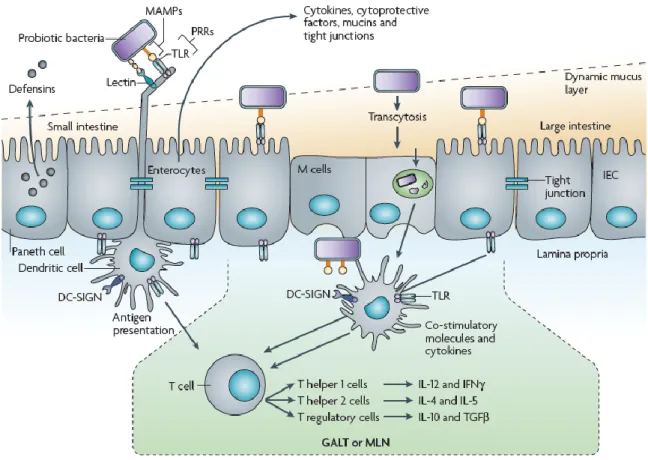
TLRs belong to a larger receptor group named ‘Pattern recognition receptors’ (PRRs), which play a critical role in initiating and regulating innate responses, by recognizing ‘microorganisms-associated molecular patterns’ (MAMPs), which are widespread and conserved. PRRs are expressed by a lot of cells including monocytes, dendritic cells, neutrophils, and epithelial cells (Medzhitov and Janeway, 2002). The interaction between MAMP and PRR results in the induction of signal cascades that develops a molecular response against the detected microorganism; this response can include the secretion of immunomodulatory cytokines, chemokines, and antimicrobial agents (Figure 1.6).
Figure 1.6. Molecular interaction of probiotic bacteria with intestinal epithelial cells and dendritic cells from the GALT. Host pattern recognition receptors (PRRs) sense the microorganism by recognizing their associated molecular patterns (MAMPs): this interaction will lead to specific molecular response, depending on the cell type. For example, Paneth cell shall produce defensins, whereas Goblet cells secrete mucins (from
19 The evoked signalling cascades usually involve nuclear factor-kB (NF-kB) and mitogen activated protein kinase (MAPK) systems, which rapidly transmit the signals to the nucleus to trigger transcription of immune-related genes (Figure 1.7).
Figure 1.7. Overview of the probiotic MAMP-PRR interaction and associated signalling events. Probiotic microorganisms interact with intestinal epithelial cells (IECs) through various surface molecules, including flagellins, cell-wall associated enzymes, lipoteichoic acids, peptidoglycan, etc. After dimerization, TLRs receptors send the signals to ‘kinases’ (MAPK pathway) through ‘adaptors’, and activate ‘transcription factors’ (AP-1, NF-kB) involved in binding specific DNA sequences. Reported IEC response includes induction of β-defensin 2, cytokine secretion, tight junction promotion, anti-apoptotic signals (from Lebeer et
20 The anti-inflammatory properties of some probiotics have been frequently associated to the inhibition of the NF-kB pathways (Petrof et al, 2004; Zhang et al, 2005). By contrast, Schlee et al, (2008) demonstrated that VSL#3 bacterial mixture and probiotic Lactobacilli up-regulate HBD-2 gene via induction of proinflammatory pathways such as NF-kB and AP-1, as well as MAPKs. Recent transcriptome analyses in in vivo models confirm that the majority of the host mucosal genes modulated by probiotic supplementation are NF-kB dependent (van Baarlen et al, 2009).
1.4.2. Relevance of the bacterial cell surface features in the interaction with the host.
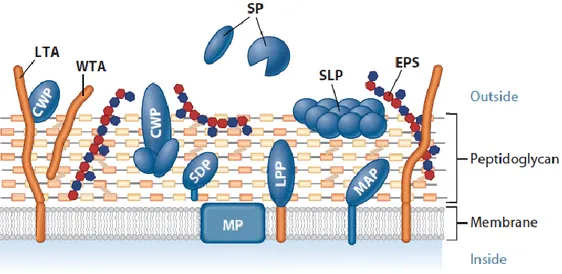
Microbial cell surface features are expected to be essential in the primary host-microbe interaction. Indeed, the different probiotics and immune-modulating properties of lactic acid bacteria seem closely related to their cell envelope composition and structure (Vinderola et al, 2004; Kleerebezem and Vaughan, 2009; Lebeer et al, 2010). The Gram-positive cell envelope of is made up by numerous characteristic structural components (Figure 1.8) that can be recognized by PPRs, induce signaling pathways and thus lead to specific health-promoting effects. MAMPs can be associated to macromolecules such as the peptidoglycan, cell wall- or membrane-associated teichoic acids, exopolysaccharides and various classes of surface proteins.
Figure 1.8. Gram-positive cell wall. Components of the cell surface macromolecules have been proposed to be directly involved in interaction with host cells. Specific MAMPs, and related host modulation properties, can be associated to: peptidoglycan (PG) layer, the predominant cell wall component; wall- and lipotheicoic acids (WTA, LTA); exopolysaccharides (EPS); and various types of surface associated proteins: secreted proteins (SP), membrane proteins (MP), cell-wall-associated proteins (CWP), sortase-dependent proteins (SDP), lipoproteins (LPP), membrane-anchored proteins (MAP), and surface layer proteins (SLP) (from
21 Subtle variations in the composition and structure of the cell wall may account for species- and strain-specific interactions with the host. In probiotic clinical application, such as treatment of IBD and allergic disorders, an intriguing goal is just to tailor these interactions, in order to achieve specific therapeutic outcomes. Indeed, by studying a L. plantarum dlt cell wall mutant, which synthesized modified teichoic acids, Grangette et al (2005) demonstrated that such specific cell surface biochemical feature might positively affect the interaction between microorganism and host, enhancing its probiotic effect in terms of increased protection from intestinal disorders. Similar results were also observed in in vitro and in vivo studies on lipoteichoic (LTA)-deficient strains of the probiotic Lactobacillus acidophilus (Mohamadzadeh et al, 2011) and Lactobacillus rhamnosus GG (Claes et al, 2010). In line with these reports, Schlee and coworkers (Schlee et al, 2007) used a genetic loss of function approach to demonstrate that the induction of human β-defensin 2 was specifically mediated by the flagellins of the probiotic E. coli Nissle 1917.
1.4.3. Host cell response - Modulation of innate defense mechanisms.
Modulation of the gut immune function seems one of the main mechanisms through which probiotics provide beneficial effects to the host. Indeed, probiotic and commensal bacteria influence the production of humoral immune factors, such as cytokines and antimicrobial agents, secreted by the gut-associated lymphoid tissue as well as by the intestinal epithelium (Borchers et al, 2009). It is thus clear that a great part of the beneficial effects of probiotics depends on their immunomodulatory abilities, both as immune-enhancing and as well as anti-inflammatory effect. Because antimicrobial peptides, mucous components, microbicidal enzymes and cytokines play key roles in the barrier and immune function of the intestinal mucosa, the expression of genes encoding such molecules has been frequently analyzed when assessing the microbial probiotic potential (Mack et al, 1999; Morita et al, 2002; Wekhamp et al, 2004).
Antimicrobial peptides (AMPs) are key effectors of the innate immune response. The
AMPs produced all along the GI tract of the host constitute a front line of chemical defence against dangerous microorganisms. This defence system functions in the airways, gingival epithelium, cornea, and in the reproductive, urinary and GI tracts. AMPs enable the innate immune system to respond in a matter of hours, well before the adaptive immune system can be sufficiently mobilized (Liévin-Le Moal and Servin, 2006).
22 Cathelicidins and defensins are the two main families of intestinal AMPs. Cathelicidins constitute a unique mammalian gene family. They are structurally organized with an N-terminal signal peptide, a highly conserved prosequence - the cathelin domain - and a variable cationic peptide at the C-terminus. The conservation of the cathelin domain is striking between species and indicates that the diverse members of this family evolved from a common ancestor gene (Bals and Wilson, 2003). LL-37 is the only cathelicidin described in humans. It is synthesized as a precursor, named ‘human cationic antimicrobial protein 18’ (hCAP18), which is then cleaved to the mature peptide by a serine protease. LL-37 can act alone and in synergy with other antimicrobial proteins (i.e., lysozyme), displaying bactericidal activities against Gram-positive and Gram-negative bacteria. LL-37 was initially found in specific neutrophil granules but is now known to also be expressed by other leukocytes, as well as keratinocytes and epithelial cells in the respiratory, urogenital, and GI tracts (Travis, et al, 2000; Sörensen et al, 2001). Additional studies suggest that expression of hCAP18 gene by human colon epithelial cells is a marker of epithelial cell differentiation (Hase et al, 2002).
Defensins are arginine-rich cationic peptides characterized by a β-sheet fold and a framework of six disulfide-linked cysteines (Lehrer, 2004). The two main defensin subfamilies are α- and β- defensins; α-defensins comprise the group of human neutrophil peptides (HNP-1 to 4) and human defensins 5 and 6 (HD-5 and HD-6). Four human β-defensins (HBD-1 to 4) have been described. Human β-β-defensins (HBD) have been isolated from many cell types, mainly epithelial, confirming that, these cells actively participate to host defence. HBD-1 is mainly expressed in the epidermis and in the epithelia of pancreas, kidney and urinary tract; HBD-2 and -3 are found in the skin and in airway epithelia; HBD-4 is expressed in testis, stomach, lung and neutrophils. HBD-2 is expressed by human intestinal epithelial cells (O’Neil et al, 1999; Lievin-Le Moal and Servin, 2006) and its transcription was shown to be activated in vitro by probiotic lactobacilli and VSL#3 mixture (Wekhamp et al, 2004; Schlee et al, 2007). These authors first suggested a novel effect of probiotics: the enhancement of the mucosal intestinal defense against pathogens through the up-regulation of defensins. Indeed, increased level of AMPs synthesized by the intestinal epithelial cells counteract adherence and invasion by pathogens.
23
Lysozyme. Lysozyme is a microbicidal enzyme directed against the β(1→4) glycosidic
bond between N-acetylglucosamine and N-acetylmuramic acid residues of peptidoglycan. Lysozyme is expressed by skin, oral mucosa and intestinal epiyhelial cells (Ganz, 2004). In fishes, probiotic water supplementations have been reported to increase lysozyme level in the skin, while dietary supplementation did not induce lysozyme in either serum or skin mucosa (Nayak, 2011).
Mucins. The mucous-gel layer, occurring at the interface between the gut lumen and the
epithelial cells, provides a physical barrier against potentially harmful bacteria and molecules, while acting as a lubricant for intestinal motility (Phillipson et al, 2008).
Microorganisms have developed diverse strategies to degrade the mucous layer (i.e., reduction of mucin disulfide bonds by Helicobacter pylori, protease activity by Candida albicans, and glycosidase activity by both oral and intestinal microbial communities) allowing them easier invasion and/or uptake of mucus-derived nutrients (De Repentigny et al, 2000; Windle et al, 2000). Mucins (i.e., glycosylated proteins located into the endomembrane system of intestinal epithelial cells or secreted into the lumen) are the main constituents of the mucous layer. Mucins are produced by specialized Goblet cells, as well as by intestinal epithelial cells all along the intestine tract (Lievin-Le and Servin, 2006). Some probiotics have been shown to increase mucin expression in vitro. Lactobacilli were found to induce mucin in human intestinal cell lines, thus blocking invasion and adherence of pathogenic E. coli strains (Mattar et al, 2002; Mack et al, 2003). In this way, probiotics may improve the barrier function of the intestinal mucosa and contribute to reduce gut permeability (Saulnier et al, 2009).
Cytokines. By optimizing the balance of pro- and anti-inflammatory cytokines and other
immune modulators, probiotics are thought to contribute to realize a healthy gut homeostasis. Cytokines are small secreted proteins which mediate and regulate immunity, inflammation, and haematopoiesis. They are produced in response to an immune stimulus, and generally act over short distances and short time spans, at very low concentration. Responses to cytokines include increasing or decreasing expression of membrane proteins (including cytokine receptors), proliferation, and secretion of effector molecules (Foster, 2001). Cytokines include ‘lymphokines’ (released by lymphocytes), ‘monokines’ (released
24 by monocytes), ‘chemokines’ (cytokines with chemotactic activities), and ‘interleukins’ (cytokines released by one leukocyte and acting on other leukocytes).
Interleukins are signaling, secreted cytokines that promote the development and
differentiation of T, B, and hematopoietic cells. The function of the immune system largely depends on interleukins, and rare deficiencies of a number of them have been described (i.e., autoimmune diseases or immune deficiency). The majority of interleukins are synthesized by helper CD4+ T lymphocytes, as well as by monocytes, macrophages, and endothelial cells. Interleukin 1 (IL-1) activates T cells; IL-2 stimulates proliferation of antigen-activated T and B cells; IL-4, IL-5, and IL- 6 stimulate proliferation and differentiation of B cells; interferon gamma (IFN) activates macrophages; IL-3, IL-7 and Granulocyte Monocyte Colony-Stimulating Factor (GM-CSF) stimulate haematopoiesis (Dinarello, 2000).
Tumor necrosis factor (TNF) is a proinflammatory cytokine contributing to recruitment
and activation of immune cells, release of cytolytic enzymes and reactive oxygen species (ROS), and exacerbation of tissue damage at inflammation sites. Increased levels of TNF are thought to contribute to the pathology of GI disorders such as Crohn’s disease (Kirman et al, 2004).
Chemokines are a large family of small (8–14 kDa) secreted chemotactic cytokines
involved in adhesion and directional homing of immune and inflammatory cells. These molecules have been divided into 4 subfamilies based on the arrangement of highly conserved cysteine residues in the amino-terminus: C, C-C, C-X-C and C-X3-C.
(Zimmerman et al, 2008). The C-C chemokines (including the macrophage inflammatory protein MIP-1α and MIP1-β) have 2 adjacent cysteines at the amino-terminal; the C-X-C chemokines (e.g., interleukin IL-8) have amino-terminal cysteines separated by an intervening amino acid (Horuk, 2007). Chemokines recruit leucocytes at the site of immune reaction. The macrophage inflammatory protein 3α (MIP-3α) is a lymphocyte directed C-C chemokine which is predominately expressed by colonic epithelial cells. Its expression level was found to be up-regulated in patients with Crohn’s disease or ulcerative colitis, suggesting that this chemokine might play an important role in the pathogenesis of human IBD (Kwon et al, 2002).
When cytokine signaling is unbalanced, serious diseases may occur in humans. The use of probiotics to restore interleukins unbalances of human and/or animals diseases, is largely
25 documented. Several studies report the probiotic-mediated suppression of human TNF production by host immune cells (Ménard et al, 2004; Lin et al, 2008). In vitro studies also demonstrated that probiotic Lactobacilli and Bifidobacteria attenuate IL-8 production by LPS, or IL-1 stimulated human intestinal epithelial cells (Candela et al, 2008). Such documented probiotic effects underlie the potential therapeutic use of selected microbes for
the treatment of IBD. Administration of probiotic L. delbruekii and L. fermentum strains to
ulcerative colitis (UC) patients, alleviated the inflammation by decreasing the colonic concentration of IL-6 and the expression of TNF-α and NF-kB p65. Therefore, probiotic supplementation could help in maintaining remission and preventing relapse of UC (Hegazy and El-Bedewy, 2010). Angiogenesis is required for wound healing, but its dysregulation is involved in GI inflammation. Bacillus polyfermenticus can promote the angiogenesis of human intestinal microvascular endothelial cells (HIMECs). In fact, the exposure of HIMECs to B. polyfermenticus increased the level of proangiogenic C-X-C chemokine IL-8, suggesting that the bacterium-mediated induction of IL-8 contributes to intestinal wound healing (Im et al, 2009).
1.5. Methodologies to study probiotics.
In order to investigate and ascertain the probiosis of microorganisms and in view of their potential therapeutic application, preliminary studies can rely on an array of in vitro assays. Indeed, in vivo approaches are often too complex and demanding, especially in the initial screenings for probiotics and for suitable food matrices. In this regards, many studies are available from the scientific literature.
1.5.1. Oro-gastrointestinal tract simulators.
Analysis of potential probiotics in in vitro multi-compartmental models that simulate the physico-chemical conditions of the human OGI tract is a prerequisite to subsequent in vivo experiments. As a result, development and implementation of such systems are highly encouraged by FAO and WHO (2002) and several recent studies have addressed this issue (Mainville et al, 2005; Masco et al, 2007; Fernández de Palencia et al, 2008; Lo Curto et al, 2011).
An ideal simulator should recreate the multi-segmentation of the human OGI tract, mimicking the events of food ingestion and digestion, and allowing for the addition of a food matrix through which the probiotic is delivered. The food matrix should primarily
26 shield the delivered microbe from the OGI hostile environment, sustaining its growth and activity. Prebiotic ingredients, that is ‘non host-digestible constituents which selectively promote growth and activity of the beneficial microbial species’ (Gibson and Roberfroid, 1995), may also be added to enhance the overall benefit of the designed functional food. Common food matrices (Table 1.6) are skim milk and soy gem powders (Mainville et al, 2005), sometimes enriched by fiber components with prebiotic action.
Table 1.6. Food matrices frequently adopted to vehicle probiotics.
The first steps of the OGI transit are associated to lysozyme and chewing stresses. Then, the models mimic the events of digestion into the stomach, from a situation of complete filling of the gastric pouch to progressive emptying. Immediately after food ingestion (full stomach condition), the matrix embedded bacteria is thought to be at pH values of 5.0 - 6.0; pH lowering (to values of 2.0 - 1.5) simulates the progressive emptying of the stomach and the digestion of food. The nature of the food affects the transit period through the stomach. Normally, food remains in the stomach between 2 and 4 hours; however, liquids empty from the stomach faster than solids, taking only about 20 minutes (Smith, 1995; Huang and Adams, 2004). The adverse conditions of the small intestine include the presence of bile salts and pancreatin. The transit time through the small intestine takes from 1 to 4 hours. In the lumen of the small intestine, pH is around 8.0. A concentration of 0.15 - 0.3% of bile salts has been recommended as a suitable concentration for selecting probiotic bacteria for human use (Smith, 1995; Huang and Adams, 2004).
Several working groups have developed different OGI tract simulators, each presenting advantages and drawbacks. Fernández de Palencia et al (2008) and Masco et al (2007) used static models in which the transit is performed in a single container recreating the entire OGI tract. The idea would be that of a method that divides the OGI tract into all its various
Food matrix Commercialized products Milk
Carbohydrate polymers Vegetables
Fruit Starch
Yogurt, fermented milks, Kefir Probiotic ice cream
Encapsulated probiotics Artichokes and olives Fruit juices
27 organs (i.e., mouth, stomach, duodenum, ileum, jejunum, colon). Both researches tested the bacterial viability using combined fluorescent probes. The advantage of such detection method is its high reproducibility and rapidity compared with conventional plate counts. However, the colorimetric methods may give non-specific results following the challenge of bacteria cells resuspended in matrix food.
Lo Curto et al (2011) and Mainville et al (2005) used dynamic OGI-simulating systems, automated through a computerized core; parameters of the OGI transit (pH, temperature, stirring, peristalsis, input of matrix) could be monitored by the operator. However these systems did not consider the interaction between intestinal endogenous microflora and the exogenous tested bacterium.
Decroos et al (2006) and De Boever et al (2000) performed the Simulator of the Human Intestinal Microbial Ecosystem (SHIME) consisting of a succession of 5 reactors (i.e., stomach, small intestine, ascending colon, transverse colon and descending colon). An inoculum prepared from human feces, and stabilized over days using a culture medium, was introduced into the SHIME. In the monitoring period, a human microbiota with Enterobacteriaceae, coliforms, Lactobacillus subsp., Staphylococcus subsp. and Clostridium subsp., was established. Such a system would allow to study potential probiotic strains interacting with the resident flora of the human intestine.
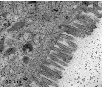
1.5.2. Methodologies to study bacterial adherence to the intestinal mucosa.
The ability to adhere to the intestinal mucosa is one of the more important selection criterion for probiotics because adhesion to the epithelial layer allows colonization. Due to obvious difficulties in performing in vivo studies, preliminary studies of potentially adherent strains are mainly based on in vitro adhesion assays. The use of at least two different systems is recommended, therefore, adhesion assays are frequently performed on both mucus and epithelial cell monolayers, representing the early and late stage of adhesion, respectively. Tissue cultures of the human colon carcinoma cell lines Caco-2 and HT-29 are the most frequently used (Salminen et al, 1996). Caco-2 cells also provide a valuable system for immunological studies (Ou et al., 2009). Tissue cultures are used not only to evaluate the level of bacterial adhesion, but also to investigate competition for binding sites with other pathogenic species (Candela et al, 2005).