Quantum Dots: Proteomics characterization of the impact on biological systems
This article has been downloaded from IOPscience. Please scroll down to see the full text article. 2009 J. Phys.: Conf. Ser. 170 012021
(http://iopscience.iop.org/1742-6596/170/1/012021)
Download details:
IP Address: 157.27.31.115
The article was downloaded on 07/11/2011 at 11:13
Please note that terms and conditions apply.
View the table of contents for this issue, or go to the journal homepage for more
Quantum Dots: proteomics characterization of the impact on
biological systems
Stefano Pozzi-Mucelli1, F. Boschi2, L. Calderan2, A. Sbarbati2 and F. Osculati1 1
Veneto Nanotech SCpA - European Centre for the Sustainable Impact of Nanotechnologies, Via San Crispino 106, 35129 Padova - Italy,
2
Department of Morphological & Biomedical Sciences, University of Verona, Strada Le Grazie 8, 37134 Verona - Italy.
E-mail: [email protected]
Abstract. Over the past few years, Quantum Dots have been tested in most biotechnological
applications that use fluorescence, including DNA array technology, immunofluorescence assays, cell and animal biology. Quantum Dots tend to be brighter than conventional dyes, because of the compounded effects of extinction coefficients that are an order of magnitude larger than those of most dyes. Their main advantage resides in their resistance to bleaching over long periods of time (minutes to hours), allowing the acquisition of images that are crisp and well contrasted. This increased photostability is especially useful for three-dimensional (3D) optical sectioning, where a major issue is bleaching of fluorophores during acquisition of successive z-sections, which compromises the correct reconstruction of 3D structures. The long-term stability and brightness of Quantum Dots make them ideal candidates also for live animal targeting and imaging. The vast majority of the papers published to date have shown no relevant effects on cells viability at the concentration used for imaging applications; higher concentrations, however, caused some issues on embryonic development. Adverse effects are due to be caused by the release of cadmium, as surface PEGylation of the Quantum Dots reduces these issues. A recently published paper shows evidences of an epigenetic effect of Quantum Dots treatment, with general histones hypoacetylation, and a translocation to the nucleus of p53. In this study, mice treated with Quantum Dots for imaging purposes were analyzed to investigate the impact on protein expression and networking. Differential mono- and bidimensional electrophoresis assays were performed, with the individuation of differentially expressed proteins after intravenous injection and imaging analysis; further, as several authors indicate an increase in reactive oxygen species as a possible mean of damage due to the Quantum Dots treatment, we investigated the signalling pathway of APE1/Ref1, a protein involved in the response to oxidative stress. Our results, although preliminary, suggest several interesting point of discussion on Quantum Dots imaging for in vivo diagnostic application, but also for a new therapeutic approach.
1. Introduction
Over the past few years, Quantum Dots have been tested in most biotechnological applications that use fluorescence, including DNA array technology, immunofluorescence assays, cell and animal biology. Quantum Dots tend to be brighter than conventional dyes, because of the compounded effects of extinction coefficients that are an order of magnitude larger than those of most dyes. Their main
Nanosafe 2008: International Conference on Safe production and use of nanomaterials IOP Publishing Journal of Physics: Conference Series 170 (2009) 012021 doi:10.1088/1742-6596/170/1/012021
advantage resides in their resistance to bleaching over long periods of time (minutes to hours), allowing the acquisition of images that are crisp and well contrasted. This increased photostability is especially useful for three-dimensional (3D) optical sectioning, where a major issue is bleaching of fluorophores during acquisition of successive z-sections, which compromises the correct reconstruction of 3D structures. The long-term stability and brightness of Quantum Dots make them ideal candidates also for live animal targeting and imaging [1]. The vast majority of the papers published to date have shown no relevant effects on cells viability at the concentration used for imaging applications; higher concentrations, however, caused some issues on embryonic development [2]. Adverse effects seem to be caused by the release of cadmium, as surface PEGylation of the Quantum Dots reduces these issues [3]. A recently published paper shows evidences of an epigenetic effect of Quantum Dots treatment, with general histones hypoacetylation, and a translocation to the nucleus of p53 [4]. Further, several Authors indicate an increase in reactive oxygen species as a possible mean of damage due to the Quantum Dots treatment. In this study, we apply a proteomic approach to investigate the impact on protein expression and networking in the liver of mice treated with Quantum Dots, to assess potential biomarkers to be used to unravel potential biological events following the treatment; more in details, we focused our attention to the signalling pathway of APE1/Ref1. APE1/Ref-1 (Apurinic/apyrimidinic endonuclease/redox effector factor-1) is a protein involved in DNA repair [5], in particular in the base excision process; during conditions of oxidative stress, the protein migrates into the nucleus, where it works also as a transcription enhancer, strengthening the DNA binding of proteins like p53, with the effect of an inhibition of the cell cycle through the inhibition of the PI3K/AKT pathway. Our results, although preliminary, suggest several interesting point of discussion on Quantum Dots imaging for in vivo diagnostic application, but also for a new therapeutic approach.
2. Materials and Methods
2.1. Optical Imaging
For all the experiments a VivoVision Systems, IVIS^® 200 Series, Imaging System for small laboratory animals, Xenogen (Xenogen Corporation, Alameda USA), was used. The system is constituted by a camera sensor back thinned, back illuminated grade CCD 1 (2.7 x 2.7 cm, -90°C), with a minimal image pixel resolution of 20 µm (pixel dimension 13.5 µm, imaging pixels 2048 x 2048), quantum efficiency >85% between 500 and 700 nm, and > 30% between 400 and 900 nm. In these experiments, this fluorescent modality was chosen, with an acquisition set of excitation filters: GFP (445-490 nm), DsRed (500-550 nm), Cy5.5 (615-665 nm), ICG background (665-695 nm), ICG (710-760 nm); and an emission filter ICG (810-875 nm). A 180W Quartz halogen 3250°Kelvin lamp at high intensity; binning factor = 8; pixel resolution = 0.125x0.125; field of view = 12.8 cm; exposition time = 1 sec; opening of diaphragm (f/stop) = 2. Images acquisition protocol concerns a first "pre" acquisition and 30 "post" acquisitions after QDs800 i.v. administration. Images were acquired and analyzed with Living Image 2.6 software and Living Image 3D software (Xenogen Corporation, Alameda USA).
2.2. Animals
Athymic /nude-nude/ mice, 3-4 weeks old, were administered with a 2 µM QDs800 solution (1:5 diluted /0.01 ml/gr mouse, i.v.), and compared with control animals, injected with PBS.
2.3. Protein extraction
Animals were sacrificed by cervical dislocation, and liver was resected and snap frozen, then homogenated in 50 mM Tris-Cl pH 8.0, 100 mM NaCl, 1% NP40, and protease inhibitor. Homogenates were then incubated on a rocker 2 hours at 4°C, centrifuged at 1000 xg at 4°C for 15 minutes, and the supernatant was recovered, and stored at -20°C.
Nanosafe 2008: International Conference on Safe production and use of nanomaterials IOP Publishing Journal of Physics: Conference Series 170 (2009) 012021 doi:10.1088/1742-6596/170/1/012021
2.4. Blood fractionation assay
2 ml blood samples were collected in heparinised tubes, and centrifuged over a Ficoll gradient to separate cells from the plasma fraction. Plasma was then fractionated according to Bechtold et al [6]; basically, fibrinogen was precipitated overnight adding a volume of 0.5 M CaCl2, and centrifuged at 990 xg for 20 minutes; supernatant was then recovered and added with 8 ml of PBS, and then with 18 ml of a solution obtained adding 50 µl of 12 N HCl to 30 ml of Ethanol. Samples were then incubated at 37°C for 30 minutes, and centrifuged at 650 xg for 5 minutes. This step essentially leads to the precipitation of glubulins. Supernatant was finally added with 2.2 ml of 0.2 M Sodium Acetate, incubated for 15 minutes at room temperature and then centrifuged again at 650 xg for 5 minutes, to precipitate albumin. All the samples were then imaged by the mean of the Optical Imager.
2.5. Immunoprecipitation and Differential Electrophoresis
Whole protein lysate (300 µg) was incubated with 3 µg of anti-APE1/Ref1 antibody (Sigma Aldrich) 2 hours at 4°C, then immunoprecipitation was carried on by the mean of the DynaMag system (Invitrogen) according to manufacturer’s protocols; electrophoresis was carried on NUPAGE 4-12% gradient gels (Invitrogen), as for all other electrophoresis analyses. Proteins were then transferred over a PVDF membrane, which was probed with an anti-p53 antibody (Invitrogen), and with a secondary Cy5-labelled antibody, and finally imaged by the mean of the Optical Imager. Differential electrophoresis was performed on 1D NUPAGE 4-12% gels, stained with Sypro Ruby; two protein bands were excised from the gels, proteins were in gel digested according to conventional protocols, and are in progress for mass spectrometry sequencing.
3. Results and discussion
Quantum Dots are semiconducting nanocrystals (2-100 nm) [6], with unique optical and electrical properties, but their more valuable property toward a biomedical application is the fluorescence spectrum, which renders them optical fluorophores for diagnostic applications. For instance, Quantum Dots can be conjugated with bioactive moieties (antibodies, ligands, oligonucleotides, …) to target particular cell populations and structures, as for tumours [1]. Bioconjugated QDots are also being explored as tools for gene delivery, as well as for tumour photodynamic therapy. Even though several applications have been proposed for these new molecules, few data are available on the biological events that follow the use of QDots in cells and living animal models. Hence, in this preliminary study we applied a proteomic approach to identify potential alterations in the protein networks of the cells and the tissues that are site of accumulation of Quantum Dots.
a) b)
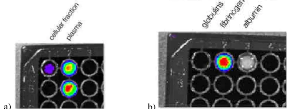
Figure 1. Blood fractionation assay results; a) Quantum Dots tends to preferentially remain in the plasmatic fraction after Ficoll gradient centrifugation and b) precipitate together with Fibrinogen, showing a low solubility or a possible binding
between fibrinogen and Quantum Dots themselves.
In this work, we used untargeted Quantum Dots, commercially available from Invitrogen, with a protective layer of PEG on the surface; this layer protects both Quantum Dots from degradation and consequent release of Cadmium ions, and should reduce accumulation in particular districts of the organism, such as liver and bone marrow [3]. We first studied the behaviour of Quantum Dots in the blood flow, to investigate their partition in the various components. Using a partitioning method, we
Nanosafe 2008: International Conference on Safe production and use of nanomaterials IOP Publishing Journal of Physics: Conference Series 170 (2009) 012021 doi:10.1088/1742-6596/170/1/012021
were able to understand that, at least in nude mice, QDots tend to remain in the plasma fraction (Figure 1a), without entering the cells. Moreover, as can be seen in the plasma fractionation assay (Figure 1b), they precipitate together with fibrinogen, which is the less soluble plasmatic protein; this point can be explained by a general poor solubility of QDots in the plasma, or by coverage of the QDots surface with fibrinogen. Chromatography assays are in progress to elucidate the proportions of these two events, since gel electrophoresis mobility assays are less likely to be performed, due to the size of QDots themselves (around 10 nm).
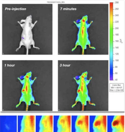
Figure 2. In vivo Optical Imaging acquisition of a mice injected with a solution of quantum dots emitting at 800 nm (Invitrogen). It is clear the accumulation in the liver area, as highlighted in the boxes below (time scale 3 hours).
To focus the molecular investigation in the actual site of accumulation, we perform a short-term distribution study of these molecules, finding that even after three hours after the intravenous injection, QDots show a strong liver accumulation, even in the presence of the PEG layer (Figure 2). We thus decide to proceed to the proteomic investigation of this organ, in a first step. Liver was resected, and processed following a non-denaturing protein extraction protocol. Proteins were separated on a 1D electrophoresis gel, stained with Sypro Ruby, and after imaging of the gels some protein bands are clearly altered (Figure 3a, red arrows), and are now in proceeding mass spectrometry investigation to sequence these proteins, to understand which molecular pathways are interested in the response to QDots treatment. As already told, a recent paper [4] showed a p53-mediated response to QDots treatment and Cadmium toxicity. Further, Fruk and co-workers [7] were able to trigger the activity of Horseradish Peroxidase by the mean of QDots light emission, proving the production of reactive oxygen species (H2O2, and O2
-) during QDots light emission process, and another recent paper, showed as QDots toxicity in aquatic organism is due to ROS production [8]. We therefore decide to characterise the role of APE1/Ref1 (Apurinic/apyrimidinic endonuclease/redox effector factor-1), a protein involved in DNA repair, in particular in the base excision process. During conditions of oxidative stress, the protein migrates into the nucleus, where it works also as a transcription enhancer, strengthening the DNA binding of proteins like p53; the effect is an inhibition of the cell cycle through the inhibition of the PI3K/AKT pathway. We checked its expression levels by the mean of western blot analysis, but in the time scale of the experiments performed we weren’t able
Nanosafe 2008: International Conference on Safe production and use of nanomaterials IOP Publishing Journal of Physics: Conference Series 170 (2009) 012021 doi:10.1088/1742-6596/170/1/012021
to identify a statistically significant change; however, and surprisingly, it is important to notice the strong increase in the binding between this protein and p53. This results points out an important role of APE1/Ref-1, which is a ‘sensor’ of the oxidative stress in the cells, in the response to radicals produced by QDots, and can be an advantage, for instance, in the case of photodynamic therapy of p53-negative tumours, whit an incremented damage due to the reduced reactivity in this molecular pathway.
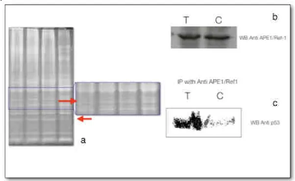
Figure 3. a) red arrows indicate differentially present protein bands in whole liver lysates, which are undergoing mass spectrometry identification; b and c) show that while the expression of APE1/Ref1 remains unchanged in the liver after the administration of Quantum Dots, its binding activity with a fundamental molecule as p53 changes dramatically, suggesting a
substantial role of APE1/Ref1 in p53-mediated Quantum Dots response. 4. Conclusion
Quantum Dots are entrusted great expectations in future biomedical applications, and in particular in diagnostics. Several studies have investigated these potential applications after the injection in animal models. To date, anyway, only limited data exist about the potential side effects of Quantum Dots after the injection within the organism. We have thus applied a molecular biology approach to investigate this impact, and even though our data are still preliminary and in need of further investigation, our results point out a possible interaction with the blood flow dynamics, with a supposed binding of Quantum Dots and fibrinogen, and the activation of a p53 mediated DNA damage response pathway. In conclusion, these preliminary achievements prompt us to proceed with our approach, as it can represent an added value to conventional toxicology characterization of nanoparticles biological impact.
References
[1] Alivisatos P 2004 Nat Biotechnol 22 47 [2] Duberdret B 2002 Science 298(5599) 1759 [3] Cho S J 2007 Langmuir 23(4) 1974 [4] Choi A O 2008 J Mol Med. 86(3) 291 [5] Fung H 2005 Mol Cell. 17(3) 463
[6] Bechtold W E 1992 Carcinogenesis 13(7) 1217 [7] Fruk L 2007 Chembiochem. 8(18) 2195
[8] Gagnè F 2008 Aquat Toxicol 86(3) 333
Nanosafe 2008: International Conference on Safe production and use of nanomaterials IOP Publishing Journal of Physics: Conference Series 170 (2009) 012021 doi:10.1088/1742-6596/170/1/012021