1
UNIVERSITÀ DEGLI STUDI DI ROMA
"TOR VERGATA"
FACOLTA' DI SCIENZE MATEMATICHE, FISICHE E NATURALI
DOTTORATO DI RICERCA IN IMMUNOLOGIA E
BIOTECNOLOGIE APPLICATE
XXI CICLO
IL-10 mediated immunological tolerance:
preclinical and clinical studies
Giorgia Serafini
Docente Guida:
Dott.ssa Rosa Bacchetta
Prof.ssa
Maria
Grazia
Roncarolo
Coordinatore:
Prof. Paolo Rossi
2
INDEX
ABSTRACT
pg. 4LIST OF PAPERS
pg. 8ABBREVIATIONS
pg. 9
INTRODUCTION
Thalassemias
pg. 11 β-thalassemia pg. 12
Stem cell transplantation for β-thalassemia pg. 13
Persistent mixed chimerism pg. 18
IL-10
pg. 20IL-10 in vivo in mouse model pg. 21
IL-10 in vivo in human pg. 23
Type
1
regulatory
T
cells
pg. 25Tr1 cells in vivo pg. 28
In vitro induction of Tr1 cells
pg. 31 Induction of Tr1 cells by immatureor semi-mature dendritic cells pg. 32 Induction of Tr1 cells by tolerogenic dendritic cells pg. 34
In vivo induction of Tr1 cells
pg. 363
PATIENTS, MATERIALS AND METHODS
pg. 42RESULTS SECTION I
High frequency of IL-10 producing T cells in the peripheral blood
of patients with PMC pg. 50
Kinetics of persistent mixed chimerism pg. 52
Characterization of T cell clones isolated from PBMCs
of the patient with PMC pg. 55
Antigen specificity of patient’s T cell clones pg. 61 Suppressive function of patient’s Tr1 cell clones pg. 65
DISCUSSION
SECTION
I
pg. 69
RESULTS SECTION II
IL-10 inhibits primary MLR proliferation and induces T-cell anergy pg. 74
DC-10 inhibit primary MLR proliferation pg. 78
DC-10 promote T-cell anergy pg. 80
Comparison between mocytes+IL-10 and tolerogenic
DC-10 in promoting T-cell anergy pg. 83
Gene expression profile of IL-10 and DC-10 anergized T cells pg. 85
DISCUSSION
SECTION
II
pg. 90
CONCLUSIVE
REMARKS
pg. 94
REFERENCES
pg. 95
4
ABSTRACT
Thalassemia Major can be cured with allogeneic hematopoietic stem-cell transplantation (HSCT). Persistent mixed chimerism (PMC) develops in around 10% of transplanted thalassemic patients, but the biological mechanisms underlying this phenomenon are poorly understood. IL-10 is an immunomodulant cytokine that plays a central role in controlling inflammation, down-regulating immune responses, and inducing immunological tolerance. Detection of high levels of IL-10, produced by PBMCs of patients with PMC, in comparison to those of patients with complete donor chimerism (CC) or normal donors (ND), prompted us to characterize the T cell repertoire in a thalassemic patient with long-term tolerance following HSCT. From the peripheral blood of the PMC patient, T cell clones of both host and donor origin could be isolated. Together with effector T cell clones, reactive against host or donor alloantigens, we identified regulatory T cell clones, with a cytokine secretion profile typical of type 1 regulatory T cells (Tr1), at high frequencies. Tr1 cell clones, both donor and host derived, were able to inhibit the function of effector T cells of either donor or host origin in vitro, suggesting a contribution by these regulatory T cells to the maintenance of PMC in vivo.
In parallel, we demonstrated that IL-10 could inhibit primary allogeneic proliferation of total PBMCs and could induce antigen-specific T-cell hyporesponsiveness. The use of tolerogenic dendritic cells (DC) differentiated in the presence of IL-10 (DC-10), as compared to monocytes+IL-10, allowed a more consistent anergy induction, even in the context of HLA-matched donors. Importantly, both monocytes+IL-10-anergized
5
and DC-10-anergized T cells preserved their ability to respond to nominal and third party antigens.
All together these results provide first, new insights regarding the mechanisms of peripheral tolerance in transplanted thalassemic patients, and secondly, offer a strong rationale for the development of a clinical protocol for the use of ex-vivo monocytes+ IL-10/DC-10-anergized T cells of donor origin as cellular therapy to promote immune-reconstitution and prevent graft-versus-host disease after HSCT.
6
RIASSUNTO
La Talassemia Major è una patologia che può essere curata con il trapianto di cellule staminali. In circa il 10% dei pazienti talassemici, dopo il trapianto si sviluppa una condizione immunologica chiamata chimerismo misto persistente, ma i meccanismi biologici alla base di questo fenomeno sono ancora poco conosciuti. L’interleuchina IL-10 è una citochina immunomodulante che gioca un ruolo centrale nel controllare i fenomeni infiammatori, abbassare le risposte immunitarie e indurre tolleranza immunologica. L’osservazione di alti livelli di IL-10, prodotta dalle PBMCs di pazienti con chimerismo misto persistente, in confronto ai livelli di IL-10 prodotta da pazienti con chimerismo completo o da donatori sani, ci ha spinto a caratterizzare il repertorio delle cellule T di un paziente talassemico con tolleranza a lungo termine dopo trapianto di midollo osseo. Dal sangue periferico del paziente con chimerismo misto persistente sono stati isolati cloni T di origine sia del donatore che del ricevente. Insieme a cloni T effettori, reattivi nei confronti di allo-antigeni del donatore e del ricevente, sono stati isolati ad alte frequenze cloni T regolatori con un profilo citochinico tipico delle cellule T regolatorie di tipo 1 (Tr1). I cloni Tr1, di origine sia del ricevente che del donatore, si sono dimostrati capaci di inibire le funzioni delle cellule effettrici di entrambi le origini in vitro, suggerendo un contributo da parte di queste cellule regolatorie nel mantenimento del chimerismo misto persistente in vivo. In parallelo, abbiamo dimostrato che l’interleuchina IL-10 può inibire la proliferazione primaria allogenica delle PBMCs, e può down-regolare la risposta delle cellule T antigene-specifica. L’uso di cellule dendritiche differenziate in presenza di IL-10 (DC-10), in paragone all’uso di monociti+IL-10, induce uno stato di anergia più
7
consistente, perfino nel contesto di donatori HLA-identici. Le cellue anergizzate, sia in presenza di monociti+IL-10, sia in presenza di DC-10, mantengono la loro capacità di rispondere ad antigeni scorrelati.
Questi risultati, forniscono nuove conoscenze riguardo i meccanismi che regolano la tolleranza periferica in pazienti talassemici trapiantati, e successivamente offrono un forte razionale per lo sviluppo di un protocollo clinico basato sull’uso di cellule T del donatore anergizzate ex vivo in presenza di monociti+IL-10/DC-10, come terapia cellulare per promuovere l’immuno-ricostituzione e prevenire la “malattia da trapianto contro l’ospite” (GvHD) dopo trapianto di midollo osseo.
8
LIST OF PAPERS
I. Giorgia Serafini, Marco Andreani, Manuela Testi, MariaRosa Battarra, Andrea Bontadini, Katharina Fleischhauer, Sarah Marktel, Guido Lucarelli, Maria Grazia Roncarolo, and Rosa Bacchetta. “Type 1 regulatory T cells are associated with persistent split erythroid/lymphoid chimerism after allogeneic hematopoietic stem cell transplantation for thalassemia.” Submitted to Haematologica Journal- Accepted.
II. Silvia Gregori, Giorgia Serafini, Claudia Sartirana, Ute Schulz, Elisabetta Zino, Khatarina Fleishhauer, Stefan Tomiuk, Uwe Janßen, Maria Grazia Roncarolo and Rosa Bacchetta. “Molecular and functional characterization of alloantigen-specific anergic T-cell suitable for cell therapy”. Manuscript in preparation.
9
ABBREVIATIONS
TM Thalassemia major RBC Red blood cell
TT Thalassemia intermedia
HSCT Hematopoietic stem-cell transplantation
BU Busulfan
CY Cyclophosphamide
GvHD Graft versus host disease CC Complete chimerism MC Mixed chimerism
RHCs Residual host cells RHCs TMC Transient mixed chimerism PMC Persistent mixed chimerism PHA Phytohemagglutinin
PBMC Peripheral blood mononuclear cell Tr1 Type 1 regulatory T cells
IBD Inflammatory bowel disease
EAE Experimental autoimmune encephalomyelitis RA Rheumatoid arthritis
Ag Antigen
NOD Nonobese diabetic SIT Specific immunotherapy APC Antigen presenting cells
10 SCID Severe combined immunodeficient Treg Regulatory T cells
GZ Granzyme
DC Dendritic cells
MHC Major histocompatibility complex T1DM Type 1 diabetes
HL Hodgkin lymphoma
HNSCC Head and neck squamous cell carcinoma TIL Tumor infiltrating lymphocytes
MLR Mixed lymphocyte reaction iDC Immature DC
mDC Mature DC
LPS Lipopolysaccharide NHP Nonhuman primates
DLI Donor lymphocyte infusions GVL Graft-vs.-leukemia activety GVT Graft-vs.- tumor activity
11
INTRODUCTION
Thalassemias
Thalassemia is a genetic disease characterized by defects in the hemoglobin production. Although originally endemic to the tropics and subtropics, it is now found worldwide as a result of migration and has become an important part of clinical practice in Europe, US and Australasia. Moreover, it is estimated that thalassemia is among the most common genetic disorders: 4.83 percent of the world’s population carry globin variants, including 1.67 percent of the population who are heterozygous for α-thalassemia and β-thalassemia (1).
The thalassemias are named according to the globin chain affected, therefore, α-globin gene mutations give rise to α-thalassemia and α-globin mutations cause β-thalassemia. Normal human hemoglobin is composed of two α-like and two β-like globin chains. Adult hemoglobin consists of hemoglobin A (α2β2) plus small amounts
of hemoglobin A2 (α2δ2) and hemoglobin F (α2γ2). Genetic mutations in one of the
globin genes (α or β) result in decreased or absent production of that globin chain and a relative excess of the other. These mutations can result in no globin production (β° or α°) or decreased globin production (β+ or α+). In addition, thalassemias can be
classified by the clinical severity. Thalassemia major (TM) syndrome requires more than eight red blood cell (RBC) transfusions per year while thalassemia intermedia
12
(TI) requires no or infrequent transfusions. Untreated TM is fatal in the first few years of life; in addition, TM and severe TI can lead to considerable morbidity affecting nearly all organ systems (2).
Beta-Thalassemia
The β-globin gene resides on the chromosome 11. More than 200 mutations on this gene are known to result in a phenotype of β-thalassemia, while, in contrast to the α-thalassemias, the disease is rarely caused by deletions. As indicated above, there are two main varieties of β-thalassemia alleles: β0 thalassemia in which no β-globin is
produced, and β+ thalassemia in which some β-globin is produced, but less than
normal (3). The clinical manifestations of β-thalassemia, corresponding to the degree of expression of the two β-globin genes that encode β-globin, are extremely various. The most severe condition is characterized by the results of the trasmission of two β0
thalassemia alleles (TM). This condition causes severe ineffective erythropoiesis, massive erythroid hyperplasia in the bone marrow and extramedullary sites, and hemolytic anemia necessitating chronic transfusions. When only one β-globin gene is affected, the resulting phenotype is milder, depending on the degree of gene expression and relative imbalance of globin chain (2).
In patients with β-thalassemia major both ineffective erythropoiesis and chronic transfusion therapy inevitably lead to iron overload resulting in multiple organ damage which causes endocrine deficiencies, liver disease, and cardiac disease with poor quality of life and increased mortality. Regular blood transfusion and iron chelation have improved both survival and quality of life and have changed a previously fatal disease with early death to a chronic, although progressive disease
13
compatible with prolonged survival (4). Up to date, allogeneic hematopoietic stem-cell transplantation (HSCT) represents the only cure for this and for other hemoglobinopathies (4-6).
Hematopoietic Stem Cell Transplantation for Thalassemia
The first two HSCT procedures for thalassemia were carried out in December 1981, in Seattle (WA) and in Pesaro (Italy) with marrow from HLA-identical relatives (5, 7). Since Pesaro group collected large experience during these years and became one of the Italian reference group for transplantation in thalassemia, I will focus my attention on the patients treated with the Pesaro protocols for bone marrow transplantation.
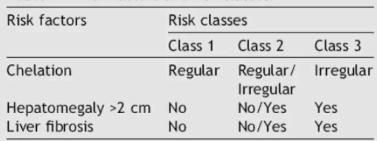
Prof. Lucarelli classified patients into three risk classes based on clinical characteristics and on liver biopsy (Table 1). The three risk factors that were considered for the classification were: iron chelation, hepatomegaly more than 2 cm and liver fibrosis.
From Lucarelli et al., Blood Reviews, 2008
14
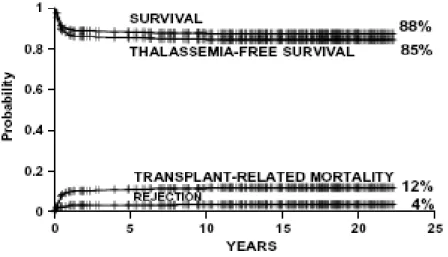
Class 1 patients had none of these adverse risk factors, class 3 patients had all three and class 2 patients had one or two adverse risk factors (Table 1). For the classes 1 and 2 the transplant protocol was similar, based on busulfan (BU) for the eradication of the hematopoietic system, and on cyclophosphamide (CY) for the suppression of the immune system. Graft versus host disease (GvHD), that could be a complication of the transplant, was prevented by the prophylactic administration of cyclosporine with or without methotrexate after the HSCT (6). The group of Prof. Lucarelli transplanted five hundred and fifteen class 1 and class 2 patients with median age of 7 years (range from 1 to 16 years) between October 1985 and August 2007. The thalassemia-free survival obtained was of 85%, the transplant-related mortality of 12%, and the incidence of rejection of 4% (4) (Figure 1).
From Lucarelli et al., Blood Reviews, 2008
Figure 1. Estimates of survival, thalassemia-free survival, non-rejection mortality and rejection for 515 class 1 and class 2 patients younger than 17 years.
15
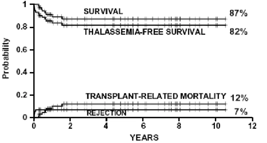
different procedures, in April 1997 a new regiment, called Protocol 26, was adopted for class 3 patients younger than 17 years. The survival, thalassemia-free survival, rejection and non-rejection mortality in 73 class 3 patients, aged less than 17 years with median age of 11 years (range 4–16 years), treated with Protocol 26 were 87%, 82%, 7% and 12%, respectively (4, 8) (Figure 2).
From Lucarelli et al., Blood Reviews, 2008
Figure 2. Estimates of survival, thalassemia-free survival, non-rejection mortality and rejection for 73 class 3 patients younger than 17 years who were treated with Protocol 26.
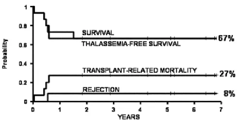
A regiment similar to the Protocol 26 was adopted for the Adult group. The probability of survival, thalassemia-free survival, rejection and non-rejection mortality in 15 high risk group patients were 67%, 67%, 8% and 27% respectively (9) (Figure 3).
16
From Gaziev et al, Ann. N.Y.Ac. Sci.,2005
Figure 3. Estimates of survival, thalassemia-free survival, non-rejection mortality and rejection in 15 adult patients treated with Protocol 26 regimen.
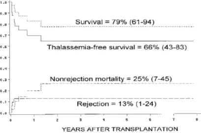
The majority of thalassemic patients (70%) do not have an HLA-identical donor within the family, therefore alternative stem cell donations have to be developed. In the last years the number of HSCT from unrelated donors has increased progressively, due to the higher number of volunteer donors worldwide. Moreover, thanks to the introduction of high-resolution molecular techniques for histocompatibility testing, the outcome of the transplant has gradually improved, reaching results comparable to those obtained from HLA-matched family donors. An analysis of the outcome of HSCT of 32 thalassemic patients transplanted with unrelated donors showed a survival, thalassemia-free survival, rejection and non-rejection mortality of 79%, 66%, 13% and 25%, respectively (10) (Figure 4) .
17
From La Nasa et al, Blood, 2002
Figure 4. Estimates of survival, thalassemia-free survival, non-rejection mortality and rejection for 32 thalassemia patients who received transplants from HLA-matched unrelated donors (between parenthesis: 95% confidence limits at 2 years).
More recently, in another study of HSCT from unrelated donors, it has been obtained comparable results with the probability of survival, thalassemia-free survival, rejection and transplant-related mortality of 70%, 70%, 4% and 30%, respectively (11) (Figure 5).
18
Figure 5. Estimates of survival, thalassemia-free survival, non-rejection mortality and rejection in 27 adult class 3 thalassemia patients transplanted from HLA-matched unrelated donors (between parenthesis: 95% confidence limit).
Persistent mixed chimerism
It is well known that when complete chimerism (CC) of the marrow donor cells is established, a successful HSCT occurred. However, the presence of mixed chimerism (MC), i.e., the coexistence of donor and host cells in the recipient, is not a rare event following transplantation. The group of Pesaro established three different levels of MC based on the percentage of residual host cells (RHCs) present in the patient after the transplant. Patients with RHCs less than 10% were included in the level 1, between 10% and 30% in the level 2, and patients with more than 30% of RHCs in the level 3. Nesci reported that the incidence of MC at 2, 6 and 12 months after the transplant was 36,5%, 34,7% and 16,7%, respectively in Pesaro experience, and that the majority of the patients at 2 and 6 months after HSCT had a level 1 of MC (less than 10% of RHCs). Moreover, they showed that the probability of rejection for patients with MC levels 1 and 2 was 19% and 17% respectively, while all the patients with MC level 3 rejected the transplant within the first year after HSCT (12). Therefore, MC represents a risk factor for graft rejection if it occurs within the first months after the transplant (transient mixed chimerism [TMC]) (13).
Nevertheless, when the coexistence of donor and host cells persisted in the marrow and in the peripheral blood for a period longer than two years (persistent mixed chimerism [PMC]), patients had a functional graft and remained blood-transfusion independent with hemoglobin levels ranging from 8.3 g/dl to 14.7 g/dl, similarly to
19
those of patients with full donor engraftment (14). These observations suggest that few engrafted donor cells may be sufficient to correct the disease phenotype in patients with thalassemia major once the tolerance between host and donor cells has been established. Battaglia analyzed the T-cell repertoire of 3 PMC patients by measuring the CDR3 length of TCR Vβ families. She showed a profound skewed repertoire with small number of specific T-cell clonotypes in the peripheral blood of these patients. After in vitro stimulation with phytohemagglutinin (PHA), the normal Gaussian distribution TCR CDR3 sizes were reestablished. This showed that the skewed repertoire in the peripheral blood mononuclear cell (PBMC) was associated with an expansion of specific T cells but not with a collapse of the whole repertoire and it could be related to an active mechanism of peripheral tolerance (15).
Up to date the mechanisms responsible for the induction and maintenance of PMC after HSCT are unknown.
20
IL-10
Since its discovery in 1990 (16) (17), IL-10 has gained increasing importance as a potent immunomodulatory cytokine that plays a central role in controlling inflammatory processes, suppressing T-cell responses and maintaining immunological tolerance (18). IL-10 directly reduces T-cell proliferation by inhibiting IL-2, TNF-α and IL-5 production (19-21), and down-regulates production of inflammatory cytokines as well as expression of costimulatory molecules on monocytes/macrophages (22). IL-10 enhances the cytotoxicity and the IFN-γ production of NK cells (23), and may have inhibitory and stimulatory effects on human CD8+ T depending on their state of activation (18) (24). IL-10 has also an
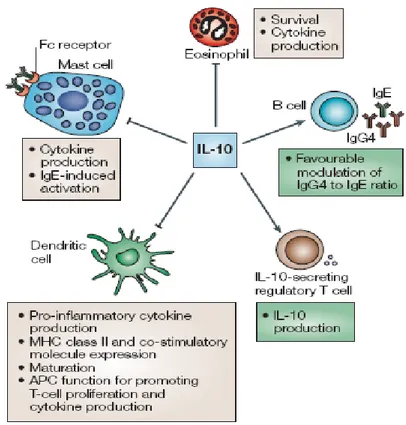
antiapoptotic effect on B cells, is involved in B-cell isotype switching and plays a role in autoimmune diseases with underlying B-cell dysregulation (25). Moreover, IL-10 not only induces long-lasting anergy in both CD4+ (26) and CD8+ T cells (24) but also the differentiation of type 1 regulatory T (Tr1) cells both in humans and mice (27) (Figure 6).
21
From Hawrylowicz CM et al. Nat Rev Immunol 2005
Figure 6. Functions of interlukin-10. IL-10 is a pleiotropic cytokine that has been shown to inhibit activation of, and cytokine generation by, mast cells, as well as survival of, and cytokine production by, eosinophils. It also inhibits antigen-presenting cell function, including the maturation of dendritic cells, the expression of MHC class II and co-stimulatory molecules and the activation of T-helper-2 cells. IL-10 has been shown to enhance immunoglobulin isotype switching in B cells, and it might promote the induction of IL-10-secreting regulatory T cells.
IL-10 in vivo in mouse model
The role of IL-10 in controlling the immune response and inducing tolerance has been demonstrated in several animal models. IL-10 deficient mice lack regulatory T cells, develop inflammatory bowel disease (IBD) (28) and a severe clinical experimental autoimmune encephalomyelitis (EAE) (29). IL-10 has been shown to be effective in inhibiting allergic inflammation in animal models. Injection of IL-10 before allergen
22
treatment induced an hapten-specific T cell tolerance in mice, inhibiting the production of proiflammatory cytokines (30). Moreover, adoptive transfer of CD4+ T cells engineered to produce IL-10, effectively inhibited the capacity of effector cells to induce allergic airway inflammation (31).
These results provided a strong rational for the use of IL-10 in vivo to prevent autoimmune diseases and allograft rejection through the induction of peripheral tolerance. Unfortunately, after the in vivo administration of IL-10 in different experimental models of autoimmune disease and transplantation, contrasting effects have been described. In models of autoimmune diseases mediated by Th1 cells, such as rheumatoid arthritis (RA), IL-10 administration before and/or after induction of the disease reduced joint inflammation (32). In an adoptive transfer model of EAE, intravenous injection of IL-10 failed to abrogate the disease (33), while the intraperitoneal treatment could inhibited antigen(Ag)-induced EAE in mice if administered at the time of immunization (34). In nonobese diabetic (NOD) mouse, daily subcutaneous administration of IL-10 before 9-10 weeks of age, prevented cellular infiltration of islet cells, delayed the onset of disease and significantly reduced the incidence of diabetes (35). Conversely, 10 transgenic NOD mice, in which IL-10 was expressed in the pancreatic islets, had severe insulitis and prominent ductal proliferation. In addition, the onset of diabetes in NOD transgenic mice was earlier than that of 14 weeks of age at the earliest in non-transgenic NOD mice (36).
Similarly, in mouse model of bone marrow transplantation, Blazar showed that IL-10 had a dose-dependent effect on the GvHD lethality, because high doses accelerated lethality, while low amounts protected mice from GvHD (37). In a mouse model of heart allograft, the effects of exogenous IL-10 on organ allograft survival were dependent on timing and dosage. IL-10 treatment prior to the transplant increased
23
heart survival while the administration at the time of the graft or after the graft, had little beneficial effect or even promoted rejection (38).
These controversial data showed that the several effects of IL-10 on the modulation of autoimmune diseases and graft rejection, depended on the site and time of administration and importantly, on the dose of cytokine injected.
IL-10 in vivo in human
Similarly to the mouse, the role of IL-10 in the control of immune response in human is well accepted. Several reports showed that IL-10 modulated many cells and effector functions associated with allergy. Lim described an inverse association between IL-10 levels and the severity of allergic and asthmatic disease, demonstrating that in asthma, the low IL10-producing haplotype was more likely to be associated with severe disease (39). In the nasal mucosa from allergic rhinitis patients, IL-10 was expressed with large variation between individuals that correlates with allergic symptoms. Patients with lower epithelial IL-10 levels had severe allergic rhinitis in comparison to patients with mild symptoms (40). Moreover, it has been demonstrated that IL-10 mRNA expression, by purified T cells of children with allergic and non-allergic asthma and children with atopic dermatitis, was strongly decreased as compared with that of healthy controls (41). Several reports indicated that IL-10 could play a pivotal role also in allergen-specific immunotherapy (SIT), efficiently used in allergy to insect venoms and allergic rhinitis. SIT was found to be associated with a decrease in IL-4 and IL-5 production and an increased in IL-10 and/or TGF-β production (42). Akdis showed that during the early phase of the bee venom-SIT, intracellular IL-10 significantly increased in the Ag-specific T cell population and activated CD4+ T
24
lymphocytes, while B cells and monocytes were responsible for IL-10 production at later time points. The data indicated that the anergic state in T cells was induced by IL-10, initially produced by the peripheral T cells themselves after high-dose of allergen administration and then followed by the modulated antigen-presenting cell (APC) population, required to maintain tolerance (43). The proliferative and cytokine responses could be reconstituted by ex vivo neutralization of endogenous IL-10, indicating that this cytokine was actively involved in promoting tolerance after allergen-SIT (43). Moreover, it has been demonstrated that therapies useful for the treatment of allergy and asthma, such as glucocorticoids, promoted the increase of IL-10 secretion by T cells (44).
In bone marrow transplantation, several works focalized their attention on the correlation between IL-10 and the absence of GvHD after HSCT. The importance of endogenous IL-10 production has been clearly demonstrated in severe combined immunodeficient (SCID) patients successfully transplanted with HLA-mismatched allogeneic fetal liver stem cells. In the absence of immunosuppressive therapy, these patients did not developed GvHD, despite the HLA disparity. Interestingly, high levels of IL-10 mRNA were detected in vivo in T cells and monocytes of these patients (45). Moreover, it has been demonstrated a positive correlation between high spontaneous IL-10 production by PBMC of patients prior to allogeneic bone marrow transplantation and a subsequent low incidence of GvHD and transplant-related mortality, compared to patients with low or intermediate IL-10 production. In patients with high cellular IL-10 production, there was also an increased in IL-10 serum levels, indicating a simultaneous occurrence of cellular as well as systemic IL-10 production (46, 47).
25
Type 1 Regulatory T cells
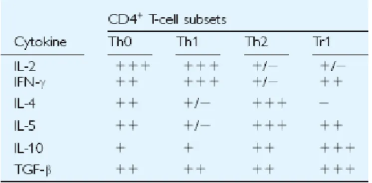
The first type of IL-10-producing Regulatory T cells (Tregs) described at the clonal level was designated Tr1 cells by Groux and colleagues. These cells are characterized by their capacity to produce high levels of IL-10 and TGF-β, and by their ability to suppress Ag-specific effector T-cell responses in vitro and in vivo via a cytokine-dependent mechanism mediated by IL-10 and TGF-β (27). Tr1 cells can be generated in vitro and in vivo upon priming of naïve T cell precursors with the Ag in the presence of IL-10. Tr1 cells can be distinguished from Th1 and Th2 cells, because they produce high levels of IL-10, TGF- β, and IL-5; low amounts of IFN-γ and IL-2; and no IL-4 (Table 2).
From Roncarolo et al., Immunol. Reviews, 2001
Table 2. The typical cytokine production profile of human CD4+ T cell subsets
Tr1 cells secrete IL-10 very soon after activation as compared to others T cell subsets. IL-10 is detectable 4 hours after activation and the highest concentration is reached 12–24 h after activation (45, 48). Depending on the experimental conditions used for
26
their induction, Tr1 cells can produce different amounts of TGF-β, IFN-γ, and/or IL-5, but their levels of IL-10 production, which represents the true hallmark of Tr1 cells, are invariably high.
Tr1 cells are anergic upon polyclonal-mediated or Ag-specific activation (27, 45). These cells do not expand significantly under standard culture conditions, this could be due to the autocrine production of IL-10, since addition of anti-IL-10 monoclonal antibody partially restores the proliferation (27, 45). Even if Tr1 cells display low proliferative capacity, they can expand in the presence of IL-2 and IL-15. Long-term culture in IL-15 does not alter the phenotype or function of Tr1 cell clones, although it enhances production of IFN-γ (49). This observation is support by the fact that activated Tr1 cells express the IL-2 receptor (IL-2R) α chain, and high levels of the IL-15Rα chain, together with both the IL-2/15Rβ and IL-2Rγ chains. Upon TCR-mediated activation Tr1 cells also express activation markers such as CD40L, CD69, CD28, cytotoxic T-lymphocyte antigen-4 CTLA-4 and HLA-DR (49). In the resting phase, human Tr1 cells express both Th1-associated (CXCR3 and CCR5) and Th2-associated (CCR3, CCR4, and CCR8) chemokine receptors (50).CCR8 is expressed at higher levels compared with Th2 cells, and upon activation, human Tr1 cells migrate preferentially in response to I-309, a ligand for CCR8 (50). It has been recently demonstrated that in a model of helminth infection the expression of CCR8 was strongly associated with IL-10-producing CD4+ T cells, which resemble Tr1 cells, suggesting a relevant role of CCR8 in vivo (51). Several reports showed that Tr1 cells do not constitutively express the transcription factor forkhead box protein 3 (Foxp3), but they can upregulated Foxp3, upon activation, to levels similar to those observed in activated CD4+CD25- T cells (52, 53). Although significant efforts have
27
other Treg subsets and from effector T cells, no specific marker for Tr1 cells has been reported so far. The low proliferative capacity of Tr1 cells is a major limitation and has hampered progress in characterizing this T cell subset in vitro.
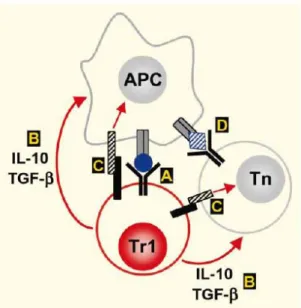
Tr1 cells are involved in down-regulation of immune responses due to their ability to produce high levels of the immunosuppressive cytokines IL-10 and TGF-β. They suppress both naıve and memory T-cell responses in vivo and in vitro (27, 45, 54). Ag-specific Tr1 cells need to be activated via the TCR to exert their suppressive function; but, once activated, they can mediate bystander suppressive activity against other Ags by the release of IL-10 and TGF-β (55, 56) (Figure 7).
From Roncarolo et al., Immunol. Reviews, 2001
Figure 7. Possible mechanisms of action of Tr1 cells. (A) Tr1 cells release IL-10 and TGF-b into the microenvironment after activation via the TCR. (B) These cytokines act on APCs and/or naive (Tn) or memory (Th) CD4+ T cells in close proximity, resulting in
indirect and direct suppression of T-cell-mediated responses. (C) Tr1 cells may also upregulate inhibitory receptors, whose ligands are expressed on APCs and/or T cells. (D) Tr1 cells mediate bystander suppression, and thus do not necessarily share the same Ag specificity as the target T cells.
28
The suppressive effects of Tr1 cells are reversed by addition of IL10 and anti-TGF-β neutralizing monoclonal antibodies (27, 45, 52), but additional mechanisms may also contribute. For example, Grossman recently demonstrated that human Tr1 cells generated in vitro by cross-linking of CD3 and CD46, a complement regulatory protein, and nTreg cells, can develop considerable cytotoxic activity, through the production of granzyme (GZ) B and GZ-A granules, respectively, in a perforin-dependent manner (57-59).
Tr1 cells in vivo
As indicated previously, the importance of IL-10 in controlling immune response has been widely demonstrated. Furthermore, up to now several works provided evidence that Tr1 cells are involved in many T cell-mediated diseases and could play a pivotal role in the regulation of transplantation tolerance in vivo.
In the setting of bone marrow transplantation, the first suggestion that human Tr1 cells were involved in maintaining peripheral tolerance in vivo came from studies in SCID patients successfully transplanted with HLA-mismatched allogeneic stem cells. From the peripheral blood of these patients it would be possible to isolate a high proportion of donor-derived T cells, specific for the host HLA Ags and able to produced high levels of IL-10 (45).
Interesting is the finding reported by Weston, who showed a correlation between high number of donor T cells producing IL-10 towards recipient allo-Ags and the absence of acute GvHD after HSCT. He suggested that a condition where IL-10 was present during repetitive presentation of the Ag in vivo could therefore predispose to less or absent grades of acute GvHD (60). Moreover, the importance of IL-10 and TGF-β
29
producing CD4+ T cells has been described in solid organ transplantation (61-62), and correlates with the spontaneous development of tolerance to kidney or liver allograft. Regarding the role of Tr1 cells in allergy, Nickel-specific Tr1 cell clones could be isolated from both peripheral blood and lesional skin of patients allergic to nickel (Ni). These Ni-specific Tr1 cells inhibited in an IL-10 dependent manner the functions of monocytes and dendritic cells (DC) and directly suppress Ni-specific Th1 responses. The results showed that Tr1 cells may have a crucial role in limiting excessive tissue reaction to haptens by blocking DC and monocytes functions and Th1 cell response. The high frequency of Tr1 cells in nonallergic donors also suggests that these cells may be important in determining whether silent immune responses or manifest allergic disease can develop (63). Akdis demonstrated an increased frequency of specific Tr1 cells with a concomitant decreased of allergen-specific Th2 cells in non-allergic subjects compared to allergic individuals. These results clearly indicated that the balance between allergen-specific Tr1 and Th2 cells was a key determinant in the development of allergic responses in humans (64).
Several works showed that Tr1 cells regulate responses to self-Ags controlling autoimmunity. Indeed, self-major histocompatibility complex (MHC)-reactive Tr1 cell clones, functionally distinct from Ag-specific T cell clones, could be isolated from the peripheral blood of healthy individuals. These cell clones inhibited proliferation of primary CD4+ T cells and tetanus toxoid-specific T-cell clones via IL-10 and TGF-β, suggesting that activated self-MHC-reactive T cells with the cytokine phenotype of Tr1 cells, may be important regulatory cells that mediate peripheral tolerance and prevent the development of autoimmunity (65). Similarly, Arif showed that autoreactive T cells specific for HLA class II molecules associated with type 1 diabetes (T1DM) development, exhibited extreme polarization toward a
pro-30
inflammatory Th1 phenotype, producing predominantly IFN-γ, in patients with T1DM. On the contrary, autoreactive T cells from non-diabetic individuals, carrying HLA class II molecules associated with T1DM, were present but polarized to produce IL-10 in response to islet peptides. The authors concluded that development of T1DM depended on the balance of autoreactive Th1 and Treg cells, that islet destruction was characterized by pro-inflammatory autoreactive T cells, while the tolerant state was characterized by autoreactive IL-10 secreting T cells (66).
Finally, some reports indicated the presence in vivo of Tr1 cells specific for tumor Ags. In the context of Hodgkin lymphoma (HL), it has been demonstrated that Hodgkin lymphoma infiltrating lymphocytes (HLIL), not only were anergic, but also could profoundly inhibit effector T cell responses. HLIL were highly enriched in IL-10-secreting Tr1 and CD4+CD25+ Treg cells, which induced a profoundly immunosuppressive environment and so provided an explanation for the ineffective immune clearance of cancer cells (67). Strauss showed that in patients with head and neck squamous cell carcinoma (HNSCC), tumor infiltrating lymphocytes (TIL) were enriched in Treg cells. These subset of cells expressed a unique phenotype distinct from that of Treg in PBMC, they produced IL-10 and TGF-β and did not require cell-to-cell contact to responder cells for suppression. Furthermore, their presence in the tumor was linked to a poor prognosis in subjects with HNSCC (68).
31
In vitro induction of Tr1 cells
Since the importance of IL-10 and Tr1 cells in controlling immune responses and inducing peripheral tolerance in vivo has been widely demonstrated, several protocols have been developed to generate Tr1 cells in vitro.
Groux demonstrated that human CD4+ T cells, activated by allogeneic monocytes in a primary mixed lymphocyte reaction (MLR) in the presence of exogenous IL-10, failed to proliferate after restimulation with the same allo-Ags. The anergic state induced by IL-10 was long-lasting (26). During the culture in the presence of IL-10, T cells became not only anergic, but differentiated in a distinct population of IL-10 producing T cells. Activation of both human and murine CD4+ T cells in the presence of IL-10 generated T cell clones with a cytokine production profile typical of Tr1 cells and different from that of Th1 and Th2 cells (27).
Although IL-10 has been shown to be indispensable for Tr1 cell induction, it is not sufficient for the differentiation of Tr1 cells in the absence of APC in vitro (69). In mouse model, it has been demonstrated that IL-10 synergized with TGF-β to promote allo-Ag hyporesponsiveness in murine CD4+ T cells. These anergized T cells were Ag-specific since they proliferated in response to nominal Ags, and in vivo transfer of these cells resulted in the prevention of lethal GvHD after bone marrow transplantation (70). In other studies, it has been also shown that Ag-specific regulatory T cells developed via the ICOS–ICOS-ligand pathway and that both the induction and the function of Treg cells depended on the presence of IL-10 and ICOS pathway. These cells produced IL-10 and had potent in vivo and in vitro inhibitory activity; when transferred into sensitized mice they blocked the development of allergen-induced airway hyperreactivity (71).
32
In human, it has been demonstrated that endogenous or exogenous IL-10 in combination with IFN-α, but not TGF-β, promoted the differentiation of CD4+ T cells
derived from cord blood, into T cells with the cytokine profile and the biological activities of Tr1 cells. In parallel, naive CD4+ T cells derived from peripheral blood
required both exogenous IL-10 and IFN-α for Tr1 cell differentiation (72).
Kemper showed that co-engagement of CD3 and the complement protein CD46 in the presence of IL-2 induced a Tr1-specific cytokine phenotype in human CD4+ T cells. These CD3/CD46-stimulated IL-10-producing CD4+ cells proliferated strongly, suppressed activation of bystander T cells and acquired a memory phenotype (59). Finally, Barrat showed that vitamin D3 and Dexamethasone (Dex) could induced human and mouse naive CD4+ T cells to differentiate in vitro into regulatory T cells. The development of these Tr1 cells was enhanced by neutralization of the Th1 and Th2 inducing cytokines IL-4, IL-12, and IFN-γ. Tr1 cells were induced in vitro stimulating naïve T cells with immunosuppressive drugs in the presence of APC, but the authors demonstrated that Vit D3 and Dex also could induce the expansion of IL-10 producing T cells when naive CD4+ T cells were stimulated with immunosuppressive drugs plus anti-CD3 and anti-CD28 only. The injection of these Tr1 cells in a mouse model of experimental autoimmune encephalomyelitis (EAE), prevented EAE with absolute abrogation of disease onset and absence of clinical signs in an IL-10 dependent manner (54).
Induction of Tr1 cells by immature or semi-mature dendritic cells
DC are the most effective APC that regulate the balance between effector T and Treg cells on the basis of the expression of specific immunogenic or tolerogenic molecules
33
and of the secretion of inflammatory or modulatory cytokines (73). Several studies indicated that Treg cells could be induced by DC both in an immature or semi-mature stage. In the steady state, presentation of the Ag by immature DC (iDC) lacking of costimulatory stimuli and unable to stimulate immunity to the self-Ags they have captured, results in tolerance (74).
Jonuleit compared the influence of different maturational states of DC on the differentiation of naive CD4+ T cells, showing that iDC and mature DC (mDC) induced completely different T cell phenotypes. Repetitive stimulation of naïve cord blood CD4+ T cells with allogenic iDC resulted in the differentiation of IL-10 producing Treg. These T cells lost their ability to produce IFN-γ, IL-2, or IL-4 and became non-proliferating T cells able to inhibit the Ag-driven proliferation of Th1 cells in a contact- and dose-dependent, but Ag-nonspecific manner (75).
Similarly, it has been reported that repeated stimulation of naïve peripheral blood CD4+ T cells with allogeneic iDC could induce the differentiation of human Tr1 cells
in vitro. In this system, after three rounds of stimulation with iDC, T cells became profoundly anergic and acquired regulatory function. These T cells secreted high levels of IL-10 and TGF-β, significant amounts of IFN-γ and IL-5, low IL-2, and no IL-4, and they suppressed T-cell responses by the production of IL-10 and TGF-β. The induction of these Treg cells could be blocked by anti-IL10R monoclonal antibody, indicating that they were Tr1 cells generated through autocrine production of IL-10 by iDC (52).
Similarly, the stimulation of iDC with TNF-α in the absence of danger signals, resulted in incompletely matured DC (semi-mature DC). The maturation by TNF- α induced high levels of MHC class II and costimulatory molecules on DC, but they remained weak producers of proinflammatory cytokines. Therefore, these
semi-34
mature DC led to the induction of peptide-specific predominantly IL-10 producing CD4+ T cells in vivo and provided complete protection from EAE (76).
Induction of Tr1 cells by tolerogenic dendritic cells
It has been demonstrated that not only immature or semi-mature DC, but also DC rendered tolerogenic with biological or pharmacological agents, could drive the differentiation of Treg cells (48, 77).
IL-10 modulates the function of APC, including DC (22, 78). After IL-10 treatment, DC display reduced surface expression of MHC class I and class II molecules and costimulatory molecules of the B7 family (79), and the release of pro-inflammatory cytokines by DC, such as IL-1β, IL-6, TNF-α and most markedly IL-12, is abolished (80).
Immature DC differentiated and treated in vitro with exogenous IL-10 displayed reduced allo-stimulatory capacity and were shown to induce anergic Ag-specific T cells (81). Furthermore, DC matured in the presence of exogenous IL-10 for the last two days of culture, showed a strongly reduced capacity to stimulate a CD4+ T cell response in allogeneic MLR in a dose-dependent manner and induced Ag-specific anergic T cells (78). These anergic T cells were able to suppress activation and function of T cells in an Ag-specific manner through a cell-cell contact mechanism (82).
Gregori recently demonstrated that IL-10 induced the differentiation of a unique subset of myeloid DC (DC-10), which were CD14+CD11c+CD11b+CD83+ HLA-DR+CD71+CD1a-, displayed a myeloid mature phenotype (CD80+CD86+) and expressed ILT2, ILT3 and ILT4. These cells secreted significantly higher levels of
IL-35
10 compared with iDC, whereas the amounts of IL-12 were low and comparable to those produced by iDC. Interestingly, this ratio of cytokine production was maintained upon activation with lipopolysaccharide (LPS) and IFN-γ. DC-10 were potent inducers of Tr1 cells in a allogenic system in vitro, more powerful than iDC, since they reproducibly induced anergic T-cell lines, with strong suppressive activity, in a single round of stimulation (Gregori et al., 2008, manuscript submitted).
A variety of immunosuppressive agents currently used in the treatment of autoimmune diseases and in the control of allograft rejection, including glucocorticoids, mycophenolate mofetil and sirolimus, have been shown to induce DC with tolerogenic phenotype and function. These agents impair DC maturation and inhibit up-regulation of costimulatory molecules, secretion of proinflammatory cytokines, in particular IL-12, and allostimulatory capacity. On the other hand, controversial effects of calcineurin inhibitors, like cyclosporine A and tacrolimus, have been reported on DC maturation, although these drugs have a clear inhibitory effect on DC, decreasing their cytokine production and allostimulatory capacity (77). Several studies reported that DC loaded with tumor Ags could also induce the differentiation of Tr1 cells. For example, exposure of DCs to myeloma cells (83), or to an adenoviral vector expressing a prostate-specific antigen (PSA) (84) could prime DC to produce IL-10. These DC primed the differentiation of Tr1 cells, which inhibit effector T cells specific for PSA.
Finally, DCs could be engineered to express tolerogenic molecules, such as IL-10, TGF-β, CTLA-4, and drove the differentiation of Tregs that mediate Ag-specific tolerance (85).
36
In vivo induction of Tr1 cells
The induction in vivo of Tr1 cells, in addition to their ex vivo generation, is certainly an attractive goal.
Treatment of mice with a killed Mycobacterium vaccae-suspension gave rise to allergen-specific regulatory T cells which conferred protection against airway inflammation. This inhibition was mediated by IL-10 and TGF-β, since antibodies against IL-10 and TGF-β completely reversed the inhibitory effect of Tr1 cells (86). The possibility to induce in vivo Tr1 cells specific for allo-Ags has been investigated by our and other groups in models of pancreatic islet transplantation (87) (88). Asiedu demonstrated that peritransplant treatment of multiple nonhuman primates (NHP) with anti-CD3 immunotoxin and deoxyspergualin induced stable rejection-free tolerance to MHC-mismatched allografts, associated with sustained elevations in serum of IL-10. In this regimen, anti-CD3 immunotoxin depleted effector T cells, whereas deoxyspergualin arrested the production of proinflammatory cytokines and the maturation of DCs. An increase in the frequency of Tr1 cells was found in the peripheral blood of long-term tolerant NHP recipients in comparison with controls. Moreover, also an increase in CD4+ CD25+Foxp3+ Tregs frequency was detected in tolerant NHP. In summary, the study showed an expansion of Treg populations in tolerant NHP recipients, suggesting that these variations should be involved in maintenance of stable tolerance (87).
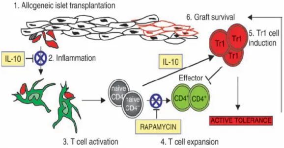
Similarly, Battaglia et al. demonstrated that an in vivo treatment with rapamycin + IL-10 was able to prevent allograft rejection in diabetic mice transplanted with allogeneic pancreatic islets. This treatment not only prevented acute allograft rejection but also led to active long-term tolerance via induction of Ag-specific Tr1 cells.
37
Administration of IL-10 alone was not able to prevent rejection. Even if IL-10 administration should down-regulated inflammation and allowed induction of Tr1 cells, it might not be sufficient to block the expansion of effector T cells. The combination of IL-10 with a compound able to down-modulate the effector phase of the immune response was required for the induction of operational tolerance in this model. Rapamycin blocked IL-2-induced expansion of effector T cells, without preventing Tr1 cell induction via TCR stimulation in the presence of IL-10 (Figure 8) (88).
Both studies suggested that induction of Ag specific long-term tolerance needed the synergistic effect of drugs that down-modulate inflammation, block effector T cells, and differentiate Tr1 cells.
38
From Roncarolo et al., Immun. Reviews, 2006
Fig. 8. Prevention of allograft rejection in vivo by Tr1 cells induced with rapamycin and interleukin-10. To prevent allograft rejection, the massive inflammation caused by the transplant must be modulated in order to reduce the recruitment and maturation of professional APC. (1). Expansion of T effector cells (Teff) must be tightly controlled and avoided, while conditions that permit Treg development and function should be preserved. Accordingly, treatment with rapamycin and interleukin-10 (IL-10) leads to allograft tolerance in diabetic mice transplanted with allogeneic pancreatic islets. Rapamycin and IL-10 have a general anti-inflammatory effect (2). Rapamycin, without inhibiting T-cell activation (3), blocks the early expansion of alloreactive T cells (4) and allows induction of Ag-specific Tr1 cells through IL-10 (5). With this combined therapy, graft survival and transplantation tolerance via induction of Tr1 cells is efficiently achieved (6).
39
Cellular therapy with Tr1 cells
At present, allogeneic HSCT is the treatment of choice for several disorders of the hematopoietic system, such as hematologic malignancies (leukemia, lymphoma, and multiple myeloma) and genetic diseases (β-thalassemia and primary immunodeficiencies) (89). Immunologic mechanisms mediated by donor derived T cells, contained along with the graft or administered separately as donor lymphocyte infusions (DLIs), make a major contribution to the eradication of malignant cells after HSCT, since donor cells not only accelerate immune reconstitution but also eliminate residual neoplastic cells [graft-vs.-leukemia activity (GVL) and graft-vs.- tumor activity (GVT)]. Unfortunately, acute and chronic GvHD which is also mediated by donor T cells and has been linked to tumor regression, remains one of the main clinical complications after allogeneic HSC transplantation (90). Thus, several studies has been made trying to suppress GvHD with maintenance of GVT activity.
The use of Treg cells as a cellular therapy after allogeneic HSC transplantation, represent a promising approach to separate the two effects. The role of natural occurring CD4+CD25+ Tregs has been extensively explored in preclinical murine models of cellular therapy (91). Edinger showed that, in a murine model of HSCT followed by engraftment of leukemia cells, CD4+CD25+ Tregs were able to prevent acute GvHD by suppressing alloreactive T-cell expansion without abrogating the anti-tumor effector functions mediated by these cells (92).
In view of the fact that Tr1 cells can be differentiate ex-vivo, the possibility to use this T cell subset as cellular therapy to prevent GvHD in vivo has been explored. The first demonstration that Tr1 cells differentiated ex-vivo with IL-10 could modulate immune responses in vivo came from Groux’s observation that Tr1 cell clones suppressed
Th1-40
mediated colitis induced in SCID mice by transfer of naïve CD4+CD45RBhi cells. He demonstrated that OVA-specific Tr1-cell clones were functional only if mice were treated with the specific OVA peptide, indicating that Tr1 cells had to be activated via the TCR in order to exert their regulatory effects (27).
In mouse model, IL-10 synergies with TGF-β to promote allo-Ag hyporesponsiveness in murine CD4+ T cells. Donor CD4+ T cells, anergized ex vivo and transferred in a preclinical model of bone marrow transplantation, were markedly impaired in inducing GvHD alloresponses to MHC class II mismatched recipients (70).
All together these results indicated that Ag-specific Tr1 cells generated ex vivo with different approaches can be used in cellular therapy to efficiently prevent GvHD in human in vivo (Figure 9).
41
From Roncarolo et al., Nature Rev. Immunol., 2007
Figure 9. Development and assessment of human cell therapy products. The steps necessary for the development of regulatory T cells as a medicinal product are shown. From cell collection to cell processing, expansion and differentiation, and final infusion into the patient, clinical good manufacturing practice (GMP) procedures need to be performed. Various quality controls are required in each manufacturing step and the final product can be released and infused into the patient only after approval by a qualified person.
42
PATIENTS, MATERIAL AND METHODS
Patients
Fourteen non-consecutive patients undergoing HSCT between 1998 and 2004 for transfusion dependent beta-thalassemia were analysed. Patients’ characteristics and indications for transplantation are shown in Table 3. Median age was 3 years and 8 months (range 2-8 years). Risk class according to Pesaro (6) was class I (8), II (3) and III (3). Twelve patients were transplanted from a HLA genotipically identical sibling, 1 from an HLA phenoidentical related donor and 1 from a 12/12 allele-level HLA-matched unrelated donor (MUD). All patients received a myeloablative conditioning regimen followed by infusion of unmanipulated bone marrow. Patients in class I-II (including #1 transplanted from an unrelated donor) were given a conditioning based on oral busulfan (Bu) 14 mg/kg and cyclophosphamide (Cy) 200 mg/kg. In addition to this, patients aged less than 4 years were conditioned with thiotepa (TT) 10 mg/kg. Patients in class III were conditioned with Bu 14 mg/kg associated with reduced dose cyclophosphamide (Cy) 160 mg/kg. Pt #11 received a BMT conditioned with Bu 14, Cy 200, TT 10 mg and antithymocyte globulin (Thymoglobulin; Genzyme- Sangstat, Lyon, France) 8 mg/kg following rejection of a first BMT. Post-HSCT GvHD prophylaxis consisted of cyclosporine, metilprednisolone and short methotrexate (93). Cyclosporine was started at 5 mg/kg intravenously day -2 through day +5 then reduced to 3 mg/kg and tapered from day +60 of 5%/week till a complete stop at +365. The desired plasmatic range was 150-250 ng/ml. Metilprednisolone was started
43
at 0.5 mg/kg iv at –1 and stopped at +30. Short methotrexate was given at 10 mg/m2 intravenously (on days +1, +3 and +6) with folinic rescue. The determination of chimerism and the analysis of the frequency of IL-10 producing T cells in the peripheral blood of patients, was performed in an extensive period of time (1 to 10 years after the transplant). Three out of five (60%) patients with CC and only one out of nine (11%) patients with PMC, developed GvHD. The study was approved by the Ethical Committee of the Policlinico Tor Vergata, Rome. Informed consent from patients was obtained according to institutional guidelines.
44
Mixed Chimerism evaluation:
A - STR typing for nucleated cells. Recipient and donor DNA samples were typed by STR and amelogenin locus using the AmpFISTR Profiler Plus kit (Applera, Foster City, CA, USA). Amplification reactions were carried out using 1-2 ng of DNA following manufacturer’s instruction. PCR products were electrophoresed on an ABI Prism 3130xl Genetic Analyzer (Applera). Informative loci for post-transplant samples were screened for quantification of donor cell percentage in mixed chimerism. For quantitative determination was applied a method based on the ratio between peak areas of donor and recipient alleles (94).
B – Erythrocytes FACS analysis. Differences in the blood groups of the patient and donor were evaluated by PCR-SSP analysis in Rhesus alleles using specific primers. The presence of donor and/or recipient erythrocytes was determined by an indirect flow cytometry analysis using a series of monoclonal antibodies (mAbs) directed against Rhesus Ags - D, C, c, E, e - (Institute Jacques Boys SA, Reims Cedex, France) in fresh peripheral blood following the manufacturer’s instructions.
TCR repertoire analysis by TcLandscape
The TCR repertoire analysis of the peripheral T cells of the patient was provided by TcLand company, Nantes, France.
Establishment of T cell clones
PBMCs from patients and normal donor were isolated by centrifugation over Ficoll-Hypaque gradients (Nycomed Amersham, Uppsala, Sweden). CD4+ T cells were purified from PBMCs by negative selection using the CD4+ T cell isolation kit (Miltenyi Biotech, Auburn, CA) according to manufacturer’s instructions. T cell
45
clones were obtained from CD4+ cells by limiting dilution at 0.3 cell/well in presence of a feeder cell mixture and soluble anti-CD3 mAb (1 μg/ml, OKT3, Jansen-Cilag, Raritan, NJ) in X-vivo 15 medium (BioWhittaker, Verviers, Belgium) supplemented with 5% pooled AB human serum (Biowhittaker), 100 U/ml penicillin/streptomycin (Bristol-Myers Squibb, Sermoneta, Italy). At day 3, IL-2 (40 U/ml, Chiron Italy, Milan, Italy) was added. T cell clones were restimulated every 14 days with feeder cell mixture and soluble anti-CD3 mAb (1 mg/ml). Between stimulations with feeder cells, T cell clones were expanded with IL-2 (40 U/ml). Once T cell clones were established, every change medium was added IL-15 (5 ng/ml, R&D System, Minneapolis, MN) as Tr1 growth factor (49).
Cytokine detection
To determine the cytokine production after polyclonal activation, T cell clones (1x10⁶ cells/ml) were activated with immobilized CD3 (10 mg/ml) and soluble anti-CD28 (1 mg/ml, PharMingen, San Diego, CA) mAbs. Supernatants were collected after 24 h for IL-2, and 48 h for IL-4, IL-10, and IFN-γ. Cytokine production was determined by ELISA according to manufacturer’s instructions (PharMingen). To test cytokine production after Ag-specific activation, T cell clones (1x10⁶ cells/ml) were plated with 1x10⁵/ml mature allogeneic DCs. Cytokine levels in cell culture
supernatants were detected using the human Th1/Th2 cytokine cytometric bead array (CBA) system (BD Biosciences, San Jose CA, USA). For intracellular cytokine production, T cell clones were activated with phorbol myristate acetate (TPA, 10 ng/ml, Calbiochem) and anti-CD3 (10 μg/ml) mAb for 6 hours. Brefeldin A (10 mg/ml, Sigma) was added for the final 3 hours. Cells were fixed with formaldehyde, permeabilized in saponin buffer (PBS 2% FCS and 0,5% saponin, Sigma, Italy) and
46
stained with PE–labeled anti–IL-2, anti–IL-4, anti–IL-10 and FITC–labeled anti–IFN-γ mAbs (BD Pharmingen). Total PBMCs were activated with TPA and ionomycin (150 ng/ml, Sigma) for 12 hours in the presence of brefeldin A. Cells were fixed and permeabilized with FOXP3 Fix/Perm buffer set (Biolegend) and stained with PE-labeled anti-IL-10 mAb. When appropriate, data were analyzed by Student’s t test.
Flow cytometry analysis
For the detection of cell surface Ags, T cells were stained with mAbs against CD3, CD4, CD8, CD25, CD16, CD56, CD19, CD25, CD28, CD14, and HLA-DR (Pharmingen or BD Biosciences). Cells were incubated with the mAb for 20 min at 4°C in PBS 2% FCS, washed twice and fixed with 0.2% formaldehyde. For the expression of granzyme-A (GZ-A) and granzyme-B (GZ-B) (BD Bioscience and Pharmingen), after surface staining, cells were fixed, permeabilized in saponin buffer and stained with GZ-A and GZ-B mAbs. Intracytoplasmic staining for human Foxp3 was performed using the anti-Foxp3 staining kit (Biolegend, San Diego, CA), according to the manufacturer’s instructions.
ELISPOT assay
IL-10 secreting T cells were enumerated by enzyme-linked immunospot (ELISPOT) assay. Fresh PBMCs were plated at 105/well in ELISPOT plates (Millipore, Bedford, MA) coated with anti–IL-10 capture mAb (clone M010, Endogen, Pierce, Rockford, US). After 48 hours of incubation, plates were washed and IL-10 producing cells were detected by anti–IL-10 detection mAb (clone M011B, Endogen, Pierce). Spots were counted by a KS ELISPOT system (Zeiss Vision, Göttingen, Germany).
47
Suppression assays
Responder cells were stimulated alone or in the presence of T cell clones (1:1 ratio) in 96-well round-bottom plates with immobilized CD3 (10 mg/ml) and soluble anti-CD28 (1 mg/ml) mAbs. After 5 days of culture, supernatants were collected for analysis of IFN-γ and TNF-α production using the CBA system (BD Biosciences). To test the suppressive capacity of Tr1 cell clones by flow cytometry, patient’s PBMCs were labeled with 5-(and-6)-carboxy fluorescein diacetate succinimidyl ester (CFSE) (Molecular Probes, Eugene, OR) before the stimulation with anti-CD3 and anti-CD28 mAbs. After 5 days of culture, proliferation of CFSE-labeled PBMCs was determined by flow cytometry, gating the responder cells for CD4+ or CD4- cells.
To test the role of endogenous IL-10 in inhibiting T cell proliferation, PBMCs were stimulated with allogeneic mature DC (10:1, T cells/DC) with or without anti–IL-10R (50 μg/mL, 3F9, R&D Systems) mAb, in complete medium in 96-well round-bottom plates. After 4 days of culture, wells were pulsed for 16 hours with 1 μCi/well of [3H]-thymidine.
DC differentiation
CD14+ monocytes were isolated as the adherent fraction after an incubation of 1 hour in RPMI 1640 (Biowhittaker) with 5% pooled AB human serum (HS) (BioWhittaker), 100 U/ml penicillin/streptomycin (Bristol-Myers Squibb), and 2mM L-glutamine (GIBCO BRL, Gaithersburg) (DC medium) at 37°C. After different washing, adherent monocytes were cultured in 10 ng/ml rhIL-4 (R&D Systems) and 100 ng/ml rhGM-CSF (R&D Systems) in DC medium alone or in the presence of 10 ng/ml of rhIL-10 (BD Bioscience, Mountain View, CA) for 5 days. After 5 days, DC differentiated in the absence of IL-10 were matured with lipopolysaccharide from E.
48
Coli (LPS) (1 μg/ml, Sigma Chemicals, St Louis, MO) for additional 2 days. At day 7, DC generated in the presence of IL-10 (DC-10), and mature DC (mDC) were collected, irradiated at 6000 Rads and used to stimulate T cells. The purity and maturation state of DC were checked by flow cytometry to determine expression of CD1a, CD14, CD83 and HLA-DR.
Mixed lymphocyte cultures (MLR) and proliferation assay
Human peripheral blood was obtained from healthy donors, haploidentical donors, and HLA-matched unrelated donors in accordance with local ethical committee approval. Peripheral blood mononuclear cells (PBMC) were separated by density gradient centrifugation over Lymphoprep (Nycomed Amersham, Uppsala, Sweden). Total PBMC were used as responder cells and CD3-depleted cells or DC as stimulators. When CD3-depleted cells were used as stimulators, responder cells were plated at 1:1 ratio: 5x10⁵/well with the same number of stimulator cells in a final volume of 1 ml, in 24-well-plates (Costar, Cambridge, MA), or 1x10⁵/well in a final 200μl in round bottomed 96-well-plates, in the presence or absence of rhIL-10 (10 ng/ml) (Figure 5A). When DC were used as stimulators, responder cells were plated at 10:1 ratio: 106/well PBMC with 105/well of DC (mDC or DC-10) in a final volume of 1 ml, in 24-well-plates (Costar), or 105/well PBMC with 104/well of DC in a final 200μl in round bottomed 96-well-plates (Figure 5B). To evaluate secondary responses, primary cultures were carried on in 24-well plates for 10 days, in the presence or absence of IL-10. At day 7 half of the medium, with or without cytokines, was replaced with fresh one. At day 10 cells were collected, washed and plated in 96-well plates with newly prepared stimulator cells (at 1:1 ratio for CD3-depleted cells or 10:1 ratio for mDC), without the addition of cytokines. After the indicated time, cells
49
were either pulsed for 16 hours with 1 μCi/well 3H-thymidine or supernatants were
collected for analysis of IFN-γ production.
Antigen-specific responses
After 10 days of culture, cells were harvested, washed and plated at 105/well in
96-well plates with 105/well autologous irradiated CD3-depleted cells in the presence of Candida Albicans (106/well heat-inactivated spores, generously provided by Proff.ssa L. Romani, University of Perugia, Italy) or Tetanus Toxoid at 5 µg/ml (Alexis Biochemicals, San Diego, CA).In parallel cells were stimulated with TPA (10 ng/ml; Calbiochem, Biosceince, La Jolla, CA) plus Ionomycin (150 ng/ml; Sigma). After the indicated time, cells were either pulsed for 16 hours with 1 μCi/well 3H-thymidine.
Statistical analysis
All analysis for statistically significant differences were performed with the student’s t test or non-parametric Wickoxon test. p values less than 0.05 were considered significant.
50
RESULTS PART 1
High frequency of IL-10 producing T cells in the peripheral blood of
patients with PMC
Cytokine production profile of freshly isolated PBMCs from transplanted patients with persistent mixed chimerism (PMC), was tested and compared to that of patients who developed complete donor chimerism (CC) post-HSCT. Five out of eight PMC patients were analyzed early after the establishment of chimerism (2 to 4 years after the transplant), whereas the other PMC patients were tested at later points (6 to 10 years after the transplant) (Table 3). The percentage of CD4+ IL-10 producing T cells after TPA/ionomycin stimulation is shown in Figure 10A. A higher proportion of IL-10 producing T cells could be detected in PMC patients (n=7) compared to CC patients (n=5) and to normal donors (n=8), with a statistically significant difference between the IL-10 levels measured in PMC patients and in CC patients (p<0.01) or in ND (p<0.01). No significant differences in the production of other cytokines (IL-2, IL-4, and IFN-γ) were observed between PMC patients and CC patients or ND (data not shown).
A higher number of IL-10 producing T cells could also be detected prior to stimulation and upon TCR mediated activation, in a PMC patient compared to ND, as shown in Figure 10B. These data indicate that constitutive and induced high IL-10 production could be detected specifically in tolerant transplanted chimeric patients, independently from the time after the transplant.
51
A
B
Figure 10. IL-10 production by T cells of transplanted thalassemic patients with persistent mixed chimerism.
(A) Cytokine production by PBMCs of patients with PMC and normal donors was determined by intracytoplasmic staining after polyclonal activation with TPA/ionomycin. The percentages of IL-10 producing T cells within the gated CD4+ T
cells for each patient and ND are indicated in the graph. (B) IL-10 producing cells of a PMC patient and of a ND were counted by ELISPOT. The IL-10 positive spots were measured in the unstimulated cultures and after αCD3/αCD28 stimulation.
52
Kinetics of persistent mixed chimerism
We further characterized the peripheral T cell repertoire in patients with PMC following HSCT. A PMC patient who received a MUD transplant eight years earlier was extensively studied for the presence and function of IL-10–producing Tr1 cells. The patient was in good clinical condition, with stable normal values for white and red blood cell counts, hemoglobin concentration and percentage of reticulocytes, over the past four years (Table 4).
Table 4. Mean blood cell counts of patient with PMC
The absolute lymphocyte number and the proportions of T cells (CD3+, CD4+, CD8+), B cells (CD19+), monocytes (CD14+), and NK cells (CD16+/CD56+), determined 76, 91, and 101 months after the transplant, were comparable with those of normal donors (Figure 11A). In addition, CD4+CD25+ and CD4+CD25+Foxp3+ T cells were present in normal proportion (18% and 5%, respectively) (Figure 11A).
53
Shortly after the HSCT, the patient showed complete donor chimerism, but one year following the transplant, the donor cells began to decrease both in the bone marrow (BM) and in the peripheral blood (PB), declining up to 50% 36 months after HSCT and remaining stable eight years following the transplant (Figure 11B). In parallel with the presence of this large proportion of residual host cells (RHCs) in the total PB, the amount of donor beta globin remained stable between 80% and 100% (Figure 11B) and the alfa/non-alfa ratio ranged between 1.21–1.7 (data not shown). The proportion of donor T lymphocytes, both in the CD4+ and CD4− subpopulations, ranged between 40% and 50%, 76 and 91 months after the transplant. These results were similar to those observed 101 months after HSCT, with proportions of donor CD3+, CD19+, and CD56+ cells of 50%, 25%, and 40%, respectively (Figure 11C). Interestingly, despite the low percentage of donor cells in the lymphoid lineages, the proportion of donor erythrocytes was 85% (Figure 11C). This indicates a predominant donor chimerism in the erythrocyte compartment, associated with a split, long-term chimerism in the lymphoid cells.
Studies of the Vβ repertoire of the peripheral T cells of the patient, performed at two time points following HSCT, revealed a stable skewed repertoire in multiple Vβ families, which clearly differed from the TCR repertoire of the donor. In particular, the expansion of a small number of specific T cell clonotypes was detected, resulting in the presence of a marked oligoclonality in the Vβ families 5.1, 7, 9, 15, 17, and 24 (Figure 11D). Importantly, after in vitro stimulation with PHA, the normal gaussian distribution in all the patient’s Vβ families was reestablished, despite the skewed repertoire of freshly isolated T cells (data not shown).