DOCTORATE
MOLECULAR MEDICINE AND MEDICAL BIOTECHNOLOGY XXX CYCLE
“Novel adenoviral vaccine encoding multiple tumor neo-antigens in combination with checkpoint blockade as a strategy for more effective
cancer treatment”
Tutor Candidate
Prof. Alfredo Nicosia Maria De Lucia
Co-Tutors Dr. Elisa Scarselli
Dr. Anna Morena D’Alise
COORDINATOR Prof. Vittorio Enrico
Avvedimento
1. INTRODUCTION
1.1 Tumor immunology: from immuno-surveillance to tumor escape……..p.1 1.2 Vaccines in cancer therapy……….. p.5 1.2.1 Tumor-associated antigens……….. p.5 1.2.2 Therapeutic cancer vaccines………p.8 1.2.3 Genetic cancer vaccines……….. p.10 1.2.4 Adeno vector based cancer vaccines………... p.14 1.3 The blockade of immune checkpoints in cancer immunotherapy…... p.16 1.3.1 Immune checkpoint receptors………. p.16 1.3.2 PD-1/PD-L1 pathway………. p.20 1.3.3 Antibody-mediated inhibition of co-inhibitory receptors………p.23 1.4 Neo-antigens as cancer immunotherapy targets……….. p.25 1.4.1 Tumor neo-antigens………. p.25 1.4.2 Personalized cancer vaccines targeting the cancer mutanome………… p.27 1.5 Combining neo-antigens-targeted cancer vaccines with checkpoint
blockade……….. p.31
1
1.
INTRODUCTION
1.1 Tumor immunology: from immuno-surveillance to tumor escape Understanding how the immune system affects cancer development and progression has been one of the most challenging questions in immunology.In the midpoint of the twentieth century, the concept that the immune system can recognize and destroy nascent transformed cells was embodied for the first time in the ‘cancer immunesurveillance’ hypothesis of Burnet and Thomas.They both speculated that lymphocytes acted as sentinels in recognizing and eliminating continuously arising, nascent transformed cells. Because of the absence of strong experimental evidence supporting the concept (not enough was understood about mouse models of immunodeficiency), this hypothesis was abandoned shortly afterwards. The concept was resurrected nearly three decades later when new data clearly showed the existence of cancer immunosurveillance and also indicated it as a component of a more general process called ‘cancer immunoediting’(Dunn et al., 2002). In cancer development, the immune system plays a dual role: on the one end it suppresses tumor growth by destroying cancer cells or by inhibiting their outgrowth, on the other end it also promotes tumor progression either by selecting for tumor cells that are more fit to survive in an immunocompetent host or by establishing conditions within the tumor microenvironment that facilitate tumor outgrowth (a phenomenon called ‘immunoediting’). This conceptual framework integrates the immune system's dual host-protective and tumor-promoting roles.
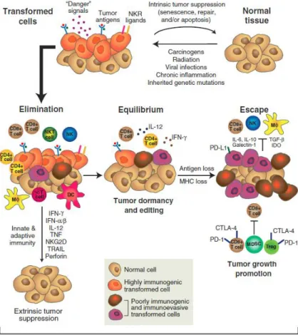
During cancer immunoediting, the host immune system shapes tumor fate in three phases, “elimination”, “equilibrium” and “escape”, through the activation of innate and adaptive immune mechanisms (Figure 1).
The elimination phase is best described as an updated version of cancer immunosurveillance, in which the innate and adaptive immune systems work together to detect the presence of a developing tumor and destroy it before it becomes clinically apparent.
Initiation of the antitumor immune response occurs when cells of the innate immune system become alerted to the presence of a growing tumor, at least in part owing to the local tissue disruption that occurs because of the stromal remodeling processes (Gopal, 2015). The consequent production of pro-inflammatory molecules, together with chemokines that may be produced by the tumor cells themselves, leads to the recruitment of cells of the innate immune system (NKT, NK, γδ T cells, macrophages and dendritic cells). Infiltrating lymphocytes such as NKT, NK or γδ T cells recognize some structures on the transformed cells and are then stimulated to produce IFN-γ.
2 Tumor cells also express stress-induced molecules such as surface calreticulin, tumor antigens in context of MHC class I molecules, and/or NKG2D ligands recognized by CD8+ effector cells and NK cells, respectively.
Figure 1. The three Es of cancer immunoediting (from Schreiber et al., 2011).
In its most complex form, cancer immunoediting consists of three sequential phases: elimination, equilibrium, and escape. In the elimination phase, innate and adaptive immune response work in synergy to eliminate the rising transformed cells. The equilibrium phase provides evidence for a tumor-sculpting role of immunity. In this phase the immune system iteratively selects and/or promotes the generation of immune evasive tumor cell variants. When the immunologically sculpted tumor expands in an uncontrolled manner in the immunocompetent host the phase of escape is accomplished.
The initial production of IFN-γ initiates a cascade of innate immune reactions leading to some tumor cell death by both immunologic and non immunologic
3 mechanisms. The INF-γ initially produced may induce a limited amount of tumor death by means of antiproliferative and apoptotic mechanisms. However, it also induces the production of some chemokines from the tumor cells themselves as well as from surrounding normal host tissues. Some of these chemokines have potent angiostatic capacities and thus they block the formation of new blood vessels within the tumor, enhancing tumor cell death. Furthermore, the transactivation of tumor-infiltrating NK cells and macrophages by reciprocal production of IFN- γ and IL-12 contributes to tumor cell death by mechanisms involving TRAIL, perforin and reactive oxygen and nitrogen intermediates.
From dead tumor cells, a source of tumor antigens becomes available to the immature dendritic cells (DCs) recruited to the tumor site. These antigens might reflect one or more of the many mutated proteins that are typical of cancer (neo-antigens) or the products of non-mutated genes that are preferentially expressed by cancer cells (tumor associated antigens).
The activated, antigen-bearing mature DCs then migrate to the draining lymphnode, where they induce the activation of naïve tumor-specific Th1 CD4+ T cells. Th1 cells facilitate the development of tumor-specific CD8+
CTL induced via cross-presentation of antigenic tumor peptides on DC MHC class I molecules. Tumor-specific CD4+ and CD8+ T cells home to the tumor site, where the cytolytic T lymphocytes destroy the remaining antigen-bearing tumor cells whose immunogenicity have been enhanced by exposure to locally produced IFN- γ (Figure 2).
4 The cancer-immunity cycle is a multistep process that involves (1) release of cancer cell antigens; (2) cancer antigen presentation; (3) priming and activation; (4) trafficking of T cells to the tumor; (5) infiltration of T cells into the tumor site; (6) recognition of cancer cells by T cells; (7) killing of cancer target cells (Chen and Mellman, 2013).
When the elimination phase is successful, it represents the complete editing process without progression to the subsequent phases.
If, however, rare tumor cell variants are not destroyed, they may survive the elimination phase and enter the equilibrium phase, in which the adaptive immune system prevents tumor cell outgrowth and also sculpts the immunogenicity of the tumor cells.
In this process, lymphocytes and IFN- γ exert potent selection pressure on the tumor cells that is enough to contain, but not fully extinguish, a tumor bed containing many genetically unstable and rapidly mutating tumor cells. During this period of Darwinian selection, many of the original escape variants of the tumor cells are destroyed, but new variants arise carrying different mutations that provide them with increased resistance to immune attack. So, in the equilibrium phase, occult tumor cells not destroyed in the elimination phase are held in a state of tumor dormancy as a consequence of adaptive immune system activity and undergo “editing”. Recent work has demonstrated that T cells play a major role in shaping the immunogenicity of developing cancers and exert this effect by at least two mechanisms. First, T cells can shape tumor antigenicity/immunogenicity through an immunoselection process by destroying tumor cells that express strong tumor-specific mutant antigens, leaving behind tumor cells that either express weaker antigens or are incapable of expressing antigens (because of mutations in antigen processing or presentation)(Matsushita et al., 2012). Second, chronic T cell attack on a tumorhas been shown to silence expression of certain tumor-specific antigens through epigenetic mechanisms in a preclinical model (Dupage et al., 2012).
When tumor cell variants selected in the equilibrium phase can grow in an immunologically intact environment they enter into the escape phase. The immune system fails to restrict tumor outgrowth and tumor cells emerge causing clinically apparent disease.
Tumors escape immune attack by a variety of complementary mechanisms of immunosuppression, many of which operate in parallel:
Reduced immune recognition: tumor cells can directly escape T-cell recognition by downregulating MHC class I, class I-like, or co-stimulatory molecules or by disabling other components of the antigen processing machinery.
5 Increased resistance or survival (such as increased expression of STAT-3
or anti-apoptotic molecule Bcl2).
Development of an immunosuppressive tumor microenvironment.
o Among paracrine mediators, release of PGE2, arginase and IDO (all T-cell suppressors), and the release of VEGF (triggered in part by intratumoral hypoxia), exert multiple direct and indirect immunosuppressive activities. These mediators may function indirectly inhibiting T-cell diapedesis from the vasculature into the tumor bed or directly suppressing effector T-cell activation while enhancing the function of regulatory T cells (Treg).
o In addition, tumor cells may upregulate surface ligands, including PD-L1, PD-L2 and other ligands that engage receptors on the surfaces of activated T cells (PD-1), causing T-cell anergy or exhaustion.
Recruitment of a variety of leukocyte subsets infiltrating tumors able to
suppress T-cell function. In addition to Treg cells (the accumulation of
which in tumors correlates with poor prognostic outcome), other suppressive lymphocyte subsets have been reported. They include IL-10 producing B cells and B regulatory cells, type II NKT cells, NK cells and γδ T cells. Myeloid lineage cells also promote immune suppression in tumors. Among these, myeloid-derived suppressor cells (MDSC) suppress T andNK cell activation, probably through several mechanisms including nitric oxide, reactive oxidative species, arginase, IL-10 andTGF-β; thereare also reports that MDSCs may specifically induce the expansion of Treg cells.
1.2 Vaccines in cancer therapy 1.2.1 Tumor-associated antigens
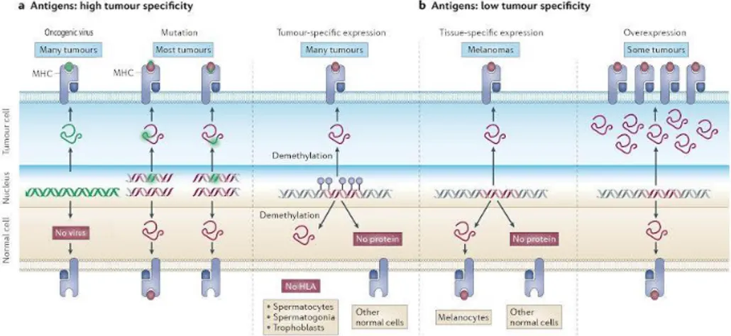
Cancer immunotherapy seeks to exploit the host’s immune system to eliminate cancer cells. In this context, cancer antigens play a critical role, as they are responsible for triggering a cancer specific immune response.At the core of the design of immunotherapy strategies lies the fact that cancer patients can produce T lymphocytes that recognize tumor-specific antigens. Ideally, cancer antigens should be highly immunogenic to induce strong immune responses and only be expressed by malignant cells for specific tumor killing. However, normal tissues may also express some. For this reason, human tumor antigens can be grouped in two main classes: 1) antigens of high tumoral specificity and 2) antigens of low tumoural specificity (Cooley et al 2014).
6 Three types of tumor antigens have the potential to elicit immune responses that are strictly tumor specific: 1) oncoviral antigens, 2) mutated antigens and 3) cancer-testis antigens. Instead, antigens of low tumor specificity include 4) differentiation antigens and 5) overexpressed/ accumulated antigens (Figure 3).
1) Approximately 12% of all human cancers worldwide are associated with oncogenic viruses. Human tumor viruses belong to a number of virus families, including the RNA virus families Retroviridae and Flaviviridae and the DNA virus families Hepadnaviridae, Herpesviridae and Papillomaviridae (McLaughlin-Drubin and Munger, 2008). They are associated to the onset of an important subset of human tumors, including cervical carcinoma, hepatocarcinoma, nasopharyngeal carcinoma and adult T cell leukemia (Parkin, 2006).
Even though human oncogenic viruses belong to different virus families and utilize diverse strategies to contribute to cancer development, they share many common features. One key feature is the tendency to establish long-term persistent infections. Consequently, they have evolved different strategies for evading the host immune response, which would otherwise clear the virus during these persistent infections.
Immunocompetent individuals normally mount a potent cytotoxic T-cell (CTL) response against infected cells expressing viral epitopes. However, oncogenic viruses generally avoid CTL surveillance by establishing latency in host cells. γ-herpesviruses express genes during the latency which can block TNF-induced pro-apoptotic signals generated by cell-mediated cytotoxic responses. Latent γ-herpesviruses remain under tight transcriptional regulation by NF-κB and histone deacetylase enzymes (HDACs). Disruption of viral latency in γ-herpesvirus lymphomas using the NF-κB and HDAC inhibitors results in lytic reactivation and cell death (Ramos and Lossos, 2011). Likewise, the chronic antigenic stimulation in HPV infections supports the establishment of an immunosuppressive microenvironment that triggers the transition from normal epithelium to cervical intra-epithelial neoplasia (CIN) and vulvar intraepithelial neoplasia (VIN) as well as their subsequent progression to invasive squamous cell carcinoma. In particular, IDO+ cells, FOXP3+ T cells, and TGFβ+ T cells increase across the disease
spectrum in parallel with a sharp decline in IFN-γ+ cells in invasive cancer (Kobayashi et al., 2008).
2) Mutated genes greatly contribute to the immunogenicity of human tumors. Gene mutations produce new antigenic peptides by changing one amino acid, by altering the phase of the reading frame or by extending the coding sequence beyond the normal stop codon. Due to their unique nature of being
7 expressed only on tumors and not on any other normal tissues, these tumor-specific TAAs can be recognized as non-self and not be subjected to central immune tolerance. These mutated proteins may play a critical role in the oncogenic process as products of mutated oncogenes or tumor suppressor genes and therefore survive immune selection in order to maintain tumor growth and proliferation. Mutated CDK4 is an example of tumor antigens proved to be oncogenic. A point mutation in CDK4 results in the loss of binding of CDK4 to the inhibitor INK4A with a consequent disrupting of the cell cycle regulation (Plaen et al., 1995).
These type of mutations that confer a selective growth advantage, thus promoting cancer development, are commonly defined ‘driver mutations’. Cancer cells also develop a large number of mutations that do not provide a growth advantage and are therefore called ‘passenger mutations’. In most cancers, there are many more passenger mutations than driver mutations, with the latter ones more frequently giving rise to the so-called neo-antigens (discussed in 1.4.1).
3) Cancer/testis (CT) antigens are a category of tumor antigens with normal expression restricted to male germ cells in the testis but not in adult somatic tissues. In some cases, CT antigens are also expressed in ovary and in trophoblast. In malignancy, this gene regulation is disrupted, resulting in CT antigen expression in a proportion of tumors of various types.The first cancer antigen reported that could be recognized by T cells, MAGE-A1, belongs to this class (van der Bruggen et al., 1991).
Cancer-germline genes are an important source of tumor-specific antigens, with more than 60 cancer-germline genes having been identified. The mechanism that leads to the activation of these genes in tumor cells involves the demethylation of their promoter, which is methylated in all normal cells except in germline cells (De Smet et al., 1996).
4) Differentiation antigens are cell type specific and shared between tumors and the normal tissue of origin (Buonaguro et al., 2011). For example, both melanoma and normal melanocytes express GP100, Tyrosinase, and Melan-A/MART-1. Other differentiation antigens include PSA, Mammoglobin-A, and carcinoembryonic antigen (CEA) overexpressed in prostate cancer, breast carcinoma, and colon cancer respectively.
5) Some proteins shared by both normal and tumor cells may display overexpression only in cancer cells thus providing an opportunity for a specific T cell response. This is because a threshold level of antigen is required for recognition by T cells. If tumor cells present an amount of peptide–HLA complexes that is above the threshold of T cell activation and if
8 normal cells do not, a specific antitumor T cell response could occur. In this way, over-expression by malignant cells overrides tolerance and triggers T cell activation. An example of overexpressed tumor antigen is the oncogene and growth factor receptor ERBB2 (also known as HER2 and NEU) which is overexpressed in many epithelial tumors, including ovarian and breast carcinomas, owing to increased transcription and to gene amplification (Ross et al., 1998).
Figure 3. Classes of human tumor antigens recognized byT lymphocytes.
a. Oncoviral antigens, mutated antigens and cancer-testis antigens show high tumor specificity. Point mutations can modify a peptide that already binds to the major histocompatibility complex or can enable a non-binding peptide to bind. Cancer-germline genes are selectively expressed in tumors and germline cells because of DNA demethylation. However, their antigens are not present on germline cells because of the lack of HLA molecules. b. Antigens of low tumor specificity include differentiation antigens and overexpressed/ accumulated antigens. In the figure, a melanocyte-specific gene is used as an example of tissue-specific gene expression. Both tumor cells and the normal tissue of origin (melanocytes) share the encoded protein. In the last panel, overexpression of particular proteins, such as ERBB2, beyond threshold levels can also trigger an antitumor immune response.
1.2.2 Therapeutic cancer vaccines
Traditionally, vaccines have been used as a preventive measure against infectious diseases, triggering the immune system to produce neutralizing antibodies against specific pathogen antigens. More recently, vaccines have been applied as therapeutic strategies, aiming to induce immune system to activate cytotoxic T cells against infected cells and cancer. However, therapeutic vaccination against established diseases has proven much more
9 challenging than prophylactic vaccination against infectious diseases, because the vaccine intervention must overcome the hurdles posed by immune evasion by having to antagonize an immune system that has been restrained by tolerizing or polarizing mechanisms that sustain the disease in a misguided attempt at self-tolerance (Melief et al., 2015).
The idea of a therapeutic cancer vaccine originated with the discovery that patients can harbor CD8+ and CD4+ T cells specific for cancer antigens expressed in their tumors (Boon at al., 2006). Therefore, vaccination might reasonably be expected to amplify the frequency and strength of these pre-existing responses against tumor antigens or perhaps induce some de novo reactivities.
Based on their format/content, cancer vaccines may be classified into several major categories, which include cell based vaccines (tumor cell lysates, irradiated whole tumor cells, DCs), protein/peptide vaccines, and genetic vaccines (DNA, RNA and viral vectors) (Guo et al., 2013). Their development is based on the concept that the introduction of various tumor antigens into the host would facilitate immune mediated clearance of tumor cells. Ideally, therapeutic vaccination aims at expanding high-avidity CD8+ T
cells that can differentiate into CTLs able to kill cancer cells and to generate long-lived memory CD8+T cells. This could be accomplished through either the priming of naïve T cells or the reprogramming of memory T cells that differentiate earlier in an environment not conducive to the generation of potent cytotoxic T cells. Indeed, cancer is a chronic disease and, as such, it is associated with skewed T cell memory, chronically activated and anergic CD8+ T cells that express programmed cell death 1 (PD-1) (Freeman et al.,
2006). In addition, vaccination should lead to the generation of long-lived memory CD8+ T cells that will act to prevent relapse (Figure 4).
Figure 4. Therapeutic vaccines act to generate protective CD8+ T cell immunity (from
10 Therapeutic vaccines are expected to prime new T cells and induce a transition from chronically activated non-protective CD8+ T cells to healthy CD8+ T cells able to (1)
generate CTLs that reject cancer and (2) provide long-lived memory CD8+ T, thereby preventing relapse.
1.2.3 Genetic cancer vaccines
Therapeutic vaccines have two objectives: priming antigen-specific T cells and reprogramming memory T cells. In this context, genetic vaccines represents a highly promising approach.
Genetic vaccination exploits the use of viral or bacterial vectors or nucleic acids to deliver one or more antigens in vivo. One major advantage of genetic vaccines is the easy delivery of multiple antigens in one vaccine and their ability to activate various arms of the immune system (Aurisicchio and Ciliberto, 2012).
DNA vaccines consists of bacterial plasmid DNA into which specific sequences are incorporated under the control of an eukaryotic promoter. Genes in DNA vaccines can encode different antigens as well as various immunomodulatory molecules to manipulate the resulting immune response, after transduction into the target cells and subsequent in vivo expression by the host’s gene expression machinery.
DNA vaccines were shown to be able to trigger both innate and adaptive immune response. The ability to stimulate the innate immune system arises from the bacterial origin of the backbone (Rice et al., 2008). The bacterial DNA appears to act as a pathogen-associated molecular pattern able to stimulate cells through Toll-like receptors (TLRs). Specifically, the hypomethilated CpG dinucleotides motif that is common in bacterial DNA, but rare in mammalian DNA, interacts with TLR9 expressed in immune cells, such as dendritic cells, B cells, and NK cells. Activation of TLR9 leads to a cascade of pro-inflammatory responses and results in the production of various cytokines. The local inflammation and increased production of cytokines from the innate immune responses can attract and activate additional immune cells, such as lymphocytes, and enhance subsequent antigen-specific immune responses.
DNA vaccines are delivered intradermically or more commonly by intramuscular injection, resulting in the transfection of keratinocytes or myocytes, respectively.
In the muscle, transfected myocytes express the vaccine-encoded antigens and act as a target for immune effector cells. In addition, they can also upregulate expression of MHC class I and co-stimulatory molecules, with
11 production of cytokines and chemokines. Consequent inflammation and production of cytokines attract professional APCs, like dendritic cells, to the transfection sites.
APCs have a dominant role in the induction of immunity of DNA vaccines by presenting vaccine-derived endogenous peptides on MHC I molecules. APCs can ‘capture’ these antigens by direct transfection or most commonly by cross presentation for example, owing to APC engulfment of apoptotic transfected cells. In addition, APCs mediate the display of peptides on MHC II molecules after secreted protein antigens from transfected cells are captured and processed within the endocytic pathway.
Antigen-loaded APCs travel to the draining lymph node via the afferent lymphatic vessel where they present peptide antigens to naïve T cells via MHC and the T cell receptor (TCR) in combination with co-stimulatory molecules, initiating an immune response and expansion of T cells. In response to peptide-bound MHC molecules and co-stimulatory secondary signals, activated CD4 T helper cells secrete cytokines during cell-to-cell interaction with B cells and bind to co-stimulatory molecules that are required for B cell activation (Figure 5).
The use of DNA plasmids in cancer immunotherapy offers several advantages. In addition to their safety, DNA vaccines allows for simple and flexible design, encoding wide range of antigens and immunomodulatory molecules. DNA vaccines are heat stable, easily stored and perfect for large scale production (Yang et al., 2014).
However, despite the promising features of DNA vaccines, they have been found to elicit immune responses less than other types of vaccines, including peptide vaccines, cellular vaccines, viral vector vaccines, and RNA vaccines. The relatively poor immunogenicity of DNA vaccines combines with other disadvantages: inefficient delivery of DNA into human cells, the need for DNA to cross both cell and nuclear membranes and be transcribed in order to allow for expression of the encoded antigen. Some of these considerations have driven a shift away from DNA vaccines and towards RNA vaccines.
12 Figure 5. Induction of cellular and humoral immunity by DNA vaccines
The optimized gene sequence of interest is inserted into a plasmid backbone, purified, and then delivered to the inoculation site. Using the host cellular machinery, the plasmid enters the nucleus of transfected myocytes (1) and of resident antigen presenting cells (APCs) (2). Here, the plasmid components initiate gene transcription, which is followed by protein production in the cytoplasm and the consequent formation of foreign antigens, that can become the subject of immune surveillance in the context of both major histocompatibility complex class I (MHC I) and MHC II proteins. The presentation of vaccine-derived endogenous peptides on MHC I molecules by APCs can follow either direct APCs transfection by the plasmid vaccine (2) or cross-presentation of cell-associated exogenous antigens (3). In addition, APCs can capture secreted protein antigens that have been shed from transfected cell, process them within the endocytic pathway and finally display peptides on MHC II molecules (4). Antigen-loaded APCs travel to the draining lymph node via the
13 afferent lymphatic vessel (5) where they present peptide antigens to naïve T cells via MHC and the T cell receptor (TCR) in combination with co-stimulatory molecules, providing the necessary secondary signals to initiate an immune response and expansion of T cells (6). In turn activated CD4 T helper cells promote B cell activation (7). ‘Armed’ lymphocytes can finally leave the draining lymph node through the efferent lymphatic system (8).Kutzler and Weiner, 2008
RNA vaccines consist in messenger RNA (mRNA) synthesized by in vitro transcription using a bacteriophage RNA polymerase and template DNA that encodes the antigen(s) of interest (McNamara et al., 2015). Once administered and internalized by host cells, the mRNA transcripts are translated directly in the cytoplasm and then, like DNA vaccines, the resulting antigens are presented to APC by major histocompatibility complex (MHC) class I and II proteins, with consequent induction of T cell-mediated immune responses. Alternatively, RNA vaccines can be constructed for the efficient production and secretion (or cell-surface expression) of extracellular antigens to stimulate B cell responses and antigen-specific antibody production. The effectiveness of RNA vaccines may also be related to the fact that RNA is known to be a potent stimulator of innate immunity (Ulmer et al., 2012).
Several techniques have been developed to improve the inherent instability of mRNA and translational efficiency and to optimize RNA vaccine delivery. One approach to induce a potent and targeted anti-tumor response is to use viruses to deliver tumor antigens to cells of the immune system.
Viral vectors are an attractive choice of antigen delivery system for cancer immunotherapy since they mimic a natural infection and provide potent danger signals, which are known to be important for the induction of an immune response (Harrop and Carroll, 2006).They enable intracellular antigen expression and induce a robust cytotoxic T lymphocyte (CTL) response, leading to the elimination of diseased cells.
Despite their efficacy, viral vectors present unavoidable problems that need to be addressed. Viral vector-based vaccines require assessment of efficacy and safety, including immunogenicity, genetic stability, ability to evade pre-existing immunity, replication deficiency or attenuation, and genotoxicity. For a high biological safety level, non- (or low-) pathogenic viruses are often selected (Ura et al., 2014).
Different viral vectors have been evaluated in cancer immunotherapy: adenoviruses, Adeno-Associated Virus (AAV), alphaviruses, flaviviruses, lentiviruses, measles virus, rhabdoviruses, retroviruses and Vaccinia Virus (VV) (Lundstrom, 2016).
14 The intrinsic properties of each virus have distinct advantages and disadvantages, which can determine their applicability in a particular therapeutic setting.
1.2.4 Adeno vector based cancer vaccines
Adenovirus vectors (Ads) are one of the most effective carriers for delivery of foreign antigens into the host cells. Ads have a large genome size and allow cloning of expression cassette for large antigens (ie: over 2000 amino acids). Furthermore, Ads do not integrate the viral genomic DNA into the hosts’ genome, which reduces the risk of insertion mutagenesis (Zhang and Zou, 2016). Compared with other viral vectors, Ads are highly immunogenic and can induce robust adaptive immune responses, offering one of the most powerful technologies for cancer vaccine applications.
Adenoviruses are double-stranded DNA viruses with a genome of ~34–43 kb, with two inverted terminal repeats at both ends as origins for DNA replication. The genes that Ads express during the life cycle are generally clustered in early genes and late genes. The early genes include E1A, E1B, E2, E3 and E4, and they are mainly responsible for facilitating the replication of Ads by changing the expression levels of related host genes. The early genes can be further classified into two types: the immediate early genes (E1A) and the delayed early genes (E1B, E2, E3 and E4).
The E1A gene is transcribed first and, with the help of cellular factors, activates transcription of the other viral genes. Deletion of E1A renders the virus replication-defective. E1A stimulates viral DNA synthesis, dysregulates cell-cycle control, promotes apoptosis and plays a role in immunoevasion by inhibiting the activity of STAT1, which is needed for activation of interferon-responsive genes.
While E1A proteins promote apoptosis, E1B proteins have antiapoptotic functions. E1B polypeptides turn off host cell protein synthesis and help to stabilize, transport, and translate selectively viral RNA.
The E2 unit encodes DNA-binding proteins and a polymerase and is essentialfor viral replication.
E3 proteins allow the virus to escape immunosurveillance by different mechanisms: i) by reducing expression of major histocompatibility complex (MHC) class I determinants; ii) by direct binding to the groove of MHC class I molecules preventing binding and export to the cell surface of peptides; iii)
by associating with TAP and thereby reducing efficient transport of peptides derived from proteolytic cleavage of de novo synthesized viral proteins from the cytoplasm tothe endoplasmic reticulum where they can associate with MHC class I molecules.
15 The E4 transcription unit encodes seven polypeptides, which affect viral transcription and a number ofhost cell functions including cell proliferation and apoptosis, in part by promoting degradation of p53. E4 is essential for nuclear export of viral RNA.
The late genes (L1-L5) are mainly responsible for the lysis of the host cells, assembly and release of the virions.
Ad virions mainly comprise two types of proteins: the capsid proteins and the core proteins. The core proteins mainly include proteins V, VII, X, and they mainly function as the DNA-associated proteins. The capsid proteins comprise Hexon, Penton, fiber, IIIa, VIII and IX.
Ads are isolated from different mammalian species, such as humans, dogs, sheep, bovines and non human primates. Among all, the human Ads and chimpanzee Ads are widely used in research or clinical studies. They have a broad tropism infecting a variety of dividing and non-dividing cells. They can be grown in human HEK293 cells and purified by CsCl gradient ultracentrifugation or chromatography, making them attractive for clinical use.
Adenoviruses are useful vectors for genetic vaccine delivery. To insert transgenes of interest viral E1 and E3 genes are commonly deleted and depending on space necessity, there is the possibility to delete also the E4 gene. By deletion of these genomic regions, the virus loses self-replication capacity, becoming replication-defective. This arrangement increases their predictability of their immunization properties and reduces unwanted side effects.
The most commonly used adenoviral vectors as genetic vaccines are derived from human adenovirus serotype 5. In particular, head-to-head comparisons with other genetic vaccine vectors (ie.: poxviruses, lentiviruses, alpha virus-based vectors and naked DNA) in animal models and the results obtained in human clinical trials, clearly showed that Ad5-based vectors currently represent one of the most potent delivery system for eliciting a CD8+ T cell
response against the encoded antigen(s). However, high titers of anti Ad5 neutralizing antibodies are commonly present in human population, impairing the immunogenicity of Ad5-based vaccines in humans.
To overcome this drawback, other human Adenovirus vectors based on rare serotypes such as Ad11, Ad24, Ad26, Ad34, Ad35, Ad48, Ad49, and Ad50 have been proposed as potential alternatives to Ad5 because antibodies present in humans rarely neutralize them. However, they showed lower immunological potency than Ad5 in mice and non-human primates. Another approach is to use non-human Adenoviruses. Indeed, Colloca et al., (2012) generated a large collection of replication defective vectors based on Ad
16 isolated from chimpanzees. Functional screenings to assess growth capability in packaging cell lines as HEK293 and PER.C6, immunological potency in mice and non-human primates and sensitivity to neutralizing antibodies present in humans allowed the identification of novel vaccine carriers inducing potent cellular immunity, suitable for vaccine delivery in humans. Among the large number of candidates screened for immunological potency by dose/response in mice, some ChAds were identified which showed immunological potency equivalent to Ad5 (ChAd3, ChAd63, ChAd83, PanAd1, PanAd2 and PanAd3). Importantly, the high level of immunogenicity of the top ranking ChAd3 and PanAd3 was confirmed in non-human primates, where they induced a level of T-cell response comparable to that of Ad5 even at low dose.
1.3 The blockade of immune checkpoints in cancer immunotherapy 1.3.1 Immune checkpoint receptors
The initiation and progression of immune responses are fine-tuned by a highly complex array of cytokines, chemokines, toll-like receptors and costimulatory molecules. Equally complex is the diversity of pathways and mechanisms employed by the immune system to regulate and/or terminate ongoing immune responses. When the regulatory mechanisms somehow fail, normal immune homeostasis is impaired leading to disastrous consequences to the host, as the onset of autoimmunity.
Co-stimulatory and co-inhibitory immune checkpoint receptors are critical modulators of the immune system, as they determine the functional outcome of T cell receptor (TCR) signalling.
Following recognition of cognate peptide–MHC complexes on APCs by the TCR, co-signalling receptors often colocalize with TCR molecules at the immunological synapse, where they synergize with TCR signalling to promote (co-stimulatory receptors) or inhibit (co-inhibitory receptors) T cell activation and function (Figure 6).
17 Figure 6. Multiple co-stimulatory and inhibitory interactions regulate T cell responses (from Pardoll, 2012).
Various ligand–receptor interactions between T cells and antigen-presenting cells (APCs) regulate the T cell response to antigen after the recognition of the cognate antigen through the TCR. These responses can occur at the initiation of T cell responses in lymph nodes or in peripheral tissues or tumors, where effector responses are regulated.
In this interactive environment, the repertoire of co-signalling receptors expressed on T cells is highly versatile and responsive to changes in overlapping spatiotemporal fashion. The costimulatory receptor CD28 and the inhibitory receptor CTLA-4 (cytotoxic T-lymphocyte-associated antigen 4) are a pivotal example. They bind the same ligands but they display distinct kinetics of expression, in order to modulate TCR signalling during the immune response (Intlekofer and Thompson, 2013).
The discovery of CD28 stimulatory receptor gave molecular confirmation to the theory according to which the TCR engagement is not itself sufficient to enact T cell clonal expansion and differentiation whereas a second signal is pivotal to drive lymphocyte clonal expansion.
18 CD28 is constitutively expressed on the surface of resting and activated T cells, while its ligands CD80 (or B7-1) and CD86 (or B7-2) belonging to the B7 ligands are expressed of the surface of APCs. The interaction between them provides a second signal to promote T cell activation, proliferation, survival and activation of effector functions.
The inhibitory receptor CTLA-4, expressed on the surface of activated T cells shares significant homology to CD28 and bounds the same B7 ligands, but their interaction counteracts CD28-mediated costimulatory signals and impairs the activation of T cells.
This opposed action is explained by a unique spatiotemporal regulation of CTLA-4.In resting T cells, CTLA-4 exhibits minimal expression and a peculiar pattern of intracellular localization and trafficking. It resides mostly within intracellular vesicles of the trans-Golgi network and endosomal compartments. Upon TCR engagement, CTLA-4 expression is induced, and intracellular vesicles containing CTLA-4 undergo relocalization to the immune synapse. At the cell surface, CTLA-4 competes with CD28 for access to B7 ligands. Compared with CD28, CTLA-4 has higher affinity and avidity for B7 ligands, which has been attributed to homodimer formation by CTLA-4 that allows for bivalent binding of B7 molecules, in contrast to the monovalent binding of B7 ligands by CD28.
The definitive role of CTLA-4 as a major negative regulator of T-cell activation was established with the description of CTLA-4−/− mice. They succumbed at three-to-four weeks of age from massive lymphoproliferation within the spleen and lymph nodes and end-organ infiltration by activated lymphocytes (Tivol et al., 1995).
Of note, CTLA-4 is constitutively expressed on Foxp3+ Treg cells that may
use CTLA-4 to mediate suppression of T effector cells. The finding that conditional deletion of CTLA-4 in Tregs, but not in other cell types, results in
impaired Treg functions appears to support this notion (Wing et al., 2008).
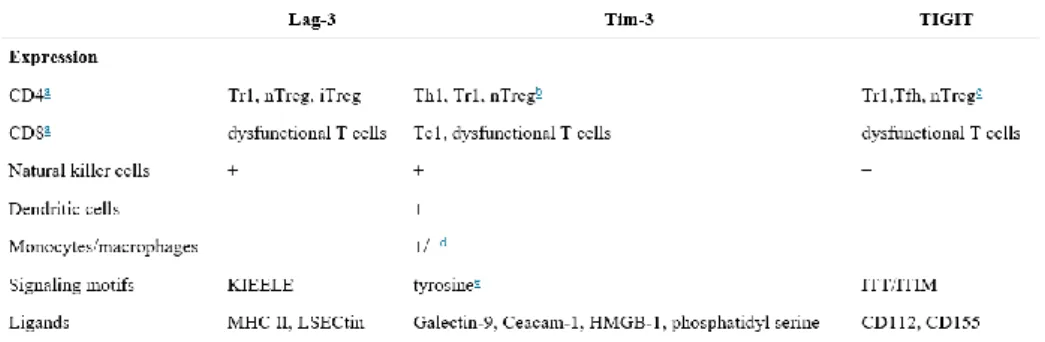
CTLA-4 together with PD-1 (discussed in section 1.3.2) are the two negative checkpoint regulators that have been most actively studied. However, multiple additional inhibitory receptors have been discovered in the recent years. TIM-3 (T-cell immunoglobulin and mucin-containing protein 3), LAG-3 (lymphocyte-activated gene-3), TIGIT (T cell immunoreceptor with Ig and ITIM domains), BTLA (B and T lymphocyte attenuator) and VISTA (V-domain Ig suppressor of T cell activation) represent the main examples. Among them Lag-3, Tim-3 and TIGIT represent the next generation of co-inhibitory receptors to be translated to the clinic since they are highly expressed on dysfunctional or exhausted T cells in chronic diseases such as chronic viral infection and cancer.
19 A comparison concerning expression, signaling mofits and ligands between them is briefly summarized in Table 1.
Table 1. Comparison of Lag-3, Tim-3 and TIGIT (from Anderson et al., 2015).
Several observations proved that these receptors have been shown to be important immune regulators in autoimmunity. Lag-3 plays a protective role in autoimmunity by dampening T helper (Th) cell responses directly through engagement of MHC-II. In addition, Lag-3 indirectly inhibits effector T cell responses via promotion of Treg-cell- and Tr1-cell-mediated suppression. In autoimmune diseases such as multiple sclerosis, Tim-3 is under-expressed on pathogenic Th1 cells. IFN-β therapy can increase Tim-3 on antigen-specific T cells directly or indirectly via promotion of IL-27 production from local antigen-presenting cells. Increased expression of Tim-3 is associated with reduction in disease relapses. TIGIT inhibits auto-pathogenic Th1/Th17 T cell responses through three different pathways: (1) TIGIT directly inhibits T cell activation and expansion; (2) TIGIT expressing effector and regulatory T cells engage CD155 on APCs thereby inducing tolerogenic APCs that secrete IL-10; (3) TIGIT promotes Treg-cell-mediated suppression through the induction of IL-10 and Fgl2, which potently and selectively suppress Th1 and Th17 cell responses.
20 1.3.2 PD-1/PD-L1 pathway
Immune checkpoints refer to a plethora of inhibitory pathways hardwired into the immune system that are crucial for maintaining self-tolerance and modulating the duration and amplitude of physiological immune responses in peripheral tissues in order to minimize collateral tissue damage. Among the inhibitory immune mediators, the pathway consisting of the programed cell death 1 (PD-1) receptor (CD279) and its ligands PD-L1 (B7-H1, CD274) and PD-L2 (B7-DC, CD273) plays an important role in the induction and maintenance of peripheral tolerance and for the maintenance of the stability and the integrity of T cells. PD-1’s immune-inhibitory function was elucidated by characterizing the autoimmune phenotype of PD-1–deficient mice, in which PD-1 deficiency leads to a loss of peripheral tolerance and the subsequent development of autoimmunity. PD-1–deficient mice develop different autoimmune diseases depending on their genetic background: C57BL/6-Pdcd1−/− mice develop lupus-like arthritis and glomerulonephritis with IgG3 and C3 deposits (Nishimura et al., 1999). BALB/c-Pdcd1−/− mice develop fetal dilated cardiomyopathy with a concomitant production of autoantibodies against cardiac troponin I (Nishimura et al., 2001).
The PD-1:PD-L1/L2 pathway also mediates potent inhibitory signals to hinder the proliferation and function of T effector cells. Similarly to CD80/CTLA-4, PD-L1/PD-1 are antagonists of CD80/CD28 co-stimulation. Engagement of PD-1 by its ligands PD-L1 or PD-L2 strongly counteracts TCR signal transduction and CD28 co-stimulation, transducing a signal that inhibits T cell proliferation, cytokine production, and cytolytic function.
Programmed death 1 (PD-1) receptor is a type I transmembrane protein preferentially expressed on activated CD4+ and CD8+ T cells, B cells,
monocytes, natural killer (NK) cells, and dendritic cells (DCs). PD-1 consists of a single N-terminal IgV-like domain, an approximately 20 amino acid stalk separating the IgV domain from the plasma membrane, a transmembrane domain, and a cytoplasmic tail containing tyrosine-based signaling motifs: an immunoreceptor tyrosine-based inhibitory motif (ITIM) followed by an immunoreceptor tyrosine-based switch motif (ITSM). Both these motifs are implicated in PD-1 immunosuppressive effects, even if the ITSM appears to be the most important for mediating PD-1 suppression of lymphocyte activation (Chemnitz et al., 2004).
Upon binding to its ligands, PD-1 becomes phosphorylated on intracellular tyrosine residues within ITIM. Subsequently, protein phosphatases, such as SHP-2 are recruited to bind to the ITSM, become activated and inhibit proximal TCR signaling events (Sheppard et al., 2004) (Figure 7).
21 Figure 7. PD-1-dependent inhibitory mechanisms. (from Arasanz et al., 2017)
PD-1 mediates its immunosuppressive activity through direct and indirect inhibitory mechanisms over TCR signalling and T cell proliferation, by inhibiting membrane-proximal T cell signaling events. (A) The direct inhibition of TCR signaling depends on the recruitment of SHP1 and SHP2 phosphatases to the tyrosine-based signaling motifs ITIM and ITSM. These phosphatases interfere with TCR signal transduction and CD28 co-stimulation by inhibiting ZAP70 and PI3K activities, respectively.(B)PD-L1 engaged PD-1 exerts an indirect inhibitory control over CD28 co-stimulation by reducing the expression levels and activities of CK2. As a result, active PTEN eliminates PIP3, shutting offAKT activation.Consequently,cell growth and survival is inhibited, because lymphocytes arrest at the G0-G1 phase.(C) PD-1 engagement regulates TCR surface expression, by promoting the expression of E3 ubiquitin ligases that ubiquitylate TCR chains. As a result, TCR is removed from the T cell surface, possibly by endocytosis. (D) Engaged PD-1 alters T cell metabolism by inhibition of ERK and PI3K-AKT activities.
PD-1 is expressed on a large proportion of tumor-infiltrating lymphocytes (TILs) from many different tumor types. Among CD4+ TILs, a generally high level of PD-1 expression is detectable on Treg cells, which can represent a
large proportion of intratumoral CD4+ T cells. Increased PD-1 expression on CD8+ TILs may either reflect an anergic or exhausted state, as has been suggested by decreased cytokine production by PD-1+ compared with PD-1– TILs from melanomas (Ahmadzadeh et al., 2009).
The ligands of PD-1 (PD-L1 and PD-L2) are type I transmembrane glycoproteins, containing IgC and IgV domains. They share about 40% of amino acid identity. PD-Ls have distinct expression patterns: PD-L1 is
22 constitutively expressed on T and B cells, DCs, macrophages, mesenchymal stem cells and bone marrow-derived mast cells (Yamaki et al., 2002). In addition, PD-L1 is expressed on a wide variety of non-hematopoietic cells. In contrast, PD-L2 expression is restricted to activated DCs, macrophages, bone marrow derived mast cells, and more than 50% of peritoneal B1 cells.
Just as PD-1 is highly expressed on TILs from many cancers, the PD-1 ligands are commonly upregulated on the tumor cell surface from many different human tumors, correlating with adverse prognosis. On cells from solid tumors, the major PD-1 ligand that is expressed is PD-L1. Two general mechanisms for the regulation of PD-L1 by tumor cells have emerged: innate immune resistance and adaptive immune resistance. In the first case, constitutive oncogenic signalling pathways in the tumor cell drive PD-L1 expression (Parsa et al., 2007). On the contrary, in adaptive immune resistance, the tumor uses the natural physiology of PD-1 ligand induction that normally occurs to protect a tissue from infection-induced immune-mediated damage in order to protect itself from an antitumor immune response. Expression of PD-L1 as an adaptive response to endogenous antitumor immunity can occur because PD-L1 is induced on most tumor cells in response to interferons (IFNs), predominantly IFNγ (Taube et al., 2012).
PD-Ls mediate potent inhibitory signals after ligation with PD-1, causing a detrimental effect on antitumor immunity by allowing the tumor cells to escape immunosurveillance.
Indeed, although the PD-1: PD-L1/L2 pathway evolved to constrain such autoreactive T cells and maintain peripheral tolerance, it has been shown to have inimical effects on antiviral and antitumor immunity.
The hypothesis that engagement of PD-1:PD-L1 pathway might dampen immune responses for tumors was confirmed by the observation that overexpression of PD-L1 on a mouse mastocytoma cell line inhibits CD8+ T cell cytolytic activity through PD-1 ligation, which intensifies tumor growth and invasiveness (Iwai et al., 2002).
When the PD-1/PD-L1 pathway is active in the tumor microenvironment, it promotes survival of cancer cells via antiapoptotic signals mediated via PD-L1 (Dong et al., 2002) and inhibits the activation of signaling pathways, which are critical for survival, expansion, and differentiation of T cells that recognize tumor antigens. The imbalanced activation of signaling events in T cells results in tumor tolerance by inhibiting T effector and memory cell generation and promoting the differentiation of TEX and Treg cells (Bardhan et
al., 2016). These observations taken together with the general findings of increased PD-1 expression by TILs and the increased PD-1 ligand expression
23 by tumor cells provided an important rationale for the capacity of antibody blockade of this pathway to enhance intratumoral immune responses.
1.3.3 Antibody-mediated inhibition of co-inhibitory receptors
The working hypothesis of immunotherapy focuses on the premise that targeting specific molecules within the complex immunological mechanisms exploited by tumor cells to evade destruction can restore the antitumor immune response. A deep understanding of the complex interrelationships between the immune system and tumor cells led to the identification of several specific immunotherapeutic targets. Among them, immune checkpoint receptors emerged as a potential target for cancer treatment. Key targets of immune checkpoint inhibitory pathways include CTLA-4 and PD-1. These two immune-checkpoint receptors have been most actively studied in the context of clinical cancer immunotherapy. However, multiple additional immune checkpoints represent promising targets for therapeutic blockade.
In 1996, Allison and colleagues gave the first demonstration in mouse models of the ability of CTLA-4 antibodies to induce therapeutic antitumor immunity. In vivo administration of antibodies to CTLA-4 resulted in the rejection of tumors, including pre-established tumors. Furthermore, this rejection resulted in immunity to a secondary exposure to tumor cells (Leach et al.,1996). These preclinical findings encouraged the production and testing of two fully humanized CTLA-4 antibodies, ipilimumab and tremelimumab. The anti-CTLA-4 monoclonal antibodies (mAbs) ipilimumab, a fully human IgG1 (BristolMyers Squibb), and tremelimumab, a fully human IgG2 (Pfizer, MedImmune), were the first immune checkpoint blocking drugs to enter clinical testing in oncology, in 2000. In 2011, ipilimumab was approved in the US and Europe as first-line therapy for advanced unresectable melanoma, based on results from two phase III trials showing significant extensions in overall survival (OS) (Hodi et al., 2010; Robert et al., 2011). On the contrary, tremelimumab showed promise in early-phase melanoma trials, but it did not meet its designated endpoint when randomized against standard chemotherapy in a first-line phase III melanoma trial (Ribas et al., 2013). Antibody blockade of CTLA-4/B7 interactions is thought to promote Teff
activation by interfering with negative signals transmitted by CTLA-4 engagement. Furthermore, these drugs have recently been postulated to have unique functions endowed by their specific isotypes, with evidence suggesting that ipilimumab may deplete Treg cells over-expressing CTLA-4
24 Information garnered from trials of anti-CTLA-4 agents fast-forwarded the development of drugs blocking PD-1 or its major ligand, PD-L1. As predicted by murine models, these drugs have heightened tumor selectivity and reduced toxicity compared to anti-CTLA-4.A number of antibodies that disrupt the PD-1 axis have entered clinical trials (Figure 8).
Figure 8. Statistics representative of number of clinical trials for PD-1 and PD-L1 inhibitor with highlight on currently for FDA approved PD-1/PDL-1 inhibitors (from Alsaab et al., 2017).
.
PD-1 is more broadly expressed than CTLA-4. Its expression is also induced on activated non-T lymphocyte subsets, including B cells and natural killer (NK) cells. Therefore, although PD-1 blockade is typically viewed as enhancing the activity of effector T cells in tissues and in the tumor micro-environment, it also probably enhances NK cell activity in tumors and tissues and may also enhance antibody production either indirectly or through direct effects on PD-1+ B cells. Furthermore, similarly to CTLA-4, PD-1 is highly expressed on Treg cells. Because many tumors are highly infiltrated with Treg
cells that probably further suppress effector immune responses, blockade of the PD-1 pathway may also enhance antitumor immune responses by diminishing the number and/or suppressive activity of intratumoural Treg cells
25 In considering that many tumor cells express multiple inhibitory ligands, and TILs express multiple inhibitory receptors that regulate immune responses at different levels and by different mechanisms, it is rational to consider that concurrent or sequential combination of immunotherapies maybe more effective than monotherapy. These considerations led to the design of combinatorial strategies based on the dual or triple blockade of immune checkpoints in order to enhance antitumor immunity.
One such approach investigated co-targeting of PD-1 and CTLA-4. Preclinical models have shown that dual blockade, as compared with blockade of either pathway alone, synergistically improves antitumor responses. In clinics, combined immune checkpoint blockade (ICB) provided unprecedented efficacy gains innumerous cancer indications, with PD-1 inhibitor nivolumab plus CTLA-4 inhibitor ipilimumab in advanced melanoma as first-ever approved therapies for combined ICB.
However, combined ICB has considerable toxicity. Thus, gains in efficacy must be balanced against a higher frequency and severity of adverse drug reactions (ADR), therefore close monitoring and high experience in diagnosis and treatment of ADR is necessary (Hassel et al., 2017).
1.4 Neo-antigens as cancer immunotherapy targets 1.4.1 Tumor neo-antigens
All tumors arise because of somatically acquired changes in the DNA of cancer cells. However, among all the somatic abnormalities present in a cancer genome some of them are triggers of cancer development, while some others have no contribution in carcinogenesis. To embody this concept, the terms 'driver' and 'passenger' mutations have been coined.
A driver mutation is causally implicated in oncogenesis. It commonly occurs in genes that directly regulate the cell cycle or apoptosis. This class includes inactivating mutations in tumor suppressor genes and activating mutations in oncogenes that confer a selective advantage to the cells that carry them.On the contrary, passenger mutations are found within cancer genomes because somatic mutations without functional consequences often occur during cell division. They are neutral with respect to cell division or death, conferring no clonal growth advantage and therefore they do not contribute to cancer development (Stratton et al., 2009).
A significant subset of passenger mutations results in neo-antigens: mutated self-peptides expressed, processed and displayed by MHC proteins on the surface of the malignant cells, and subsequently recognized by autologous T cells as ‘non-self’ antigens. Because normal tissues do not possess these
26 somatic mutations, neo-antigen-specific T cells are not subject to central and peripheral tolerance, and lack the ability to induce normal tissue destruction. As a result, neo-antigens appear to represent ideal targets for T cell-based cancer immunotherapy.
The majority of relevant cancer somatic mutations are non-synonymous single nucleotide variants (SNVs) which change the amino acid translated by the respective codon. Other types of relevant mutations are insertions and deletions (indels), gene fusions and mutations in splice donor or acceptor sites of the open reading frame (ORF) of the resulting mRNA. Thereby, shifts may occur in the ORF and give rise to longer neo-antigen stretches harboring multiple immune recognition motifs (Vormehr et al., 2016).
A single altered amino acid may affect T-cell recognition in three ways (Figure 9):
o by creating an anchor amino acidby which the peptide acquires the ability to bind to an MHC molecule (Duan et al.2014);
o (II) by changing the T-cell receptor (TCR) binding properties resulting in a conformationally altered MHC-peptide complex, which is recognized by a different T cell clone not affected by central tolerance (Yadav et al. 2014)
o (III) by altering processing of the respective protein and its routing through MHC loading compartments, e.g. an altered proteasomal cleavage site preserving a ligand which normally would be degraded (Spierings et al., 2003, Pierce at al., 2001).
Figure 9. SNVs introduce neo-antigens through distinct mechanisms.
Mutations affecting anchor positions (I) or TCR facing residues (II) can create neo-antigens. Furthermore, novel epitopes can occur if a mutation alters the processing of a protein or the transport of a peptide into the ER (III).
27 1.4.2 Personalized cancer vaccines targeting the cancer mutanome
Neo-antigens represent potent targets for cancer immunotherapy vaccines, as they differentiate cancer from normal cells and can potentially be recognized as ‘mutated self-antigens’ by the mature T-cell repertoire, escaping central immune tolerance. Their systematic targeting by vaccine approaches, however, has been hampered by the fact that every patient’s tumor possesses a unique set of mutations (‘the mutanome’) that must first be identified. Indeed, it is now appreciated that cancer is a patient-specific disease, where no two tumors are alike.
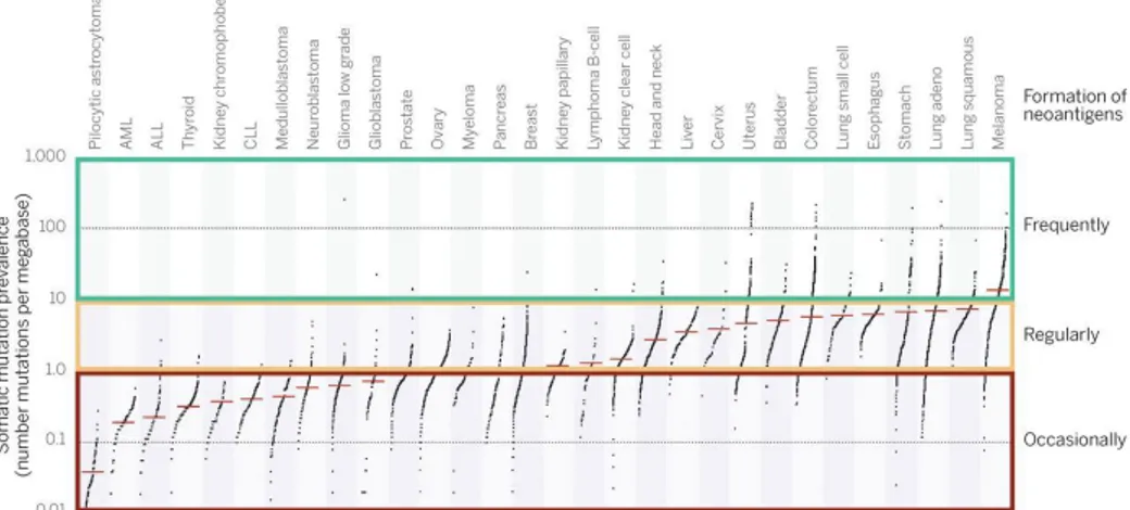
With the development of deep-sequencing technologies, it has become feasible to identify the mutations present within the protein-encoding part of the genome (the exome) of an individual tumor with relative ease. However, only a very small fraction of the non-synonymous mutations in expressed genes leads to the formation of a neo-antigen for which CD4+ or CD8+ T cell reactivity can be detected within tumor-infiltrating lymphocytes. The figure 10 depicts indeed the categories that indicate current estimates of the likelihood of neo-antigen formation in different tumor types, on the basis of their mutational load.
Figure 10. Estimate of the neo-antigen repertoire in human cancer (from Schumacher and Schreiber, 2015)
Data depict the number of somatic mutations in individual tumors. Every dot represents a sample while the red horizontal lines are the median numbers of mutations in the respective cancer types. Categories on the right indicate current estimates of the likelihood of neo-antigen formation in different tumor types.
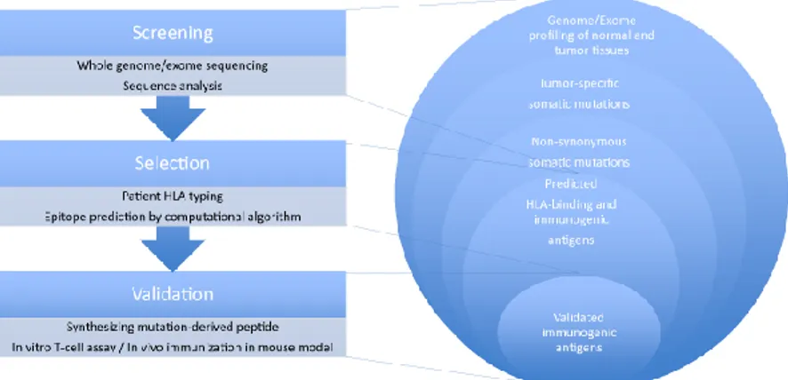
28 Taken this into consideration, starting from the whole exome and transcriptome of tumor, computational algorithms are necessary for the prediction of likely neo-antigens to use for a personalized therapy in the clinical setting (Figure 11).
Figure 11. Cancer exome–based identification of neo-antigens.
The process of identifying cancer neo-antigens for targeted cancer immunotherapy consists of three steps: screening, selection, and validation of the candidate neo-antigens. First, the whole genome/exome sequence profile is comprehensively screened to identify tumor-specific somatic mutations (cancer neo-antigens) by massive parallel sequencing of tumor and normal tissues, respectively. When available, RNA sequencing data are used to focus on mutations in expressed genes. Second, computational algorithms are used for predicting the affinity of the mutation-derived peptides with the patient’s own HLA and/or TCR. Alternative ways of epitope selection include “minigene” library screening and utilizing mass spectrometry analysis. Third, synthetic mutated peptides and wild-type peptides are used to validate the immunogenicity and specificity of the identified antigens by in vitro T-cell assay or in vivo immunization.
The identification of neo-antigens based on cancer exome data has been documented in a variety of experimental model systems and human malignancies. In 2012, two independent reports in mouse models provided a first preclinical proof that cancer exome–based approach can be used to identify neo-antigens that can be recognized by T cells.
Schreiber and colleagues demonstrated the feasibility of identifying spontaneously immunogenic tumor rejection antigens and their anti-tumor potency. Using massively parallel sequencing (MPS) and the MHC class I epitope prediction algorithm (IEDB algorithm), they characterized expressed mutations in highly immunogenic methylcholanthrene-induced sarcomas derived from immunodeficient Rag2-/- mice. Using class I prediction
29 algorithms, they identified mutant spectrin-β2 as a potential rejection antigen of the d42m1 sarcoma and validated this prediction. They also demonstrated that cancer immunoediting of d42m1 occurs via a T-cell-dependent immunoselection process that promotes outgrowth of pre-existing tumor cell clones lacking highly antigenic mutant spectrin-β2 and other potential strong antigens (Matsushita et al., 2012).
In the same year, Castle and colleagues presented a personalized immunotherapy approach to target the full spectrum of an individual tumor mutanome. They performed MPS and used the NetMHC algorithm to identify target neo-antigens for designing a cancer vaccine against B16F10 murine melanoma. They identified 962 non-synonymous somatic point mutations, 563 of which were actually expressed in tumor genes. They then selected 50 mutations for in vivo validation of immunogenicity and specificity, by administering either mutated or wild-type synthetic long peptides to the experimental mice. Approximately one third (16/50) showed the induction of a T-cell response, two of which were confirmed to have antitumor effects in both prophylactic and therapeutic settings, thereby qualifying mutated epitopes that include single amino acid substitutions as effective vaccines (Castle et al., 2012).
In 2015, the same group performed new vaccination studies employing the RNA vaccine technology. By using mRNA encoding mutated peptides identified in three different mouse tumor models (B16F10, CT26, 4T1 cancer cell lines)they revealed that a significant portion of mutations (21-45%) were immunogenic. Surprisingly, most neo-antigens were recognized by CD4+ T cells (70–94%) and this subset controlled growth of advanced, highly aggressive mouse tumors. Building on these data they developed an in silico approach to extract the therapeutically effective vaccine candidates out of the dozens or hundreds of mutations which are typically identified by NGS (Kreiter et al., 2012).
Yadav et al. employed another approach for the identification of immunogenic neo-antigens in two tumor cell lines of MC-38 and TRAMP-C1 (Yadav et al.2014). They used mass spectrometry analysis combined with whole-exome/transcriptome sequencing. Of 1290 and 67 mutations expressed in MC-38 and TRAMP-C1, respectively, 170 and 6 were predicted to bind MHC-class I molecule by the NETMHC-3.4 algorithm. On the other hand, only 7 and 0, respectively, were shown to be present on the MHC-class I molecule by mass spectrometry. Two of the seven antigens were structurally predicted to be immunogenic, and both actually showed strong anti-tumor responses in vitro. Their study suggested that utilizing mass spectrometry, as another filter it is possible to reduce the burden of validation assays, which are extremely laborious, thereby simplifying the neo-antigen discovery process.
30 All the encouraging results obtained by several pre-clinical studies have promoted the use of personalized cancer vaccines in clinics, showing glimmers of success. Recently, two small clinical trials showed effective anti-tumor activity of vaccines tailored to match a patient’s mutanome.
One group, led by Catherine Wu, evaluated a personalized peptide vaccine in a phase I study in patients with previously untreated high-risk melanoma after surgical resection (Ott et al., 2017). For each person, they formulated a vaccine that contained up to 20 protein fragments corresponding to the identified tumor mutations. Of six vaccinated patients, four had no recurrence at 25 months after vaccination, while two with recurrent disease were subsequently treated with anti-PD-1 therapy and experienced complete tumorregression, with expansion of the repertoire of neo-antigen-specific T cells. Indeed, they demonstrated that vaccination with neo-antigens both expands pre-existing neo-antigen-specific T-cell populations and induces a broader repertoire of new T-cell specificities in cancer patients, tipping the intratumoral balance in favor of enhanced tumor control.
The second group, led by Ugur Sahin, treated 13 melanoma patients with the RNA-based poly-neo-antigen approach (Sahin et al., 2017). Ten selected mutations per patient were engineered into two synthetic RNAs, each encoding five linker-connected 27mer peptides with the mutation in position 14 (pentatope RNAs). Eight patients who had no visible tumors at the time of vaccination remained tumor-free more than a year later. The remaining five participants' tumors had spread by the time they received the vaccine. Two of the five patients with metastatic disease experienced vaccine-related objective responses. One of these patients had a late relapse. A third patient developed a complete response to vaccination in combination with PD-1 blockade therapy.
These studies provide a strong rationale for further development of neo-antigens-targeted personalized cancer vaccines, alone and in combination with checkpoint blockade or other immunotherapies.