proliferation (Fig. 4B, c and d). Collectively these results are consistent with the hypothesis that increased FGF production by the Hand2-null uterine stroma stimulates epithelial proliferation by activating the FGFR-ERK1/2 pathway.
The ERK1/2-dependent phosphorylation of epithelial ERa at Ser118is critical for the tran-scriptional activation of ERa (11). Administra-tion of either PD173074 (Fig. 4C, a to d) or PD184352 (Fig. 4C, e to h) to Hand2-null uterine horns blocked the phosphorylation of epithelial ERa at Ser118
and the expression of Muc-1. This result supported our view that elevated signaling by FGFR-ERK1/2 pathway in Hand2d/duteri is responsible for phosphorylation and activation of ERa in epithelial cells, which promotes persistent expression of Muc-1 and which in turn creates a barrier that prevents embryo attachment.
Earlier studies using tissue recombinants pre-pared with uterine epithelium and stroma isolated from neonatal wild-type and PR-null mice indi-cated that the stromal PR plays an obligatory role in mediating the inhibitory actions of P on E-induced epithelial cell proliferation (18). However, the mechanism of this stromal-epithelial
communi-cation remained unknown. Our study has delineated a pathway in which Hand2 operates downstream of P to regulate the production of FGFs, mito-genic paracrine signals that originate in the stro-ma and act on the FGFR(s) in epithelium to control its E responsiveness (fig. S15). The anti-proliferative action of P in uterine epithelium is of clinical significance, because the breakdown of this action underpins E-dependent endometrial cancer (19). Hand2, therefore, is an important fac-tor to be considered for hormone therapy to block the proliferative actions of E in the endometrium.
References and Notes
1. C. A. Finn, L. Martin, J. Reprod. Fertil. 39, 195 (1974). 2. D. D. Carson et al., Dev. Biol. 223, 217 (2000). 3. C. Y. Ramathal, I. C. Bagchi, R. N. Taylor, M. K. Bagchi,
Semin. Reprod. Med. 28, 17 (2010).
4. L. Martin, R. M. Das, C. A. Finn, J. Endocrinol. 57, 549 (1973). 5. I. C. Bagchi, Y. P. Cheon, Q. Li, M. K. Bagchi, Front.
Biosci. 8, s852 (2003).
6. H. Pan, Y. Deng, J. W. Pollard, Proc. Natl. Acad. Sci. U.S.A. 103, 14021 (2006).
7. I. C. Bagchi et al., Semin. Reprod. Med. 23, 38 (2005). 8. D. Srivastava et al., Nat. Genet. 16, 154 (1997). 9. A. B. Firulli, Gene 312, 27 (2003).
10. Materials and methods are available as supporting material on Science Online.
11. S. Kato et al., Science 270, 1491 (1995).
12. G. A. Surveyor et al., Endocrinology 136, 3639 (1995). 13. K. Y. Lee et al., Nat. Genet. 38, 1204 (2006). 14. V. P. Eswarakumar, I. Lax, J. Schlessinger, Cytokine
Growth Factor Rev. 16, 139 (2005).
15. R. Iwamoto, E. Mekada, Cytokine Growth Factor Rev. 11, 335 (2000).
16. M. Koziczak, T. Holbro, N. E. Hynes, Oncogene 23, 3501 (2004). 17. D. B. Solit et al., Nature 439, 358 (2006).
18. T. Kurita et al., Endocrinology 139, 4708 (1998). 19. J. J. Kim, E. Chapman-Davis, Semin. Reprod. Med. 28,
81 (2010).
20. We thank M. Laws for genotyping and Y. Li for immunohistochemistry. This work was supported by the Eunice Kennedy Shriver National Institute of Child Health and Human Development, NIH, through U54HD055787 as part of the Specialized Cooperative Centers Program in Reproduction and Infertility Research. The Gene Expression Omnibus (GEO) microarray accession number is GSE25881. Supporting Online Material
www.sciencemag.org/cgi/content/full/331/6019/912/DC1 Materials and Methods
Figs. S1 to S15 Table S1 References
7 September 2010; accepted 15 December 2010 10.1126/science.1197454
Distinct Properties of the XY
Pseudoautosomal Region Crucial
for Male Meiosis
Liisa Kauppi,1Marco Barchi,2,3Frédéric Baudat,2* Peter J. Romanienko,2 Scott Keeney,1,4† Maria Jasin2†
Meiosis requires that each chromosome find its homologous partner and undergo at least one crossover. X-Y chromosome segregation hinges on efficient crossing-over in a very small region of homology, the pseudoautosomal region (PAR). We find that mouse PAR DNA occupies unusually long chromosome axes, potentially as shorter chromatin loops, predicted to promote double-strand break (DSB) formation. Most PARs show delayed appearance of RAD51/DMC1 foci, which mark DSB ends, and all PARs undergo delayed DSB-mediated homologous pairing. Analysis ofSpo11b isoform–specific transgenic mice revealed that late RAD51/DMC1 foci in the PAR are genetically distinct from both early PAR foci and global foci and that late PAR foci promote efficient X-Y pairing, recombination, and male fertility. Our findings uncover specific mechanisms that surmount the unique challenges of X-Y recombination.
M
eiotic recombination, initiated by pro-grammed double-strand breaks (DSBs), promotes homologous chromosome (homolog) pairing during prophase I (1). A subset of DSBs matures into crossovers that physicallyconnect homologs so that they orient properly on the first meiotic spindle. Because sex chromo-some recombination and pairing are restricted to the PAR (2), at least one DSB must form within this small region, and the homologous PAR must be located and engaged in recombination to lead to a crossover. Accordingly, the PAR in males ex-hibits high crossover frequency (2, 3), but sex chromosomes also missegregate more frequently than autosomes (4). Nevertheless, X-Y nondisjunc-tion is rare, which suggests that there are mecha-nisms that ensure successful X-Y recombination. X-Y pairing is more challenging than auto-somal pairing, as it cannot be mediated by mul-tiple DNA interactions along the length of the chromosomes. We used fluorescence in situ
hy-bridization (FISH) (5) to compare timing of mei-otic X-Y and autosomal pairing in mice (Fig. 1). At leptonema, when DSBs begin to form and only short chromosome axis segments are pre-sent, PAR and autosomal FISH probes were most-ly unpaired. By earmost-ly to mid-zygonema, when axes elongate and homologs become juxtaposed, dis-tal ends of chr 18 and 19 were paired in ~50% of nuclei; by late zygonema, these regions were paired in nearly all nuclei (Fig. 1B and fig. S1). In contrast, the X and Y PARs were rarely paired before pachynema (Fig. 1B); hence, X-Y pairing is delayed compared with that of autosomes.
DSBs precede and are required for efficient homolog pairing in mouse meiosis (6, 7). Nucleus-wide (“global”) foci of DSB markers RAD51/ DMC1 peak in number at early to mid-zygonema (Fig. 2A) (8, 9). Because stable X-Y pairing oc-curs late, we asked whether PAR DSB kinetics is also delayed (Fig. 2B and fig. S2). More than half of cells had no RAD51/DMC1 focus in the PAR before late zygonema (Fig. 2C), distinct from global patterns. Only when global foci were already declining did the majority of cells (~70%) display PAR foci (Fig. 2C and fig. S2i). We interpret the lack of PAR foci to indicate that DSBs have not yet formed. Thus, we propose that PAR DSB formation and/or turnover are under distinct temporal control. We cannot ex-clude the alternative possibility that PAR DSBs have formed but are cytologically undetectable, for example, because RAD51/DMC1 have not yet been loaded onto DSB ends or because foci have already turned over. In either case, DSB dy-namics and/or processing differs on the PAR.
Most sites marked by PAR RAD51/DMC1 foci appeared incapable of mediating stable pairing before early pachynema (~70% of late zygotene
1Molecular Biology Program, Memorial Sloan-Kettering Cancer
Center, New York, NY 10065, USA.2Developmental Biology
Program, Memorial Sloan-Kettering Cancer Center, New York, NY 10065, USA.3Department of Public Health and Cell
Bio-logy, Section of Anatomy, University of Rome Tor Vergata, 00133 Rome, Italy.4Howard Hughes Medical Institute, Memorial Sloan-Kettering Cancer Center, New York, NY 10065, USA. *Present address: Institute of Human Genetics, CNRS, 34090 Montpellier, France.
†To whom correspondence should be addressed. E-mail: [email protected] (M.J.); [email protected] (S.K.)
nuclei had foci, but <20% showed PAR pairing) (Figs. 1B and 2C). The number of PAR foci per cell also increased over time. In leptonema and early to mid-zygonema, most cells with a PAR RAD51/DMC1 focus had only one (typically on X), whereas by late zygonema, two PAR foci were often present (both X and Y) (Fig. 2C). Foci on both PARs could represent two independent DSBs. If so, then having more than one X-Y recombination interaction may stabilize pairing, similar to multiple interactions that stabilize pair-ing of autosomes (10). Alternatively, foci on both PARs could represent the two, separated ends of a single DSB (11, 12)—with one focus marking the broken PAR and the second focus marking the other PAR (fig. S3A). In this“ends-apart” model, nuclei that have two PAR foci are those in which the X and Y PARs have successfully en-gaged each other. However, we found that most such nuclei showed no evidence of a preferential X-Y spatial relationship (fig. S3B), and most PAR pairing occurred abruptly at the zygonema-to-pachynema transition, i.e., after the stage when many cells displayed two PAR foci (compare Figs. 1B and 2C). Sex body formation (13) may facilitate this sudden completion of X-Y pairing by providing homology-independent X-Y prox-imity that simplifies the homology search.
The haploid mouse genome averages fewer than one DSB per 10 Mb (Fig. 2A), whereas the
Fig. 2. Distinct temporal and structural properties of the PAR. (A) Nucleus-wide RAD51/DMC1 foci in spermatocytes (bars show meansT SD). (B) Assay for PAR DSB formation. IF against RAD51/DMC1 and SYCP3 (i) and FISH (ii) with probes shown in Fig. 1Ai on a leptotene spermatocyte nucleus. Scale bar, 10mm. (iii) Magnified views of Y and X PARs from frames in (i) (5) and an overlay of the PAR FISH signal with SYCP3 (right), here with a RAD51/DMC1
focus only on the X PAR. (C) Nuclei (%) with one or two PAR RAD51/DMC1 foci. (D) Axis/loop segments as a determinant of DSB potential [after (15)]. Only one homolog is shown. DNA organized on a longer axis into more and smaller loops (i) has more DSB potential than if the same DNA is organized on a shorter axis into fewer and larger loops (ii). (E) Examples of chromatin extension (gray brackets in insets); see also Table 1. Scale bar, 5mm. Fig. 1. Late PAR pairing during male meiosis. (A) FISH assay for pairing. (i and ii) Example of
im-munofluorescence (IF) and two sequential rounds of FISH on a late zygotene spermatocyte nucleus. Nuclei stained with an antibody against axis protein SYCP3 were subjected first to PAR FISH (i), then to distal chr 18 and distal chr 19 FISH (ii). Scale bar, 10 mm. (B) Nuclei (%) with unpaired and paired (≤2 mm apart) FISH signals. Chromosome synapsis status was also recorded at sites of paired signals.
<1 Mb PAR (14) undergoes one or two DSBs (Fig. 2C), which is 10 to 20 times the genome average. We speculated that distinct higher-order chromosome structure could render the PAR more conducive to DSB formation. Meiotic recombi-nation is proposed to occur within DNA segments residing in chromatin loops that become tran-siently tethered to chromosome axes (15). Loop density per micrometer of axis is constant (16) and produces an inverse relation between loop size and axis length (17). DNA arranged into small-er loops may have highsmall-er DSB potential (Fig. 2D) (18); indeed, autosomal crossover frequency in male mice correlates with axis length (19). We found that PAR axes were disproportionately long relative to DNA length and incorporated ~1 Mb permm of axis (Table 1A). At the distal ends of
chr 18 and 19 (regions with relatively frequent crossing-over) (19), DNA content was 10 to 13 Mb permm and correlated well with axis length, i.e., the distal ~10% of DNA occupied ~10% total axis length (Table 1A). The≥10-fold difference between PAR and autosome axes is of the mag-nitude expected for a region that experiences more than 10 times as many DSBs. Axes of non-PAR portions of the X and Y had a DNA content more like autosomes (≥14 Mb per mm) (fig. S4). Long PAR axes predict short chromatin loops. As a proxy for loop size, we measured FISH signal extension from axes for probes in the PAR and autosomal subtelomeric regions (Fig. 2E and Table 1B). PAR FISH signals were substan-tially more compact at all stages (about one-third to one-seventh as extended), consistent with smaller
loops. Thus, chromosome structure could be one factor that facilitates high-frequency DSB forma-tion in the PAR.
The distinct temporal and structural features outlined above raised the possibility that mech-anisms ensuring efficient PAR recombination and pairing may be under different genetic con-trol from autosomes. Characterization of a variant of SPO11, the evolutionarily conserved meiotic DSB catalyst (1), validated this hypothesis (Fig. 3). Two major mRNA splicing isoforms in mice and humans are Spo11a and Spo11b (7, 20–22) (Fig. 3Ai and fig. S5). Spo11b is expressed early in meiosis, when most DSBs are formed, whereas Spo11a is expressed later (7, 20, 23) (Fig. 3Aii and fig. S6A). Thus, SPO11b is likely responsi-ble for most DSB formation.
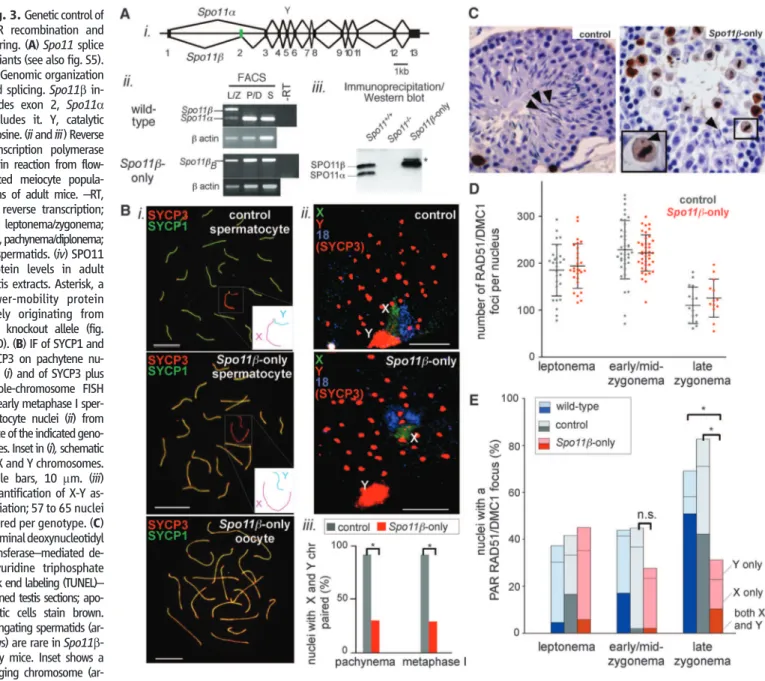
Fig. 3. Genetic control of PAR recombination and pairing. (A)Spo11 splice variants (see also fig. S5). (i) Genomic organization and splicing.Spo11b in-cludes exon 2, Spo11a excludes it. Y, catalytic tyrosine. (ii and iii) Reverse transcription polymerase chain reaction from flow-sorted meiocyte popula-tions of adult mice.–RT, no reverse transcription; L/Z, leptonema/zygonema; P/D, pachynema/diplonema; S, spermatids. (iv) SPO11 protein levels in adult testis extracts. Asterisk, a lower-mobility protein likely originating from the knockout allele (fig. S6D). (B) IF of SYCP1 and SYCP3 on pachytene nu-clei (i) and of SYCP3 plus whole-chromosome FISH of early metaphase I sper-matocyte nuclei (ii) from mice of the indicated geno-types. Inset in (i), schematic of X and Y chromosomes. Scale bars, 10 mm. (iii) Quantification of X-Y as-sociation; 57 to 65 nuclei scored per genotype. (C) Terminal deoxynucleotidyl transferase–mediated de-oxyuridine triphosphate nick end labeling (TUNEL)– stained testis sections; apo-ptotic cells stain brown. Elongating spermatids (ar-rows) are rare in Spo11b-only mice. Inset shows a lagging chromosome
(ar-rowhead) in a TUNEL-positive cell. (D) RAD51/DMC1 focus counts in spermatocytes from control andSpo11b-only mice (bars show means T SD). (E) Nuclei (%) with PAR RAD51/DMC1 foci in mice of the indicated genotypes; for each genotype, 41 to 55 nuclei were scored per stage. *P ≤ 0.0002 (two-tailed Mann-Whitney test); n.s., not significant (P = 0.09).
We generated transgenic mice expressing Spo11bBcDNA (fig. S5) from a meiosis-specific
promoter (24) (fig. S6B). Tg(Xmr-Spo11bB)
tran-script expression overlapped with Spo11b mRNA appearance in wild type (Fig. 3Aii and fig. S6, A and C). In testis extracts of Spo11–/– Tg (Xmr-Spo11bB)
+/+
(hereafter,“Spo11b-only”) mice, SPO11bBprotein approximated the total level of
SPO11 in wild type (Fig. 3Aiii). The transgene did not cause obvious meiotic phenotypes in mice heterozygous at the endogenous Spo11 locus [i.e., Spo11+/– Tg(Xmr-Spo11bB)+/+], and
these mice were used as controls. The profound meiotic defects of Spo11–/– mice [no recombi-nation, failure of homolog pairing and synapsis, and infertility (6, 7, 25)] were mostly rescued by Tg (Xmr-Spo11bB) in both sexes: Autosomal
homol-ogous pairing, synapsis, and MLH1 focus for-mation (a crossover marker) appeared normal (Fig. 3Bi and fig. S7A). Moreover, ovaries of Spo11b-only mice contained abundant primordi-al follicles (fig. S7B), and Spo11b-only femprimordi-ales were fully fertile with normal litter sizes. Thus SPO11bBsupports autosomal crossing-over,
pair-ing, and synapsis, and (in females) full meiotic progression and accurate chromosome segrega-tion. Male meiosis was not fully rescued, how-ever. Although sex bodies formed (fig. S7C), the X and Y failed to pair and synapse in ~70% of spermatocytes (Fig. 3B). Spo11b-only testis sec-tions showed numerous apoptotic metaphase I cells (Fig. 3C), many with a lagging chromosome (Fig. 3C, inset, and fig. S7D), consistent with spindle checkpoint-induced apoptosis (9, 26, 27), triggered by the failure of nonrecombinant X and Y to orient properly on the metaphase I spindle. Few postmeiotic cells were formed, and testis sizes were reduced (Fig. 3C and fig. S7, D and E), so that although some Spo11b-only males pro-duced offspring, most were infertile.
Nucleus-wide numbers and timing of RAD51/ DMC1 foci were indistinguishable between Spo11b-only and control males (Fig. 3D and fig. S7F), which indicated that the X-Y pairing defect cannot be attributed to reduced global DSB lev-els. Similarly, the frequency of PAR RAD51/ DMC1 foci in leptonema was not affected (Fig. 3E). In contrast, the percentage of late zygotene nuclei with a PAR focus was reduced in Spo11b-only males, consistent with a defect in a late-forming DSB population (PAR-specific, or possibly in-cluding a small subset of autosomal DSBs). About 70% of late zygotene nuclei lacked PAR foci (Fig. 3E), which was similar to the percentage of cells with X-Y pairing failure (Fig. 3Biii). Thus, the few PAR foci that form early in both wild-type and Spo11b-only males seem to per-sist until late zygonema (fig. S4, discussion), at which time recombination-mediated X-Y pair-ing occurs. We propose that a lack of late PAR DSBs is the cause of infertility in Spo11b-only males. In females, two fully homologous X chro-mosomes make PAR recombination dispensable.
Spo11a is the only splice variant missing from Spo11b-only mice that is known to be develop-mentally regulated, and its expression in wild type correlates with the timing of late PAR DSBs as inferred from the appearance of RAD51/DMC1 foci. It is thus possible that SPO11a, by itself or in combination with SPO11b, is needed for DSB formation in late zygonema. In this scenario, late-forming PAR DSBs are genetically separable from both global DSBs and early-forming PAR DSBs, and the surge of late-forming PAR DSBs is crucial for efficient X-Y pairing and fertility. PAR recombination occasionally fails in humans, as evidenced by paternally inherited sex chromo-some aneuploidies [e.g., Klinefelter’s or Turner syndromes (28)]. Because Spo11 isoforms are conserved, we speculate that variation in Spo11
splicing patterns may be a human X-Y non-disjunction susceptibility trait.
References and Notes
1. F. Cole, S. Keeney, M. Jasin, Genes Dev. 24, 1201 (2010).
2. F. Rouyer et al., Nature 319, 291 (1986). 3. P. Soriano et al., Proc. Natl. Acad. Sci. U.S.A. 84,
7218 (1987).
4. Q. Shi et al., Am. J. Med. Genet. 99, 34 (2001). 5. Materials and methods are available as supporting
material on Science Online.
6. F. Baudat, K. Manova, J. P. Yuen, M. Jasin, S. Keeney, Mol. Cell 6, 989 (2000).
7. P. J. Romanienko, R. D. Camerini-Otero, Mol. Cell 6, 975 (2000).
8. X. Ding et al., Dev. Cell 12, 863 (2007). 9. M. Barchi et al., PLoS Genet. 4, e1000076 (2008). 10. B. M. Weiner, N. Kleckner, Cell 77, 977 (1994). 11. N. Hunter, in Molecular Genetics of Recombination,
A. Aguilera, Rothstein, R., Eds. (Springer-Verlag, Heidelberg, 2007), vol. 17, pp. 381–442. 12. A. Storlazzi et al., Cell 141, 94 (2010).
13. P. S. Burgoyne, S. K. Mahadevaiah, J. M. Turner, Nat. Rev. Genet. 10, 207 (2009).
14. J. Perry, S. Palmer, A. Gabriel, A. Ashworth, Genome Res. 11, 1826 (2001).
15. Y. Blat, R. U. Protacio, N. Hunter, N. Kleckner, Cell 111, 791 (2002).
16. D. Zickler, N. Kleckner, Annu. Rev. Genet. 33, 603 (1999).
17. E. Revenkova et al., Nat. Cell Biol. 6, 555 (2004). 18. N. Kleckner, A. Storlazzi, D. Zickler, Trends Genet. 19,
623 (2003).
19. L. Froenicke, L. K. Anderson, J. Wienberg, T. Ashley, Am. J. Hum. Genet. 71, 1353 (2002).
20. P. J. Romanienko, R. D. Camerini-Otero, Genomics 61, 156 (1999).
21. S. Keeney et al., Genomics 61, 170 (1999). 22. M. A. Bellani, K. A. Boateng, D. McLeod, R. D. Camerini-Otero,
Mol. Cell. Biol. 30, 4391 (2010).
23. M. J. Neale, J. Pan, S. Keeney, Nature 436, 1053 (2005). 24. P. J. Romanienko, dissertation, Cornell University Ithaca,
NY (1997).
25. M. Di Giacomo et al., Proc. Natl. Acad. Sci. U.S.A. 102, 737 (2005).
26. S. Eaker, J. Cobb, A. Pyle, M. A. Handel, Dev. Biol. 249, 85 (2002).
Table 1. Chromosome axis lengths and chromatin extension in PARs and distal ends of chr 18 and 19. Axis lengths are means T SD and FISH signal extensions are meansT SD, for the number of observations in parentheses. Probe size is the size of the bacterial artificial chromosome (5).
(A) DNA content versus chromosome axis lengths (late zygonema) Chromosome Total size
(Mb) Probe-distal region (Mb) Total chromosome axis (mm) Probe-distal axis
Length (mm) DNA content (Mb/mm) Y 95 0.7 4.2T 0.7 (17) 0.7T 0.2 (20) 1
X 167 0.7 12.7T 2.8 (13) 0.8T 0.2 (23) 1 18 91 8 6.1T 0.9 (11) 0.6T 0.1 (12) 13 19 61 6 5.1T 0.5 (11) 0.6T 0.1 (10) 10 (B) Length of chromatin extension from axes
Locus Probe size (kb)
FISH signal extension (mm) Leptonema Early to
mid-zygonema Late zygonema Pachynema Y PAR
146 0.5T 0.2 (25) 0.6T 0.3 (21) 0.6T 0.3 (21) 1.2T 0.5 (23)* X PAR 0.6T 0.4 (23) 0.7T 0.5 (17) 0.6T 0.5 (20)
Distal chr 18 207 2.2T 0.8 (26) 3.2T 1.5 (31)* 4.5T 1.9 (35)* 5.4T 2.2 (21)* Distal chr 19 182 2.3T 0.9 (18) 3.6T 2.0 (33)* 5.3T 3.0 (40)* 5.9T 2.3 (23)*
27. T. Odorisio, T. A. Rodriguez, E. P. Evans, A. R. Clarke, P. S. Burgoyne, Nat. Genet. 18, 257 (1998). 28. H. Hall, P. Hunt, T. Hassold, Curr. Opin. Genet. Dev. 16,
323 (2006).
29. This work was supported by NIH grant R01 HD040916 (M.J. and S.K); International Grants in Cancer Research (AIRC) [My First AIRC Grant (MFAG) grant 4765], Italian Ministry for Education, University and Research (MIUR), the Lalor Foundation, and the
American-Italian Cancer Foundation (AICF) (M.B.); and the Charles H. Revson Foundation (F.B.). We thank M. Leversha [Memorial Sloan-Kettering Cancer Center (MSKCC)], P. Bois (Scripps Florida), and K. Manova (MSKCC) for valuable advice and protocols. We are grateful to Keeney and Jasin lab members, especially I. Roig, E. de Boer, and F. Cole, and to N. Hunter (University of California, Davis) for insightful comments.
Supporting Online Material
www.sciencemag.org/cgi/content/full/331/6019/916/DC1 SOM Text
Materials and Methods Figs. S1 to S7 References
28 July 2010; accepted 21 December 2010 10.1126/science.1195774
Classic Selective Sweeps Were Rare
in Recent Human Evolution
Ryan D. Hernandez,1* Joanna L. Kelley,1Eyal Elyashiv,2S. Cord Melton,1Adam Auton,3 Gilean McVean,3,41000 Genomes Project, Guy Sella,2† Molly Przeworski1,5,6†‡
Efforts to identify the genetic basis of human adaptations from polymorphism data have sought footprints of“classic selective sweeps” (in which a beneficial mutation arises and rapidly fixes in the population). Yet it remains unknown whether this form of natural selection was common in our evolution. We examined the evidence for classic sweeps in resequencing data from 179 human genomes. As expected under a recurrent-sweep model, we found that diversity levels decrease near exons and conserved noncoding regions. In contrast to expectation, however, the trough in diversity around human-specific amino acid substitutions is no more pronounced than around synonymous substitutions. Moreover, relative to the genome background, amino acid and putative regulatory sites are not significantly enriched in alleles that are highly differentiated between populations. These findings indicate that classic sweeps were not a dominant mode of human adaptation over the past ~250,000 years.
H
umans have experienced myriad adapta-tions since the common ancestor with chimpanzees and more recently have adapted to a wide range of environments. Efforts to infer the molecular basis of these adaptations from polymorphism data have largely been guided by the“classic selective sweep” model, in which a new, strongly beneficial mutation in-creases in frequency to fixation in the population [reviewed in (1, 2)]. In this scenario, the allele ascends rapidly enough in frequency for there to be little opportunity for recombination to un-couple it from its genetic background, such that its rise sweeps out variation at linked sites, re-ducing linked neutral diversity in the popula-tion and distorting allele frequencies and patterns of linkage disequilibrium (3). In humans, the ef-fects of sweeps are expected to persist for ap-proximately 10,000 generations or about 250,000 years (4).Identifying the footprint of a sweep against a noisy genomic background is challenging, be-cause patterns of genetic variation reflect the ef-fects of multiple modes of natural selection as well as of demographic history, mutation, and recombination. To date, applications of statistical tests based on the sweep model have led to the identification of more than 2000 genes as po-tential targets of positive selection in the human genome (2) and to the suggestion that diversity patterns in ~10% of the human genome have been affected by linkage to recent sweeps [e.g., (5)]. The list of functionally characterized cases of genetic adaptations is short, however, and the false discovery rate of selection scans is poten-tially high (6). Thus, it remains unknown whether the well-documented cases are typical of human adaptations, or whether they represent rare in-stances where the genetic architecture of the ad-aptation was conducive to classic sweeps (7, 8), with most adaptations occurring by other modes (e.g., polygenic selection and selection on stand-ing variation).
Two main lines of evidence have been ad-vanced in support of the hypothesis that classic selective sweeps were common. First, regions of low recombination, in which a single sweep should have a larger span, exhibit lower diversity (after correcting for variation in mutation rates) relative to regions of high recombination (9–11). Regions of low recombination also show greater differen-tiation between populations (12), as expected from local adaptation or, for some parameters, from the fixation of globally advantageous alleles (13).
Second, under the sensible assumption that ami-no acid and conserved ami-noncoding sites are enriched among targets of adaptation, one would expect that the signal of selection would be most clearly visible at or around such sites [e.g., (10, 14)]. Consistent with this expectation, diversity levels decrease with the number of human-specific sub-stitutions at amino acid or conserved noncoding sites (in 200- to 600-kb windows) (10), and genic regions show an enrichment of alleles that are highly differentiated between populations relative to nongenic regions (15, 16). These patterns are informative but are only indirectly related to theo-retical predictions. Moreover, some—possibly all— of these patterns may instead result from purify-ing selection actpurify-ing on deleterious mutations at linked sites (“background selection”) (9–11, 16–18). To evaluate the importance of classic sweeps in shaping human diversity, we analyzed rese-quencing data for 179 human genomes from four populations, collected as part of the low-coverage pilot for the 1000 Genomes Project (19). These data overcome ascertainment biases arising in the study of genotyping data, with ~99% power to detect variants with a population frequency above 10% for 86% of the euchromatic genome (19).
We examined the extent to which selection affects diversity levels at linked sites by calcu-lating the average diversity as a function of ge-netic distance from the nearest exons, collating all exons across the genome (fig. S1). To estimate neutral diversity levels, we focused only on non-conserved noncoding and fourfold degenerate sites (11). To correct for systematic variation in the mutation rate, we divided diversity by human– rhesus macaque divergence [to which the contri-bution of ancestral polymorphism is minor (11)]. Our estimate of relative diversity appears little affected by the low fold coverage of individuals or variation in sequencing depth (fig. S2, C to E). Scaled diversity levels are lowest near exons (Fig. 1A and fig. S3), recovering half the drop by 0.03 to 0.04 cM, depending on the population, and 80% by 0.07 to 0.1 cM [see (20)]. Given that diversity is scaled by divergence, the trough in scaled diversity around exons does not reflect systematic variation in mutation rates as a func-tion of the distance from exons, strong purifying selection on the sites themselves (which would decrease both diversity and divergence), or weak selection near exons (which should inflate, not decrease, diversity levels divided by divergence). Rather, the trough provides evidence for the effects of directional selection at linked sites, extending over 100 kb.
1Department of Human Genetics, University of Chicago, Chicago,
IL 60637, USA. 2Department of Ecology, Evolution and
Behavior, Hebrew University of Jerusalem, Givat Ram, Jerusalem 91904, Israel.3Wellcome Trust Centre for Human Genetics, University of Oxford, Oxford OX3 7BN, UK.
4Department of Statistics, University of Oxford, Oxford OX1
3TG, UK.5Department of Ecology and Evolution, University of Chicago, Chicago, IL 60637, USA.6Howard Hughes Medical
Institute, University of Chicago, Chicago, IL 60637, USA. *Present address: Department of Bioengineering and Ther-apeutic Sciences, University of California, San Francisco, CA 94143, USA.
†These authors contributed equally to this work. ‡To whom correspondence should be addressed. E-mail: [email protected]