The Importance of Cone-Beam Computed Tomography in
Clinical Research: Gender Differences and Bilateral Symmetry of
Permanent Teeth Anatomy in a Saudi Arabian Population
By
Mohammed Mashyakhy BDS, MSc
The Department of Oral and Maxillo Facial Sciences
Sapienza, University of Rome
© Copyright by Mohammed Mashyakhy 2019
A thesis submitted in conformity with the requirements
ii
The Importance of Cone-Beam Computed Tomography in
Clinical Research: Gender Differences and Bilateral Symmetry of
Permanent Teeth Anatomy in a Saudi Arabian Population
Mohammed Mashyakhy
Doctor of Philosophy 2019
The Department of Oral and Maxillo Facial Sciences
Sapienza, University of Rome
ABSTRACT
Aims and Objectives: To retrospectively explore the relationship of maxillary and mandibular permanent dentition taking into consideration the number of roots, their respective quantity of root canals, the bilateral symmetry of root canal morphology and root canal configurations between both genders, in a Saudi Arabian population.
Materials and Methods: This study comprised of 208 subjects (48% males and 52% females) with a mean age of 28.74±9.56 years. The CBCT images of the recruited subjects were evaluated for all permanent teeth except third molars. A careful examination was obtained by optimal visualization using all software features to investigate the differences between both genders and to evaluate the bilateral symmetry of number of roots, number of canals and root canal system configurations. The data was analyzed using SPSS 25. Cohen’s Kappa test was used for reliability and bilateral symmetry, while, Chi-squared test was used for the differences
iii between both genders in relation to the study variables. A P-value < 0.05 was considered significant.
Results:
Gender differences: A total of 5254 maxillary and mandibular permanent teeth were evaluated.
In relation to the number of roots, there were no significant differences between the genders for maxillary and mandibular teeth (P= 0.064) as well as for maxillary and mandibular teeth separately (P= 0.315 and P= 0.100, respectively). Significant difference was found between males and females in relation to number of canals of maxillary teeth (P= 0.014). For mandibular teeth, the significant level of difference was at the cut-off point (P= 0.050). For both arches the distribution among both genders was not significant (P= 0.082). Conversely, the difference between both genders with regard to canal configuration of maxillary roots was highly statistically significant (P< 0.001). For mandibular teeth, difference between males and females in relation to canal configuration of anterior and premolar teeth was significant (P= 0.016) while, the same was not significant when related mesial roots of 1st and 2nd molars (P= 0.205).
However, a greater significance was found when distal roots of 1st and 2nd molars were
compared (P< 0.001).
Bilateral symmetry:
The Bilateral symmetry of number of roots was 100% in maxillary centrals, laterals, canines, 1st molars, and 2nd molars. In the mandibular arch however, it was 100% in mandibular centrals,
and 2nd premolars only. The most frequent asymmetry was exhibited by the maxillary 1st
premolars (14.9%). In relation to number of canals, the bilateral symmetry was 100% in maxillary centrals and laterals only. The commonest asymmetry was found in maxillary 2nd
molars (18.9%). When canal configuration was assessed, the bilateral symmetry was found to be 100% in maxillary centrals and laterals. However, the most frequent asymmetry was found in maxillary 2nd premolars (32.2%).
iv Conclusion:
No significant differences were found between both genders in relation to the number of roots. Regarding number of canals, significant differences were detected only in 3 out of 14 groups of teeth. Overall, females had lower number of canals than males. Canal configuration was also governed by gender in this study. Bilateral symmetry was more evident when number of roots were assessed than the canal configurations
Clinical relevance: The influence of gender should be considered when root canal morphology is assessed and henceforth root canal treatment is to be performed. Clinicians should be aware of these variations particularly when treating contralateral teeth in the same individual as well. Also, the utilization of CBCT could aid in proper dental therapy for teeth when conventional 2-D radiographs are inconclusive.
v
ACKNOWLEDGEMENTS
It was a great honor to be a student of Prof. Gianluca Gambarini, his invaluable guidance, support, encouragement and dedication throughout this research project was invaluable. Also, being a student at the Sapienza, University of Rome was a dream that came true.
My parents, for unlimited and unconditional love and support.
My wife, she has been always there backing me up with patience.
My Kids, There presence is a pure happiness
My family, my people, brothers and sisters, friends; thanks for believing in me, you were of great support and would love to share this success with you.
My friend Dr. Amal for her inspiration during the write up of my thesis.
Special respect and gratitude to my eldest brother: Yahya Mashyakhy who was always on
my side supporting me financially and morally throughout under and post graduate studies. My words are not enough to thank him, ALLAH BLESS him always and proud of such a brother.
vi
TABLE OF CONTENTS
Abstract ii Acknowledgements v Table of Contents vi I. GENERAL INTRODUCTION 1 1. Endodontic Objectives 12. Roots Canal Anatomy And Classification Systems 4
3. Methods Used To Study Root Canal Anatomy 6
4. 3D Cone-Beam Computed Tomography 7
5. Root Canal Anatomy Differences Between Genders 10
6. Bilateral Symmetry Of Root Canal Anatomy 12
7. Anatomical Variations In A Saudi Arabian Population 13
II. OBJECTIVES AND NULL HYPOTHESES 15
1. Objectives 15 2. Null Hypotheses 15 III. METHODOLOGY 16 1. The Sample 16 2. CBCT Scans 16 3. Data Analysis 17 IV. RESULTS 18 V. DISCUSSION 36 VI. CONCLUSIONS 44 VII. REFERENCES 45
VIII. PUBLISHED- SUBMITTED- PRESENTED RESEARCH 55
1 I. GENERAL INTRODUCTION
I.1 Endodontic objectives:
Endodontics is a branch of dentistry specialized in the morphology, physiology, and pathology of the human dental pulp and periapical tissues. It includes science and practices within these areas and is currently termed as root canal therapy or pathodontia. About a century ago, Dr. Harry B. Johnston came up with the term “Endodontics” from the Greek word: “en” meaning in or within, "odous" meaning tooth: “The process of working within the tooth” So, endodontics recently is solidified to focus on two areas:
1. Morphology, physiology, and pathology of the human dental pulp and periapical tissues
2. Etiology, diagnosis, prevention, and treatment of disease of the pulp and associated periapical conditions. (1)
Scientific and clinical opinions about endodontics vary widely worldwide, however, they all agree in one basic statement; that cleaning and shaping of the canal is the most important aspect of endodontic therapy. (2–7) Disinfecting the root canal system (RCS) with irrigation in combination with root canal debridement is considered one of the most important factors in the prevention and treatment of endodontic pathoses. (8) The rationale of endodontics is based on simple biologic principles. Significant reduction or complete elimination of microorganisms and infected necrotic pulp tissue are critical factors for success. (9)
Practically, this procedure is not simple, since the pulp tissue is enclosed inside a hard tissue ‘dentin” in a RCS. The defense mechanism inside the RCS it not fully equipped by the body’s immune system because of the special nature of teeth and lack of bilateral circulation in the
2 pulp tissue. Hence due to this and various other factors, most of which are bacterial in nature, the pulp tissue can go into vascular necrosis and/or infectious necrosis. Consequently, irritants and bacterial byproducts escape the root exit to cause lesions known as Lesions from Endodontic Origin (LEO). Root canal treatment (RCT) then is indicated and basically diseased teeth and peri-radicular tissue can be managed if diagnosed properly. (2,3)
Successful RCT after proper diagnosis and treatment planning is based on applying knowledge of tooth anatomy, root morphology and performing adequate chemo-mechanical cleaning, shaping and filling of the RCS three dimensionally. (10) Throughout the years, many instruments and techniques were used for mechanical cleaning and shaping to address the complexity of anatomical variations of the RCS. The real challenge is dealing with the reality that a root canal system is a three dimensional object which include fins, isthmuses, lateral canals and other complex structures that need to be cleaned and filled. Therefore, clinicians should be aware of the complexities of the “pulp space” rather than thinking about the RCS as a straight canal from orifice to apex.(2,3) It is well known that after performing an adequate cleaning, shaping and establishing a coronal seal endodontic treatment yields great success.(11) Subsequently, it is henceforth appropriate to summarize the main principles of cleaning and shaping the RCS when performing RCTs:
Principles of Cleaning and Shaping:
Tooth anatomy and morphology, the endodontic instruments and irrigation solution play a crucial role in cleaning the RCS. Even with contemporary instrumentation, complex RCS including lateral and accessory canals, canal curvatures, canal wall irregularities, fins, and isthmuses make total debridement virtually impossible. Since the current methods are not able to touch and debride all aspects of RCS, reducing the amount of irritants to a significant number within the RCS is therefore the main goal of cleaning. (12)
3 The overall goals of successful RCT are to facilitate cleaning and provide space for obturation. It is imperative to shape the canal accordingly if the latter is to be achieved. The main objectives of shaping are to maintain or develop a continuously tapering funnel from the canal orifice to the apex which allows for the achievement of general goals of thorough treatment. (13) Schilder et al. established certain constant principles for this, however, RCS are varied among different teeth and each one should be considered individually.(2) The principles are as follows:
1. Root canal preparation should develop a continuously tapering funnel from the root apex to the coronal access cavity.
2. The cross-sectional diameter of the preparation should be narrower at every point apically and wider at each point as the access cavity is approached.
3. The preparation should occupy as many planes as are presented by the root and the canal. 4. The preparation should flow with the shape of the original canal.
5. The apical foramen should remain in its original spatial relationship both to the bone and to the root surface.
6. The apical opening should be kept as small as is practical in all cases.
The knowledge of common root canal morphology and its frequent variations is a basic requirement for success during root canal procedures.
4 I.2 Root Canal Anatomy and Classification Systems:
RCS can take numerous configurations and presents many shapes. A thorough knowledge of human teeth morphology, proper understanding and interpretation of dental x-rays, and various other similarly fundamental endodontic procedures are essential for successful RCT. With the presence of a complex anatomy, utilizing magnification and illumination is a huge aid to manage such cases. (14)
Peters et al. (15) in a Micro-Computed Tomography (µCT) study demonstrated that, the geometry of the untreated root canals had more influence on the final changes produced by instrumentation techniques, hence, even after instrumentation about 35-40% of the RCS was not treated properly. This finding stresses the significance of the canal morphology. It is well established that a root with a tapering canal and a single foremen is the exception rather than the rule since the earliest relevant works on this by Preiswerk in 1912, Fasoli and Arlotta in 1913, plus Hess and Zurcher in 1917, to more recent studies. (19–22)
A number of studies have shown wide a variety of internal morphology of the RCT including: c-shaped canals, loops, accessory canals, fins, deltas and etc. So, clinicians should be aware of these variations and treat every single tooth as having a complexly unique anatomy keeping in mind that the RCS may branch, divide and rejoin again. (16–23) Because the great number of variations in the root canal anatomy, investigators came up with classification systems to ease the process of identification, understanding and communication in the dental community. Weine F.S. et al. (24), was the first to classify root canal configurations into four types:
• Type I (1-1): Single canal runs from orifice to apex, • Type II (2-1): Two canals unite into one at apex,
5 • Type III (2-2): Two canals from orifice to apex with no connection, and
• Type IV (1-2): One canal divides into two.
Vertucci et al. (25) in his study found a much more complex canal system and categorized them into eight configurations. (Figure 1) His classification is the most famous and widely used in endodontics, and it is as follows:
• Type I (1-1): Single canal runs from orifice to apex.
• Type II (2-1): Two canals arise from pulp chamber which unite in its course into one. Type III (1-2-1): One canal arises from pulp chamber and during its course splits into two. These two canals again unite into one before exiting from apex.
• Type IV (2-2): Two canals run separately from orifice to apex.
• Type V (1-2): One canal arises from floor of pulp chamber and during its course divides into two.
• Type VI (2-1-2): Two canals start from pulp chamber, during its course; they unite into one and then again divide into two before exiting from root apex.
• Type VII (1-2-1-2): One canal leaves the pulp chamber which divides and again unites into each other in due course and finally divide into two before exiting from apex.
• Type VIII (3-3): Three canals leave the pulp chamber and run independently towards the apex.
Caliskan et al. (19) reported similar canals system configurations in a Turkish population compared to Vertucci, however, there are some variations that could be related to variations in ethnicity. Kartal and Yanikoglu (21) found two new canal configurations in mandibular
6 anterior teeth which are not mentioned previously. Also, Gulabivala et al. (20) in a Burmese population reported seven additional canal configurations in mandibular first molars. Sert and Bayirli (22) studied root canal configurations in a large number of teeth (2800) in a Turkish population. 99% of their results were in agreement with the Vertucci classification, while, the remaining 1% (36 teeth) represented 14 additional canals configurations. Some teeth with unusual anatomies had their own classification, for example maxillary molars with four roots, maxillary premolars with three canals, the middle mesial canal, and distolingual root in mandibular molars. (33) Also, Kottoor et al. (34) and Albuquerque et al. (35) suggested a new system to classify RCS in maxillary and mandibular molars, respectively. In addition to in
vitro studies, many case reports have reported a variety of complex canal configurations. (36)
Other studies in different races and populations have reported wide varieties and differences in canal anatomy as well. (37–41) Contemporary literature reveals inconsistencies with the older one regarding the classification of the RCS of several tooth types. Advances in 3-D imaging technology has confirmed this variations and shown more complex anatomy and many canal configurations which did not fit in any presently utilized classification. (42–45) So, based on results from old and recent anatomical studies using µCT technology, a highly complex RCS is evident, and a new simple classification system is needed.(46) Most recently, a new canal configuration system has been proposed by Ahmed et al.(47) This classification aims to provide a simple, accurate and practical system that allows students, dental practitioners and researchers to classify root and root canal configurations. It is basically naming the tooth number, number of roots and root canal configuration types as one figure, without going in many details and abnormalities for the ease of the process.
7 I.3 Methods Used to Study Root Canal Anatomy:
During the past few decades, many techniques have been developed to evaluate external and internal anatomy of the teeth that include plastic resin injections, radiography, histology, Scanning Electron Microscopy (SEM), clearing of samples with ink injection, conventional Computed Tomography and µCT.(48,49) All these methodological approaches provided useful information to clinicians, however, inherent limitations of these techniques have encouraged the search for newer methodologies that could potentially help to read the anatomy in vivo. Recently, Cone Beam Computed Tomography (CBCT) has been widely used as an improvement of the diagnostic tools by the practitioners. CBCT now provides the clinician with the ability to observe an area in three different planes and thus to acquire three-dimensional (3-D) information and many studies have reported the usefulness of in vivo CBCT analysis in determining root canal anatomy. (50–53) So, with the help of 3-D technology, root morphology can be visualized in 3-D including the number of root canals, allowing a thorough understanding of the true morphology of the RCS. (54)
The need to understand dental anatomy in 3-D is not limited to non-surgical RCT, it is also effective for endodontic surgery which require a thorough knowledge of tooth anatomy and root canal morphology so that microorganisms and pulp tissue can be accessed, removed and root ends be managed properly. (55) With the availability of CBCT as a clinical methodology to diagnose and interpret the RCS properly, dentist are able to identify many more irregular and challenging root canal anatomies before initiating the RCT.(56,57) CBCT as a 3-D technology proved its ability to show comparable details to old methodologies: such as canal staining and clearing techniques for identification of the root canal morphology. (58)
8 I.4 3D Cone-Beam Computed Tomography:
The first 3-D technology to study tooth morphology was conventional medical tomography, and it failed because of the poor resolution.(59) More recently, µCT became popular in dental research because of its high resolution, which allows a precise 3-D reconstruction to evaluate external and internal root anatomy.(60,61) Followed by more studies using the same technology with different applications and methodologies to evaluate root canal morphology
in vitro. (15,56,62–67) Despite the high anatomical details µCT provides, so far, these
techniques are time consuming and not applicable in the dental office. It remains a research tool
and cannot be used for human imaging in vivo. More recently, CBCT machines have become
affordable and available for dental offices, which offer immediate high diagnostic details. (68,69) CBCT is the only in vivo method which is capable of providing high quality anatomical details in a 3-D manner compared to the µCT.
CBCT can be simply defined as an extra-oral 3-D imaging system dedicated to explore dento-maxillo-facial structures. (70) This modality uses a cone beam shaped acquisition of images of the entire volume as it rotates around the anatomy of interest. CBCT offers high-resolution, isotropic images that allow effective evaluation of root morphology and detection of any fine abruptions in the RCS. The resolution of conventional 2-D radiographs (18 microns) is superior to CBCT, however, the availability of 3-D information is paramount in characterization of RCS. 2-D grayscale images, whether conventional film based or digital, are poor representations of the pulpal anatomy, they underestimate canal structure greatly and often cannot accurately visualize periapical changes. In contrast, CBCT helps the clinician to view the tooth and pulpal structures in thin slices in all three anatomic planes: axial, sagittal, and coronal. (71) CBCT scans provide the practitioners with many advantages: such as the ability to change the vertical or horizontal angulation of the image in real time and 3-D surface rendering. Also, it provides thin-slices of the
9 can be studied in multi-planar reformation which help to get a high details in a 3-D manner for better diagnosis. Another important feature of CBCT is no structural superimposition, so any object can be evaluated clearly. (72–77)
Currently, there are over 40 CBCT scanners on the market. Researchers and practitioners should be aware that, the results of research on a specific CBCT scanner may not be applicable to another one. That because every machine is different with regard to their specifications, exposure settings, effective dosages and image quality. The principles of radiation protection must be adhered to IRMER 2000, Holroyd & Gulson 2009 and Patel & Horner 2009.(78–80) As with any ionizing radiation imaging device, the radiation dose must be kept ‘as low as reasonably achievable’. (ICRP 2007) When the benefits overweigh the risk for the sake of patient’s good and after getting approval from the subject, a CBCT scan may be taken.(81) The radiation exposure dose from CBCT is 10 times less than that of conventional CT scans during maxillofacial exposure (68µSv compared to 600µSv of conventional CT), with great dimensional accuracy.(82) More recent CBCT machines have reduced exposure time and lowered radiation doses compared to conventional CT. In addition, the field of view can be as small as 4x4cm, but still have very good spatial resolution in all three planes. (83–86) CBCT has high accuracy and sensitivity and can capture the maxilla and mandible in a single rotation of the X-ray source.(84,87,88) CBCT helps in researching and understanding the root canal morphology. It also helps the clinician identify all canals in a given tooth and even to project the smallest access cavity possible prior to endodontic treatment. (89)
CBCT is capable of providing sub-millimeter resolution (2 \line pair/mm) images of higher diagnostic quality, with shorter scanning times (~60s). Increasing availability of CBCT machines in dental offices will provide clinicians with immediate 3-D representation of the maxillofacial structures with minimal distortion and reduced radiation hazards. A high
10 correlation was found between histological sections and CBCT images, which makes CBCT viable for anatomical studies.
I.5 Root Canal Anatomy Differences Between Genders:
Differences between genders regarding anatomical variations and incidence of some disease is well documented in medicine.(90,91,92) In dentistry, differences between males and females regarding root morphology and association with gender specific diseases has also been reported.(53,93,94,95) In dental literature, many anatomical studies addressed different variations in root canal morphology according to ethnic background as well. (19,20,24,96–101) Alas, the data on both ethnicity and gender in relation to dentistry is scarce and therefore inconclusive in this day and age. (22,102) Very few studies have reported differences in the morphology of RCS, or number of roots in specific groups of teeth with regards to gender differences. (103-105)
CBCT studies performed to evaluate C-shaped canal configurations in maxillary molars and mandibular molars showed that females presented with a significantly higher prevalence rate compared to males in this anatomical variation. (93, 107,108) Another study to evaluate root canal morphology of upper first molars, reported a higher presence of two canals in mesio-buccal roots in males. (53) Also, in another CBCT study to evaluate fused-rooted maxillary molars and merged canals, females had a significantly higher prevalence of such anatomy. (108) A comprehensive literature search was conducted to find articles that evaluated the relationship between root canal anatomy and gender, and we were able to retrieved several articles comparing the root canal system anatomy only in a specific group of teeth.
11 The first in vitro study reported gender differences in all groups of teeth tested. It was published in 2004 by Sert and Bayirli and gave informative data about the Turkish population. (22) In another study, 1400 male and 1400 female extracted mandibular and maxillary permanent teeth were investigated and after evaluation, it was concluded that gender ought to be considered as a factor when performing the preoperative evaluation of non-surgical endodontic treatment.
There were only two published studies that addressed the association of genders with root canal system anatomy in all groups of teeth. The first comprehensive in vivo CBCT study was published in 2018, with a sample size of 12,325 teeth (4597 males and 7728 females) from 670 patients in Portuguese population. (102) A comparison between genders regarding the number of roots, number of root canals, and the RCS configuration was conducted. They concluded that differences are present within genders; with females showing lower numbers of roots per tooth and higher numbers of Vertucci type I configurations, while males were associated with a higher rate of three RCS configurations. The other in vivo CBCT study was published in 2019 in a Malaysian sub-population, which showed in general no significant differences in genders except few group of teeth. (109) In maxillary teeth, males showed significantly higher number of canals in second premolars and second molars. While in the mandibular arch, females showed a significantly higher prevalence of C-shaped configuration in second molar.
Very few in vivo CBCT anatomical studies addressed only a specific group of teeth in regard to gender, but not the whole arch, therefore a comprehensive picture could not be portrayed as to if gender did have a predilection on the configuration of the RCS. (53,110,111) Since majority of the in vivo studies were not elaborate, the information retrieved from them is too fragmented to put into theory. (53,103,110–116) Conducting in vivo studies with big sample
12 sizes and for all teeth in the same individuals can fill the gaps and provide clinicians with precise results.
In a Saudi Arabian population, the tendency of maxillary first molars having 4 canals is higher in males (117) For mandibular canines, males showed a higher percentage of 2 roots compared to females, while no differences in canal configurations were observed. (120) In regards to C-shaped canal morphology in mandibular molars, Alfawaz et al. reported higher prevalence in females. (107)
I.6 Bilateral Symmetry of Root Canal Anatomy:
Literature only provides minimal data regarding bilateral symmetries in numbers of roots, number of canals and RCS configurations. (121–126) Anatomical symmetry of some groups of teeth with some variations in ethnical background and sample size have been reported however. Mandibular incisors, mandibular canines, mandibular and maxillary premolars have been investigated with regards to bilateral symmetry of root anatomy and canals morphology by means of CBCT in a limited number of studies. (121–125) In addition to this, other in-vivo CBCT studies were performed on root numbers and canals morphology of maxillary and mandibular molars to compare right with left side of the same tooth groups in the participants. (125,126) Even though, these studies have some potential limitations such as: only a specific group of teeth were evaluated, the available data presented a great range of symmetry and asymmetry between right and left teeth in the same patients.
In the Saudi Arabian population, Alqedairi et al. conducted a CBCT study and found bilateral symmetries in number of roots and canal configurations to be 91.2% and 85.3% for
13 first and second maxillary premolars, respectively. (118) Another in vivo CBCT study in the same population, performed by Mashyakhy et al. evaluated bilateral symmetry in mandibular first molars and reported 100% symmetry in number of roots, 56.4% in number of canals, and for canal system configurations it was 54.1% in mesial roots and 47.6% in distal roots. (119) One of the most recent CBCT studies on mandibular canines reported that 97.7% of the teeth showed symmetrical number of roots and canal configurations. (120)
Knowledge about bilateral symmetry could be of clinical relevance when root canal treatment (RCT) is indicated in two contralateral teeth in the same individual. Up to our best knowledge, no study so far addressed bilateral symmetry of all groups of teeth in the same individuals and certainly not in a Saudi Arabian population.
I.7 Anatomical Variations in the Saudi Arabian Population:
Root canal morphology were studied widely in different populations showing some variations and similarities. (127) Most of these studies were done in Asian and western populations, where the findings did not match the Saudi population with a Middle Eastern ethnic background. Ahmed (2015) in a review article on the Saudi population, reported only 23 relevant studies that included laboratory, clinical/laboratory and case reports. (128) To date, only one in vitro CBCT study on maxillary first premolars and 4 in vivo CBCT studies on maxillary first molars, maxillary premolars and mandibular first and second molars have been reported. (107,117–119)
All these studies were carried out with different methodologies (in vitro and in vivo techniques) in the Saudi Arabian population in different teeth groups. They showed, in general,
14 an agreement with international teeth morphological studies. The drawbacks of most of these studies were a lack of homogeneity, small sample sizes, lack of information regarding gender and location of teeth in the jaw. In addition, these studies did not report all teeth groups but focused on some groups of teeth and certain anatomical variations. Also, only a few of them investigated the impact of gender, age and bilateral symmetry on root canal morphology.
Therefore, the current in vivo CBCT study focused on all teeth groups with a large sample size. Gender differences and bilateral symmetry were addressed for better understanding of teeth morphology and its association with the variables.
15 II. OBJECTIVES AND HYPOTHESES
Ii.1. Objectives
The objectives of this in vivo CBCT study in a Saudi Arabian population were to:
- Evaluate the external anatomy and internal morphology of all permanent teeth except 3rd molars in regard to:
a. Number of roots b. Number of canals
c. Canal system configurations according to the Vertucci classification
- Gender influence on teeth morphology
- Bilateral symmetry of teeth morphology within the same patients
II.2. Null Hypotheses:
- The first null hypothesis was that there is no difference between genders in regard to the number of roots, number of canals and canal configurations.
- The second null hypothesis was that there is no difference between right and left teeth within the same patients in regard to the number of roots, number of canals and canal configurations.
16 III. METHODOLOGY
III.1 The Sample:
A total of 208 Saudi Arabian patients (100 (48%) males and 108 (52%) females) with mean age of 28.74±9.56 years (median= 26 years) ranging from 17 to 59 years, with (5254) maxillary and mandibular teeth were tested in this study. More details about the screened, excluded and evaluated maxillary and mandibular teeth are presented in Tables 1-3. The CBCT scans were retrieved from the database of College of Dentistry, Jazan University, Jazan, Saudi Arabia from the period 2016 to 2018. The study protocol was approved by the local institutional review board (IRB: REC39/6-S011). Teeth with fully developed roots and closed apices were included in the study. Previously root treated or root canal treatment–initiated teeth, teeth with periapical lesions, calcification or resorptions, and distorted CBCT images were excluded.
III.2 CBCT Scans:
The CBCT machine used in this retrospective cross-sectional in vivo study was 3D Accuitomo 170 (MORITA, Japan) and the scanning parameters were constant for all patients as follows: FOV 170_120 mm, 90 Kv, 5-8 mA, 17.5 seconds exposure time and 0.25mm voxel size. All CBCT images were processed and reconstructed using Morita’s i-Dixel 3D imaging software. Serial axial, coronal and sagittal sections were acquired to evaluate number of roots, number of the canals and root canal system configuration on the basis of Vertucci’s classification. First, the sectioning was oriented to be parallel to the long axis of root canal with 1mm slice thickness. Then the projections were examined by scrolling the images in
coronal-17 apical direction for axial sections and from mesial to distal for parasagittal sections. A careful examination was obtained by optimal visualization using all the software features, such as zooming, change in contrast and brightness. The author evaluated all the scans twice with 4-week intervals.
III.3 Data Analysis:
The collected data were introduced to the Statistical Package of Social Sciences software program for Windows (SPSS V25; IBM, Chicago, IL), coded, and analyzed. The primary outcome of this study was to identify the number of roots, number of canals and canal system configurations of all permanent teeth except 3rd molars. Differences by gender and bilateral
symmetry (right and left) were assessed for the above-mentioned variables. The results were expressed as frequencies and percentages with 95% confidence interval (CI). The Z-test was used for differences in the independent proportions, Chi-squared test was used for the differences between both genders (male and female) and Cohen’s Kappa test was used for bilateral symmetry. Kappa test was also used for intra-rater reliability. Level of significance for all statistical tests was set at p-value < 0.05.
18 IV. RESULTS
For inter-rater reliability, two readings of 30% of the study sample were taken with an interval period of 4 weeks. Cohen’s Kappa test revealed agreement of measurement with a value of 0.85 and P < 0.001.
Iv.1. External Anatomy and Internal Morphology:
IV.1.1. Maxillary Teeth:
Central Incisors
A total of 384 maxillary central incisors were evaluated. All teeth (100%) had one root, one canal, and Vertucci type I (Table 4).
Lateral Incisors
Three hundred and eighty six maxillary lateral incisors were screened for the purpose of this study. All of them (100%) had one root, one canal, and Vertucci type I (Table 5).
Canines
A total of 384 maxillary canine teeth were evaluated. All teeth (100%) had one root. Out of them, 380 (99.0%) teeth had one canal, and 4 (1.0%) teeth had 2 canals. Similarly, 380 (99.0%) teeth had Vertucci type I, and 4 (1.0%) teeth had Vertucci type III (Table 6).
First Premolars
Among all screened teeth, 351 maxillary first premolars were investigated. Out of them, 143 (40.7%) teeth had one root, 202 (57.5%) teeth had 2 roots, and 6 (1.7%) teeth had 3 roots. Regarding number of canals, majority of teeth (93.2%) had 2 canals, 3.7% had one canal, 2.6% had 3 canals, and only 0.6% (2 teeth) had 4 canals. Different Vertucci types were observed in
19 the maxillary first premolars. About two thirds (63.8%) of teeth had Vertucci type IV, 14.8% had Vertucci type V, 7.7% had Vertucci type III, and 6.8% had Vertucci type II. Other different types of canal configuration were found in 2.8% of teeth. There were three teeth with 2 roots, 3 canals, and C-shape configuration (Table 7).
Second Premolars
Out of 359 evaluated maxillary second premolars, 316 (88.0%) teeth had one root, and 43 (12.0%) teeth had 2 roots. One canal was observed in 137 (38.2%) teeth while, 2 canals in 219 (61.0%) teeth, and 3 teeth were found with extra canal (had 3 canals). More than one third (38.2%) of teeth had Vertucci type I, 19.2% had Vertucci type IV, 15.3% had Vertucci type III, and 12.3% had Vertucci type V. Three teeth (0.8%) had different types of canal configuration. More details are presented in Table 8.
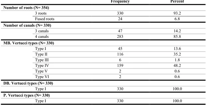
First Molars
A total of 354 maxillary first molars were evaluated. Out of them, 24 (6.8%) teeth had fused roots and 330 (93.2) teeth had non–fused roots (3 roots). Amongst the teeth with non-fused roots, 283 (85.8%) teeth had 4 canals and 47 (14.2%) teeth had 3 canals. Nearly half of teeth (48.2%) had Vertucci type IV in mesio-buccal roots, 35.2% had Vertucci type II, and 13.6% had Vertucci type I. Vertucci types III, V, and VI were found in less percentages (1.8%, 0.6%, and 0.6%, respectively). Regarding canal configuration in disto-buccal and palatal roots, all teeth (100%) had Vertucci type I (Table 9).
Amongst teeth with fused roots (n= 24), 6 (25%) teeth had one root and 18 (75%) teeth had 2 roots. Out of them, 22 (91.7%) teeth had non-merged canals and 2 (8.3%) teeth had merged canals and C-shaped configuration. For teeth with non-merged canals (n= 22), 17 (68.2%) teeth had 4 canals and 7 (31.8%) teeth had 3 canals. For canal configuration in mesio-buccal roots,
20 7 (31.8%) teeth had Vertucci type I, 10 (45.5%) teeth had Vertucci type II, 4 (18.2%) teeth had Vertucci type IV, and only one (4.5%) tooth had Vertucci type V, All teeth (100%) had Vertucci type I in disto-buccal and palatal roots (Table 10).
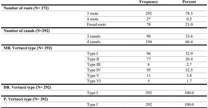
Second Molars
A total of 372 maxillary second molars were investigated. Out of them, 78 (21.0%) teeth had fused roots, 292 (78.5%) had non-fused roots, and 2 (0.5%) teeth had extra palatal root (had 4 roots). Amongst teeth with non-fused roots (n= 292), 98 (33.6%) had 3 canals and 199 (66.4%) teeth had 4 canals. All teeth (100%) had Vertucci type I in disto-buccal and palatal roots. However, regarding canal configuration in mesio-buccal roots, 32.9% of teeth had Vertucci type I, 32.5% had Vertucci type IV, and 26.4% had Vertucci type II. All other Vertucci types (III, V, and VI) were found in 8.2 % of teeth (Table 11).
Amongst teeth with fused roots (n= 78), 53 (67.9%) teeth were with non-merged canals and 25 (32.1%) teeth had merged canals and C-shaped configuration. Regarding number of roots, 24 (30.8%) teeth had one root (out of them, 17 teeth had merged canals) and 54 (69.2%) teeth had 2 roots (out of them, 8 teeth had merged canals). Out of teeth with non-merged canals (n= 53), 38 (71.7%) teeth had 3 canals and 15 (28.3%) teeth had 4 canals. All teeth with non-merged canals (100%) had Vertucci type I in disto-buccal and palatal roots. Whilst, out of them, 71.7% had Vertucci type I, 17.0% had Vertucci type IV, 9.4% had Vertucci type II, and 1.9% had Vertucci type V in mesio-buccal root (Table 12).
21 IV.1.2. Mandibular Teeth:
Central Incisors
A total of 410 mandibular central incisors teeth (right and left) were evaluated. All teeth (100%) had one root. Out of the total sample, there were 302 (73.7%) teeth had one canal and 108 (26.3%) teeth had 2 canals. Similarly, there were 302 (73.7%) teeth found with Vertucci Type I and 108 (26.3%) teeth with Vertucci Type III (Table 13).
Lateral Incisors
A total of 412 mandibular lateral incisors (right and left) were evaluated. Out of them, 410 (99.5%) teeth had one root, and only 2 (0.5%) teeth had 2 roots. Teeth with one canal accounted 285 (69.2%) teeth while, 127 (30.8%) teeth were found with 2 canals. Vertucci type I was found in 285 (69.2%) teeth while, 123 (29.8%) teeth had Vertucci type III, and only 4 (1.0%) teeth had Vertucci type V (Table 14).
Canines
Out of 410 mandibular canines (left and right) evaluated by CBCT, 339 (97.3%) teeth had one root, 11 (2.7%) teeth had 2 roots. Whereas 372 (90.7%) teeth had one canal, and 38 (9.3%) teeth had 2 canals. Vertucci type I was found in 372 (90.7%) teeth while, 25 (6.1%) teeth had Vertucci type III, and 13 (3.2%) teeth had Vertucci type V (Table 15).
First Premolars
Amongst the evaluated teeth there were 397 mandibular first premolars. Out of them, 395 (99.5%) teeth had one root and only 2 (0.5%) teeth had 2 roots. More than two thirds of the sample (69.5%) had one canal, 117 (29.5%) had 2 canals, and only 4 (1.0%) teeth had 3 canals. Regarding canal configuration, 276 (69.5%) teeth had Vertucci type I, 25 (6.3%) teeth had
22 Vertucci type III, 92 (23.2%) teeth had Vertucci type V, and only one (0.3%) tooth had Vertucci type VII. However, 3 (0.8%) teeth had different type of configuration. Only 6 (1.5%) had C-shape configuration. All of them had one root, and out of them 4 teeth had 2 canals and 2 teeth had 3 canals. Similarly, 4 teeth had Vertucci type V and 2 teeth had different types of canal configuration (Table 16).
Second Premolars
Three hundred and seventy nine mandibular second premolars were evaluated. All teeth (100%) had one root. Out of them, 367 (96.8%) teeth had one canal, 8 (2.1%) teeth had 2 canals, and 4 (1.1%) teeth had 3 canals. The majority of the sample (96.8%) had Vertucci type I, 6 (1.6%) teeth had Vertucci type III, 3 (0.8%) had Vertucci type V, and 3 (0.8%) teeth had different types of canal configuration. Out of the sample, there were 3 (0.8%) teeth had one root, 3 canals, and C-shape configuration (Table 17).
First Molars
A total of 290 mandibular first molars were evaluated. The majority (94.5%) had 2 roots, and 16 (5.5%) teeth had 3 roots. Only 2 (0.7%) teeth had 2 canals, 187 (64.5) teeth had 3 canals, and 101 (34.8%) teeth had 4 canals. More than half of the sample (57.9%) had Vertucci type IV in mesial canals. Whereas, 105 (36.2%) teeth had Vertucci type II. However, Vertucci types I, III, and V were found in less proportions. In contrast, 200 (69.0%) teeth had Vertucci type I in distal canals, 50 (17.2%) teeth had Vertucci type III while, Vertucci types II, IV, and V were found in less proportions. There were 14 (4.8%) teeth had 3 roots and 4 canals (Table 18).
Second Molars
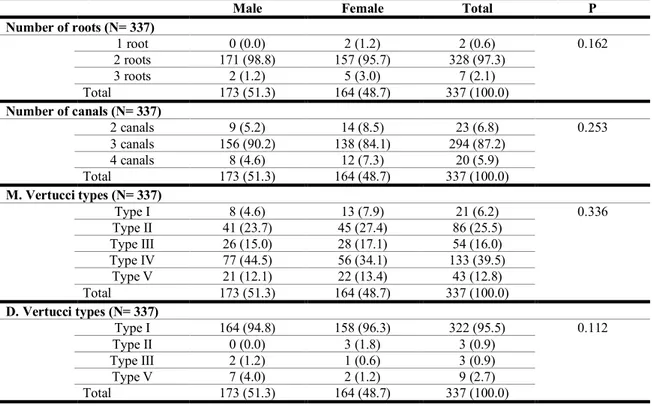
A total of 366 mandibular second molars were evaluated. Out of them, 328 (89.6%) teeth had 2 roots, 31 (8.5%) teeth had one root (2 teeth had fused roots without C-shape configuration
23 and 29 teeth had fused roots with C-shape configuration), and 7 (1.9%) teeth had 3 roots. Amongst teeth with normal canals (N= 337), there were 294 (87.2%) teeth had 3 canals, 23 (6.8%) teeth had 2 canals, and 20 (5.9%) teeth had 4 canals. Frequency of Vertucci types in mesial canals was the highest for Vertucci type IV (39.5%), followed by Vertucci type II (25.5%), and the least was for Vertucci type I (6.2%). Dissimilarly, Vertucci type I was the most frequent type in distal canals (95.5%), and no Vertucci type IV was detected (Table 19).
IV.2. Gender Differences:
IV.2.1. Maxillary and Mandibular Teeth:
In general, there were no significant differences between both genders in relation to number of roots of maxillary and mandibular teeth separately (P= 0.315 and P= 0.100, respectively) as well as no significant difference was found for all maxillary and mandibular teeth together (P= 0.064). However, significant difference was found between males and females in relation to number of canals of maxillary teeth (P= 0.014) where teeth with 1 or 3 canals were found more frequent in females than in males while, teeth with 2 or 4 canals were found more frequent in males than in females. For mandibular teeth, the significant level of difference between males and females in relation to number of canals was at the cut-off point (P= 0.050). For all maxillary and mandibular teeth together, the distribution among both genders in relation to number of canals was not significant (P= 0.082) (Table 20 & 21) (Figure 2 & 3).
Difference between both genders with regard to canal configuration of maxillary roots was highly statistically significant (P< 0.001). Roots with Vertucci type I, III, and V were more frequent in females than in males while, roots with Vertucci type II, IV, VI, and VII were more frequent in males than in females. For mandibular teeth, difference between males and females
24 in relation canal configuration of anterior and premolar teeth was significant (P= 0.016) while, the difference was not significant when related to the canal configuration of mesial roots of 1st and 2nd molars (P= 0.205). However, the difference between males and females was highly significant when related to canal configuration of distal roots of 1st and 2nd molars together (P< 0.001) (Figures 4 & 5).
IV.2.2. Maxillary Teeth:
Central Incisors
In comparison between males and females, 200 (52.1%) teeth were found in females and 184 (47.9%) teeth in males. However, the significance of difference could not be computed because all teeth (100%) in males and all teeth (100%) in females had one root, one canal, and Vertucci type I (Table 22).
Lateral Incisors
Although number of teeth in females was higher than in males (200 (52.1%) teeth in females compared to 184 (47.9%) teeth in males), the significance of difference was not applicable because all teeth (100%) in females and all teeth (100%) in males had one root, one canal, and Vertucci type I (Table 23).
Canines
There were 200 (52%) teeth in females and 180 (48%) teeth in males. All teeth (100%) in both genders had one root. All teeth (100%) in females had one canal while, in males, 180 (97.8%) teeth had one canal, and 4 (2.2%) teeth had 2 canals, with no significant difference between both genders (P= 0.052). Similarly, all teeth (100%) in females had Vertucci type I while, 180
25 (97.8%) teeth in males had Vertucci type I, and 4 (2.2%) teeth had Vertucci type III. No significant difference between both genders was found (P= 0.052) (Table 24).
First Premolars
Out of 176 (50.1% of all maxillary first premolars) teeth in females, 53.4% had 2 roots, 45.5% had one root, and 1.1% (2 teeth) had 3 roots. However, 108 (61.7%) out of 175 teeth in males had 2 roots, 36% had one root, and 2.3% had 3 roots. The difference between both genders was not statistically significant (P= 0.161). The majority of teeth in both genders (96.0% of teeth in females and 90.3% of teeth in males) had 2 canals. No teeth in females and 2 teeth in males had 4 canals. Teeth with one canal and 3 canals were found in less percentages. Similarly, no significant difference was found regarding number of canals (P= 0.125). Significant difference was found between both genders in relation to Vertucci types (P< 0.001). More than half of teeth (59.7%) in females had Vertucci type IV, followed by Vertucci type V (21.0%), and Vertucci type III (11.4%). In males, 68.0% of teeth had Vertucci type IV, followed by Vertucci type II (10.3%), and Vertucci type V (8.6%) (Table 25).
Second Premolars
Distribution of teeth among both genders was approximately similar (50.7% in males compared to 49.3% in females). The majority of teeth in both genders had one root with no significant difference (P= 1.000). About two thirds (65.9%) of teeth in males and about half of teeth (55.9%) in females had 2 canals with statistically significant difference (P= 0.046). Vertucci type I was found in 62 (34.1%) teeth in males followed by Vertucci type IV which was found in 41 (22.5%) teeth. However, Vertucci type III was found in 31 (17.5%) teeth in females followed by Vertucci type IV which was found in 28 (15.8%) teeth. Even though, no significant difference between males and females was found (P= 0.064) (Table 26).
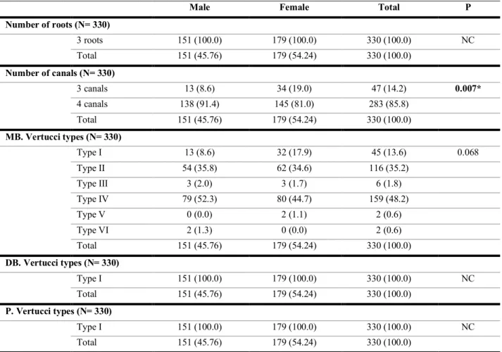
26 First Molars
In comparison between both genders, all teeth in males (n= 151, representing 45.76% of the total sample) and all teeth in females (n= 179, representing 54.24% of the total sample) had 3 roots and Vertucci type I in disto-buccal and palatal roots. Test of significant was not applicable. However, significant difference between males and females was found with regards to number of canals (p= 0.007). Teeth with 4 canals were found in higher percentage in males than in females (91.4% in males compared to 81.0% in females) while, 19.0% of teeth in females and 8.6% of teeth in males had 3 canals. Regarding Vertucci types in mesio-buccal roots, the majority of teeth in males and females (52.3% and 44.7%, respectively) had Vertucci type IV followed Vertucci type II (35.8% of teeth in males and 34.6% of teeth in females). No significant difference between both genders was found (P= 0.068). More details are shown in Table 27.
Second Molars
All teeth in males (n= 144, representing 49.0% of the total sample) and all teeth in females (n= 148, representing 51.0% of the total sample) had one root, and Vertucci type I in disto-buccal and palatal roots. A significant difference was found between both genders in relation to number of canals (P= 0.047). More than two thirds (72.2%) of teeth in males had 4 canals and 27.8% had 3 canals while, 60.8% of teeth in females had 4 canals and 39.2% had 3 canals. The most frequent Vertucci type in mesio-buccal roots in males was Vertucci type IV (43.8% of teeth), followed by Vertucci type I (26.4% of teeth), and Vertucci type II (25.7% of teeth). However, this is was not the case in females where the most frequent Vertucci type in mesio-buccal roots was Vertucci type I (39.2% of teeth), followed by Vertucci type II (27.0% of teeth), and Vertucci type IV (21.6% of teeth). Highly significant difference between males and females was found (P< 0.001). More details are illustrated in Table 28.
27 IV.2.3. Mandibular Teeth:
Central Incisors
Regarding comparison between males and females, both genders had all their mandibular central incisors with one root. Test of significance could not be computed. Females had more teeth with one canal (170 teeth, representing 79.4% of teeth in females) compared to males who had 132 teeth (67.3% of teeth in males) with one canal. However, males had higher number of teeth with 2 canals (64 teeth, representing 32.7% of teeth in males) than females who had only 44 teeth (20.6% of teeth in females) with 2 canals. The difference was statistically significant (P= 0.007). Similarly, there was significant difference (P= 0.007) in relation to Vertucci classification among both genders with the same percentages applied (Table 29).
Lateral Incisors
Amongst 214 teeth in females, there were 213 (99.5%) teeth with one root, and only one tooth (0.5%) was found with 2 roots. Similarly, amongst 198 teeth in males, there were 197 (99.5%) teeth with one root, and only one tooth (0.5%) was found with 2 roots. No significant difference was observed (P= 1.000). One hundred and fifty two (71.0%) teeth in females and 133 (67.2%) in males had one canals while, 62 (29.0%) teeth in females and 65 (32.8%) teeth in males had 2 canals, with no significant difference (P= 0.455). Regarding Vertucci classification, 133 (67.2%) teeth in males had Vertucci type I, 63 (31.8%) teeth had Vertucci type III, and only 2 (1.0%) teeth had Vertucci type V. In females, 152 (71.0%) teeth had Vertucci type I, 60 (28.0%) teeth had Vertucci type III, and only 2 (1.0%) teeth had Vertucci type V. No significant difference was found between both genders (P= 0.698) (Table 30).
28 Canines
Amongst 197 mandibular canines in males there were 195 (99.0%) with one root and 2 (1.0%) teeth with 2 roots while, amongst 213 mandibular canines in females there were 204 (95.8%) teeth with one root, and 9 (4.2%) teeth with 2 roots. No significant difference was found between both genders (P= 0.064). One hundred and eighty four (93.4%) mandibular canines in males had one canal while, 188 (88.3%) mandibular canines in females had one canal. No significant difference was observed between both genders (P= 0.088). Vertucci type I was more frequent in males than in females (93.4% compared to 88.3%) while, Vertucci types III and V were more frequent in females than in males. The significant level of difference between both genders was near the cut-off point (P= 0.049) (Table 31).
First Premolars
Although teeth with one root were higher in females than in males (100% compared to 98.9%), no significant difference between both genders was found (P= 0.224). Similarly, no significant difference (P= 0.229) was found between both genders with regard to number of canals. However, significant difference (P= 0.012) was found between both genders in relation to canal configuration. Teeth with Vertucci types I and V were higher in females than in males while, teeth with Vertucci type III were higher in males (Table 32).
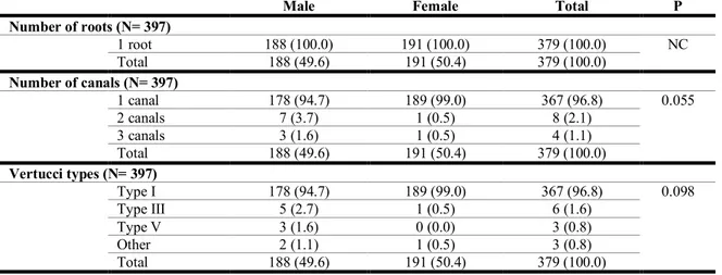
Second Premolars
Regarding comparison between both genders, all teeth (100.0%) in males and all teeth (100.0%) in females had one root. No significant difference (P= 0.055) was found between both genders with regard to number of canals although teeth with one canal were higher in females compared to males (99.0% compared to 94.7%, respectively). Similarly, no significant
29 difference (P= 0.098) was found in relation to canal configuration. Even though teeth with Vertucci type I in females were 189 (99.0%) and 178 (94.7%) in males (Table 33).
First Molars
Most teeth in both genders had 2 roots and 3 canals with no significant differences (P= 0.305 and P= 0.987, respectively). Similarly, no significant difference (P= 0.471) was found between both genders with regard to Vertucci types in mesial canals. However, significant difference (P= 0.005) was found between both genders in relation to Vertucci types in distal canals, with more frequent of Vertucci types II and III in females and Vertucci type V in males (Table 34).
Second Molars
As illustrated in Table 35, no significant differences were found between both genders in relation to number of roots (P= 0.162, with higher proportion of teeth with 2 roots in males), number of canals (P= 0.253, with higher proportion of teeth with 3 canals in males), Vertucci types in mesial canals (P= 0.336, with higher proportion of teeth with Vertucci type IV in males and Vertucci type II in females), and Vertucci types in distal canals (P= 0.112, with higher proportion of teeth with Vertucci type I in females).
30 IV.3. Bilateral Symmetry:
IV.3.1. Maxillary Teeth:
The results of the total bilateral symmetry of the maxillary teeth are presented in Tables 36 & 37.
Central Incisors
For bilateral symmetry, 185 (88.9% out of the total sample) participants had maxillary central incisors in both sides (right and left). The total bilateral symmetry was 100%, all 185 participants (100%) had one root, one canal, and Vertucci type I in both sides (Table 38 & Figure 6).
Lateral Incisors
For bilateral symmetry, 184 (88.5% out of the total sample) participants had right and left maxillary lateral incisors. The total bilateral symmetry was 100%, all 185 participants (100%). All participants (100%) had one root, one canal, and Vertucci type I in both sides (Table 39).
Canines
As shown in Table 40, all the 183 (88.0% of the total sample) participants, who had maxillary canines in both sides, had one root in both right and left sides (100% bilateral symmetry). However, bilateral symmetry for number of canals was 98.9% (P< 0.001) where 180 (98.4%) participants had one canal in both sides, and one participant (0.5%) had 2 canals in both sides. Similarly, the bilateral symmetry for Vertucci types was 98.9% (P< 0.001). Out of 183 participants, 180 (98.4%) participants had Vertucci type I in both sides while, one (0.5%) participant had Vertucci type III in both sides.
31 First Premolars
Table 41 shows the bilateral symmetry among participants with both right and left maxillary first premolars. Out of 208 participants, 162 (77.9%) participants had maxillary first premolars in both sides. Bilateral symmetry for number of roots was 85.1% (P< 0.001) while, it was 93.2% (P< 0.001) for number of canals, and 83.0% (P< 0.001) for Vertucci types. A bit more than half of participants (51.2%) had 2 roots in both sides while, 90.8% of participants had 2 canals in both sides, and 59.3% of participants had Vertucci type IV in both sides. The other sub-categories were found in less percentages.
Second Premolars
Bilateral symmetry according to the study variables is shown in Table 42. Out of 208 participants, 165 (79.3%) participants had maxillary second molars in both right and left sides. Bilateral symmetry for number of roots was 93% (P< 0.001). The majority of participants had one root in both sides while, 8.6% had 2 roots in both sides. Bilateral symmetry for number of canals was 83% (P< 0.001). About half (52.7%) of participants had 2 canals in both sides, 29.7% had one canal, and one participant (0.6%) had 3 canals in both sides. Bilateral symmetry for canal configuration was less frequent, accounting for 112 participants (67.8%, P< 0.001). Less than one third of participants (29.7%) had symmetrical Vertucci type I, 6.7% had symmetrical Vertucci types II and V, 9.1% for Vertucci type III, 14.5% for Vertucci type IV, and 0.6% for Vertucci types VI and other canal configuration (Figure 7).
First Molars
One hundred and forty three out of 208 participants (68.8%) had maxillary first molars in both sides (right and left). Bilateral symmetry for number of roots and Vertucci types in disto-buccal and palatal roots was 100% each. All participants had 3 roots and Vertucci type I in
disto-32 buccal and palatal roots in both sides. Out of 143 participants, 137 participants had bilateral symmetry for number of canals (95.8%; P< 0.001). Eighteen (12.6%) participants had symmetrical 3 canals, and 119 (83.2%) participants had symmetrical 4 canals. Only Vertucci types I, II, and IV were symmetrical in mesio-buccal roots. Out of 143 participants, 116 participants had symmetrical Vertucci types in mesio-buccal roots in both sides (81.1%; P< 0.001). This was 17 (11.9%) participants for Vertucci type I, 40 (28.0%) for Vertucci type II, and 59 (41.3%) for Vertucci type IV (Table 43).
Second Molars
Out of the evaluated participants (n=208), 127 (61.1%) participants had bilateral maxillary second molars. All participants had 100% bilateral symmetry for number of roots and Vertucci types (type I) in disto-buccal and palatal roots. However, bilateral symmetry for number of canals was 81.1% (P< 0.001). There were 30 (23.7%) participants which had 3 canals and 73 (57.5%) participants which had 4 canals in their both right and left maxillary second molars. In relation to canal configuration in mesio-buccal roots, the bilateral symmetry was 69.3% (P< 0.001) where 23.7% of participants had symmetrical Vertucci type I, 16.5% had symmetrical Vertucci type II, and 25.2% had Vertucci type IV. The other Vertucci types were found in less percentages (Table 44).
IV.3.2. Mandibular Teeth:
The results of the total bilateral symmetry of the mandibular teeth are presented in Tables 45-47.
Central Incisors
For bilateral symmetry, out of 208 participants, 205 (98.6%) participants were found with bilateral (both right and left sides) mandibular central incisors. In general, bilateral symmetry
33 for number of roots was 100% while, it was 91.2% (P< 0.001) for number of canals, and also 91.2% (P< 0.001) for Vertucci classification. All of participants (100%) had one root in both right and left sides. Out of them, 142 (69.3%) participants had one canal in both sides, and 45 (21.9%) participants had 2 canals in both sides. Similarly, significant symmetry of Vertucci types was found with the same percentages applied (Table 48).
Lateral Incisors
Out of 204 (98.1% of all participants) participants with both right and left mandibular lateral incisors, 202 (99.0%) participants had one root in both sides. Bilateral symmetry for number of canals was 85.8% (P< 0.001), where 127 (62.3%) participants had one canal in both sides, and 48 (23.5%) participants had 2 canals in both sides. For Vertucci classification, bilateral symmetry was 85.3% (P< 0.001), where 127 (62.3%) participants had Vertucci type I in both sides, 46 (22.5%) participants had Vertucci type III, and one (0.5) participant had Vertucci type V in both sides (Table 49).
Canines
Two hundred and two out of 208 participants (97.1%) had both right and left mandibular canines. The total bilateral symmetry for number of roots was 95.5% (P= 0.023) where the majority of participants (95.1%) had one root in both sides while, only one participant (0.5%) had 2 roots in both sides. Bilateral symmetry for number of canals was 91.1% (P< 0.001) where 86.1% of participants had one canal in both sides, and only 4.9% of participants had 2 canals in both sides. Regarding canal configuration, the total bilateral symmetry was 90.1% (P< 0.001) where 86.1% of participants had Vertucci type I in both sides, 3.5% of participants had Vertucci type III in both sides, and only one participant (0.5%) had Vertucci type V in both sides (Table 50 & Figure 8).
34 First Premolars
Amongst the 208 participants, 194 (93.3%) participants had mandibular first premolars in both sides (right and left). Among them, the total bilateral symmetry for number of roots was 99% where 192 (99%) participants had one root in both sides while, no participant had 2 roots in both sides. The total bilateral symmetry for number of canals was 87.1% (P= 0.001) where 122 (62.9%) participants had one canal in both sides, 46 (23.7%) participants had 2 canals in both sides, and only one (0.5%) participant had 3 canals in both sides. Regarding canal configuration, the total bilateral symmetry was 83.5% (P< 0.001) where 122 (62.9%) participants had Vertucci type I in both sides, 5 (2.6%) participants had Vertucci type III in both sides, and 35 (18.0%) participants had Vertucci type V in both sides. However, Vertucci type VII was found in only one (0.5%) participant in one side (right side, and no symmetry was found for the other types of canal configuration (Table 51).
Second Premolars
Out of 208 participants, there were 179 (86.1%) participants had mandibular second premolars in both sides (right and left). All participants (100.0%) had one root in both sides. Bilateral symmetry for number of canals was 83.7% (P< 0.001) where 171 (95.5%) participants had one canal in both sides, 2 (1.1%) participants had 2 canals in both sides, and only one (0.6%) participant had 3 canals in both sides. The total bilateral symmetry for canal configuration was 82.7% (P< 0.001). Vertucci type I in both sides was found in 171 (95.5%) participants while, Vertucci type III in both sides was found in one (0.6%) participant only. No bilateral symmetry was for Vertucci type V. Also, the other different types of canal configuration were not symmetrical among the participants (Table 52).
35 First Molars
One hundred and twenty one participant (58.2% of 208 participants) had mandibular first molars in both sides. Bilateral symmetry for number of roots was 99.2% (P< 0.001) while, it was 89.3% (P< 0.001) for number of canals, 87.6% (P< 0.001) for Vertucci types in mesial canals, and 82.6% (P< 0.001) for Vertucci types in distal canals. One hundred and fourteen (94.2%) participants had 2 roots in both sides, and 6 (5.0%) participants had 3 roots in both sides. No symmetry was found for teeth with 2 canals while, 60.3% of participants had 3 canals in both sides and 28.9% of participants had 4 canals in both sides. Similarly, no symmetry was found among participant regarding Vertucci types I and III in mesial canals while, more than half (52.9%) of participants had symmetrical Vertucci type IV, 31.4% of participants had symmetrical Vertucci type II, and only 4 (3.3%) participants had symmetrical Vertucci type V. The majority of participants (63.6%) had bilateral symmetry of Vertucci type I in distal canals. Whereas less proportions of participants had bilateral symmetry for the other Vertucci types in distal canals (Table 53).
Second Molars
Amongst 208 participants, 155 (74.5%) participants had both right and left mandibular second molars. For them, the total bilateral symmetry for number of roots was 98.1% (P< 0.001) where most participants (96.2%) had 2 roots in both sides. Regarding number of canals, the total bilateral symmetry was 92.9% (P< 0.001) where the majority of participants (84.5%) had 3 canals in both sides, 4.5% of participants had 4 canals and 3.9% of participants had 2 canals in both sides. The total bilateral symmetry for Vertucci types in mesial canals was 74.2% (P< 0.001). About one third (34.2%) of participants had Vertucci type IV in both sides, 18.7% and 9.7% of participants had Vertucci types II and III, respectively, and the least symmetrical type was Vertucci type I (3.9%). In relation to Vertucci types in distal canals, the total bilateral
36 symmetry was 97.4% (P< 0.001) where the majority of participants (94.8%) had symmetrical Vertucci type I while, the least symmetrical Vertucci type was type II (0.6% of participants), and no Vertucci type III was found in the right side among participants (Table 54).
37 V. DISCUSSION
Gender Differences:
In the present study, a comparison between males and females regarding number of roots, number of root canals, and root canal configurations according to Vertucci’s classification was performed. Regarding number of roots, no significant differences were found between genders in all 14 groups of teeth. This is in agreement with an in vivo CBCT study of all permanent dentition in a Malaysian sub–population. (109) However, another comprehensive in vivo CBCT study in a Portuguese population reported significant differences between genders in four of 14 tooth groups with females showing lower numbers of roots per tooth in maxillary first premolars, and second molars while, mandibular canines showed the opposite. (102) In a Saudi population, anatomical studies using in-vivo CBCT are consistent with our findings with no significant differences between genders in number of roots of maxillary premolars, maxillary first molars, mandibular canines, and mandibular first molars. (117-120) Two other
in vivo CBCT studies in different populations reported some significant differences between
genders in number of roots of maxillary and mandibular first and second premolars, where both found that males had higher number of 2-rooted premolars, while females had higher number of single-rooted premolars. (105,130) Also, the same was found in other CBCT studies on maxillary and mandibular molars, where females had lower number of roots compared to males. (113,114,131) In the literature, there is a tendency of females having a lesser number of roots per tooth. Many studies support the latter stance but there are ones that say otherwise. (111–113)
Generally, the internal canal morphology follows the external anatomy of the root, and that might have an impact on the lower number of roots in different genders. In regards to number of canals in the present study, only 3 groups out of 14 teeth groups (two in maxillary teeth and
38 one in mandibular teeth) showed significant differences between genders. Maxillary first and second molars had 4 canals with higher percentage in males than in females. However, in mandibular teeth groups, only central incisors showed statistically significant differences (P= 0.007), where males had higher number of teeth with 2 canals compared to females. In a study of all permanent dentition in a Malaysian sub-population (109), only 2 groups of teeth (second premolars and second molars) showed that males had significantly higher number of canals compared to females while the rest of teeth groups had no significant differences. In the Saudi population, some studies reported similar results to our findings, where in maxillary first molars males had a significant higher number of canals compared to females. (117) However, other studies reported that maxillary premolars, mandibular canines, and mandibular first molars had no significant differences between genders. (118)
In the present study, we also investigated the association between genders with RCS configurations. In total, Maxillary teeth showed highly statistically significant differences (P< 0.001) between both genders with regard to canals configurations where, roots with Vertucci type II, IV, VI, and VII were more frequent in males than in females while, roots with Vertucci type I, III, and V were more frequent in females. Whereas, only 2 groups (first premolars and second molars) out of 7 teeth groups of maxillary teeth showed statistically significant differences between both genders in relation to canals configurations. These findings are generally consistent with a study in a Portuguese population where all teeth showed higher prevalence of Vertucci type I configurations in females, being highly statistically significant in both maxillary premolars. (102) However, in a Malaysian subpopulation there were no statistically significant differences between genders regarding RCS configurations in all teeth groups. (109) In addition, a study of maxillary first premolars in a Saudi population (118) showed no differences between genders in regards to canal configuration, while the findings the German subpopulation study (130) is in agreement with our results where Vertucci type I