UNIVERSITÀ DEGLI STUDI ROMA TRE
DIPARTIMENTO DI SCIENZESCUOLA DOTTORALE IN BIOLOGIA
SEZIONE BIOLOGIA APPLICATA ALLA SALUTE DELL’UOMO Ciclo XXVII
OPTIMIZATION OF DENDRITIC CELL BASED
IMMUNOTHERAPIES
OTTIMIZZAZIONE DELLE IMMUNOTERAPIE
BASATE SU CELLULE DENDRITICHE
PhD student: Luciano Castiello
a.a.: 2014-2015Supervisor: Dr Filippo Belardelli Docente guida: Prof. Maria Marino Coordinatore: Prof. Paolo Visca
To Giulia,
for always believing in me
To my family,
for their constant and doubtless support
To Eleonora, Marianna and Filippo
for driving me along this path with their teachings
INDEX
SUMMARY ... I RIASSUNTO ... V 1. BACKGROUND
1.1 Dendritic Cells and their role in shaping immune response ... 1
1.2 Dendritic Cells subsets: localization and function ... 2
1.3 Monocyte-derived Dendritic Cells: pathways of differentiation and maturation ... 3
1.4 Dendritic Cell Based Immunotherapies ... 4
1.5 Open questions in immunotherapy based on monocyte-derived Dendritic Cells IFNa-DC ... 6
1.6 Novel strategies for generation of monocyte-derived Dendritic Cells ... 8
1.6.1 LPS/IFN-gamma mature Dendritic Cells ... 8
1.6.2 IFN-alpha Dendritic Cells ... 9
2. AIM ... 11
3. IDENTIFICATION OF FACTORS AFFECTING LIg-DC AND CANDIDATE BIOMARKERS OF LIg-DC CONSISTENCY AND VARIABILITY 3.1 Introduction ... 13
3.2 Results ... 15
3.2.1 Role of manufacturing, intra-donor variability and inter-donor variability in consistency of LIg-DC ... 15
3.2.1.1 Study design and LIg-DC production ... 15
3.2.1.2 Variability of LIg-DC expression of surface markers ... 15
3.2.1.3 Variability of LIg-DC at gene expression level ... 16
3.2.1.4 Origin of LIg-DC variability ... 18
3.2.1.5 Role of sources of variation on single-gene expression levels ... 18
3.2.2 Identification of candidate markers for quality assurance of LIg-DC ... 24
3.2.2.1 Selection of quality assurance gene markers ... 24
3.2.2.2 Identification of candidate markers of consistency of LIg-DC ... 24
3.2.2.3 Identification of candidate markers of variability/potency of LIg-DC ... 27
3.2.2.4 Characteristics of candidate markers are conserved in an independent clinical LIg-DC product dataset ... 27
4. IDENTIFICATION OF CANDIDATE BIOMARKERS OF LIg-DC EFFICACY IN PROSTATE CARCINOMA PATIENTS
4.1 Introduction ... 35
4.2 Results ... 37
4.2.1 LIg-DC induced clinical and immunological response in prostate carcinoma patients ... 37
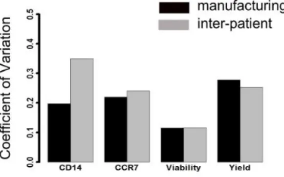
4.2.2 LIg-DC showed high level of variability on CD14 and CCR7 surface expression ... 39
4.2.3 DC transcriptomes clustered according to patient ... 41
4.2.4 Class comparison failed showing differences between RespDC and NonRespDC ... 42
4.2.5 Weighted Gene Coexpression Analysis revealed the presence of 8 modules in DC ... 43
4.2.6 Low expression of module 2 genes correlated with clinical and strong immunological responses ... 44
4.2.7 Module 2 was a tolerogenic DC module ... 48
4.2.8 Low IL-10 concentrations correlated with strong immunological response ... 49
4.2.9 Module 2 expression correlated with CD14, IL-10, MDC and MCP-1 secretion ... 50
4.2.10 Module 2 genes are co-expressed also in other DC ... 53
4.3 Discussion ... 54
5. CHARACTERIZATION OF IFNa-DC USED IN METASTATIC MELANOMA PATIENTS AND THEIR LOT-TO-LOT VARIABILITY 5.1 Introduction ... 59
5.2 Results ... 61
5.2.1 IFNa-DC showed variability in final viability, phagocytosis and surface expression of key markers ... 61
5.2.2 Gene expression profiling revealed changes occurring upon differentiation from monocytes ... 61
5.2.3 Module 2 genes were not co-expressed in IFNa-DC ... 65
5.2.4 IFNa-DC showed a different pattern of variability in cytokine Secretion ... 65
5.3 Discussion ... 67
6. CONCLUSION ... 69
7. REFERENCES ... 73 APPENDIX... Materials and Methods available on CD
I
SUMMARYDendritic cells (DC) are professional antigen presenting cells that continuously sample the environment; capture and process antigens; and transmit gathered information to T cells (Steinman & Banchereau 2007; Geissmann et al. 2010; Steinman & Cohn 1973). In presence of danger signals, DC become activated and trigger an inflammatory response against processed antigens, otherwise they remain in an immature state that lead to immune tolerance (Steinman 2003). Similarly, DC can interact and activate both B cells and NK (Batista & Harwood 2009; Steinman & Banchereau 2007). Given their pivotal role in shaping the immune system, DC are considered among the most promising cell-based immunotherapeutic approach in cancer setting aiming at activating an immune response against tumor associated antigens (TAA) (Steinman & Banchereau 2007). Several strategies have been developed to directly target DC in vivo by conjugating TAA with antibodies specific for DC receptor. Similarly more recently initial results have been collected on the administration of ex vivo activated circulating DC loaded with TAA (Wimmers et al. 2014). However, by far the most explored approach is vaccination with ex vivo generated monocyte-derived DC (Palucka & Banchereau 2013). Over twenty years ago, in fact, it was discovered that monocytes cultured with GM-CSF and IL-4 differentiate into DC and that upon maturation these cells were capable of activating T cells against specific antigens (Sallusto & Lanzavecchia 1994). Since then many other strategies to differentiate monocyte into DC have been developed, each one generating DC with a different phenotype, but none has conquest a general consensus on which one is endowed with ideal phenotype and should be used clinically (Kalinski et al. 2009).
Despite the large number of studies and clinical trials on ex vivo-generated DC vaccines, clinical results so far obtained were disappointing. Even though these vaccines proved to be safe and capable of inducing immune and clinical response in patient with melanoma, prostate carcinoma, glioma, and renal cell carcinoma, the overall response rate was usually below 15% (Datta et al. 2014). Many reasons have been hypothesized for such low response rates, among which the generation of DC with suboptimal potency is considered the most relevant. It’s not known yet how to generate the most potent DC; furthermore, differences in clinical setting, study design, sources of antigens, and route of administration make it almost impossible to compare results from previously conducted trials in order to clearly delineate the shared determinants of in vivo efficacy of DC-based vaccines.
Differently from previous attempts to optimize DC by modifying differentiation and/or maturation procedures, this project explores the possibility to identify factors affecting DC potency/efficacy in vivo in order to gain knowledge of molecular determinants essential for specific DC types
II
and that can thus be used for quality assessment of manufactured DC. Therefore, this project aimed at identifying factors affecting DC consistency and candidate molecular biomarkers of consistency, potency and efficacy of GMP manufactured DC.
In the first part of this project, we therefore analyzed factors affecting DC consistency as well as genes and proteins mostly affected by these factors. We analyzed a specific type of DC that is being tested clinically that are DC differentiated by GM-CSF and IL-4 and matured with LPS and IFN-gamma (LIg-DC). We showed that even when highly standardized procedures are used to generate LIg-DC, manufacturing, intra-donor and inter-donor related factors may affect DC phenotype with the last one being the most relevant (Castiello et al. 2013). Interestingly, these three factors mainly affected expression level of different genes and, while intra-donor variability diminished during differentiation (probably because of strong differentiation signals), inter-donor variability increased upon differentiation/maturation. Additionally, we observed that, while most of the well-known and usually tested DC markers (e.g., CD80, CD86, CD83, HLA-DR) did not show any differences in expression among LIg-DC generated at different times from different donors, the expression of several genes and the levels of several key secreted cytokines and chemokines showed significant variability among LIg-DC products. In particular, among top variable genes, many are likely to be functional important for LIg-DC and their expression correlated with the levels of inflammatory IL-12 as well as other key chemokines, such as MDC, MIG and CXCL10.
Then, in order to analyze whether such variability can be responsible of functional differences in vivo, we characterized from a molecular as well as immunophenotypic point of view LIg-DC vaccines administered to stage D0 prostate cancer patients. We observed a strong correlation between DC phenotype and development of clinical and immunological response in patient after vaccination. In particular, we identified a 303-gene signature made up of several well-known tolerogenic DC factors, such as CD14 and IL-10, that was capable to discriminate DC of patients that later showed clinical/immunological response from the ones of non-responders. The differential expression of CD14 and IL-10 was confirmed at the proteomic level and we also observed that MCP-1 and MDC protein levels correlated with the expression of the tolerogenic gene signature. Even though IL-10 secretion levels were able to predict strong immunological responses, it was only by combining CD14, IL-10, MCP-1 and MDC protein measures that it was possible to obtain an index able to replace the tolerogenic gene expression signature in its ability to discriminate both clinical and strong immunological responses.
III
In the final part of the project, we explored whether monocyte-derived DC differentiated in presence of GM-CSF and interferon-alpha (IFNa-DC) show patterns of variability similar to LIg-DC and whether biomarkers of efficacy are shared with LIg-DC. The DC used in this study were also manufactured to sustain a phase I clinical study aiming at activating an immune response in advanced melanoma patients. Even IFNa-DC were showing pretty invariable expression levels of major histocompatibility complex class I and class II molecules, as well as co-stimulatory receptors CD80 and CD11c marker. However, we did observe high variation in the level of expression of CD86, CD40, CD83 and CD1a among IFNa-DC made from different patients. At gene expression level, instead, we did not observe the existence of the tolerogenic signature we detected in LIg-DC, but even in these cells immune response genes showed high level of variability, therefore pointing to functional differences in DC vaccines. Also proteomic analysis suggested that lot-to-lot variability shown by IFNa-DC affects different cytokines and chemokines compared to LIg-DC.Altogether, this project developed a methodological framework for the identification of biologically-relevant quality control markers of DC by combining genomic and proteomic analysis. When applied to clinical DC, such approach was able to identify genes and proteins that correlated with clinical and immunological response and that can therefore be used as efficacy biomarkers of LIg-DC. However, as highlighted from the analysis of IFNa-DC, such newer markers are specific for DC used. On a broader range, these results strongly support the need for in-depth analysis of DC for the identification of newer quality assessment markers and factors essential for DC activity in vivo. Once identified, these markers can be used for the advancement of DC immunotherapies and to foster their implementation in clinic.
V
RIASSUNTOLe cellule dendritiche (DC) sono cellule specializzate nella presentazione degli antigeni che analizzano costantemente il microambiente in cui si trovano, catturano e processano antigeni, e in base a questi ultimi regolano l’attività dei linfociti T (Steinman & Banchereau 2007; Geissmann et al. 2010; Steinman & Cohn 1973). Solo in presenza di segnali di pericolo, infatti, le DC si attivano e innescano una risposta infiammatoria contro gli antigeni che hanno processato; alternativamente queste cellule rimangono in uno stato immaturo di solito legato al sostenimento della tolleranza immunologica (Steinman 2003). Inoltre, le DC possono interagire e attivare anche i linfociti B e le cellule NK (Batista & Harwood 2009; Steinman & Banchereau 2007). Proprio alla luce del loro ruolo centrale nel dirigere il sistema immunitario, le DC sono considerate la base di uno degli approcci più promettenti tra le immunoterapie cellulari in campo oncologico, il cui obiettivo è di attivare una risposta immunitaria contro antigeni tumorali (TAA) (Steinman & Banchereau 2007). Diverse strategie sono state sviluppate per dirigere TAA sulle DC direttamente in vivo, coniugando gli antigeni con anticorpi specifici per recettori delle DC. Altri gruppi invece stanno analizzando l’uso di DC circolanti che possono essere isolate dal sangue e che vengono brevemente attivate e caricate di antigeni ex vivo prima di essere somministrate al paziente. Tuttavia, l’approccio di gran lunga più studiato è la vaccinazione con DC ottenute ex vivo dal differenziamento di monociti circolanti (Palucka & Banchereau 2013). Da oltre vent’anni, infatti, è noto che i monociti differenziano in DC se messi in coltura con GM-CSF e IL-4 e che queste DC - previa maturazione - sono in grado di attivare i linfociti T contro specifici antigeni (Sallusto & Lanzavecchia 1994). Da allora molte altre strategie per il differenziamento e la maturazione delle DC sono state sviluppate, ognuno capace di generare DC con un peculiare fenotipo. Ciò nonostante nessuna di queste strategie ha conquistato il consenso generale per essere la più indicata per un uso clinico (Kalinski et al. 2009).
Nonostante il gran numero di studi e trial clinici con DC, i risultati clinici finora ottenuti con questo approccio sono stati insoddisfacenti. Infatti pur mostrando di essere in grado, in alcuni pazienti, di attivare una risposta immunitaria e clinica contro diversi tumori, la percentuale di risposta osservata è stata mediamente inferiore al 15% (Datta et al. 2014). Diverse cause sono state ipotizzate per spiegare questi insuccessi, e tra queste la generazione di DC con funzionalità subottimali è considerata la più importante. È ancora in discussione, infatti, come generare DC che inducano una spiccata attività infiammatoria una volta inoculate nei pazienti, e l’eterogeneità degli studi finora condotti non permette di comparare i diversi
VI
risultati e determinare esaustivamente le caratteristiche necessarie per l’efficacia di queste cellule in vivo.
Diversamente dai precedenti tentativi di ottimizzare le DC modificando le strategie di differenziamento/maturazione, questo progetto esplora la possibilità di identificare i fattori che condizionano la funzionalità in vivo delle DC con l’obiettivo di identificare gli elementi molecolari essenziali per l’attività delle DC e che quindi possono essere usati per i controlli di qualità delle DC prodotte. Quindi, questo progetto mira a identificare i fattori che influiscono sul fenotipo finale delle DC e i biomarcatori molecolari candidati per valutare la consistenza, la potenza e predire l’efficacia di DC prodotte per studi clinici.
Nella prima parte del progetto, abbiamo analizzato quali fattori influenzano la consistenza delle DC, e i geni e le proteine che ne sono principalmente condizionati. Per questo studio abbiamo utilizzato un tipo specifico di DC che è attualmente in fase di sperimentazione clinica, ovvero di DC differenziate in presenza di GM-CSF e IL-4 e maturate con LPS e Interferon-gamma (LIg-DC). I risultati ottenuti mostrano come anche quando protocolli altamente standardizzati vengono utilizzati per generare le LIg-DC, differenze intra-donatore, inter-donatore, e di processamento delle cellule condizionano il fenotipo delle DC generate e, tra questi, le differenze inter-donatore sono le più rilevanti (Castiello et al. 2013). Questi tre fattori, inoltre, condizionano i livelli di espressione di geni differenti e mentre la variabilità intra-donatore diminuisce durante il differenziamento dei monociti in DC quella inter-donatore risulta più elevata nelle DC rispetto ai monociti di partenza. In aggiunta, abbiamo osservato che sebbene i livelli di espressione dei principali marcatori delle DC si mantengano costanti in modo indipendente dal donatore(es. CD80, CD86, CD83, HLA-DR), l’espressione di numerosi geni così come i livelli di secrezione di citochine e chemochine importanti per la funzionalità delle DC mostrino un’elevata variabilità tra le LIg-DC prodotte da diversi donatori. In particolare, molti dei geni tra i più variabili sono stati descritti come importanti per la funzionalità delle DC e la loro espressione correla con i livelli di secrezioni della citochina infiammatoria IL-12 e delle chemochine MDC, MIG e IP10, tutte essenziali per l’interazione con le altre cellule del sistema immunitario.
Poi, al fine di comprendere se tale variabilità potesse essere responsabile di una diversa funzionalità in vivo, abbiamo analizzato LIg-DC usate per vaccinare pazienti con carcinoma prostatico in stadio D0. Dai risultati è emersa una forte correlazione tra il fenotipo delle DC e lo sviluppo di una risposta clinica e immunologica nei pazienti dopo vaccinazione. In particolare, abbiamo identificato 303 geni – in gran parte già descritti come tipici di DC tollerogeniche come CD14 e IL-10– capaci di discriminare le DC somministrate ai pazienti che successivamente avrebbero risposto alla
VII
vaccinazione da quelle di pazienti “non responders”. La diversa espressione di CD14 e IL-10 è stata anche confermata a livello proteico, insieme a quella di MDC e MCP-1. Tuttavia sebbene i livelli di espressione di IL-10 fossero sufficienti a discriminare DC capaci di indurre forti risposte immunologiche, è stato solo combinando le quattro proteine (CD14, IL-10, MDC, MCP-1) che siamo stati in grado di ottenere una capacità predittiva paragonabile a quella dei 303 geni.Al fine di stabilire l’applicabilità di questi marcatori di efficacia in vivo anche per altre DC, nella parte finale del progetto abbiamo poi cercato di comprendere se anche nelle DC differenziate in presenza di GM-CSF e interferon-alpha (IFNa-DC) fosse possibile osservare gli stessi schemi di variabilità osservati nelle LIg-DC. Anche in questo studio abbiamo analizzato DC che erano state prodotte per uno studio clinico di fase I, stavolta mirato a vaccinare pazienti con melanoma metastatico avanzato. Anche le IFNa-DC hanno mostrato un pressoché invariabile livello di espressione di alcuni marcatori ben noti delle DC, quali CD90, CD11c e i complessi maggiori di istocompatibilità di classe 1 e 2, anche se livelli di variabilità elevati sono stati registrati nell’espressione di CD86, CD40, CD83 e CD1a. A livello genico, invece, pur non osservando la presenza dell’impronta tollerogenica osservata nelle LIg-DC, anche nelle IFNa-DC è stata rilevata un’elevata variabilità in molti geni della risposta immunitaria, suggerendo anche qui l’esistenza di potenziali differenze funzionali tra le diverse DC analizzate. Anche a livello proteico, poi, l’analisi dei livelli di secrezione di citochine e chemochine ha messo in luce come le IFNa-DC siano caratterizzato da una diversa variabilità rispetto alle LIg-DC.
In generale, questo progetto ha sviluppato un approccio metodologico per l’identificazione di nuovi controlli di qualità delle DC, basato sulla combinazione di analisi genomiche e proteomiche. Quando tale approccio è stato utilizzato su DC usate in clinica, esso ha permesso di identificare geni e proteine che correlavano con lo sviluppo di una risposta clinica e immunologica e che quindi possono essere usati come biomarcatori di efficacia delle LIg-DC. Tuttavia, come evidenziato dall’analisi delle IFNa-DC, questi biomarcatori non sono condivisi universalmente da tutte le DC. Aldilà dell’aver identificato nuovi biomarcatori delle LIg-DC, questi risultati evidenziano la necessità di analisi approfondite delle DC per l’identificazione di nuovi marcatori e fattori essenziali per l’attività delle DC in vivo. Una volta identificati, questi nuovi marcatori possono essere utilizzati per controlli di qualità più affidabili e quindi facilitare l’implementazione clinica di queste cellule.
1
1. BACKGROUND
1.1 Dendritic Cells and their role in shaping immune response
Dendritic cells (DC) are bone marrow-derived cells that are present in all tissues in order to sample the environment and transmit gathered information to the adaptive immune system (Steinman & Banchereau 2007; Geissmann et al. 2010; Steinman & Cohn 1973). To initiate an immune response DC present the captured antigen, which is in the form of peptide–major histocompatibility complex (MHC) molecule complexes, to naive T cells in lymphoid tissues (Steinman & Banchereau 2007).
Normally, DC in peripheral tissues are immature. These immature DC have the ability to efficiently capture antigens; they can express low levels of co-stimulatory molecules; and have a limited capacity for secreting cytokines. Non-activated (immature) DC can present self-antigens to T cells (Steinman 2003), which leads to immune tolerance either through T cell deletion or through the differentiation of regulatory or suppressor T cells. However, DC promptly respond to environmental signals and differentiate into mature DC that can efficiently launch immune responses. Maturation is associated with the down-regulation of antigen-capture activity, the increased expression of surface MHC class II molecules and co-stimulatory molecules, the ability to secrete cytokines, as well as the acquisition of CCR7, which allows migration of the DC into the draining lymph node (Trombetta & Mellman 2005). However, DC maturation alone does not result in a unique DC phenotype. In fact, depending on the type of maturation signal, DC acquire distinct phenotypes that are distinct in both expression of co-stimulatory/co-inhibitory molecules as well as type and amount of secreted cytokines and chemokines.
Depending on the interaction with DC, naive CD4+ T cells and CD8+ T cells can differentiate into antigen-specific effector T cells with different functions. In fact, CD4+ T cells can become T helper 1 (TH1) cells, TH2 cells, TH17 cells or T follicular helper (TFH) cells that help B cells to differentiate into antibody-secreting cells, as well as regulatory T (TReg) cells that down-regulate the functions of other lymphocytes. Naive CD8+ T cells can give rise to effector cytotoxic T lymphocytes (CTLs). The type of T cell response — for example, CD4+ helper T cells or CD8+ CTLs — is at least partly linked to the subset of DC that presents the antigen (Banchereau & Steinman 1998). DC can also interact with cells of the innate immune system, including natural killer (NK) cells, phagocytes and mast cells (Steinman & Banchereau 2007; Banchereau & Steinman 1998).
DC also have an important role in controlling humoral immunity. They do so both directly by interacting with B cells and indirectly by inducing the
2
expansion and differentiation of CD4+ helper T cells (Batista & Harwood 2009). These key properties of DC, which allow the activation of both arms of the adaptive immune system (that is, cellular and humoral) and which launch the immune response, render DC the central candidates for antigen delivery and therapeutic vaccination against cancer.
1.2 Dendritic Cells subsets: localization and function
Both mice and humans have two major subsets of DC: myeloid DC (mDC; also known as conventional DC and classical DC) and plasmacytoid DC (pDC). Different subsets of human DC in the blood can be distinguished by the differential expression of three cell-surface molecules: CD303 (also known as BDCA2 and CLEC4C), CD1C (also known as BDCA1) and CD141 (also known as BDCA3 and thrombomodulin) (Dzionek et al. 2000). CD303+ pDCs represent a front line of anti-viral immunity owing to their ability to secrete large amounts of IFNα in response to virus encounters (Siegal et al. 1999). Their presynthesized stores of MHC class I molecules may allow a rapid initial CD8+ T cell response to viral infections. pDC-derived IFNα may also promote the immunogenic maturation of other subsets of DCs, thus helping to activate novel T cell clones. Human CD141+ DCs share with mouse CD8+ DCs the high capacity to capture exogenous antigens for presentation on MHC class I molecules (known as cross-presentation). CD141+ DCs express XCR1, which is the receptor for the chemokine XCL1 (also known as lymphotactin) that is produced by NK cells and activated CD8+ T cells (Bachem et al. 2010). Thus, mouse CD8+ DCs and human CD141+ DCs are equipped for the generation of CD8+ T cell-mediated immune responses. The unique functions of CD1C+ DCs also continue to be analyzed.
The human skin hosts two main subsets of mDC: epidermal Langerhans cells and dermal interstitial DC (dermal DC) (Valladeau & Saeland 2005). The dermal DC can be further subdivided into CD1a+ DC and CD14+ DC. Human CD14+ DC can directly help activated B cells, as well as induce naive T cells to differentiate into cells with the properties of TFH cells. CD14+ DC may thus be specialized for the development of humoral responses (Ueno et al. 2010). Langerhans cells are more efficient in cross-presenting peptides from protein antigens to CD8+ T cells and can prime the differentiation of CD8+ T cells into effector CTLs.
The development and homeostasis of tissue-resident DC subsets in steady state conditions (that is, when there is no infection or activation of the immune system) is dependent on the activation of the receptor tyrosine kinase FLT3 and of the macrophage colony-stimulating factor 1 receptor (M-CSFR; also known as CSF1R). However, inflammatory processes, such as those initiated by microbial invasion, substantially alter the populations of
3
DC subsets. The origin of DC that are recruited to sites of inflammation is still under investigation, although it is clear that monocytes can give rise to inflammatory DC in vivo (Cheong et al. 2010).1.3 Monocyte-derived Dendritic Cells: pathways of differentiation and maturation
Over twenty years ago, Sallusto and Lanzavecchia described for the first time that immature DC can be generated culturing monocytes in vitro in presence of granulocyte macrophage colony stimulating factor (GM-CSF) and interleukin-4 (IL-4) (Sallusto & Lanzavecchia 1994). These cells (IL4-DC) showed typical dendritic morphology, expressing high levels of major histocompatibility complex (MHC) class I and class II molecules, CD1 family, and other co-stimulatory molecules. Even functionally, these cells proved to be highly stimulatory in mixed leukocyte reaction (MLR) and were also capable of triggering naive T cells. Since then, many different protocols have been developed to differentiate monocytes into DC, each one leading to DC with a unique phenotype.
Given their functional similarity, both IL-13 and IL-7 can substitute IL-4, even though signaling in different ways. In fact, 13DC are similar to IL-4DC both phenotypically and functionally, whereas IL-7DC express CD21, the complement receptor type 2. Even functionally, these cells proved to be more effective than IL-4DCs in eliciting proliferative responses of CD4 and CD8 T cells, and stronger T cell cytotoxicity (Takahashi et al. 1997). Originally described for its anti-viral activity, type I interferon (IFN) exerts important effects on the immune system, including promotion of cellular and humoral responses, by virtue of its adjuvant effects on antigen-presenting cells (Belardelli 1995). As high amounts of IFN-α can be physiologically produced in response to infectious agents and inflammatory stimuli, this cytokine may be among the factors signaling danger to circulating monocytes, thus enabling them to rapidly differentiate into DC. In line with this hypothesis, it has been previously demonstrated that highly active partially mature DC can be generated from monocytes after a single step of 3-day culture with IFN-α and GM-CSF (IFNa-DC) (Santini et al. 2000). IFNa-DC proved to be more effective than immature DC generated in the presence of GM-CSF and IL-4 in inducing a Th-1 type of immune response and CD8+ T cell responses against defined antigens in different models (Lapenta et al. 2006; Santodonato et al. 2003). Although antigen uptake and endosomal-processing capabilities were similar for IFNa-DC and IL-4DC, and both DC types efficiently cross-presented soluble antigens to the specific CD8+ T cell clone, IFNa-DC were superior in cross-presenting low amounts of viral antigens. This property correlated with enhanced potential to express the specific subunits of the IL-23 and IL-27 cytokines.
4
Depending on the specific cytokine combination, blood monocytes can also give rise to Langerhans cells. In this respect, it has been reported that blood monocytes differentiated into LCs by replacing IL-4 with IL-15 (Mohamadzadeh et al. 2001). This cytokine cocktail gives rise to CD1a+, HLA-DR+, CD14− DCs, a proportion of which express LC markers, such as E-cadherin, Langerin and CCR6. Accordingly, IL-15DC, but not IL-4DC, migrated in response to CCL20. However, IL-15DCs cannot be qualified as genuine LCs because, despite Langerin expression, they do not express Birbeck granules (Mohamadzadeh et al. 2001).
Independently of the differentiation protocol used, DC can be matured by different stimuli. Mimicking signal microenvironment sensed by DC in vivo, DC have been matured in vitro using either inflammatory or tolerogenic compounds. Tolerogenic DC can be generated by maturing DC with IL-10 alone or in combination with IL-6, dexamethasone, or TGF-beta1 (Torres-Aguilar et al. 2010). Alternatively, tolerogenic DC have been generated by co-culturing DC with immunosuppressive cells, such as T regulatory cells and mesenchymal stromal cells (Pulendran et al. 2010). However, many more signals can be used to generate DC with inflammatory phenotype. Maturation of DC can be achieved by triggering Toll-like receptors (TLR) or through inflammatory cytokines. Triggering of TLRs on DC is thought to be critical for their functional maturation to immunogenic DC and the priming of naïve T cells in response to infection, and therefore coupling innate and adaptive immunity. Because of its bacterial origin and its predominant role as a pathogen associated pattern (PAP), LPS is recognized by TLR4 and represents a prototypical model of DC maturation. LPS-DC are endowed with strong chemotactic and immune activating properties (Castiello et al. 2011). Polyinosinic:polycytidylic acid (Poly I:C) is another of the most studied maturation signals in DC. Poly I:C is structurally similar to double-stranded RNA, which is present in some viruses, and is recognized by TLR3. Similarly to LPS, also Poly I:C induces maturation of DC with a strong immunogenic phenotype (Möller et al. 2008). Type I and II IFN, Tumor Necrosis Factor alpha (TNFa), IL-6 and IL-1b are all inflammatory cytokines that are capable to induce DC maturation either alone or in combination (Castiello et al. 2011). Differently from PAP, these cytokine do not trigger TLR, but activate different downstream signaling pathways, therefore leading to DC with different phenotypes.
1.4 Dendritic Cell Based Immunotherapies
Given their pivotal role in activating an antigen specific immune response, DC have been considered among the ideal cell-based immunotherapies. Over the last twenty years, many studies focused on three different DC-based
5
immunotherapies: in vivo targeting of DC, ex vivo activation of circulating DC and ex vivo generation of monocyte-derived DC.Pioneering studies from Steinman and Nussenzweig demonstrated the feasibility of targeting antigens to DC in vivo by coupling the desired antigen to an antibody recognizing a DC receptor (Bonifaz et al., 2002; Hawiger et al., 2001; Soares et al., 2007a). Importantly, in the absence of adjuvants, (e.g., targeting antigens to DEC205+ DC) in vivo induces antigen-specific tolerance, which can be used as treatment against autoimmune diseases such as type I diabetes (Steinman, 2012). Administration of these complex vaccines with DC-activators such as TLR3, TLR7-8, or CD40 agonists enables the maturation of DC and thus the establishment of immunity rather than tolerance (Steinman, 2012). The induced immunity was shown to be protective in a number of diseases including various infections (malaria, HIV) and cancer (Steinman, 2012; Tacken and Figdor, 2011), but moving in vivo DC-targeting to human trials requires considerable work yet because activator to use, DC subset to target, and specificity of targeting in vivo have still to be optimized (Datta et al. 2014).
Due to the low occurrence of naturally circulating DC in blood, conclusive clinical evidence on their usability for immunotherapy is lacking. Only recently, thanks to technological advancement in cell separation techniques, some encouraging results have been achieved using either pDC and mDC. In both cases, circulating DC subsets are selected by magnetic separation kits and cultured with activating cytokines for short period of time (i.e., overnight). Then, cells are loaded with tumor associated antigens and injected back to patient. Interestingly, one clinical trial selecting/activating pDC in melanoma patients showed induction of cancer-specific immune response in 7 of 15 treated patients and similar encouraging results were obtained selecting CD1c+ mDC (Wimmers et al. 2014).
Thanks to the accessibility to high amount of monocytes, injection of ex vivo generated monocyte-derived DC has been under the spotlight as the most promising and ready to implementation immunotherapy. Since the discovery of monocyte differentiation into DC over 300 clinical trials have been performed, but the heterogeneity of cells used, sources of antigen, dose and site of injection makes impossible to clearly summarize results so far obtained. By far the majority of the studies involved conventional IL-4DC used immature or matured by different maturing compound or cocktails loaded with tumor associated antigens (either single peptides, whole proteins or tumor lysates). To date, none of these study showed conclusive results of efficacy of such approach, even though both clinical and immunological responses have been observed (Engell-Noerregaard 2009). Furthermore, more recently some studies were published utilizing DC injected intra-tumorally after chemotherapy or radiotherapy, therefore aiming at an in situ
6
loading of antigens released by therapy induced cell death (Yu et al. 2003; Kolstad et al. 2014; Tanaka et al. 2005). These studies showed preliminary promising results, but larger studies have to be performed to assess efficacy of this other approach. Altogether, even though a large body of research and clinical experimentation has been performed, monocyte-derived DC immunotherapies have still to be optimized and many parameters to be more deeply analyzed.
1.5 Open questions in immunotherapy based on monocyte-derived Dendritic Cells
Even though a large number of clinical trials has be conducted utilizing ex vivo-generated DC vaccines, several controversies linger. First, the optimal DC phenotype, and the differentiation/maturation protocol utilized therein remains contentious. In particular, given the complexity of DC biology and the wide array of molecular players essential for T cell activation, a consensus on optimal DC phenotype and the procedure to obtain it is still lacking (Castiello et al. 2011). Among molecular markers of ideal DC, it is increasingly recognized that abundant production of IL-12p70 during DC maturation ex vivo, as well as “burst” secretion during DC-activated Th interaction in vivo (via CD40-CD40L in lymphoid organs), are critical for the induction of CTL responses and Th1-polarized immunity (Strioga et al. 2013). In addition to IL-12p70 elaboration, other desirable functions of immunogenic DC include non-exhaustive capacity, expression of chemokines enhancing tumor microenvironment infiltration of T effector cells (e.g., CXCL9/10), low IL-10 secretion following restimulation with CD40L, and enhanced migratory ability to lymph nodes. Several cytokine cocktails have been proposed to achieve optimal DC characteristics, but other factors have also to be finely tuned, such as time of stimulation and cytokine concentrations. Also, most of the studies focused only on how to optimize DC in order to have the best activation of T cells; however there is increasing evidence that also DC interaction with B cells and NK is important for effective activation of immune system, but the ideal DC phenotypes for these interactions are mostly unexplored (Bray et al. 2011; Qi et al. 2006). Additionally, it has to be considered that the ideal DC phenotype may differ depending on tumor setting, type of antigen used, site of injection, and patient immune status, thus opening the door to multiple “ideal DC phenotypes”, each one for a specific setting/use.
Second, the ideal strategy for DC antigen-loading is not universally agreed upon. The most common approach has been loading with tumor-associated peptides or whole recombinant tumor proteins (Palucka & Banchereau 2012). Other modalities of DC loading (engineered fusion proteins, autologous/allogeneic tumor cells, tumor cell-lysate, DC-tumor hybrids, and
7
DNA- or mRNA-transfected) have also emerged (Strioga et al. 2013). Even though clinical studies are still evaluating all of these strategies, the use of tumor lysate is thought to be the most promising one given the wide antigenic repertoire the DC can process and thus present. As already mentioned, another strategy is also emerging pointing to in situ loading of intra-tumorally injected DC, usually following chemo/radiotherapy (Kolstad et al. 2014; Tong et al. 2001; Akutsu et al. 2007; Finkelstein et al. 2012). Similarly to tumor lysate, this approach allows a wide antigenic repertoire, and compared to tumor lysate has the advantage of being more simple (given that tumor lysate has not to be processed ex vivo), but has the drawback that DC have to overcome tolerogenic tumor microenvironment in order to activate immune system.Third, the optimal route for DC administration remains controversial. Historically, with intradermal/subcutaneous injection techniques, DC trafficking to regional lymph nodes was considered critically important to their function. Indeed, maturation cocktails (e.g., PGE2-containing) were designed to optimize trafficking ability (Strioga et al. 2013). However, depending on differentiation/maturation strategy and DC maturation status at time of injection, alternative routes might be more effective. Ultrasound-guided intranodal injection, which co-localizes DC1-derived IL-12p70 “burst” with the anatomic site of T-cell sensitization, has emerged as a feasible solution (Bedrosian et al. 2003) ideal in case of DC impaired migration ability or advanced maturation.
Fourth, ex vivo-generated DC vaccines, like all other cell therapies, is challenging because of their considerable lot-to-lot and patient-specific variability that in most cases has yet to be sufficiently quantified and characterized (Stroncek et al. 2010). In fact, while each cell therapy lot has to be tested for identity, purity and potency among other tests, feasibility issues dictate these tests to be focused on a handful of factors and, therefore, they cannot assure an exhaustive characterization of each lot. Identity testing aims to ensure the manufactured cells show a defined phenotype, purity testing evaluates the absence/low level of cell contaminants. Potency testing, instead, assesses one biologically-relevant activity of the product and therefore is more controversial because can be tested with relatively-easy and low-informative assays (such as the secretion of IL-12 or phagocytosis ability) or lengthy and complex high-informative ones (such as the ability to activate tumor specific immune response in animal models). An accurate characterization of DC should ideally assay all the factors affecting their in vivo biological functions: antigen processing and presentation, expression of co-stimulatory signals, absence or reduced expression of co-inhibitory signals, lymph node migration, and secretion of activating cytokines and chemokines. These are all essential features of potent DC and should be
8
thoroughly tested. Since it is impossible to routinely evaluate each product for every cell function using cellular assays, the identification of reliable biomarkers of identity, consistency and potency of cell therapies is highly encouraged by regulatory agencies beginning in the earliest phases of clinical development of the cellular product (Hinz et al. 2006; Vatsan et al. 2013). 1.6 Novel strategies for generation of monocyte-derived Dendritic Cells As previously stated, a wide array of strategies to generate monocyte-derived DC has been developed and tested, but no general consensus exists on which one leads to ideal DC. In the next two sections, two novel strategies that represent promising candidates and that were used and analyzed for the PhD research project are discussed more in details.
1.6.1 LPS/IFN-gamma mature Dendritic Cells
Maturation of conventional IL-4DC with clinical grade LPS and IFN-gamma generated DC with strong immunostimulatory activity (LIg-DC) (Dohnal et al. 2009; Felzmann et al. 2005; Hüttner et al. 2005; Vopenkova et al. 2012). In fact, it has been shown that LIg-DC exhibit fully mature phenotype, the highest IL-12p70 production and stimulate T cell proliferation as well as their specific cytotoxic activity (Vopenkova et al. 2012). However, it has been observed that IFN-gamma and high-IL-12p70 production reduced migratory ability of DC, and thus LIg-DC do not migrate well. In particular, donor-dependent differences were observed in DC migratory capacity, suggesting that DC migration does not depend only on the maturation strategy used, but also on individual characteristics of the donor. Also, it was noted that timing of maturation play a critical role for this kind of cells. In fact, while only two hours were sufficient to induce maturation and potent immunostimulatory ability, cultivation of DC in the presence of both maturing agent for longer than 24 hours generated DC that were unable to release IL-12p70 and were less effective in triggering anti-tumor immunity(Hüttner et al. 2005).
Two clinical studies using LIg-DC have been published. In the first one, 22 pediatric cancer patients were vaccinated with LIg-DC pulsed with tumor cell lysate and keyhole limphet hemocyanin (KLH, as positive control). Following immunization, the majority of patients responded positively to KLH in a delayed-type hypersensitivity (DTH) test. In addition, three of six intra-nodally treated patients responded to the tumor Ag in the DTH test(Dohnal et al. 2007). In the second study, twenty-seven subjects with HER-2/neu over-expressing ductal carcinoma in situ of the breast were enrolled in a neoadjuvant immunization trial of LIg-DC pulsed with six HER-2/neu promiscuous MHC class II-binding peptides, plus two additional HLA-A2.1 class I-binding peptides. Interestingly, sensitization of Th cells to at least 1 class II peptide was observed in 22 of 25 evaluable subjects, while
9
eleven of 13 HLA-A2.1 subjects were successfully sensitized to class I peptides (Koski et al. 2012).1.6.2 IFN-alpha Dendritic Cells
As already mentioned culture of monocyte with GM-CSF and IFN-alpha generate DC showing a semi-mature phenotype and endowed with potent functional activities (IFNa-DC) (Santini et al. 2000; Farkas et al. 2008; Paquette et al. 1998; Santini et al. 2009). In fact, these cells produce mostly T-helper-1 (Th-1) cytokines and chemokines, express toll-like receptors (TLRs) 1 to 8, show migratory response to chemokines, and are capable of stimulating Th-1 polarized immune responses after injection into severe combined immunodeficient mice reconstituted with human peripheral blood leukocytes(Santini et al. 2009; Farkas et al. 2008). Of interest, in a variety of in vitro and in vivo preclinical models, IFNa-DC proved to be superior with respect to IL-4DC in inducing potentially protective immune responses . Notably, IFNa-DC exert a direct cytotoxic effect on tumor cells (Santini et al. 2000), are capable to take up apoptotic cells through the scavenger receptor Lectin-like oxidized-LDL receptor-1 (LOX-1) (Parlato et al. 2010) and present their antigens to CD8+ T cells, thus leading to an efficient cross-priming of these cells(Santodonato et al. 2003; Tosi et al. 2004; Lapenta et al. 2006). In addition, IFNa-DC are capable of expanding both Th1 and Th17 responses as a result of the production of cytokines such as 23 and IL-12(Santini et al. 2011). Remarkably, IFNa-DC do not require TLR triggering to induce antigen specific cytotoxic T lymphocytes and to stimulate allogeneic CD4+ T cells (Bracci et al. 2008). Interestingly, it was also shown that IFNa-DC can mediate TRAIL-dependent cytotoxicity of tumor cells and that even human CD11c(+) blood DCs express TRAIL after stimulation with IFN-alpha, thus acquiring the ability to kill TRAIL-sensitive tumor cell targets (Fanger et al. 1999; Santini et al. 2000; Servet et al. 2002). Of special note, a cell population resembling IFNa-DC was identified in immune cell infiltrates in Molluscum contagiosum virus-induced cutaneous lesions undergoing spontaneous regression (Vermi et al. 2011). Accordingly, a type I IFN signature associated with pDC infiltration was demonstrated in both keratinocytes and inflammatory cells. All these features make IFNa-DC highly promising new candidates for the development of more effective DC-based strategies of cancer immunotherapy (Farkas & Kemény 2011; Bracci et al. 2013).
11
2. AIM
Dendritic cells (DC) play a key role in the activation of immune system by presenting antigens to T cells and, by so, generating an antigen-specific immune response (Ueno et al. 2010). For this reason, several attempts have been done so far in order to develop effective immunotherapeutic approaches that consist of ex vivo generated fully-functional DC to be infused in patients in order to induce an antigen specific T cell expansion (Palucka & Banchereau 2012). DC can be generated ex vivo by culturing monocytes in presence of differentiating cytokines (such as GM-CSF, IL4, IL15 and IFNa) to obtain immature DC and maturing them with single agents or cocktails of agents (such as TNFa, LPS, IFNg, CD40L, IL6, IL1) (Kalinski et al. 2009). Once generated, DC are usually pulsed with tumor antigens and injected into patients (usually intranodally or intradermally) or directly injected intratumorally aiming at in situ antigen loadings usually after chemo/radiotherapy. Clinical results, mainly from studies in cancer patients, clearly showed the feasibility and efficacy of this approach even if the overall response rate is below the 15% (Engell-Noerregaard 2009). Several possible reasons have been hypothesized to justify such low response rate, among which the suboptimal generation of DC able to activate an anti-inflammatory Th1-polarized T cell response is considered the main bottleneck, even if there is no general consensus on how to improve DC function and which factors are responsible for the discrepancies between preclinical and clinical results (Castiello et al. 2011). Furthermore, cell based immunotherapies would strongly benefit of new markers for quality control assessment. Since many more factors are responsible for the function and effectiveness of cellular therapies than those of drugs and other biological products, an in depth evaluation of the characteristics of all newly developed cellular therapies is needed (Stroncek et al. 2010).
Differently from previous attempts to optimize DC by modifying differentiation and/or maturation procedures, this project explores the possibility to identify factors affecting DC potency/efficacy in vivo in order to gain knowledge of molecular determinants essential for DC function and that can thus be used for quality assessment of manufactured DC. Therefore, this project aimed at identifying factors affecting DC consistency and candidate molecular biomarkers of consistency, potency and efficacy of GMP manufactured DC. The project initially focused on a preclinical setting in order to evaluate feasibility of this approach by analyzing factors affecting DC consistency by combining genomic and proteomic approaches. Successively, given that the only reliable indicators of DC potency/efficacy derive from results in humans, the project focused on analyzing DC from two
12
clinical trials. The two trials utilized two different DC (in one case DC were differentiated with GM-CSF and IL-4 and matured with LPS and IFN-g; in the other one DC were only differentiated in presence of GM-CSF and IFN-a), therefore allowing the analyze whether factors affecting DC potency/efficacy are shared among different DC or are unique to each type of DC.
13
3. IDENTIFICATION OF FACTORS AFFECTING
REPRODUCIBILITY OF LIg-DC AND CANDIDATE
BIOMARKERS OF LIg-DC CONSISTENCY AND
VARIABILITY
3.1 Introduction
Monocyte-derived dendritic cells (DC) have been used in vaccine trials and represent one of the most promising approaches in inducing a targeted immune response(Palucka & Banchereau 2012). However, despite of twenty years of research and clinical experimentation in different settings, response rate are still pretty low, ranging from 5% to 15%(Engell-Noerregaard 2009). Reasons behind these results have to be identified in the complexity of DC biology as well as the lack of knowledge on best antigens, adjuvants and route of administration(Kalinski et al. 2009). In particular, DC function depends strongly on several factors, such as the differentiation process, maturation stimulus and duration of the manufacturing processing. In facts, monocytes can be differentiated using different cytokines leading to DC showing differences both on phenotype and functional activity. Since the initial discovery that monocyte can differentiate into DC when cultured in presence of GM-CSF and IL-4 (Sallusto & Lanzavecchia 1994), it has been shown that simple culture in presence of only GM-CSF leads to tolerogenic DC(Conti & Gessani 2008), whereas differentiation in presence of GM-CSF and IL-15 leads to DC more similar to Langherans cells(Dubsky et al. 2007) and that replacing IL-4 with IFN-alpha leads to DC with a semi-mature phenotype and potent co-stimulatory activity (Santini et al. 2000; Santodonato et al. 2003; Santini et al. 2009). Also the length of differentiation has a role in DC activity: several groups have shown that reducing the length of DC differentiation from the classical 7 days to 3-4 days leads to more potent DC(Dauer et al. 2003). Lastly, even maturation stimulus has a central role in shaping DC function, but even if there are over a dozen of different maturation cocktails no consensus exists on which signal leads to more potent DC(Castiello et al. 2011). Therefore, all of these factors drive DCs to develop a specific qualitative and quantitative immune activation, ranging from strong pro-inflammatory Th1 response to regulatory T cell induction. Even though several elements are known to affect the function of monocyte-derived DCs, the best methods for manufacturing DCs and for characterizing key DC functions are yet to be defined.
On the other hand, quality control is becoming a critical part of cellular therapy(Stroncek et al. 2010). Major aspects of ensuring product consistency and quality involve process control, adhering to standardized procedures, using GMP grade reagents, training staff and validating instruments and
14
equipment. However, the final product characterization is the main critical aspect of ensuring product consistency, especially for assuring that every production lot exceeds determined minimum standards. DC product characterization is performed at each step of the manufacturing process and at lot-release. Final products are evaluated for identity, sterility, purity, consistency, stability and potency; the latter being a quantitative measure of a product-specific biological activity that is linked to a relevant biological property. Feasibility issues dictate that actual product characterization must be a balance of what should and what can be tested (e.g., time needed for functional assays, lack of animal models). However, since many more factors are responsible for the function and effectiveness of DC-based therapies than those of other drugs, an in depth evaluation of the characteristics of newly developed cellular therapies is extremely desirable and needed for the identification of markers that are able to easily reveal characteristics relevant to important biological functions.
Additionally, even if high level of variation in DC phenotype has been observed even when using the same procedure of differentiation, factors behind such variability have never been analyzed(Stroncek et al. 2010).The understanding of these factor may thus play an essential role in the implementation of DC immunotherapies in clinical practice by defining molecular biomarkers of DC with increased potency, thus driving the development of newer protocols and procedure for DC-based immunotherapies. In fact, a prerequisite for the implementation of DC immunotherapies, as well as other cell based therapies, is the ability to consistently produce high quality products. Therefore, the project started analyzing what are the main factors affecting consistency of DC generated under GMP and whether such variability may hinder functional differences relevant to DC biology.
To dissect factors affecting DC consistency, the project focused on understanding how much manufacturing, intra-donor and inter-donor related causes have a role in DC variability. To test these factors we evaluated DC generated at different times from aliquots of the same monocyte collection (manufacturing related variability), DC generated from monocytes deriving from leukapheresis collected at different times from the same donors (intra-donor related variability), and DC generated from monocytes of different donors (inter-donor related variability). In addition, starting monocytes and intermediate DC products (i.e., immature DC) were included in the study to make possible the selection of candidate molecular markers of DC consistency and variability. In order to have a wide DC characterization we combined standard flow cytometry of known DC surface markers with gene expression profiling and protein secretion profiling of a broad panel of cytokines and chemokines.
15
Regarding the type of DC to analyze in this study, DC differentiated in presence of IL-4 and GM-CSF for 3 days and matured with LPS and IFN-g (LIg-DC) were selected because of their promising Th1-polarization phenotype and their use in multiple clinical trials at National Institute of Health(Shin et al. 2008; Jin et al. 2010).3.2 Results
3.2.1 Role of manufacturing, intra-donor and inter-donor variability in consistency of LIg-DC
3.2.1.1 Study design and LIg-DC production. When reviewing the overall manufacturing process, we realized that many factors could affect LIg-DC. Among these factors we decided to evaluate the effects of manufacturing variability, inter-donor variability and within-donor time-dependent fluctuations in the starting material (intra-donor variation) on the features of the final DC product. Manufacturing-related variability was tested by generating 5 DC preparations on different days using 5 aliquots from the same starting material. Inter-donor variation was assessed by studying DC derived from 9 different healthy donors. Intra-donor variation was assessed by preparing DC starting from 5 monocytes preparations derived from as many apheresis products from the same donor. Although it could be argued that the evaluation of a larger number of samples should lead to a more robust model for the description of manufactured products’ characteristics, the conditions that we defined are sufficient for the early-stage progressive assay implementation.
LIg-DC were manufactured according to standard GMP procedures established to support phase I/II vaccine trials at the NCI, NIH Bethesda, Maryland (NCI-09-C-0139, NCI-08-C-0051 and NCI-07-C-0206) and the clinical DC product release criteria were used to evaluate the quality of each experimentally manufactured product. All LIg-DC passed release criteria by showing cell viability >70%, CD83 expression >80%. Sterility tests were not performed given that cells were not use as clinical products.
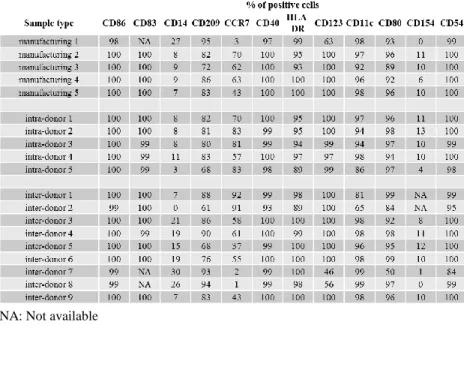
3.2.1.2 Variability of LIg-DC expression of surface markers. More than 95% of the cells in the final product were CD80+, CD86+, CD83+, CD209+, HLA-DR+, CD40+, CD54+, CD123+, CD11c+ as individually assessed by flow cytometry (see Table 3.1). Other markers were showing more variable levels of expression
When assessed for variability among the three tested conditions (i.e., manufacturing, intra-donor and inter-donor related variability), the majority of surface markers did not show any appreciable variation among different
16
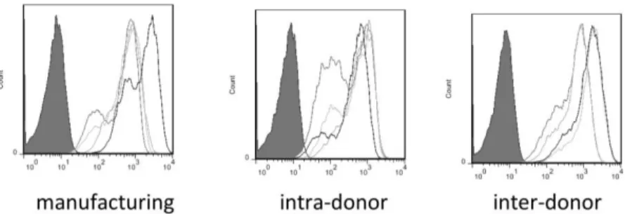
DC. Levels of variations were only observed for CD14, CCR7 and CD54 expressionlevels. While CD14 and CCR7 were expressed only by a proportion of DC, CD54 was expressed by all DC but at a very different intensity. In particular, manufacturing- and inter-donor-related variability did affect both CD14 and CCR7 expression levels (Figure 3.1), while intra-donor showed lower level of variation. Instead, even though >90% of the LIg-DC were CD54+, we observed high variability in all three conditions tested when the signal intensity was evaluated (Figure 3.2).
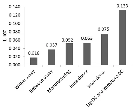
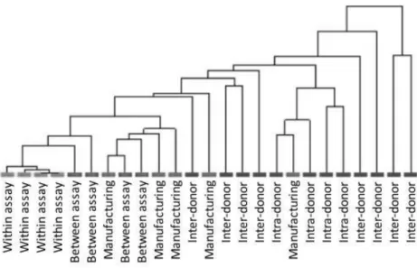
3.2.1.3 Variability of LIg-DC at gene expression level. Next, instead f focusing on few markers, gene expression profiles were examined by using Agilent Microarray technology. However, in order to appraise the confounding effect due to the variability of gene arrays, replicate samples were tested to estimate the within assay variability (shown by replicate samples of RNA amplified, labeled and hybridized on the same day) and the between assay variability (replicate samples of RNA prepared and hybridized on different days). The Coefficient of Variation (CV) was calculated for the samples used for assessing assay variability. The within assay and between assay samples showed CV median values of 6.1% and 14.4%, respectively (data not shown). These values are consistent with those obtained by the MicroArray Quality Control project for intra-site repeatability and reproducibility and indicated sufficient reliability for clinical and regulatory purposes(Shi et al. 2006). Then, we calculated the Intra-class Correlation Coefficient (ICC) for each possible source of variability that could impact the final product (assay, manufacturing, intra-donor and inter-donor). As expected, the within and between assay ICCs were much greater than manufacturing, intra-donor and inter-donor ICCs indicating that assay variability had a low confounding effect in our experimental approach (Figure 3.3), thus reconfirming the role of high-throughput gene expression analysis for the assessment of manufactured cell products. Interestingly, manufacturing, intra-donor and inter-donor factors all affected the final product. Although inter-donor samples show the lowest ICC value (0.925), it should be noted that manufacturing (0.948) and intra-donor variation (0.947) also played a significant role in final product consistency. This was also demonstrated by unsupervised hierarchical clustering and similarity matrix analysis of the samples based on the entire dataset (Figure 3.4). However, to place results into context and correctly assess the biological value of the obtained ICCs we also evaluated the ICC of an artificial class made up of a mixed cell populations that are known to show functional and molecular differences (i.e., immature DC andLIg-DC). We selected immature DC and mDC from 5 donors, and obtained an ICC value of 0.867. The ICC for the mixed immature and LIg-DC population was, as expected, less than the ICC
17
associated with LIg-DCs. However, since the ICC for mixed cells was similar to the inter-donor ICCs, these results indicate that LIg-DCs from different donors show a degree of variability that likely reflects functional differences.Table 3.1 Expression level of LIg-DC surface markers
NA: Not available
Figure 3.1 Variability of CD14 and CCR7 surface expression levels. On the left, percentages of CD14+ cells measured on LIg-DC evaluating manufacturing-related variability (n=5), intra-donor-related variability (n=5) and inter-donor-related variability (n=9).On the left, percentages of CCR7+ cells measured on LIg-DC as above.
18
Figure 3.2 Variability of CD54 surface expression levels. Histograms of CD54 expression levels on LIg-DC evaluating manufacturing-related variability (n=5), intra-donor-related variability (n=5) and inter-intra-donor-related variability (n=9). Histogram of isotype labeled cells is shown with a grey surface
3.2.1.4 Origin of LIg-DC variability. In order to determine if the observed LIg-DC variability was due to differences already present in the cellular starting material (i.e., the monocytes), or due to variability introduced during the manufacturing process, we evaluated the ICC values of the source material, monocytes, both for intra- and inter-donor variability. We assessed the variability of monocytes at the very beginning of the manufacture process (i.e., thawing and washing of the monocytes). As shown in Figure 3.5, the early steps of monocyte manufacturing had a lower impact on variability (ICC=0.955). Interestingly, we observed a lower value for intra-donor variability in the starting monocytes (ICC=0.938) than in the final product, mDCs, suggesting that in vitro culture decreases initial differences; whereas the inter-donor variability increased in LIg-DCs (ICC of monocytes was 0.939), indicating that differences due to genetic make-up increased during manufacturing.
3.2.1.5Role of sources of variation on single-gene expression levels. After characterizing the degree of consistency of the final cell product related to intra-donor, inter-donor and manufacturing factors using the entire gene expression data set, we focused on single gene variability in order to understand whether these three factors affect variability in expression of the same specific genes (i.e., whether the three factors might affect the same or different pathways/functions of the final cell product). To address this point, we calculated the assay-adjusted manufacturing, intra-donor and inter-donor variances for each gene (see Methods). We then ranked the genes according to variability and selected the most variable genes for each factor (one
19
percent or 344 genes per factor for a total of 877 genes due to some overlap among the three sets). As depicted on Figure 3.6, the three factors mainly affected different genes. However, it is important to note that 138 genes were present among the most variable genes of more than one factor (hypergeometric p-value < 10-10). This result indicated that even if the three factors affect mostly different genes, a subset of genes showed a strong susceptibility to more than one factor.Next, we checked whether the variability shown by these genes was already present in the starting monocytes. Monocyte manufacturing, intra-donor and inter-donor variances were calculated for each of the 877 genes. The analysis did not show any pre-existing differences in the monocytes (Figure 3.6c, d).
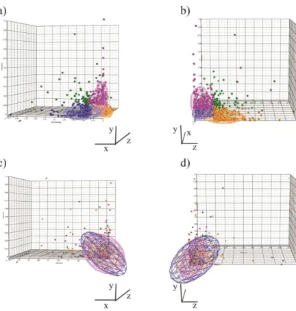
Figure 3.3 Intra-class Correlation Coefficient of gene expression profiles.Bar plot showing the 1-intra-class correlation coefficient calculated LIg-DC assessing the different sources of variability. “Within assay” and “Between assay” were included to evaluate the microarray-related confounding effect, whereas the “LIg-DC and immature DC” group was included to show the ICC value of a class showing high variability and well-known functional differences
20
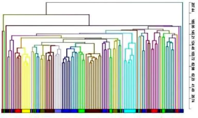
Figure 3.4 Unsupervised hierarchical clustering of LIg-DC gene expression profiles. Whole dataset was used to cluster LIg-DC samples. Pearson correlation was used to calculate distances.
21
Figure 3.5 Comparison of Intra-class Correlation Coefficient of LIg-DC and monocytes.Bar plot showing the 1-intra-class correlation coefficient calculated onLIg-DC and starting monocytes assessing the different sources of variability22
Figure 3.6 Genes with the greatest assay-adjusted manufacturing, intra-donor and inter-donor variability. (a) (b) Three-dimensional plots of the 877 genes whose expression was most variable in the DC gene expression data set in at least one factor. Each genes is represented according to its assay adjusted variances in the DC dataset: manufacturing related variability (x-axis), intra-donor related variability (z-axis) and inter-donor related variability (y-axis). Genes whose expression was most variable in more than one factor are represented in green. Genes most variable in LIg-DC manufacturing,intra-donor and inter-donor samples are shown in blue, purple, and orange, respectively. For each factor, ellipsoids are depicted to include 2 standard deviations from the mean value of each of the three factors. Each panel shows a different perspective.(c) (d) Three-dimensional plots of the assay adjusted variances of the same 877 genes in monocytes.