Clinical Study
Postnatal Growth in a Cohort of Sardinian Intrauterine
Growth-Restricted Infants
Maria Grazia Clemente,
1Giampiero Capobianco,
2Paolo Mattia Galasso,
1Francesco Dessole,
2Giuseppe Virdis,
2Maria Grazia Sanna,
3Mauro Giorgio Olzai,
3Lino Argiolas,
4Salvatore Dessole,
2and Roberto Antonucci
11Pediatric Clinic, Department of Surgical, Microsurgical and Medical Sciences, University of Sassari, Sassari, Italy
2Gynecologic and Obstetric Clinic, Department of Surgical, Microsurgical and Medical Sciences, University of Sassari, Sassari, Italy
3Neonatology and Neonatal Intensive Care Unit, Azienda Ospedaliero-Universitaria of Sassari, Sassari, Italy
4Italian Federation of Pediatric Physicians, Rome, Italy
Correspondence should be addressed to Maria Grazia Clemente; [email protected] Received 1 February 2017; Revised 2 May 2017; Accepted 17 May 2017; Published 20 June 2017 Academic Editor: Enrico Grisan
Copyright © 2017 Maria Grazia Clemente et al. This is an open access article distributed under the Creative Commons Attribution License, which permits unrestricted use, distribution, and reproduction in any medium, provided the original work is properly cited.
Recent studies have shown that infants with intrauterine growth restriction (IUGR) undergo catch-up growth during infancy. The aim of our study was to evaluate the postnatal growth in a cohort of IUGR infants born in a tertiary-level Obstetric University Hospital of Northern Sardinia. An observational retrospective study was conducted on 12 IUGR (group A) and 12 control infants (group B) by measuring the anthropometric parameters of weight (𝑊), length (𝐿) and head circumference (HC) from birth to the 3rd postnatal year. At birth, significant differences were found between group A and group B with regard to all the auxological parameters (𝑊, mean 1846.6 versus 3170.8 g, p < 0.0001; HC, 30.1 versus 34.4 cm, p < 0.0001; 𝐿, mean 43.4 versus 49.4 cm, p < 0.0001). During the 1st year, 8 of 12 (70%) IUGR infants exhibited a significant catch-up growth in the 3 anthropometric parameters and a regular growth until the 3rd year of follow-up. The majority but not all infants born with IUGR in our series showed significant postnatal catch-up growth essentially during the first 12 months of life. An improved knowledge of the causes of IUGR will help to develop measures for its prevention and individualized treatment.
1. Introduction
A combination of environmental, genetic, and epigenetic factors, still partially unknown, can be responsible for a condition in which a fetus is unable to reach its genetically determined growth potential: this condition is defined as intrauterine growth restriction (IUGR) [1, 2]. The IUGR fetus begins to lose its growth potential during the first trimester of pregnancy, mainly as a result of uterine hypoperfusion often associated with thin umbilical cord [1, 2]. The causes can be maternal, fetal, or placental. Preeclampsia, pathologic conditions of the umbilical arteries, maternal smoking, and unbalanced diet are known risk factors of IUGR [3–6]. It is essential to diagnose IUGR by ultrasound scan before the 28th week of gestation and to monitor its evolution through-out pregnancy. In this regard, Doppler flow measurement of the fetal vessels (namely, umbilical artery, ductus venosus,
and middle cerebral artery) has been found to be particularly helpful [7]. The circulatory status of the fetus is assessed especially in the middle cerebral artery, to determine the appropriate timing of delivery, that needs to be neither too early nor too late for a better outcome and prognosis [7, 8].
Clinical studies have shown that IUGR is a condition associated not only with an increased perinatal mortality, but also with significant morbidity later in life, includ-ing short stature, metabolic syndrome, and neurocognitive impairment [9–11]. The “symmetric” form of IUGR, defined by significant reduction of all anthropometric parameters including a small head circumference for gestational age, is associated with a worst prognosis compared to the “asym-metric” form of IUGR, in which the head circumference is within the normal range, and a favorable, complication-free postnatal course is generally observed [12, 13]. Among term infants, morbidity and mortality are 5-30-fold higher in low Volume 2017, Article ID 9382083, 5 pages
Table 1: Main parameters of the enrolled newborns, both group A and B.
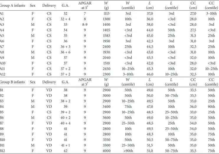
Group A infants Sex Delivery G.A. APGAR
at 5 𝑊 (g) 𝑊 (centile) 𝐿 (cm) 𝐿 (centile) CC (cm) CC (centile) A1 F CS 32 7 1115 3rd 37,0 3rd 27,0 3–10th A2 F CS 32 + 4 8 1300 10th 36,0 <3rd 28,0 10th A3 M CS 33 8-9 1400 3rd 38,0 <3rd 28,0 3rd A4 F CS 34 9 1405 <3rd 44,0 50th 27,5 <3rd A5 M CS 35 9 1583 <3rd 45,0 25th 31,5 25th A6 F CS 36 9 1950 3rd 42,5 3rd 31,0 3rd A7 F CS 36 + 3 9 2400 25th 44,5 10th 32,5 25th A8 M CS 36 + 6 9 1930 <3rd 43,0 <3rd 31,0 10th A9 M CS 37 9 2040 <3rd 43,5 <3rd 32,0 10th A10 F CS 37 9 1510 <3rd 42,0 <3rd 28,0 <3rd A11 F CS 37 + 2 9 2450 10–25th 45,3 10th 32,0 10–25th A12 F CS 37 + 2 9 2300 3–10th 46,0 10–25th 32,5 10th
Group B infants Sex Delivery G.A. APGAR
at 5 𝑊 (g) 𝑊 (centile) 𝐿 (cm) 𝐿 (centile) CC (cm) CC (centile) B1 F VD 38 9 2900 50th 49,0 50th 33,5 50th B2 F VD 38 9 3000 50th 50,0 50–75th 33,5 50th B3 M VD 38 + 2 9 2900 10–25th 49,5 50th 33,0 25th B4 M VD 39 9 3400 75th 47,0 10th 36,0 90th B5 F CS 39 + 2 9 2900 10–25th 48,5 25–50th 33,0 25th B6 M CS 40 + 2 9 3600 50th 49,0 10–25th 35,0 50th B7 F VD 40 + 4 9 2900 25–50th 48,5 25th 34,0 50th B8 F VD 41 9 2800 10th 49,5 25–50th 34,0 50th B9 F VD 41 9 2800 10th 48,3 10th 35,0 75th B10 F VD 41 9 3350 50th 50,5 50–75th 35,0 75th B11 M VD 41 + 1 9 3500 25–50th 51,5 50th 35,0 50th B12 F VD 42 9 4000 >90th 51,0 50–75th 35,5 75th
birth weight infants (LBWI) compared to infants with birth weight within the normal range (10th–90th centile) [12, 13].
The postnatal catch-up growth begins as soon as infants move to a more favorable environment and becomes evident during the first months of life. However, not all IUGR infants exhibit a postnatal catch-up growth, likely depending on the underlying causative factor/s and genetic diversity [14].
The present study reports results from a 3-year follow-up of a cohort of Sardinian IUGR infants with special emphasis on the postnatal catch-up growth.
2. Study Population and Methods
2.1. Study Population. In the year 2013, a total 27 IUGR diagnoses were made among infants born in the Gynecologic and Obstetric Clinic of the University of Sassari, Italy. Gesta-tional age (GA) was defined on the basis of ultrasonographic estimation (Voluson E8 ultrasound system, GE Healthcare, Fairfield CT, USA) performed at the time of the first scan, as recommended (SIEOG Italian guidelines), and at about 20, 28, and 36 weeks’ gestation [15]. Distributions of all measure-ments were similar to previously reported reference cohorts (data not reported). At the 20-week scan, details about med-ical history and demographic characteristics of the pregnant women were collected retrospectively. At this time, women were also informed about fetal anatomy and biometric
measurements, as well as uterine and umbilical artery Doppler flow velocimetry data [15]. Ultrasonographic mea-surements of fetal biparietal diameter, head circumference, abdominal circumference, and femur length were performed according to standard techniques. The Hadlock equations and reference standard were used to calculate the fetal weight (EFW) centile, and EFW values less than the 10th centile defined the IUGR [15]. At the 36 week scan, pregnant women were informed about previously undiagnosed placenta prae-via, severe oligohydramnios, a previously undiagnosed fetal abnormality, or noncephalic presentation [15]. Women were selected for additional, clinically indicated scans in the third trimester of pregnancy as per routine clinical care, using local and national guidelines (e.g., SIEOG guidelines). The indi-cations for cesarean section (CS) included a not reassuring cardiotocography (85%) and a reversed end diastolic flow of umbilical artery at ultrasound evaluation (15%).
As 13 families moved out of the Sassari province and 2 newborns unfortunately deceased, the access to postnatal data was available for 12 IUGR infants (F : M = 8 : 4), enrolled as group A. Twelve term infants with a birth weight greater than 2,500 g (F : M = 8 : 4) were enrolled as a control group (group B). The parents of all the infants enrolled in this study provided informed consent.
Group A and group B newborns’ main parameters are shown in Table 1, ordered by GA (column 4). Among group
Table 2: Weight centile categories of IUGR (group A) infants at 12 months of postnatal life.
Male Female Total
<3rd centile 1 3 4
3rd–50th centile 2 3 5
>50th centile 1 2 3
Total 4 8 12
Table 3: Length centile categories of IUGR (group A) infants at 12 months of postnatal life.
Male Female Total
<3rd centile 1 3 4
3rd–50th centile 2 4 6
>50th centile 1 1 2
Total 4 8 12
Table 4: Head Circumference centile categories of IUGR (group A) infants at 12 months of postnatal life.
Male Female Total
<3rd centile 0 3 3
3rd–50th centile 3 3 6
>50th centile 1 2 3
Total 4 8 12
A, 3 of 12 infants (25%; Table 1, A1–A4) were born preterm and with a very low birth weight (VLBW), ranging from 1115 to 1400 g. The remaining group A, namely, 5 (41,6%) late preterm (Table 1, A5–A8) and 4 (33,3%) at term infants (Table 1, A9–A12), were all but one born with low birth weight (LBW), ranging from 1510 to 2450 g, and one with VLBW (1405 g).
All group A but only 2 group B infants had CS births (Table 1, B5-B6).
2.2. Methods. This is an observational study conducted by retrospective collection of the measures of weight (𝑊), length (𝐿), and head circumference (HC), at birth and at 3-month intervals during the first year, then annually in the second and third years of follow-up (𝑊 and 𝐿). All values were plotted and recorded in the growth charts as follows: (1) weight to age, (2) length to age, and (3) head circumference to age (Center for Disease Controls, Atlanta, GA, USA).
2.3. Statistical Analysis. Student’s𝑡-test was used to compare groups, considering significant a value of p< 0.05.
3. Results
At birth, significant differences were found between group A and group B infants with regard to all anthropometric parameters considered in this study (𝑊, mean 1846.6 versus 3170.8 g, p< 0.0001; HC, 30.1 versus 34.4 cm, p < 0.0001; 𝐿, mean 43.4 versus 49.4 cm, p< 0.0001).
During the first year of life, a significant catch-up growth led to cover the differences in𝐿 (mean 72.6 versus 76.5 cm,
Birth Birth
Weight Catch-up growth from birth to 36-month follow-up
≤3rd centile >3rd centile Length 36 m 12 m 12 m 36 m 0 10 20 30 40 50 60 70 80 90 100 G ro u p A infa n ts (%)
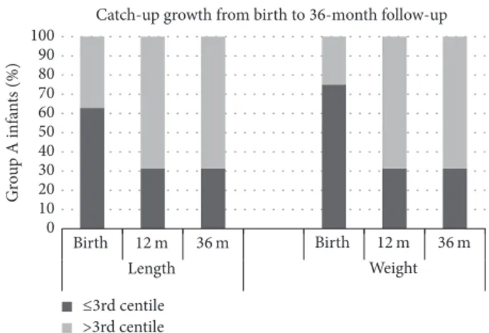
Figure 1: Percentage of group A infants below and above the 3rd centile cut-off for length (left panel) and weight (right panel) at birth, at 12 months (12 m) and 36 months (36 m) of age.
p = ns) and to reduce those in 𝑊 (mean 7861.0 versus 9165.0 g, p = 0.02) and HC (mean 43.5 versus 45.7 cm, p = 0.04) between the two study groups. At the age of 1 year, 8 (70%) group A infants were comparable to group B infants with respect to the 3 anthropometric parameters (Figure 1). However, analysis of data from individual patients revealed that 4 of 12 (30%) IUGR infants (Table 1, A2, A6, A8, and A10) did not exhibit catch-up growth during the first postnatal year with minimal improvement during the second and third years of follow-up (Figure 1). Categories of centiles for weight, length, and head circumference of IUGR infants at 12 months of life are reported in Tables 2, 3, and 4.
It deserves a note that among those who did not show postnatal catch-up growth, the only IUGR infant born at term (A10, Table 1) was discovered to be affected by the rare Pallister-Killian syndrome, caused by tetrasomy of chro-mosome 12p which is characterized by facial dysmorphism, rhizomelic limb shortness, and small hands and feet, along with corpus callosum hypoplasia. Moreover, during the postnatal years of follow-up, one of the IUGR preterm infants (A2; GA = 32 + 4; Table 1) showed failure to thrive, and it is currently under pediatric endocrinology evaluation.
4. Discussion
The majority of IUGR infants in our series showed significant postnatal catch-up growth during the first 12 months of life, and regular growth until 3 years of age.
Several studies in literature have reported on the postnatal catch-up growth in preterm IUGR and SGA infants, but only a few studies exist on term IUGR infants [1, 10–12].
One study conducted in North America (USA) on 42 IUGR infants has calculated growth velocity, which was significantly higher in IUGR infants compared to the control group (3.58 kg/m2 versus 2.36 kg/m2) during the first 12 months of life [13].
Another study, conducted in North Europe on 73 IUGR newborns, found catch-up growth in up to 90% of cases during the first year of life; 7% of infants among those who did
not have significant catch-up growth exhibited neurological and cognitive impairment [3].
This study was not a clinical trial and was also limited by both its retrospective, observational design and the small sample size. Even with these limitations of the study, our results further confirm those reported by others. All the term IUGR infants but the one affected by Pallister-Killian syndrome exhibit a catch-up growth. More than half of the preterms IUGR did show also a significant catch-up growth, and it was significantly greater during the first 12 months of life [9].
It was not possible for us to determine for each single case whether maternal or fetal factors played a role in the development of IUGR, as well as the role played by genetic, epigenetic, and environmental factors, or likely the complex combination of multiple factors on the catch-up growth and outcome during the postnatal life.
Interesting, the recent personalized medicine approach through the Newborn Individualized Developmental Care and Assessment Program (NIDCAP) has been the focus of a study conducted on preterm infants born with severe IUGR by a multidisciplinary research working group of Harvard University [16, 17]. The NIDCAP was shown to be effective in ameliorating the neurobehavior, electrophysiology and brain structure outcomes compared to IUGR controls [16, 17]. At least 2/3 of our IUGR infants required special assistance at the Newborn Intensive Care Unit (NICU). We can therefore speculate that also our infants compromised by severe IUGR who showed postnatal catch-up growth might have had significant benefit from an individualized developmental care approach during NICU stay.
Moreover, methods of infant feeding (breast-feeding ver-sus formula feeding) and other nutritional factors (including iron, zinc, and vitamins) might play a critical role in the catch-up growth during the first months of life [18, 19] and would deserve further, more extensive, investigation.
Abbreviations
CS: Cesarean section EFW: Estimated fetal weight GA: Gestational age HC: Head circumference
IUGR: Intrauterine growth restriction
𝐿: Length
LBWI: Low birth weight infants
NIDCAP: Newborn Individualized Developmental Care and Assessment Program
NICU: Newborn Intensive Care Unit PROM: Premature rupture of the membrane SGA: Small for gestational age
VD: Vaginal delivery VLBW: Very low birth weight
𝑊: Weight.
Conflicts of Interest
The authors declare that there are no conflicts of interest regarding the publication of this paper.
Authors’ Contributions
The first three authors (Maria Grazia Clemente, Giampiero Capobianco, and Paolo Mattia Galasso) contributed equally and wrote the first draft of the paper. Francesco Dessole, Giuseppe Virdis, and Giampiero Capobianco selected IUGR infants for the study, did the prenatal ultrasound evalua-tion, and analyzed the gestational auxological data, Maria Grazia Sanna and Mauro Giorgio Olzai did the perinatal physical evaluation of the study population, and Maria Grazia Clemente, Paolo Mattia Galasso, and Lino Argiolas collected and analyzed the perinatal and postnatal clinical records and anthropometric data. Salvatore Dessole and Roberto Antonucci were co-senior authors. All participated in writing the paper. The final version was approved by everyone.
Acknowledgments
The study was partially funded by the School of Biomedi-cal Sciences, Addressing Gender Medicine (Man, Woman, and Child), Sassari University, Italy, and partially by the Autonomous Region of Sardinia, Italy. The authors are grateful to the pediatric endocrinologist Dr. Carlo Burrai for providing clinical postnatal information of one of the IUGR infants.
References
[1] G. Puccio, M. Giuffr´e, M. Piccione, E. Piro, G. Rinaudo, and G. Corsello, “Intrauterine growth restriction and congenital malformations: A retrospective epidemiological study,” Italian Journal of Pediatrics, vol. 39, no. 1, article no. 23, 2013.
[2] S. Tabano, P. Colapietro, I. Cetin et al., “Epigenetic modulation of the IGF2/H19 imprinted domain in human embryonic and extra-embryonic compartments and its possible role in fetal growth restriction,” Epigenetics, vol. 5, no. 4, pp. 313–324, 2010. [3] L. T. Sehested and P. Pedersen, “Prognosis and risk factors for intrauterine growth retardation,” Danish Medical Journal, vol. 61, no. 4, p. A4826, 2014.
[4] G. Banderali, A. Martelli, M. Landi et al., “Short and long term health effects of parental tobacco smoking during pregnancy and lactation: a descriptive review,” Journal of Translational Medicine, vol. 13, article 327, pp. 10–1186, 2015.
[5] R. S. Mayor, K. E. Finch, J. Zehr et al., “Maternal high-fat diet is associated with impaired fetal lung development,” American Journal of Physiology - Lung Cellular and Molecular Physiology, vol. 309, no. 4, pp. L360–L368, 2015.
[6] G. M. Fadda, G. Capobianco, A. Balata et al., “Routine second trimester ultrasound screening for prenatal detection of fetal malformations in Sassari University Hospital, Italy: 23 years of experience in 42,256 pregnancies,” European Journal of Obstetrics Gynecology and Reproductive Biology, vol. 144, no. 2, pp. 110–114, 2009.
[7] I. Aditya, V. Tat, A. Sawana, A. Mohamed, R. Tuffner, and T. Mondal, “Use of Doppler velocimetry in diagnosis and prognosis of intrauterine growth restriction (IUGR): A Review,” Journal of Neonatal-Perinatal Medicine, vol. 9, no. 2, pp. 117–126, 2016.
[8] G. M. Fadda, P. L. Cherchi, D. D’Antona et al., “Umbilical artery pulsatility index in pregnancies complicated by insulin-dependent diabetes mellitus without hypertension,” Gynecologic and Obstetric Investigation, vol. 51, no. 3, pp. 173–177, 2001. [9] A. A. Baschat, “Neurodevelopment after fetal growth
restric-tion,” Fetal Diagnosis and Therapy, vol. 36, pp. 136–142, 2014. [10] I.-K. Sung, B. Vohr, and W. Oh, “Growth and
neurodevelop-mental outcome of very low birth weight infants with intrauter-ine growth retardation: Comparison with control subjects matched by birth weight and gestational age,” The Journal of Pediatrics, vol. 123, no. 4, pp. 618–624, 1993.
[11] A.-K. Von Beckerath, M. Kollmann, C. Rotky-Fast, E. Karpf, U. Lang, and P. Klaritsch, “Perinatal complications and long-term neurodevelopmental outcome of infants with intrauterine growth restriction,” American Journal of Obstetrics and Gynecol-ogy, vol. 208, no. 2, pp. 130–e6, 2013.
[12] M. Hack, M. Schluchter, L. Cartar, M. Rahman, L. Cuttler, and E. Borawski, “Growth of very low birth weight infants to age 20 years,” Pediatrics, vol. 112, no. 1, Pt 1, pp. e30–e38, 2003. [13] T. L. Crume, A. Scherzinger, E. Stamm et al., “The Long-term
impact of intrauterine growth restriction in a diverse US cohort of children: The EPOCH study,” Obesity, vol. 22, no. 2, pp. 608– 615, 2014.
[14] E. Llurba, A. A. Baschat, O. M. Turan, J. Harding, and L. M. McCowan, “Childhood cognitive development after fetal growth restriction,” Ultrasound Obstet Gynecol, vol. 41, no. 4, pp. 383–389, 2014.
[15] U. Sovio, I. R. White, A. Dacey, D. Pasupathy, and G. C. Smith, “Screening for fetal growth restriction with universal third trimesterultrasonography in nulliparous women in the Pregnancy Outcome Prediction (POP) study: a prospective cohort study,” Lancet, vol. 386, no. 10008, pp. 2089–2097, 2015. [16] H. Als, F. H. Duffy, G. B. McAnulty et al., “Is the Newborn
Individualized Developmental Care and Assessment Program (NIDCAP) effective for preterm infants with intrauterine growth restriction,” Journal of Perinatology, vol. 31, no. 2, pp. 130–136, 2011.
[17] H. Als, F. H. Duffy, G. McAnulty et al., “NIDCAP improves brain function and structure in preterm infants with severe intrauterine growth restriction,” Journal of Perinatology, vol. 32, no. 10, pp. 797–803, 2012.
[18] M. Toumba, A. Hadjidemetriou, M. Topouzi et al., “Evaluation of the auxological and metabolic status in prepubertal children born small for gestational age,” Journal of Pediatric Endocrinol-ogy and Metabolism, vol. 18, no. 7, pp. 677–688, 2005.
[19] R. T. Yamada and C. R. Leone, “Intrauterine growth restriction and zinc concentrations in term infants during the first month of life,” Journal of the American College of Nutrition, vol. 27, no. 4, pp. 485–491, 2008.
Submit your manuscripts at
https://www.hindawi.com
Stem Cells
International
Hindawi Publishing Corporationhttp://www.hindawi.com Volume 2014
Hindawi Publishing Corporation
http://www.hindawi.com Volume 2014
INFLAMMATION
Hindawi Publishing Corporation
http://www.hindawi.com Volume 2014
Behavioural
Neurology
Endocrinology
International Journal of Hindawi Publishing Corporationhttp://www.hindawi.com Volume 2014
Hindawi Publishing Corporation
http://www.hindawi.com Volume 2014
Disease Markers
Hindawi Publishing Corporation
http://www.hindawi.com Volume 2014
BioMed
Research International
Oncology
Journal ofHindawi Publishing Corporation
http://www.hindawi.com Volume 2014
Hindawi Publishing Corporation
http://www.hindawi.com Volume 2014 Oxidative Medicine and Cellular Longevity Hindawi Publishing Corporation
http://www.hindawi.com Volume 2014
PPAR Research
The Scientific
World Journal
Hindawi Publishing Corporation
http://www.hindawi.com Volume 2014
Immunology Research
Hindawi Publishing Corporation
http://www.hindawi.com Volume 2014
Journal of
Obesity
Journal ofHindawi Publishing Corporation
http://www.hindawi.com Volume 2014
Hindawi Publishing Corporation
http://www.hindawi.com Volume 2014 Computational and Mathematical Methods in Medicine
Ophthalmology
Journal ofHindawi Publishing Corporation
http://www.hindawi.com Volume 2014
Diabetes Research
Journal ofHindawi Publishing Corporation
http://www.hindawi.com Volume 2014
Hindawi Publishing Corporation
http://www.hindawi.com Volume 2014
Research and Treatment
AIDS
Hindawi Publishing Corporation
http://www.hindawi.com Volume 2014 Gastroenterology Research and Practice
Hindawi Publishing Corporation
http://www.hindawi.com Volume 2014