UNIVERSITY OF SASSARI
Department of Biomedical Sciences
International PhD School in Life Sciences and Biotechnologies
Curriculum Biochemistry, Physiology and Molecular Biology
Cycle XXX
Director: Professor Leonardo A. Sechi
The role of mechanical stress in progression
of intima hyperplasia associated to vein
coronary bypass grafts disease
Supervisor:
Prof. Gianfranco Pintus
Co-Supervisor: Dr. Gaia Spinetti
PhD Candidate: Davide Maselli
Dr. Davide Maselli, “The role of mechanical stress in progression of intima hyperplasia associated
to vein coronary bypass grafts disease.”, PhD thesis in Biochemistry, Physiology and Molecular
Biology of International PhD School in Life Sciences and Biotechnologies, University of Sassari.
Abstract
Coronary artery bypass grafting is a surgical procedure introduced to restore the blood circulation into the myocardium after an ischemic event. Despite progress in the use of arterial conduits, saphenous vein (SV) remains one of the most used vessels for the bypass. A short time after bypass implantation SV undergoes intima hyperplasia (IH) that progressively reduce its patency. One trigger cause of IH is the hemodynamic changes in the blood flow with higher sheer stress on the endothelial layer a radial deformation on the wall of the vein.
The aim of this project was to understand the role of saphenous vein progenitors (SVP) in the progression of IH. These cells with high differentiation potential are the pericytes of vasa vasorum in the tunica adventitia.
By in vitro and ex vivo models of mechanical stress we demonstrated the susceptibility of SVPs to the strain that causes a cytoskeletal reorganization and the acquisition of a potential migratory phenotype. SVPs showed the stimulus-related up-regulation of Amphoterin-Induced Gene And Open Reading Frame 2 (AMIGO2), that may have a role in the mechanical activation via prosurvival and migratory effects.
For the first time has been described the presence of AMIGO2 in SVPs and its relationship with mechanical stress. Migratory phenotype acquisition and AMIGO2 overexpression demonstrate how SVPs are potential targets for further study of IH.
Dr. Davide Maselli, “The role of mechanical stress in progression of intima hyperplasia associated
to vein coronary bypass grafts disease.”, PhD thesis in Biochemistry, Physiology and Molecular
Biology of International PhD School in Life Sciences and Biotechnologies, University of Sassari.
Acronyms and Abbreviations
AdvSca1 Sca1-positive vascular progenitor cells population AMIGO Amphoterin-Induced Gene And Open Reading Frame Angpt-2 Angiopoietin 2
BBB Blood–Brain-Barrier
BM Basal Membrane
CABG Coronary Artery Bypass Grafting
CEACAM1 Carcinoembryonic Antigen-Related Cell Adhesion Molecule 1
CNS Central Nervous System
DMEM Dulbecco Modified Eagle‟s Medium
ECM Extracellular Matrix
ECs Endothelial Cells
EPCs Endothelial Progenitor cells
ET-1 Endothelin-1
FGF Fibroblast Growth Factor
HAECs Human Aortic Endothelial cells
HEK Human Embryonic Kidney
HSCs Hematopoietic Stem cells
HUVECs Human Umbilical Vein Endothelial cells ICAM Intracellular Adhesion Molecule
IGF Insulin like Growth Factor
IH Intima Hyperplasia
Dr. Davide Maselli, “The role of mechanical stress in progression of intima hyperplasia associated
to vein coronary bypass grafts disease.”, PhD thesis in Biochemistry, Physiology and Molecular
Biology of International PhD School in Life Sciences and Biotechnologies, University of Sassari.
IPA Ingenuity Pathway Analysis
LAD Left Anterior Descending
LCX Left Circumflex Artery
MAPKs Mitogen-Activated Protein Kinases MCP-1 Monocyte Chemoattractant Protein-1
MS Multiple Sclerosis
MSCs Mesenchymal Stromal cells
NFATc2 Nuclear Factor of Activated T-cells cytoplasmic 2 NFkappaB Nuclear Factors kappa B
NG2 Neuron-Glial 2
NI Neointima
NO Nitric Oxide
NT No-touch Technique
NVU Functional Neurovascular Unit
P/S Penicillin/Streptomycin
PCs Pericytes
PDGF Platelet-Derived Growth Factor
PDGFRβ Platelet-Derived Growth Factor Receptor β
PIT Pathologic Intimal Thickening
PVDF Polyvinylidene Difluoride
RA Rheumatoid Arthritis
RBC Red Blood Cell
RCA Right Coronary Artery
ROS Reactive Oxygen Species
Dr. Davide Maselli, “The role of mechanical stress in progression of intima hyperplasia associated
to vein coronary bypass grafts disease.”, PhD thesis in Biochemistry, Physiology and Molecular
Biology of International PhD School in Life Sciences and Biotechnologies, University of Sassari.
Shh Sonic hedgehog
SLRP Small Leucine-Rich Proteoglycans
Sp1 Specificity protein 1
SSREs Shear Stress Response Elements
SV Saphenous Vein
TdT Terminal deoxynucleotidyl Transferase
TNF-α Tumor Necrosis Factor-α
VECAM Vascular Endothelial Cell Adhesion Molecule VW-MPSCs Vascular Wall-resident Multipotent Stem Cells
VZ Vasculogenic Zone
Dr. Davide Maselli, “The role of mechanical stress in progression of intima hyperplasia associated
to vein coronary bypass grafts disease.”, PhD thesis in Biochemistry, Physiology and Molecular
Biology of International PhD School in Life Sciences and Biotechnologies, University of Sassari.
Table of contents
INTRODUCTION ... 1
1.1CORONARY CIRCULATION ... 2
1.2ATHEROSCLEROSIS ... 3
1.3CORONARY ARTERY BYPASS GRAFTING: ARTERIAL AND VEIN CONDUIT ... 5
1.3.1INTERNAL MAMMARY ARTERY ... 5
1.3.2RADIAL ARTERY ... 6
1.3.3GASTROEPIPLOIC ARTERY ... 6
1.3.4SAPHENOUS VEIN ... 6
1.4INTIMAL HYPERPLASIA IN SAPHENOUS VEIN ... 8
1.4.1THE SURGICAL SIDE: NO-TOUCH METHOD ... 8
1.4.2INTIMAL HYPERPLASIA: HEMODYNAMIC FACTORS ... 9
1.5INTIMAL HYPERPLASIA: CELLULAR FACTORS ... 10
1.6INTIMAL HYPERPLASIA: THE LAYER CHANGES ... 12
1.6.1TUNICA INTIMA ... 12
1.6.2TUNICA MEDIA ... 12
1.6.3TUNICA ADVENTITIA ... 13
1.6.4PERIVASCULAR ADIPOSE TISSUE ... 14
1.6.5VASA VASORUM ... 14
1.7PERICYTES ... 16
1.7.1MORPHOLOGY ... 16
1.7.2MARKERS ... 17
1.8FUNCTIONS OF PERICYTES ... 19
1.8.1CONTRIBUTION OF PERICYTES TO ENDOTHELIAL BARRIER ... 19
1.8.2ANGIOGENESIS ... 20
1.8.3CONTRACTILE FUNCTION ... 20
1.8.4BASEMENT MEMBRANE FORMATION ... 21
1.8.5MULTIPOTENCY OF PERICYTES ... 21
1.9VASCULAR PROGENITOR CELLS IN HUMAN SAPHENA ... 23
1.10AMIGO2, A NEW MODULATOR OF PERICYTES BIOLOGY ... 26
1.10.1THE AMIGO FAMILY ... 26
Dr. Davide Maselli, “The role of mechanical stress in progression of intima hyperplasia associated
to vein coronary bypass grafts disease.”, PhD thesis in Biochemistry, Physiology and Molecular
Biology of International PhD School in Life Sciences and Biotechnologies, University of Sassari.
1.10.3AMIGO2 AND INFLAMMATION ... 28
1.10.4AMIGO2 AND TUMORIGENESIS ... 29
1.10.5AMIGO2 ACTIVATION IN ENDOTHELIAL CELLS ... 31
AIMS OF THE STUDY ... 32
METHODS ... 36
3.1PATIENTS RECRUITMENT ... 37
3.2CELL ISOLATION ... 37
3.2.1VEIN OMOGENATION FOR CELL ISOLATION ... 37
3.2.2MAGNETIC SORTING AND CULTURE ... 37
3.3CELL CHARACTERIZATION ... 39 3.3.1FLOW CYTOMETRY ... 39 3.3.2IMMUNOCYTOCHEMISTRY ... 39 3.3.3DOUBLING TIME ... 41 3.3.3DOUBLING TIME ... 41 3.4IN VITRO STRETCHING ... 42
3.5GENE EXPRESSION ANALYSIS ... 43
3.5.1TOTAL RNA ISOLATION ... 43
3.5.2RNA QUANTIFICATION ... 43
3.5.3RNA REVERSE TRANSCRIPTION ... 44
3.5.4RNA-SEQUENCING ANALYSIS ... 44
3.5.6GENE ONTOLOGY ... 45
3.5.7REAL-TIME QPCR ... 45
3.6OVEREXPRESSION ... 47
3.6.1LIPO-MEDIATED TRANSFECTION ... 47
3.6.1.1 Bacterial transformation and selection ... 47
3.6.1.2 DNA isolation ... 48
3.6.1.3 Cell transfection ... 48
3.6.2LENTIVIRAL TRANSDUCTION ... 50
3.6.2.1 Bacterial transformation and selection ... 50
3.6.2.2 Lentivirus preparation ... 51
3.6.2.3 Cell culture ... 51
Dr. Davide Maselli, “The role of mechanical stress in progression of intima hyperplasia associated
to vein coronary bypass grafts disease.”, PhD thesis in Biochemistry, Physiology and Molecular
Biology of International PhD School in Life Sciences and Biotechnologies, University of Sassari.
3.6.2.5 Cell infection ... 52
3.7WESTERN BLOT ... 53
3.7.1PROTEIN EXTRACTION AND QUANTIFICATION... 53
3.7.2GEL ELECTROPHORESIS ... 53
3.7.3ANTIBODY HYBRIDIZATION ... 54
3.8IN VITRO SCRATCH ASSAY ... 55
3.9BIOREACTOR FOR VEIN STIMULATION... 56
3.10IMMUNOHISTOCHEMISTRY ... 58
RESULTS ... 59
4.1SVPS ISOLATION AND CHARACTERIZATION ... 60
4.2FACS ANALYSIS ... 61
4.3GROWTH CURVE IN STATIC CONDITIONS ... 62
4.4STRETCHING AFFECTS ORIENTATION AND PROLIFERATION RATE OF SVPS ... 63
4.5MECHANICAL STRAIN CHANGE SVPS TRANSCRIPTOME... 64
4.6MECHANICAL STRESS INDUCES MIGRATORY PHENOTYPE IN SVPS ... 67
4.6.1CANONICAL PATHWAYS ... 67
4.6.2SPECIFIC FUNCTIONS ... 68
4.6.3REGULATED GENES ... 70
4.6.4VALIDATION OF GENES EXPRESSION ... 71
4.7AMIGO2OVEREXPRESSION ... 73
4.7.1LIPO-MEDIATED TRANSFECTION ... 73
4.7.2LENTIVIRAL TRANSDUCTION ... 75
4.8AMIGO2 AFFECTS DOUBLING TIME AND MIGRATION OF SVPS ... 76
4.9AMIGO2 IN SITU ANALYSIS OF EX VIVO STRAIN-TREATED VEINS ... 77
DISCUSSION ... 80
LIST OF REFERENCES ... 85
Dr. Davide Maselli, “The role of mechanical stress in progression of intima hyperplasia associated
to vein coronary bypass grafts disease.”, PhD thesis in Biochemistry, Physiology and Molecular
Dr. Davide Maselli, “The role of mechanical stress in progression of intima hyperplasia associated
to vein coronary bypass grafts disease.”, PhD thesis in Biochemistry, Physiology and Molecular
Biology of International PhD School in Life Sciences and Biotechnologies, University of Sassari. Myocardial infarction occurs when the arteries that surrounded the cardiac muscle are obstructed. The stenosis hampers the oxygenated blood flow to cardiomyocytes that start to die and by the time the necrotic cells are replaced with non-functional scar tissue. Coronary Artery Bypass Grafting (CABG)is a surgical technique introduced more than 50 years ago to restore blood flow in the ischemic area of the heart. Several vessels can be used as a bridge upstream and downstream of the obstruction, one of the most used is the great saphenous vein (SV) even if it is not free from contra-indications. SV bypass, in fact, progressive thickening due to the accumulation of cells in the intima layer. This arterialization process of the vein named intima hyperplasia (IH) starts few hours after the bypass implantation, and in 10-15 years caused the occlusion of 60% of the SV lumen with consequent need of a stent implantation or a re-intervention. IH involves loss of typical cells phenotype in the vessels and the beginning of proliferation, migratory and invasion events. This project investigates the cellular and molecular basis of IH associated with vein graft disease, using a multidisciplinary approach focused on mechanobiology.
1.1 Coronary circulation
Coronary arteries are the arteries that supply oxygenated blood to the cardiac cells (cardiomyocytes). Cardiomyocyte metabolism relies almost exclusively on oxidative phosphorylation carried on mitochondria that are abundant in these cells. Oxidative phosphorylation requires oxygen, making cardiac myocytes extremely vulnerable to ischemia. The coronary arteries are distal to the aortic valve, initially running along the external surface of the heart (epicardial coronary arteries) and then penetrating the myocardium (intramural arteries). The intramural arteries gradually decrease by diameter, forming a dense network of capillaries enveloping individual cardiac muscle cells. The three major epicardial coronary arteries are the left anterior descending (LAD) and the left circumflex (LCX) arteries, both rising from branches of the left (main) coronary artery, and the right coronary artery (RCA). Branches of the LAD are called “diagonal” and “septal perforators," and those of the LCX is "obtuse marginals." Most of the blood flow to the myocardium during ventricular diastole, when the microcirculation is not compressed by cardiac contraction. Over time, coronaries may undergo an obstructive pathological condition named atherosclerosis, in which heart arteries are occluded by cholesterol deposits. These diseases fall into the broader group of cardiovascular diseases and contribute to making them the leading cause of death in the world.[1]
Dr. Davide Maselli, “The role of mechanical stress in progression of intima hyperplasia associated
to vein coronary bypass grafts disease.”, PhD thesis in Biochemistry, Physiology and Molecular
Biology of International PhD School in Life Sciences and Biotechnologies, University of Sassari.
1.2 Atherosclerosis
Atherosclerosis is a pathological condition in which a plaque builds up in the luminal side of the artery reducing blood flow and lowering the oxygenation of downstream tissues. At the injury site, the arterial wall becomes hard, stiff and swollen due to calcium and fat deposits and heavily infiltrated with inflammatory cells. Atherosclerosis is a dynamic multifactorial process that starts with a pathologic intimal thickening (PIT), which is characterized by the presence of a cellular lipid deposition within the intima, close to the media.[2]Subsequently, inflammatory cells (macrophages and T-lymphocytes) from the blood infiltrate the fat deposit in the attempt to remove it. The more advanced stage of the atherosclerotic lesion, known as fibroatheroma, is characterized by the presence of a lipid-rich necrotic core (mainly cholesterol and cholesterol esters) encapsulated by fibrotic tissue.[3]
Any artery of the body can be affected, and different diseases may develop as a consequence:
Carotid Artery Disease: occurs when atherosclerosis affects the carotid arteries. On each
side of the neck, there are two pairs of carotid arteries: the external carotid artery (ECA) supplies blood to the face, scalp, and neck while the internal carotid artery (ICA)supplies the brain. Atherosclerosis involves frequently the ICA at the branching site. and is responsible for strokes when the blood flow is completely blocked or transitory ischemic attacks (TIA) when the blood flow is reversibly blocked.
Peripheral Artery Disease: is a pathological condition in which there is an obstructive
lesion located in the arteries of the lower extremities with hypoperfusion of the legs. The consequences range from numbness to pain, infections or gangrene and an increased chance of experiencing a heart attack.
Chronic Kidney Disease: occurs when renal arteries are occluded. Over time such an
occlusion causes loss of kidney function that leads to neuromuscular symptoms, peripheral neuropathies, hypertension and metabolic disorder.
Coronary Artery Disease: occurs when an atherosclerotic plaque builds up in one of the
coronary arteries. The reduction of blood flow to the heart can cause angina (chest pain or discomfort) or heart attack. A heart attack consists in the death of the cardiac muscle due to prolonged severe ischemia. It can occur at any age, but the probability rises progressively with age and when predispositions to atherosclerosis are present. The complete coronary arterial occlusion is a step by step phenomenon which starts with a sudden
Dr. Davide Maselli, “The role of mechanical stress in progression of intima hyperplasia associated
to vein coronary bypass grafts disease.”, PhD thesis in Biochemistry, Physiology and Molecular
Biology of International PhD School in Life Sciences and Biotechnologies, University of Sassari. change in the atherosclerotic plaque. This may consist of intraplaque hemorrhage, erosion or ulceration, rupture or fissuring. When sub-endothelial collagen is exposed, the platelets start to adhere and aggregate to form micro thrombi. Vasospasm is stimulated by mediators released from platelets and the coagulation pathway is activated. Within minutes, the thrombus evolves to completely occlude the lumen of the vessel.
In most cases, heart attack produces permanent damage to the myocardium. This occurs when its perfusion is severely reduced for an extended time, usually at least 2 to 4 hours. Necrosis begins in a small zone of the myocardium beneath the endocardial surface in correspondence to the ischemic site and it is usually complete within 6 hours. The extent of myocardial damage depends on the heart portion supplied by the occluded artery (Figure 1).[1]
Figure 2 The coronary arteries and their obstruction. At the top is a schematic representation of the
coronary arteries, the left coronary artery is divided into the left anterior descending (LAD) and left circumflex (LCX), and the right coronary artery (RCA). An atherosclerotic plaque in the LAD that has ev olved in a thrombus. At the bottom is the scheme of myocardial necrosis progression of the artery. Ischemia starts spreading in the zone of myocardium beneath the obstructed artery. The whole region depends on the occluded vessel for perfusion in the area at risk of necrosis. Image from Pathologic Basis of Disease. 6th ed. Philadelphia, WB Saunders, 1999.
Figure 1 The coronary arteries and their obstruction. At the top is a schematic representation of the
coronary arteries, the left coronary artery is divided into the left anterior descending (LAD) and left circumflex (LCX), and the right coronary artery (RCA). An atherosclerotic plaque in the LAD that has evolved in a thrombus. At the bottom is the scheme of myocardial necrosis progression of the artery. Ischemia starts spreading in the zone of myocardium beneath the obstructed artery. The whole region depends on the occluded vessel for perfusion in the area at risk of necrosis. Image from Pathologic Basis of Disease.6th ed. Philadelphia, WB Saunders, 1999.
Dr. Davide Maselli, “The role of mechanical stress in progression of intima hyperplasia associated
to vein coronary bypass grafts disease.”, PhD thesis in Biochemistry, Physiology and Molecular
Biology of International PhD School in Life Sciences and Biotechnologies, University of Sassari.
1.3 Coronary artery bypass grafting: arterial and vein conduit
A surgical procedure commonly used to avert a heart attack and restoring blood flow is the Coronary artery bypass grafting (CABG). CABG consists in using vascular grafts as a bridge that connects ascending aorta and one or more coronary arteries, upstream and downstream of a severe lesion with a stenosis of about 70-75%.[4] The severity of the lesion is evaluated by a coronary angiography. With this kind of obstruction, the heart muscle cannot receive the adequate blood or oxygen supply it needs, and this leads to further pathologic conditions such as angina or heart attack, where permanent heart damage occurs.[5, 6]However, not all of the coronary branches can undergo revascularization because of the size limitations: the vessel diameter should be greater than 1-1.5mm. Despite the number of CABG procedures carried out across the majority of industrialized countries between 2000 and 2012 decreased, CABG is still one of the most common types of open-heart surgery to treat coronary disease.[7]
The success of the surgery is related to the choice of the vessel used as bypass arteries as detailed in next paragraphs. Currently, the internal mammary artery (IMA), the radial artery (RA) and gastroepiploic artery (GA) and veins like the saphenous vein (SV) have been used.[8, 9] The conduit evaluation could be based on a combination of clinical, angiographic or other imaging procedures in patients with CABG, but traditionally the graft patency has been a high priority for surgeons.[10]
1.3.1 Internal mammary artery
IMA rises up from the first portion of the subclavian artery, descend below, then transverse the first rib to enter the thorax in the first intercostal space and ends at the 6th intercostal space. The use of IMA as bypass is now the “gold standard” of coronary artery revascularization.[11] The size matches well with most coronary arteries. It needs a single point of anastomosis and both (right and left IMA) could be used as bypass graft.[12] Several studies showed the importance of its use on the longevity of patients who are operated on for coronary artery disease.[13, 14] In general, IMA graft patency is better than SV patency,[15] the main limitations associated with this conduit are an increased incidence of mediastinitis in patients with diabetes mellitus and poorer results, compared to SV, when bypassing target vessels with less than 70% stenosis.[16, 17]
Dr. Davide Maselli, “The role of mechanical stress in progression of intima hyperplasia associated
to vein coronary bypass grafts disease.”, PhD thesis in Biochemistry, Physiology and Molecular
Biology of International PhD School in Life Sciences and Biotechnologies, University of Sassari.
1.3.2 Radial artery
The RA is the lateral branch of the bifurcation of the brachial arteries. It extends from the elbow crease to the palm of the hand. The RA was proposed as a coronary artery bypass for the first time by Carpentier et al. in1973[18] with favorable early results. It was soon abandoned due to short-term occlusion rate (35–50%) and spasm.[19] Currently, with surgical techniques that preserve muscle layer and with an adequate pharmacologic postoperative prophylaxis, most of the limitations can be prevented; and the RA represents the second elective arterial graft after the IMA.[20] The RA is routinely harvested from the non-dominant arm because of concerns that RA harvesting could adversely affect the sensory and motor function of the hand.[21] Some studies suggested comparable patency rates between the RA and SV.[22]
1.3.3 Gastroepiploic artery
The GA derives from the gastroduodenal artery, runs along the greater curvature of the stomach, between the layers of the gastrocolic ligament and ends anatomising with the left gastroepiploic artery, a branch of the splenic artery. GA has been used as CABG since 1986. This conduit has the main advantage of having a coronary-sized fit. The issues related with the GA grafting are the postoperative effects due to the laparotomy incision to harvest the artery, the risk of an incisional hernia, the difficulties in obese patients, and the risk of development of atherosclerosis in celiac and gastroduodenal arteries.[23] Randomized controlled trials have shown that the SV has better patency than GA both in early (6 months) and midterm (3 years) results when used for myocardial revascularization.[24]
1.3.4 Saphenous vein
The great saphenous vein is a superficial and subcutaneous vein of the leg, located 2cm anterior to the medial malleolus (where it is easily visible) and ascends posteriorly up to the tibial border before emptying into the femoral vein through the saphenofemoral junction. It receives numerous tributaries and contains 10-20 valves. The vein grafts must be placed in a reversed position to allow free blood flow via the venous valves.
The first use of SV as a bypass conduit dates back to 1967 when Favoloro et al. reported using SV to restore coronary artery blood flow in 171 patients.[25]
The SV is currently the most frequently used conduit in patients undergoing CABG, because it is easy to harvest with minimal side effects, a diameter that fits wits the
Dr. Davide Maselli, “The role of mechanical stress in progression of intima hyperplasia associated
to vein coronary bypass grafts disease.”, PhD thesis in Biochemistry, Physiology and Molecular
Biology of International PhD School in Life Sciences and Biotechnologies, University of Sassari. coronary arteries and the possibility to be used for multiple. However structural changes occur. The increase in the vein wall thickness, namely intima hyperplasia, lead to the reduction in vein patency in the early months after CABG; an additional 1% to 5% of the bypass occludes each year beyond the first year. Therefore, the early patency rate of SV around 80-90% decreases to 50-60% 10 years after CABG.[26]
The choice of the conduit is still a matter of surgical preference and each surgeon must make decisions considering the balance between risks and benefits for each patient. The difference between the patency of vein and arterial grafts start becoming significant after 7 to 8 years and many surgeons prefer vein grafts over GA and RA due to their medium-term patency.[27] That is why, despite all the associated problems, SV is still an important conduit.
Figure 2 Number of bypass and type of conduit. These coronaries can undergo a bypass because their
diameters are: the LAD, the RCA, the LCX and the first diagonal artery of the LAD. (A) Single bypass performed with left IMA to restore the flux in the LAD. With a single point of anastomosis, IMA represents the gold standard in case of single graft. (B-C) Double and quadruple bypass performed with left IMA and SV, RA or GA: to restore the flow in the LAD and respectively in the RCA (B) and in RCA, in the first diagonal artery of the LAD and LCX (C). Images from en.wikipedia.org
Dr. Davide Maselli, “The role of mechanical stress in progression of intima hyperplasia associated
to vein coronary bypass grafts disease.”, PhD thesis in Biochemistry, Physiology and Molecular
Biology of International PhD School in Life Sciences and Biotechnologies, University of Sassari.
1.4 Intimal hyperplasia in saphenous vein
Intima hyperplasia (IH) is the formation of a thick layer of neo-intima inside the lumen of SV, which can lead to vessel occlusion. IH is a physiologic healing response to injury of the blood vessel wall. The causes of IH can be traced to the consequence of technical surgical factors occurring during SV harvesting[28] and the influence of the new hemodynamic forces like the pulsatile flow and shear stress.[29] These lead to in situ damage to the endothelial layer, smooth muscle cells, and fibroblast activation and the release of growth factors.[30]
1.4.1 The surgical side: no-touch method
en the vein is stripped out its surrounding tissue, vascular spasm often occurs, the endothelial layer can be particularly damaged.[28] The no-touch technique (NT) for SV graft preparation has been used since the beginning of the „90s,[31] the procedure requires that the SV is extracted from its bed with its perivascular fat pedicle and all its side branches are ligated (Figure 3).[32-34] No venous spasm occurs because the perivascular tissue is preserved and neither flushing nor manual dilation is required.[33]With the NT technique, it is possible to avoid direct surgical trauma. The technique also preserves the vein's cushion of perivascular fat and adventitial vasa vasorum.[35]
Figure 3 No-touch technique. Representative image of SV harvesting with the conventional method (CONV)
and NT. No venous spasm occurs and the vein's tunica adventitia and cushion of perivascular fat are preserved. Scale bars = 2.5 mm for left panels and 10 mm for right panel. Image from Dashwood et al., 2013.
Dr. Davide Maselli, “The role of mechanical stress in progression of intima hyperplasia associated
to vein coronary bypass grafts disease.”, PhD thesis in Biochemistry, Physiology and Molecular
Biology of International PhD School in Life Sciences and Biotechnologies, University of Sassari.
1.4.2 Intimal hyperplasia: hemodynamic factors
SV has venous morphofunctional characteristics and is designed to respond to linear flow and low pressure: it has a thin wall and a large lumen that help the venous to accommodate large shifts in volume with minimal change in pressure, the range is usually between 5 and 25 mmHg.[36] Arteries instead have a thick tunica media and a quantity of collagen and elastic fibers that limit their ability to expand but which provides greater resistance to vessels exposed to arterial flow that can change from 70 to 140 mmHg. So when the vein is connected to the arterial circulation in a CABG, the blood flow rate in SV is 5–10 times higher never experienced before.[37] Thus the vein needs to adapt structurally and functionally to the new local biomechanical environment.[38]The higher pressure, causes a shear stress of approximately 3-6 dynes/cm2, instead of 0.2 dyne/cm2 normally funded in veins; and the pulsatile flow causes a radial and circumferential deformation (tangential stress) (Figure 4),[39] It is known that these forces induce an increase of apoptosis with subsequent cell proliferation at the beginning.[40]
Figure 4 New hemodynamic conditions of SV. The coronary high pressure and the pulsatile flow leads to
two different types of stress on the SV wall: high fluid shear stress that causes mainly ECs damage and pulsatile wall stress which involves the full thickness of the vessel's wall. While the sheer stress has the same direction of flow, pulsatile stress stretches the SV perpendicularly causing a cyclic deformation of the vessel wall.
High fluid shear stress Pulsatile wall stress
Coronary
Flow
Saphenous Vein
Flow
Dr. Davide Maselli, “The role of mechanical stress in progression of intima hyperplasia associated
to vein coronary bypass grafts disease.”, PhD thesis in Biochemistry, Physiology and Molecular
Biology of International PhD School in Life Sciences and Biotechnologies, University of Sassari.
1.5 Intimal hyperplasia: cellular factors
Carrell and Guthrie used the SV as a graft for arterial reconstruction in 1906 and for the first time described the IH as a “glistening substance similar in appearance to the normal endothelium”.[41] Since then several studies have been carried out to understand the cellular components involved in the IH. Endothelial cell (ECs) damage is considered the first step of the process that leads to IH.[42] It is possible to recognise three different stages in its evolution distributed over time but partly overlapping: inflammatory phase, cellular proliferation phase and remodelling phase (Figure 5).[43]
Inflammatory phase (few hours or days after surgery). The single layer of ECs is easily
damaged due to the new hemodynamic condition.[44, 45]The damaged endothelium recruits platelets and a deposition of fibrin is observed. The adhesion of platelets and then leukocytes and other inflammatory cells is mediated by selectins as intracellular adhesion molecule (ICAM) and vascular-endothelial cell adhesion molecule (VECAM) expressed by the activated ECs. The endothelial denudation leads to a loss of growth-inhibitory effect of the nitric oxide (NO) produced by EC that normally maintains the quiescence of SMCs and ECs themselves so a phase of intense replication begins.[46]
Cellular proliferation phase (days to weeks after surgery). This phase is marked by the
production of growth and chemotactic factors from the activated cells of the injury site. Monocyte adhesion is an early event mediated by monocyte chemoattractant protein-1 (MCP-1). Then monocytes differentiate into tissue resident macrophages under the influence of macrophage colony stimulating factor (M-CSF).[45, 47] Activated SMCs and ECs start to produce interleukins (IL-1, IL-6, IL-8) and tumour necrosis factor-α (TNF-α) that reinforce inflammatory cell recruitment.[48, 49]Other growth factors are induced in SV graft like endothelin-1 (ET-1), platelet-derived growth factor (PDGF), fibroblast growth factor (FGF) and insulin-like growth factor (IGF), all of which promote the proliferation and migration of SMCs.[50, 51]It has also been demonstrated that the transforming growth factor-β1(TGF-β1)/connective tissue growth factor (CTGF) pathways mediate a conversion
of fibroblasts to myofibroblasts in the adventitia layer.[52, 53]
Remodelling phase (months to year after surgery). This phase involves extra-cellular
matrix (ECM) protein degradation and resynthesis. In a physiological condition, elastic fibres and laminae in the vessel wall prohibit SMCs proliferation and prevent intimal hyperplasia.[54]When the endothelial protective factors are lost, NO in particular, metalloproteinases (MMPs) production and proteoglycan synthesis are induced. Inflammatory cells can produce MMPs and other proteases as well, moreover activated
Dr. Davide Maselli, “The role of mechanical stress in progression of intima hyperplasia associated
to vein coronary bypass grafts disease.”, PhD thesis in Biochemistry, Physiology and Molecular
Biology of International PhD School in Life Sciences and Biotechnologies, University of Sassari. macrophages secrete cytokines that upregulate MMP gene expression in vascular cells.[55] SMCs switch from a quiescent/contractile to a migratory/secretory phenotype that starts to produce MMPs. MMP-3 is considered the protease that supports the SMC-migration the most.[56] It has also been shown that the proliferative SMCs at the superficial layers of the NI have a high concentration ofMMP-9.[57] The lost integrity of the ECM allows the migration of SMCs and myofibroblasts in the intimal layer, in which they subsequentially start the synthesis and deposition of extracellular matrix. The NI grows over the layer of platelets and fibrin resulting in a progressive increase in intimal thickness, which leads to the obstruction of the lumen and the graft failure.[58]
Figure 5 Intimal hyperplasia evolution. The endothelial layer is easily damaged due to the new hemodynamic
condition. Within hours, the luminal surface is covered by platelets, fibrin and white blood cells that start to infiltrate intima. SMCs in the media and fibroblasts in the adventitia are activated and start migrating towards the intima, resulting in formation of IH. Growth factors and cytokines released by cells in the vessel induce ECM protein degradation and resynthesis. That resulting in further growth of the intimal hyperplasia. Under atherogenic conditions, macrophages can uptake lipids and become foam cells. A necrotic core is formed by dying foam cells, apoptosis, and cholesterol deposits. The atherosclerotic process is depicted in the upper part of the illustration. Image from de Vries et al., 2015.
Dr. Davide Maselli, “The role of mechanical stress in progression of intima hyperplasia associated
to vein coronary bypass grafts disease.”, PhD thesis in Biochemistry, Physiology and Molecular
Biology of International PhD School in Life Sciences and Biotechnologies, University of Sassari.
1.6 Intimal hyperplasia: the layer changes
The SV placed into the coronary circulation is subjected to an increase in wall, sheer and tangential stress due to arterial hemodynamics. All layers are involved, and they respond according to the type of stimulus they receive and the phenotype of their cells.
1.6.1 Tunica intima
The tunica intima is essentially a monolayer of endothelial cells abutting directly on a basal lamina. The basal lamina layer can be divided into two layers due to their different aspects seen by the electron microscope. The clear layer closer to the epithelium is called lamina lucida, while the electro-dense layer is called lamina densa. The lamina densa membrane consists of collagen IV coated with Perlecan, a heparansulfate proteoglycan. The lamina lucida is made up of laminin, integrins, entactins, and dystroglycans.[59]
The conventional technique of harvesting leads to damage of intima integrity, while the NT method, with minimal surgical trauma, retains an essentially normal architecture of the cells.[60] However it has been shown that shear stress is sufficient to activate transcription factors related to the inflammatory response including nuclear factors kappa B (NF kappa B), nuclear factors of activated T-cells cytoplasmic 2 (NFATc2), and specificity protein 1(Sp1).[61] Hemodynamic forces can increase the production of reactive oxygen species (ROS) from NADPH oxidases, nitric oxide synthase (NOS), cytochrome P450, and the mitochondrial electron transport chain.[62] In addition, endothelium-dependent permeability is decreased in a vein under arterial flux.[38] As a result of all these processes inflammation is induced with subsequent endothelial damage or denudation, all mechanisms that trigger IH.
1.6.2 Tunica media
SMCs and elastin are the main component of tunica media. The elastin is arranged in fenestrated sheets (lamellae) between which are collagen fibres (mostly type I and III) and thin layers of amorphous of proteoglycans rich ECM. Thin elastic fibres connect the lamellae into a three-dimensional continuous network and connect the lamellae with the SMCs.[63] The shape of SMCs was observed to be dynamic during the development of the IH and the changes are closely associated with functional modulation. As previously discussed, SMCs under particular conditions can switch their phenotype from quiescent/contractile to migratory/secretory (Figure 6). It has been shown that when in IH the SMCs lose their spindle shape and there is a desmin decrease with a contemporary
Dr. Davide Maselli, “The role of mechanical stress in progression of intima hyperplasia associated
to vein coronary bypass grafts disease.”, PhD thesis in Biochemistry, Physiology and Molecular
Biology of International PhD School in Life Sciences and Biotechnologies, University of Sassari. increase of vimentin expression, which is associated with the secretory phenotype [64] and the reduction of cytoskeleton-associated protein smoothelin, a protein expressed in contractile SMCs.[65]Moreover, physiologically mostly of SMCs are highly aligned in the circumferential direction (perpendicular to the flow of blood), as well as ECM fibres. As a consequence of its passive extension or over-extension, the inner region of the media become straight and co-aligns close to the long axis of the vein; such an event contributes hyperplasia.[66]
1.6.3 Tunica Adventitia
The tunica adventitia is the outer layer of the vascular wall, it consists of loose connective tissue (mostly collagen), fibroblasts and mast cells. There are also small vessels and nerves that serve to provide nutrition and functional molecules to intima and media because the diffusion from the blood through the lumen is inadequate in larger vessels such as the SV.
Several studies describe how the fibroblasts present in adventitia undergo activation by mechanical stimuli. This is an early phenomenon in which the cells start to proliferate and migrate into the neointima by 7–14 days after grafting. Apoptosis occurs simultaneously Figure 6 Phenotypic plasticity of SMCs. Mature SMCs can switch between quiescent/contractile to a
migratory/secretory phenotype. The cell on the right represents the quiescent/contractile one, with abundant fibres in the cytoplasm and many focal adhesions to connect to ECM to regulate the tension in the vessel wall. On the left the cell dedifferentiated in the migratory/secretory phenotype, whose main purpose is to repair the vessel wall by proliferation, migration, and synthetic activity. Image from Yuan, 2015.
Dr. Davide Maselli, “The role of mechanical stress in progression of intima hyperplasia associated
to vein coronary bypass grafts disease.”, PhD thesis in Biochemistry, Physiology and Molecular
Biology of International PhD School in Life Sciences and Biotechnologies, University of Sassari. with proliferation and migration and in part limits neointimal thickness.[67] The migration is associated with differentiation of fibroblasts to myofibroblasts, which migrate through the injured media of the graft to the neointima. Perivascular fibroblasts transiently acquired α-smooth muscle actin (α-SMA), and some of them expressed desmin during migration toward the vessel lumen in the initial phase of the injury.[68] These processes contribute to remodeling of the graft and the consequent increase in diameter.[69] The adventitia in the SV, like in the other large vessels, is innervated by the autonomic sympathetic, parasympathetic and sensory nerves. There is evidence of neural remodeling being time-related in SV bypass with an increasing number of nerves in the adventitia at 1 month that diminished at 6 months.[70] Many neurotransmitters that possess mitogenic properties may play a role in various stages of graft failure.[71]
1.6.4 Perivascular adipose tissue
The cushion surrounding the SV not only provides postoperative mechanical support to protect the vessel against arterial hemodynamic [72, 73] but it is also a source of adipokines which might contribute to the improved success of the grafts. One of the most important adipokines is Leptin that is demonstrated as a vasodilator in both human SV and internal mammary artery. Leptin-mediated vasodilatation is not dependent on an intact endothelium and is likely to be at least in part due to smooth muscle hyperpolarization.[74] It has been shown that TNF-α is involved in rapid phenotypic changes in adipose tissue surrounding the vessels following endovascular injury and that TNF-α stimulated VSMC proliferation and migration in vitro.[75, 76]Little is still known regarding the interaction between the vascular nerves and the perivascular fat although the influence of the sympathetic nervous system on adipocytes is well established with norepinephrine being the predominant neurotransmitter involved.[77]
1.6.5 Vasa vasorum
As mentioned previously, vasa vasorum is a complex of small vessels necessary to feed the cells of the media and adventitia. The SV vasa system is extensive and significantly more prolific compared to that in other vessels used for CABG, for example, the IMA and RA.[78] There is also a link between the vasa vasorum and the mechanical properties of the vessel wall. Increased luminal pressure causes deformation of the wall, inducing a shape-change, where the initially circular vasa rapidly take on an elliptical shape, causing a reduction in vessel wall irrigation [79] that leads to subsequent loss of elasticity. After the bypass surgery vasa vasorum are disconnected from the blood flow but in patients
Dr. Davide Maselli, “The role of mechanical stress in progression of intima hyperplasia associated
to vein coronary bypass grafts disease.”, PhD thesis in Biochemistry, Physiology and Molecular
Biology of International PhD School in Life Sciences and Biotechnologies, University of Sassari. undergoing CABG using the NT technique of harvesting the SV, this reconnection occurs instantly since filling of the vasa vasorum is observed at the removal of the arterial clamps. It has also been described a proliferation of native vasa vasorum after the graft implantation.[80]
Has been described by Campagnolo et al. a population of cells surrounding the SV vasa vasorum showing the typical pericytes (PCs) markers and multipotential differentiation capability.[81]Further studies on human samples in an ex vivo system showed that these cells undergo activation after 7 days of arterial flow stimulation showing an increase of PCs and vascular progenitor cell markers like NG2, CD44 and SM22α and the acquisition of migratory ability and an increased proliferative rate.[82]
Dr. Davide Maselli, “The role of mechanical stress in progression of intima hyperplasia associated
to vein coronary bypass grafts disease.”, PhD thesis in Biochemistry, Physiology and Molecular
Biology of International PhD School in Life Sciences and Biotechnologies, University of Sassari. This PhD project tried to describe the effect of mechanical stress on this population of cells around the vasa vasorum of the SV. How their activation occurs and how the activate state can have a role in the development of IH. The following chapters will briefly describe the essential characteristics and functions of PCs and the main features about the PCs population in SV discovered in 2010 by Campagnolo et al.
1.7 Pericytes
PCsare SMC-like contractile cells implicated in the regulation of microvasculature tone and vascular permeability.[83]They also play an important role during capillogenesis and angiogenesis, where, under the control of platelet-derived growth factor B (PDGF-B), they control proliferation and differentiation of endothelial buds.[84]PCs' modulation processes include the initial patterning of vascular networks, the regulation EPCs proliferation, and differentiation, regulation of capillary size and ECM synthesis. [85] Studies of genetic knockout mice have demonstrated that lack of pericytes leads to endothelial hyperplasia and abnormal vascular morphogenesis.[86]
1.7.1 Morphology
In 1873 Rouget described cells in frogs as “non-pigmented adventitial cells” that had encircled capillaries with cytoplasmic processes. He detailed their contractile phenotype and their ability to control capillary diameters. In 1923, the anatomist Zimmermann renamed these cells as “pericytes” (peri: around, cyte: cell).[87] Electron microscopy helped to better understand their morphology and position and pericytes have been
Figure 7 Pericyte morphology. On the left, electron micrograph scan of a brain capillary with ECs, PCs and red
blood cell (RBC). PCs have a prominent nucleus and limited cytoplasm embedded in the basement membrane. Image from PathologyOutlines.com.On the right electron micrograph scan of pericytes attached to the long axis of capillaries, embracing ECs. The plump nuclear area (N)is visible, a primary process (1) of the PCs paralleling the long axis of the capillary, secondary processes (2) encircling the capillary, tertiary processes (3) extending from the secondary process. Image from Shepro et al.,1993.
Dr. Davide Maselli, “The role of mechanical stress in progression of intima hyperplasia associated
to vein coronary bypass grafts disease.”, PhD thesis in Biochemistry, Physiology and Molecular
Biology of International PhD School in Life Sciences and Biotechnologies, University of Sassari. described as cells residing around the endothelial layer of capillaries, having a huge nucleus oriented towards the abluminal side of the vessel and with elongated structures used to directly interact with the underneath endothelium (Figure 7).[88] Depending on the vascular bed their precise function and organ milieu, PCs can exhibit a wide spectrum of morphologies, from typical flat and stellate shape in the central nervous system to a more rounded shape in the kidneys.[89]PCs are embedded in the same basement membrane of the ECs and they directly contact each other at discrete points of interruption.[90] PCs and ECs interact in different ways. When pericyte protrusions are inserted in invaginations of the endothelial membrane.The interaction is called peg-and-socket. Peg-and-socket contacts contain gap junctions rich in Connexin 43 and N-cadherin- and adherens junctions β-catenin-based,[91] which are mainly involved in ion and molecule exchange.[92] PCs and ECs are also connected by adhesion plaque always in correspondence with holes in the basement membrane. Adhesion plaques anchor PCs to the matrix of fibronectin produced by ECs via integrins with the cytoskeletal actin.[93] It is thought that these junctions may be the mediators of the mechanical signals through ECs and PCs.[92] These basic features cannot be extended to immature, aging or pathological vessels. Changes in morphology and biology of pericytes are known in kidney injury,[94] aging human hippocampus,[95] diabetic ischemia [96] and retinopathy.[97]
1.7.2 Markers
Because of their dynamic expression profile, generally, PCs are identified by a combination of markers, most of them related to a specific organ or type of vessel. The most common membrane markers are platelet-derived growth factor receptor β (PDGFRβ), CD146, aminopeptidases A and N (CD13), endoglin (CD105), and neuron-glial 2 (NG2). The cytosolic markers mostly reflect their contractile activation and are αSMA, non-muscle myosin, desmin, vimentin, and nestin. In general PCs from capillaries are NG2+/α-SMA−, whereas pericytes from arterioles and venules are respectively NG2+/α-SMA+ and NG2−/α-SMA+, but all equally express CD146 and the PDGFR-β.[87]
In 1988 Nayak et al had used what seemed to be a promising membrane marker, 3G5 ganglioside antigen to identify retinal capillary pericytes in humans.[98] The usefulness of this marker was demonstrated also in cardiac muscle and human dermis.[99] Later, studies were performed on granulating tissues from decubitus ulcers and showed that 3G5 antigen-expressing cells exhibited the characteristic structure of mature mast cells. This finding was consistent with the fact that mast cells initially increase in number around blood vessels during inflammation. Further analyses with double immunofluorescent
Dr. Davide Maselli, “The role of mechanical stress in progression of intima hyperplasia associated
to vein coronary bypass grafts disease.”, PhD thesis in Biochemistry, Physiology and Molecular
Biology of International PhD School in Life Sciences and Biotechnologies, University of Sassari. staining using anti-tryptase and anti-chymase antibodies (tryptase and chymase are serine proteinase contained in mast cells) definitively showed that most 3G5 cells were mast cells even in normal skin.[100]This marker is not used to identify PCs anymore.
Another marker that seems promising is the regulator of G protein signaling 5 (RGS5).[89] This marker may play a role in proliferation and recruitment of SMCs and PCs during vascular remodeling.[16] A limitation for the PCs identification could be its expression on SMCs in larger size vessels.[101]
In the last few years, the use of a single target for pericyte identification has not been the main goal anymore. It is known how the marker expression is dependent on the type of tissue and related to the cells state.
NG2 was reported as a marker of perivascular cells along arterioles and absent in the wall of venules. Thus, NG2 could be considered as a potential marker that differentiates venous SMCs and PCs from other capillary- and arteriole-associated perivascular cells.[102]However, αSMA is still a good marker for the study of PCs when used in combination of additional pericyte markers [103] or to detect their activation state.[104]
Dr. Davide Maselli, “The role of mechanical stress in progression of intima hyperplasia associated
to vein coronary bypass grafts disease.”, PhD thesis in Biochemistry, Physiology and Molecular
Biology of International PhD School in Life Sciences and Biotechnologies, University of Sassari.
1.8 Functions of pericytes
Despite the basic structure and position around the ECs, the morphology, biology and density of PCs vary between organs depending on the stringency of endothelial barrier function. The ratio PCs/ECs changes from 1:100 in the human skeletal muscles up to 1:3 and 1:1 in the central nervous system and the retina, where the para-cellular and transendothelial flow is strictly regulated.[83] PCs density appears to also relate to the rate of endothelial turnover. In tissues where the ECs turnover decreases their coverage is higher. PCs density rise with the increase of orthostatic blood pressure and larger coverage in the lower body parts.[89]
1.8.1 Contribution of pericytes to endothelial barrier
The strict contact of PCs with endothelial cells suggests their role in the development and maintenance of blood barriers. The most stringent endothelial barrier is the blood-brain-barrier (BBB) in the central nervous system (CNS). It consists of physical blood-brain-barriers to the passive transport of proteins and compounds from the blood to the tissue. It has a continuous complex of tight junctions that ensure the isolation from extracellular environment; combined with reduced vesicular exchange; and several specialized transporters for molecular trafficking.[105]The constituents of the BBB include extracellular matrix, astrocytes and pericytes and is mainly formed by direct cell–cell contact. In the CNS, pericytes are in complex structures composed of microglia cells, neurons, ECs, and astrocyte that together form the functional neurovascular unit (NVU).[106] PCs recruitment is necessary for BBB formation during embryogenesis and determines the permeability of CNS vessels during development. PCs regulate the formation of tight junctions and vesicle trafficking. They also have an inhibitory effect on the expression of molecules that increase vascular permeability of ECs. Their integrity also prevents dysfunction and neuroinflammation during CNS injury and disease.[107] PCs secret Wnt and Hedgehog ligands thus contributing to endothelial barrier formation. In particular, Wnt7a and Wnt7b can bind to the endothelial membrane receptor Frizzled 4 (FZD4) and co-receptor LRP5/6, which leads to the induction of claudin-3 expression with an improvement of the tight junctions‟ formation.[108, 109]As shown in mature BBB, paracrine signalling between pericytes and ECs mediated by TGFβ and angiopoietin 1 (Angpt-1) contribute to endothelial barrier maintenance.[110, 111]
Dr. Davide Maselli, “The role of mechanical stress in progression of intima hyperplasia associated
to vein coronary bypass grafts disease.”, PhD thesis in Biochemistry, Physiology and Molecular
Biology of International PhD School in Life Sciences and Biotechnologies, University of Sassari.
1.8.2 Angiogenesis
PCs contribute to the formation of new vessels and play an important role in their sprouting and stabilization. The initial phase of vascular formation provides the ECM degradation via secretion of matrix MMPs. When both ECs and PCs are activated, they can produce several MMPs like MMP2, MMP3 and MMP9 to support ECs migration outward into the ECM.[112]Angiopoietin 2 (Angpt-2) leads the detachment of PCs from the endothelial layer loosening the EC junctions. In this phase, PCs change their quiescent phenotype, shorten their processes, increase volume and start to proliferate.[113]The sprouting is led by a group of ECs called tip cells. ECs migration following the tip cells forms vascular sprouting by activating proliferating cells. The vessel then undergoes active lumen formation.[112] The ECs release chemokines, such as PDGFβ to recruit PCs from surrounding mesenchymal precursors or from the vessel wall.[114] In order to initiate this vascular stabilization process PCs start to produce ECM, that will form the basement membrane; and start to release paracrine factors as TGFβ and Angpt-1 that promote endothelial maturation, blood-barrier formation, suppress EC proliferation and migratory response.[88, 89] TGFβ is also produced by ECs and may trigger the TGFβ receptor 2 in PCs, inhibiting their proliferation and stimulating the acquisition of a contractile phenotype. Moreover, TGFβ activates Notch pathway and facilitates the pericyte attachment by upregulation of N-cadherin.[93]
1.8.3 Contractile function
PCs may take contractile phenotype, which they probably do not possess physiologically, following activation by ischemic condition or by excess reactive oxygen species (ROS) generation. Their contraction-related proteins αSMA, desmin, and vimentin get in touch with actin filament bundles located on the endothelial side. Activation of endothelin-1 receptors increase the intracellular concentration of ions calcium and lead to constriction of pericytes, which coincides with alignment of F-actin and intermediate filaments.[115]PCs located at the transition of arterioles to capillaries express a high concentration of contractile proteins, which suggests they might be acting as pre-capillary sphincters.[88] PC contraction contribute to increase of vessel wall stiffness in high or increased blood pressure condition. Several ligands can operate a diameter adaptation of PCs: calpain, histamine, serotonin, angiotensin 2, endothelin-1, and α2-adrenoceptors are known vasoconstrictor. While, nitric oxide, cholinergic agonists, and β2-adrenoceptors produce vasodilation.[116, 117]
Dr. Davide Maselli, “The role of mechanical stress in progression of intima hyperplasia associated
to vein coronary bypass grafts disease.”, PhD thesis in Biochemistry, Physiology and Molecular
Biology of International PhD School in Life Sciences and Biotechnologies, University of Sassari.
1.8.4 Basement membrane formation
The basement membrane is defined as a thin sheet of ECM located in blood vessels, mainly composed of laminin, collagen type IV, fibronectin and the proteins perlecan and nidogen 1 and 2. During angiogenesis, ECs produce MT1-MMP and MMP14 proteases that digest the surrounded matrix forming vascular guidance tunnels.[118]These are empty spaces within ECM allows the motility of ECs and PCs and used as a guide for angiogenesis. PCs recruitment and allocation upon the abluminal surface of the new forming vessels occurs under the stimulus of PDGF-BB and HB-EGF produced by ECs. ECs-PCs interaction in the site of new vessel formation is crucial for basement membrane production. Deposition of fibronectin and nidogen-1 at the beginning, followed by perlecan and laminin isoforms are the events that start tube maturation.[119] Expression of integrins α5β1, α6β1, α3β1 and α1β1 by both ECs and PCs stabilise the vessel by binding the newly deposited matrix. Finally, TIMP-3 production by PCs inhibits MMPs proteolytic activity and controls collagen type IV deposition and stability, avoiding further morphogenesis processes.[120]
1.8.5 Multipotency of pericytes
PCs are a cell population that share part of their origin with the MSCs,[121] but respect them seems to have a higher differentiation potential. PCs can trans-differentiate into typical cells of mesenchymal lineage like adipocytes, chondrocytes, osteocytes, myocytes, and neural cells [122] which may contribute to regenerative mechanisms following tissue injury. PCs play an important role in mediating inflammation under pathological conditions, several studies have shown that PCs can differentiate into immune cells such as dendritic cells [123] and macrophage-like cells.[124] It is known that there is a strength activated response to ischemia/hypoxia. In the brain, consequentially to an ischemic stroke (cerebral infarction) the PCs can differentiate in neural cells, vascular cells, and microglia and they can produce all the components of NVU.[125]PCs from different types of muscular tissue show wide differentiation potential in vitro and in vivo, while retaining some specificity of their origin tissue. PCs resident in the skeletal muscle for instance contribute to myofiber regeneration. Dellavalle et al isolated PCs from human skeletal muscle and transplanted them in dystrophic immunodeficient mice. These cells could generate myofibers expressing human mini-dystrophin.[126]Microvascular PCs within human myocardium exhibit angiogenic behaviours in response to hypoxia and seem to have cardiomyogenic potential in pathological and healthy myocardium in vivo, although only for a small percentage.[127]It has also been reported that PCs can regenerate human epidermal cells
Dr. Davide Maselli, “The role of mechanical stress in progression of intima hyperplasia associated
to vein coronary bypass grafts disease.”, PhD thesis in Biochemistry, Physiology and Molecular
Biology of International PhD School in Life Sciences and Biotechnologies, University of Sassari. during skin tissue regeneration.[128] PCs may play a role in bone marrow balance as well. In this tissue PCs are identified as CD34-/CD45-/CD146+ cells, localized around the sinusoids, capillaries and small arterioles. A different role for the two population has been suggested; perisinusoidal cells seem to activate HSCs to egress into vasculature, while the periarteriolar maintain the dormant pool of HSCs in their niche. PCs can recreate bone and marrow stroma in orthotopic and heterotopic transplantation. Ex vivo data show that perivascular cells support stemness and differentiation stimuli in human HSC,[129] while in vivo data in an immunodeficient mice model demonstrate that /CD271+/CD45-/CD146+ cells from bone marrow are able to induce the formation of bone, adipocyte, fibroblasts and capillaries.[130]
Dr. Davide Maselli, “The role of mechanical stress in progression of intima hyperplasia associated
to vein coronary bypass grafts disease.”, PhD thesis in Biochemistry, Physiology and Molecular
Biology of International PhD School in Life Sciences and Biotechnologies, University of Sassari.
1.9 Vascular progenitor cells in human saphena
In 2010 Campagnolo et al., described a population of progenitor cells present in the tunica adventitia of the saphenous vein, around the vasa vasorum. (Figure 8) These cells, named saphenous progenitor cells (SVPs), were isolated from aged patients with coronary artery disease by their specific surface markers expression CD34pos and CD31neg. SVPs freshly isolated showed poor expansion capability but because of stimulation with serum, they become proliferating and started to express typical markers of mesenchymal/pericyte lineage. This phenotype might originate from serum-dependent stimulation that induces the expression or expansion of mesenchymal/pericyte clones within the freshly isolated population. Indeed, a vasculogenic zone (VZ) persists in adult veins although the progenitors inside appear more differentiated and with a limited plasticity. In culture, SVPs stop expressing CD34 but maintain the partial stemness as indicated by their expression of Sox2, the clonogenic capacity, and mesenchymal differentiation attitude.[131, 132] SVPs have a strong relation with ECs both in co-culture systems and in vivo, establishing multiple interactions. They acquire the migration ability and stimulate angiogenesis and proliferation of ECs. The SVPs transplantation accelerates blood flow recovery of ischemic limbs in a mouse, but this effect is mainly due to paracrine mechanisms. This was hypothesized because no SVP-derived vascular structures were detected but just proliferating SVPs (Figure 9). SMCs could be an additional target for the paracrine effect because in vitro studies have been documented with increased viability of SMCs exposed to SVPs conditioned medium.[81]
SVPs represent a target of cellular biology with high clinical relevance because they are available during bypass surgery, easily accessible, without additional risk for the patient. However, later studies have opened a new frontier in cellular biology for autologous cell therapy. It has been shown that diversities in the epigenetic profile of SVPs can significantly impact on the recovery outcomes in a model of peripheral ischemia.[133]
Dr. Davide Maselli, “The role of mechanical stress in progression of intima hyperplasia associated
to vein coronary bypass grafts disease.”, PhD thesis in Biochemistry, Physiology and Molecular
Biology of International PhD School in Life Sciences and Biotechnologies, University of Sassari. Figure 8 In situ identification of SVPs. (Di) Low-magnification pictures show a section of saphenous vein with
adventitia highlighted by dashed lines. A single vasa vasorum is shown at higher magnification stained for progenitor/EC marker CD34 (red; Dii), EC marker vWF (green; Diii), and nuclei (DAPI, blue, Div). Representative confocal image merge reveals the presence of CD34+vWF- cells located in the perivascular zone of the vasa vasorum (white arrowheads in Dv). Furthermore, few CD34+ cells (green; E and F) coexpressed NG2 (red, E) or PDGFRβ (red, F). Double-positive cells are indicated by white arrows. Higher-magnification panels in E and F show adventitial CD34+/CD31- cells coexpressing NG2 or PDGFRβ. Scale bar=50μm (D) and 10μm (E and F). Image from Campagnolo et al., 2010.
Dr. Davide Maselli, “The role of mechanical stress in progression of intima hyperplasia associated
to vein coronary bypass grafts disease.”, PhD thesis in Biochemistry, Physiology and Molecular
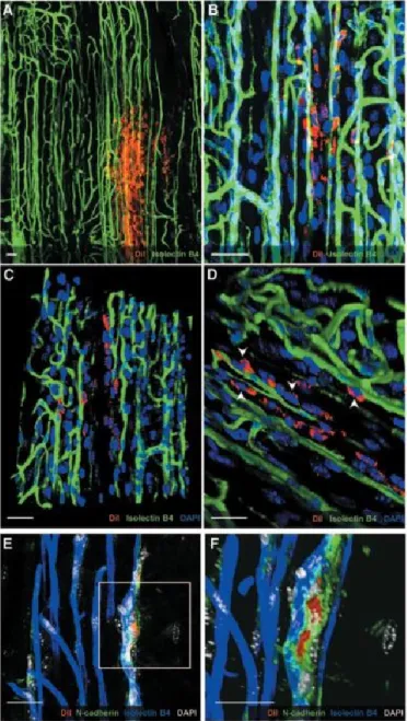
Biology of International PhD School in Life Sciences and Biotechnologies, University of Sassari. Figure 9 Interaction of SVPs with the host vasculature. Confocal images showing SVPs (red) surrounding
the host vasculature (isolectin B4; green; A and B). 3D reconstruction shows physical contact between SVPs and ECs (C and D; white arrowheads). Direct interaction was also confirmed by N-cadherin staining (green), which accumulated in the junction between SVPs (red) and capillary ECs (blue; E and F). Nuclei were stained with DAPI (blue in A through D; white in E and F). Scale bar=25μm. Image from Campagnolo et al., 2010.
Dr. Davide Maselli, “The role of mechanical stress in progression of intima hyperplasia associated
to vein coronary bypass grafts disease.”, PhD thesis in Biochemistry, Physiology and Molecular
Biology of International PhD School in Life Sciences and Biotechnologies, University of Sassari.
1.10 AMIGO2, a new modulator of pericytes biology
The Amphoterin-Induced Gene And Open Reading Frame (AMIGO) protein family is described below. In subsequent chapters will be shown as one component of the AMIGO family, AMIGO2, is involved in the SVPs mechano-sensitivity, resulting as a possible marker of the activated PCs status.
1.10.1 The AMIGO family
In 2003 a novel family of transmembrane proteins was described for the first time by Kuja-Panula et al. then named Amphoterin-Induced Gene And Open Reading Frame (AMIGO). This family is composed of three proteins: AMIGO, AMIGO2, and AMIGO3; that shows a clear homology and almost identical length and domain organization (Figure 10).[134]
Figure 10 AMIGO proteins. (A) Alignment of the primary structure of the three AMIGOs. The identical amino
acids between all AMIGOs are highlighted in red with white letters and similar amino acids are highlighted in red with black letters (B) The hypothetic tertiary structure of the proteins. (C) Alignment of the LRRs domains of Slit1, Nogo, and AMIGO1. The identical amino acids between Slit1 and Nogo receptor compared with the AMIGO1 are highlighted in red with white letters and similar amino acids are highlighted in red with black letters. Image from Kuja-Panula et al., 2003.