Philosophiae Doctor Degree in Cellular and Molecular
Biology – XVII
thcycle
The role of Aip4/Itch, a member of Nedd4 family
E3 ligases, in the ubiquitination of proteins
involved in RTKs endocytosis
ABSTRACT...7
INTRODUCTION...8
UBIQUITIN–MORE THAN JUST A SIGNAL FOR PROTEIN DEGRADATION...9
The ubiquitin pathway...9
The ubiquitin-activating enzyme, E1...11
The ubiquitin-carrier proteins (ubiquitin-conjugating enzymes), E2S. 11 The E3 ligases...11
RING Finger Ubiquitin Ligases...12
HECT- Type E3s...13
Aip4/Itch...14
The proteasome...17
Mono, multi or poly–ubiquitination...17
ENDOCYTOSIS MACHINERY...20
Clathrin-dependent receptor endocytosis...21
Constitutive endocytosis...22
Regulated endocytosis...23
The proteins involved in RTK endocytosis machinery...26
Proteins involved in sorting to the clathrin-coated vesicle...26
AP2...26
Dynamin...27
Amphiphysin...27
Endophilin...28
RalBP1 and POB1...28
Sorting in the multivesicular body (MVB)...29
Cbl...29
Eps15...30
Hrs...31
STAM2...32
ErbB RECEPTOR: STRUCTURE AND FUNCTION...34
ErbB1...35
ErbB2...36
ErbB3 and ErbB4...36
ErbBs family and cancer...37
ErbB1...38
AIM OF THE WORK...40
EXPERIMENTAL PROCEDURES...41
Constructs...41
Eukaryotic cell lines...42
Antibodies...43
Cell culture and transfection...43
Induction with EGF...44
Immunoprecipitation and western blotting analysis...44
GST-fusion proteins purification...44
In vitro ubiquitination...45
Pull-Down assays...45
Indirect immunofluorescence...46
Endocytosis assay...46
In vivo proteasome-dependent degradation assay...47
RESULTS...48
cDNA constructs...48
The SH3 domain of Amphiphysin I binds a proline rich peptide of Itch in vitro and in vivo...49
Itch does not ubiquitinate Amphiphysin I and POB...51
Ubiquitination of STAM2 by Itch, in vivo and in vitro...53
Proteasome inhibitor does not stabilize Itch-ubiquitinated STAM...55
Pull-Down between GST-Itch and different constructs of [35S] labeled STAM2...57
Immunoprecipitation between Itch and STAM2 in cells...59
Effects of Itch on endogenous STAM2...60
Itch binds and multi-ubiquitinates Hrs...62
Itch-WW domains recognize the 234-237 peptide of Hrs...65
KIAA0439 does not ubiquitinate HRS and STAM2...67
KIAA0439 does not ubiquitinate HRS and STAM2...69
Itch overexpression caused a perinuclear accumulation of EGF-TRIC ...70
Co-localizzation of endogenous Itch with endogenous STAM2, HRS or EPS15...72
Co-localizzation of endogenous Itch with endogenous Endophilin...72
DISCUSSION AND CONCLUSIONS...77
ABSTRACT
We have used a recently developed genomic approach (WISE: Langraf et al.; 2004), to search the whole proteome for proteins containing peptides that could bind to the SH3 domains of amphiphysin I and endophilin, two proteins implicated in vesicle trafficking. Among the inferred candidate ligands we focused on the protein Itch, a HECT domain ubiquitin ligase that was recently shown to participate in ubiquitination processes affecting the internalization of the EGF receptor. Itch can be co-immunoprecipitated with Amphiphysin I, in vivo, suggesting that this interaction could promote the enzymatic activity of Itch in endocytic vesicles and endosome compartments. We are systematically testing a number of proteins involved in receptor endocytosis to see whether they are targets of the ubiquitination activity of Itch, in vivo or in vitro. We demonstrated that Itch is implicated in the binding and ubiquitination of three proteins HRS, STAM2, Eps15 that form a multivalent complex essential in sorting ubiquitinated proteins into different vesicular pathways.
INTRODUCTION
Cells are continuously exposed to diverse external stimuli, ranging from soluble endocrine and paracrine factors to signaling molecules on neighboring cells. The cell must interpret these extracellular signals to produce an appropriate developmental or proliferative response. Receptors of the tyrosine kinase (TK) family play principal roles in these processes, as they integrate a multitude of external stimuli with specific internal signals and responses, ultimately allowing the cell to respond correctly to its environment. Receptor tyrosine kinases (RTKs) bind soluble extracellular growth factors and induce the activation of intracellular signaling pathways. These signal transduction mechanisms regulate a wide range of biological outcomes, including cellular proliferation, differentiation, motility and survival. The induction of the appropriate biological response requires signaling of the correct magnitude and kinetics. Further, disregulated activation of many RTKs through mechanisms including mutations, overexpression, structural rearrangements, disruption of autocrine/paracrine loops and inactivation of regulatory constraints, is implicated in multiple human neoplasias (Blume-Jensen et al., 2001). Thus, the controlled attenuation of these signaling pathways plays a central role in maintaining signaling homeostasis, thereby preventing over stimulation that could lead to cellular transformation. Termination of RTK signaling occurs via endocytosis and lysosomal degradation, processes in part regulated by ligand-induced receptor ubiquitination (Dikic et al., 2003).
However, RTK endocytosis should not be considered as an on/off switch for signaling through protein destruction, but rather a fine-tuning mechanism that ensures the appropriate biological outcome through modulation of signal strength and duration (Marshall, 1995). Further, internalization of receptors from the plasma membrane may enable their signaling through a distinct set of intracellular substrates and effectors (Wiley et al., 2001).
UBIQUITIN–MORE THAN JUST A SIGNAL FOR PROTEIN DEGRADATION
The ubiquitin pathway
The conserved 76-residue polypeptide ubiquitin fulfills essential functions in eukaryotes through its covalent conjugation to other intracellular proteins. Substrates marked with a polymer of ubiquitins (a polyubiquitin chain) are selectively targeted to a multisubunit ATP-dependent protease known as the 26 proteasome (Hershko et al., 1998), whereas certain substrates marked with one or a few ubiquitins are targeted for endocytosis, ultimately resulting in proteolysis in the lysosome/yeast vacuole (Hicke, 1999). Ubiquitination regulates a host of critical cellular functions, frequently by mediating the selective degradation of master regulatory proteins by proteasomes. The progression of the cell cycle (Koepp et al., 1999), the induction of the inflammatory response (Ghosh et al., 1998), and antigen presentation (Rock. et al., 1999) are just a few of the many processes regulated by ubiquitin/proteasome-dependent proteolysis.
Not surprisingly, disregulated ubiquitin-dependent proteolysis has been implicated as a causative factor in cancer and several inherited diseases (Glickman et al., 2002). Degradation is not the only fate possible for ubiquitin-tagged proteins; ubiquitination also regulates certain processes by mechanisms that, although poorly understood, do not appear to involve proteolysis. These processes include ribosomal function (Spence et al. 2000), postreplicational DNA repair (Spence et al. 1995), the initiation of the inflammatory response (Deng et al. 2000), and the function of certain transcription factors (Kaiser et al., 2000). Ubiquitination usually results in the formation of a bond between the C-terminus of ubiquitin (G76) and the -amino group of a substrate lysine residue. This reaction requires the sequential actions of three enzymes: (a) an activating enzyme (E1) that forms a thiol ester with the carboxyl group of G76, thereby activating the C terminus of ubiquitin for nucleophilic attack; (b) a conjugating enzyme (E2) that transiently carries the activated ubiquitin molecule as a thiol ester; and (c) a ligase (E3) that transfers the activated ubiquitin from the E2 to the substrate (or ubiquitin) lysine residue. This three-step mechanism initiates all known ubiquitination reactions, independent of whether the substrate-bound ubiquitin(s) will signal proteasomal proteolysis, endocytosis, or some other fate (Figure 1).
Figure 1. The proteasome pathway. (1) Activation ofubiquitin by the ubiquitin-activating enzyme, E1. (2) Transfer of the activated ubiquitin moiety to a member of the Ubiquitin carrier protein (ubiquitin-conjugating enzymes; Ubc's) family of enzymes, E2. (3) Transfer of activated ubiquitin from E2 to a Cys residue on E3 (in the case of the HECT domain family of ligases) or directly to the substrate (in most other cases; not shown). (4) Complex formation between the substrate and a specific binding site (BS) on the ubiquitin ligase, E3, followed by transfer of the first ubiquitin moiety to an internal Lys residue of the substrate and subsequent formation of a polyubiquitin chain. E3 can be either a monomer, a homodimer, or a component of a larger multimeric complex (as depicted in the Figure). (5) The polyubiquitinated substrate binds to the ubiquitin receptor (Ub-R) subunit of the 19S complex and is then degraded to short peptides (6) with the release of free and reutilizable
Substrate E2 Ub E2 E3 peptides Ub Ub Ub Ub Recycling Ubiquitin Ub E1 Substrate Ub E2 Ub Ub Ub Ub n Ub Proteasome 26S Ub ATP AMP+PPi E1 Substrate E2 Ub E2 E3 peptides Ub Ub Ub Ub Recycling Ubiquitin Ub E1 Ub E1 Substrate Ub E2 Ub Ub Ub Ub n Ub Ub Ub Ub Ub n Ub Proteasome 26S Ub ATP AMP+PPi E1 1 2 3 4 5 6 7
ubiquitin (7). Free E3 is also recycled. The 26S proteasome is composed of two 19S regulatory complexes attached at each side to the barrel-shaped 20S catalytic complex.
The ubiquitin-activating enzyme, E1
E1 activates ubiquitin, via a two-step intramolecular and ATP-dependent reaction, to generate a high-energy E1-thiol-ester-ubiquitin intermediate (Fig. 1). The activated ubiquitin moiety is then transferred to E2. The yeast genome encodes for a single ubiquitin-activating enzyme, UBA1. Inactivation of this gene is lethal. The protein contains a nuclear localization signal. The enzyme is phosphorylated, a modification that was suggested to play a role in its cell cycle-dependent nuclear localization. However, the physiological relevance of this modification has not been further substantiated (Checkanover et al., 2000).
The ubiquitin-carrier proteins (ubiquitin-conjugating enzymes), E2S E2s catalyze covalent attachment of ubiquitin to target proteins, or, when acting along with HECT domain E3s, transfer of the activated ubiquitin moiety to a highenergy E3-ubiquitin intermediate. They all share an active site ubiquitin-binding Cys residue and are distinguished by the presence of a UBC domain required for binding of distinct E3s. In few cases, they can also interact with the substrate. The physiological significance of this interaction is not known. Eleven ubiquitin conjugating enzymes (Ubc1–8, 10, 11, 13) have been identified in the yeast genome. Two additional enzymes, Ubc9 and Ubc12, are members of the UBC family, although they conjugate the ubiquitin-like proteins Smt3 and Rub1, respectively, and not ubiquitin. Many more E2s have been described in higher organisms. Typically, each E2 interacts with a number of ligases, thus being involved in targeting numerous substrates. The number and variety of different E2s in mammalian species is much greater (Pickart, 2001).
The E3 ligases
The ubiquitin ligases are the key regulatory determinants in the ubiquitination reaction, analogous to kinases in phosphorylation reactions.
Ubiquitin ligases comprise two major families (Pickart, 2001). The first family, characterized by the zinc-binding RING (Really Interesting New Gene) finger domain and related domains, promotes ubiquitination by simultaneously binding the substrate and an E2 (Joazeiro et al., 2000). The
second family, defined by the HECT (homologous to E6-AP carboxy-terminus) domain, participates directly in catalysis by forming an obligate thiolester bond with ubiquitin during the ubiquitination reaction (Huibregtse et al. 1995). Because E3s carry the specificity information of the ubiquitination machinery, understanding the function of this class of enzymes is critical for understanding events regulated by ubiquitin in vivo.
RING Finger Ubiquitin Ligases
Ubiquitin ligases that carry a RING finger domain exist as multisubunit complexes, or as monomers with substrate binding information and E3 activity built into the same molecule (Joazeiro et al., 2000). Genome sequencing suggests that RING finger E3s may far out number their HECT domain counterparts, and the identification of E3 activity in domains related to the RING finger, the PHD (plant homeodomains) and U-box (UFD2-homology domain) lengthens the list. The RING finger directly interacts with an E2 and the substrate. RING finger ligases do not function as enzymes per se but instead activate E2s to modify specific substrates. RING finger ligases have emerged as key regulators of neuronal function in many places For example, the drosophila Highwire plays an important role in synaptic development and disruption of the E3 ubiquitin-protein ligase activity of parkin is probably the cause of protein aggregation in Parkinson’s disease (DiAntonio et al., 2004).
Depending on whether the RING finger is present in the form of a functional domain in a single protein or in the form of a subunit in a protein complex, the RING-type E3s are subdivided into single protein E3s and multisubunit E3s.
Single protein ring-type E3S: In this subgroup, the RING finger constitutes a functional portion of a single protein, in which a protein interaction domain or domains are also present for the substrate recruitment. One of the earliest identified E3s of this subgroup is Cbl. Cbl is a 120 kDa protooncogene product that is comprised of an N-terminal tyrosine kinase– binding (TKB) domain, a RING finger, and C-terminal proline-rich sequences and tyrosine phosphorylation sites. Cbl acts as an E3 Ub ligase, whose RING finger recruits Ub-loaded E2, and its TKB domain binds to tyrosine phosphorylated receptor tyrosine kinases. The crystal structure of Cbl RING-E2 complex further supports a role of Cbl in Ub conjugation, in
which the Cbl RING finger forms a shallow grove on to which the two loops of UbcH7 bind. More interestingly, the E2-binding grove in the Cbl RING domain is quite similar to that in the HECT domain of E6-AP, and UbcH7 uses the same structural elements for interaction with both domains, even though there is no similarity in amino acid sequences between a RING finger and a HECT domain.
Multisubunit ring-type E3s: This subgroup consists of a superfamily of E3s including the SCF (Skp1-Cullin 1-F box protein), the APC (Anaphase-promoting complex), and the VCB (VHL-elongin C/elongin B). All three E3s contains an 100 amino acid RING finger, Roc1/Rbx1 or Apc11, which together with its binding component, Cullin in the SCF or VCB E3s, or Apc2 in the APC, forms a core enzymatic structure for Ub-charged E2 recruitment (Liu, 2004).
HECT- Type E3s
Studies on human papillomaviruses led to the discovery of the viral E6-associated protein (E6-AP), which forms a complex with the oncogenic E6 to induce the degradation of the p53 tumor suppressor. Later on, the same group (Liu et al.; 2004) identified a family of proteins that have a highly conserved region of 350 amino acids similar to the C-terminus of E6-AP, named the HECT domain. In the C-terminus of the HECT domain, there exists a conserved active cysteine residue, which forms a high-energy thioester bond with Ub and constitutes a necessary step for the Ub transfer to the substrate. Interestingly, E6-AP seems not to be the physiological E3 ligase because it does not induce p53 degradation in HPV uninfected cells. Recent studies have demonstrated that Src family kinases are the potential targets for E6-AP. Notably, genetic analysis has linked the disruption of the maternal copy of E6-AP with Angelman syndrome, a genetic neurological disorder. E6-AP also acts as a coactivator for steroid hormones through ligase-dependent or -independent mechanisms.
With the exception of E6-AP, other HECT domain–containing E3 ligases often contain an N-terminal Ca2C-binding, protein kinase C-related C2 domain, followed by multiple WW domains, in addition to the C-terminal HECT domain. WW domains derive their name from the presence of two highly conserved tryptophan (W) residues, which are spaced 20–22 amino acids apart. They normally contain 38–40 amino acids in a triple-stranded
-sheet and are found in proteins that participate in cell signaling or regulation. These domains are implicated in mediating protein-protein interactions by binding to proline-rich motifs or phosphoserine- and phosphothreonine-containing elements in their binding partners. One of the well-studied WW domain–containing HECT-type E3s in mammalian cells is Nedd4, which is implicated in the regulation of the epithelial NaC channel in the kidney and other tissues, and whose deletion is related to the human Liddle’s syndrome, a hereditary form of hypertension.
The WW domains of Nedd4 associate with a PPXY motif in the channel protein, and mutations of the PPXY motif were found in the patients with Liddle’s syndrome (Liu, 2004).
Recently, a number of proteins have been discovered that share the same modular structure as Nedd4 and appear o be part of a family of ubiquitin-protein ligases. Some of these ubiquitin-proteins have been implicated in a variety of cellular functions.
The most closely related protein to human Nedd4 is the KIAA0439. This putative protein shares approximately 78% sequence similarity with human Nedd4 and it has a Xenopus homologue. KIAA0439 and Nedd4 proteins both play a redundant role in EnaC regulation (Harvey, 2001). Three human Nedd4-like proteins, WWP2/Aip2, WWP1/Aip5 and Aip4/Itch, have been cloned recently as molecules that interact with atrophin-1, a protein containing five PY motif, and they share a similar domains architecture (Wood, 1998) (Figure 2).
Aip4/Itch
Aip4/Itch was identified through the study of the agouti locus on mouse chromosome 2, and mutation at this locus results in a wide variety of coat color alterations (Hustad et al., 1995). The agouti protein induces the production of yellow pigment by melanocytes and thus determines the amount of yellow present in the hair. One of these mutations, 18H, which displays a darker coat, causes immunological disorders. The most obvious disorder is ear and skin scarring due to constant itching (thus called itchy mice) starting from 16-week-old or older mice. The mutant mice have enlarged spleens and lymph nodes, possibly due to lymphocyte hyperproliferation.
cDNA cloning showed that the Itch gene encodes 854 amino acids protein with a molecular weight of 113 kDa. It consists of an N-terminal
protein kinase C-related C2 domain, four WW protein-protein interaction domains, and a C-terminal HECT Ub ligase domain. Therefore, Itch is a member of the HECT domain-containing E3 Ub protein ligases. The data based on itchy mice clearly suggest a critical role of E3 ligase or ubiquitination in the regulation of the immune system. Itch-/- T cells showed slightly enhanced T cell proliferation and IL-2 production upon anti-CD3 engagement. In aging mice, Itch-/- T cells displayed increased cell surface expression of CD69 activation marker, suggesting a chronic activation of T cells in the absence of Itch. Interestingly, increased serum levels of IgG1 and IgE in itchy mice as well as a T helper cell type 2 (Th2)-biased differentiation were observed in itchy mice (Fang et al., 2002). The molecular mechanism underlying Itch-mediated T cell differentiation was further explored by using biochemical approaches. Itch was found to associate with Jun-B, through a PPXY motif in Jun-B and the WW domains of Itch, and to promote Jun-B ubiquitination. Interestingly, Jun-B has been implicated in the gene regulation of Th2 cytokines such as IL-4 (Li et al., 1999). In Itch-/-T cells, the rate of Jun-B degradation was reduced, in parallel with increased nuclear translocation and DNA binding activity of Jun-B. Moreover, Jun appears stabilized in the presence either of a proteasome inhibitor or of an inhibitor of the lysosomal protein degradation , suggesting a possible role for the lysosome pathway in Jun degradation, Itch mediated (Fang et al., 2004).
New evidences show a possible role of Itch in the regulation of RTK endocytosis. Indeed, Itch ubiquitinates Endophilin A1 and interestingly, overexpressed Itch co-localizes with markers of the endosomal system in a C2 domain-dependent manner. Moreover, upon EGF stimulation, Endophilin A1 translocates to an EGF-positive endosomal compartment where it colocalizes with Itch. EGF treatment of cells stimulates Endophilin A1 ubiquitination. This interaction may be involved in ubiquitin-mediated sorting mechanisms operating at the level of endosomes (Angers et al., 2004).
Moreover, Itch binds Cbl proteins and these E3 ligases are involved in EGFR signaling and ubiquitination. Both, Itch and Cbl, become phosphorylated on tyrosines following epidermal growth factor stimulation. In addition, Cbl-C increases the ubiquitination of EGFR, and the coexpression of the WW domains of AIP4/Itch exerts a dominant negative effect on EGFR ubiquitination. Finally, coexpressing Cbl-C and AIP4/Itch induces a down-regulation of EGFR signaling (Courbard et al., 2002).
The proteasome
The proteasome is a large, 26S, multicatalytic protease that degrades polyubiquitinated proteins to small peptides. It is composed of two subcomplexes: a 20S core particle (CP) that carries the catalytic activity and a 19S regulatory particle (RP). The 20S CP is a barrel-shaped structure composed of four stacked rings, two identical outer -rings and two identical inner -rings. The eukaryotic - and -rings are composed each of seven distinct subunits, giving the 20S complex the general structure of 1–71– 71–71–7. The catalytic sites are localized to some of the -subunits. Each extremity of the 20S barrel can be capped by a 19S RP. One important function of the 19S RP is to recognize ubiquitinated proteins and other potential substrates of the proteasome. An ubiquitin-binding subunit of the 19S RP has indeed been identified; however, its importance and mode of action have not been discerned. A second function of the 19S RP is to open an orifice in the -ring that will allow entry of the substrate into the proteolytic chamber. Also, because a folded protein would not be able to fit through the narrow proteasomal channel, it is assumed that the 19S particle unfolds substrates and inserts them into the 20S CP. Both the channel opening function and the unfolding of the substrate require metabolic energy, and indeed, the 19S RP contains six different ATPase subunits. After degradation of the substrate, short peptides derived from the substrate are released, as well as reusable ubiquitin (Checkanover et al., 2000).
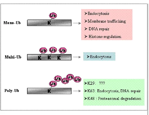
Mono, multi or poly–ubiquitination
The addition of a single ubiquitin to a substrate is defined as monoubiquitination. Moreover, several lysine residues in the substrate can be tagged with single ubiquitin molecules, giving rise to multiple monoubiquitination (Haglund et al., 2003 and Mosesson et al., 2003). Finally, ubiquitin contains seven lysine residues, which can also be targeted by another ubiquitin in an iterative process, known as polyubiquitination, that leads to the formation of a ubiquitin chain attached to a single lysine Pickart, 2001) (Figure 3). It is now clear that different types of ubiquitin conjugates are involved in the regulation of different cellular processes (Hicke, 2001). Monoubiquitination is implicated in the endocytosis of plasma membrane proteins, the sorting of proteins to the multivesicular body
(MVB), budding of retroviruses, DNA repair, histone activity and transcriptional regulation. By contrast, polyubiquitin chains formed via the C-terminal glycine and lysine 48 of two ubiquitins have a well characterized role in targeting proteins for degradation by the 26S proteasome, whereas ubiquitin chains formed through lysine 29 or lysine 63 are involved in other cellular functions, including DNA repair and endocytosis (Weissman, 2001). A major unanswered question concerns how the decision is made by the ubiquitin machinery as to whether to mono- or polyubiquitinate a substrate. One possibility is that different subsets of ubiquitin ligases have specificity for the two different modifications. For example, the ubiquitin ligase Mdm2 mediates monoubiquitination of p53, whereas p300 has been suggested to promote p53 polyubiquitination (Grossman et al., 2003). Alternatively, an individual ubiquitin ligase might mediate either mono- or polyubiquitination, depending on the nature of the substrate or on other molecular specifiers, such as proteins interacting with the E3 in different subcellular locations. Such an example is Cbl, a ubiquitin ligase known to direct polyubiquitination and proteasomal degradation of cytoplasmic proteins, including Sprouty, as well as Src and Abl tyrosine kinases (Yokouchi et al., 2001). However, Cbl can also direct monoubiquitination of RTKs and of the Cbl-associated adaptor protein CIN85 in the endocytic pathway (Haglund et al., 2002).
The type of ubiquitin modification might also be determined by ubiquitin-binding proteins. Recently, a role for ubiquitin-interacting domains (such as the ubiquitin-interacting motif, UIM, or the Cue1-homologous domain, (CUE) in the determination of monoubiquitination of endocytic proteins has been proposed based on the frequent monoubiquitination of proteins containing these domains. UIM- or CUE-containing proteins might transiently bind to ubiquitin-loaded E3 ligase and, once ubiquitin is transferred to a UIM/CUE-containing protein, the E3 ligase might dissociate, yielding a monoubiquitinated substrate (Di Fiore et al., 2003). Alternatively, following ubiquitination, the binding of UIM/CUE domains to ubiquitin might sterically hinder the formation of ubiquitin chains because the major polyubiquitination site in ubiquitin, lysine 48, is masked in the UIM/CUE-ubiquitin complex (Shekhtman et al., 2002; Kang et al. 2003). Finally, ubiquitination is a dynamic and reversible process, and the rapid removal of ubiquitin is mediated by the activity of de-ubiquitinating enzymes (Wilkinson, 2000). It is therefore possible that a balance between activity and subcellular localization of de-ubiquitinating enzymes and ubiquitin ligases determines whether a specific protein becomes mono- or
polyubiquitinated. Clearly, further experimental effort is needed to assess whether one or a combination of these mechanisms determines the type of ubiquitin modification in vivo, and thus the fate and function of ubiquitin-tagged substrates.
ENDOCYTOSIS MACHINERY
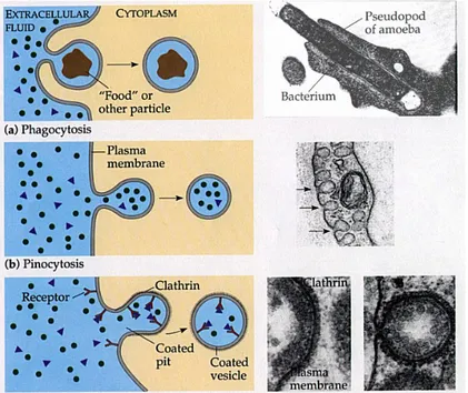
Several morphologically and functionally different types of endocytic pathways exist in animal cells (Waterman et al., 2001):
1) Phagocytosis represents the uptake of solid particles (>0.5 diameter) that must bind to specific plasma membrane receptors capable of triggering their own endocytosis, usually by causing the formation of F-actin-driven pseudopods that envelop the bound particle.
2) Pinocytosis is an enables uptake of extracellular fluid, macromolecules and solutes bound specifically or non-specifically to the plasma membrane. 3) Receptor-mediated endocytosis, is the selective adsorptive uptake of specific macromolecules bound to plasma membrane receptors (Figure 4). This process involves constitutive formation of small (60.2 diameter) vesicles, which is usually preceded by the formation of clathrin-coated vesicles (CCVs). In addition, exist at least two types of clathrin independent endocytosis: caveolar endocytosis and membrane ruffling (Parton et al., 2003).
Clathrin-dependent receptor endocytosis
The capability of clathrin-coated vesicles (CCVs) to selectively sequester protein cargo into a membrane vesicle is dependent upon three major components. Firstly, the self assembling property of the clathrin coat, which is contingent upon the triskelion shape of clathrin and its intrinsic ability to form a polyhedral lattice. Secondly, the well-established AP2 adaptor complex which is drawn into the lattice and triggers CCV formation at the plasma membrane, while incorporating transmembrane molecules by associating with the cytoplasmic domains of these proteins (Figure 5). Thirdly, dynamin, a GTPase responsible for fission of the vesicle from the plasma membrane resulting in the detachment of the CCV from the donor membrane (Brodsky et al., 2001). In essence, the formation of the CCV may provide the initial cargo selection function. The coated vesicle then undergoes clathrin shedding and fuses with an acceptor compartment forming the early endosome, where the second major sorting event occurs. Although it remains disputed whether trafficking from the early endosome to the next compartment in the endocytic pathway involves vesicular transport or maturation, the cargo proteins proceed into a compartment referred to as either the late endosome or the multi-vesicular body (MVB). This compartment is characterized by different protein composition, low luminal pH, and multiple internal vesicles. Finally, the cargo that is not recycled back to the plasma membrane is degraded in the lysosome.
In general, clathrin-dependent receptor endocytosis can be functionally subdivided into two pathways: the constitutive pathway that is dependent on short linear internalization motifs within the endocytic cargo, as exemplified by transferrin receptor (TfR) and catalytically inactive RTKs, and regulated endocytosis, which depends on additional, phosphorylation mediated interactions, as is the case for EGFR and other RTKs.
Figure 5. The assembly and disassembly of a clathrin coat. The assembly of the coat is thought to introduce curvature into the membrane, which leads in turn to the formation of uniformly sized coated buds. The adaptins bind both clathrin triskelions and membrane-bound cargo receptors, thereby mediating the selective recruitment of both membrane and cargo molecules into the vesicle. The pinching-off of the bud to form a vesicle involves membrane fusion; this is helped by the GTP-binding protein dynamin, which assembles around the neck of the bud. The coat of clathrin-coated vesicles is rapidly removed shortly after the vesicle forms.
Constitutive endocytosis
TfR and other constitutively endocytosing proteins such as the low-density lipoprotein (LDL) are are internalized by virtue of their constitutive binding to AP2, and subsequent recruitment to clathrin-coated pits. In contrast to ligand-induced endocytosis, receptors undergoing constitutive complex (Gammie et al 1995); these determinants are internalization signals located within the cytoplasmic domains of transmembrane proteins. A variety of internalization motifs have been identified in mammalian cells and may be sub-categorized into two groups (Wendland et al., 1998). The first group is characterized by an essential tyrosine, such as the YXXØ motif (where X is any amino acid and Ø is an amino acid with a bulky hydrophobic group), or NPXY. The second class of internalization signals typically contains a dileucine sequence.
Regulated endocytosis
Clathrin triskelions assemble at the membrane and form a polyhedral lattice. Upon activation, EGFR molecules cluster over clathrin-coated regions of the plasma membrane. These loaded clathrin-coated regions invaginate in a dynamin dependent manner to form endocytic vesicles. The CCV sheds clathrin and fuses with an internal vesicle to form the early endosome, which proceeds along the endocytic pathway to the late endosome through a mechanism that involves both regulatory proteins and phosphoinositides (De Camilli et al., 1996). Endocytic vesicle maturation is concomitant with a reduction in the internal pH and the accumulation of hydrolytic enzymes. Early and late endosomes are defined by the kinetics with which cargo arrives in these compartments, as well as morphologically, as early endosomes are often localized at the cell periphery and are more tubular, whereas late endosomes are more spherical and closer to the nucleus. Segregation in late endosomes involves invagination of the limiting membrane and budding into the lumen to form internal vesicles, and thus this sorting compartment is called multivesicular bodies (MVB). It is of interest to note that internalized EGFRs can still associate with signaling proteins to activate intracellular effectors; however its sorting into lumenal vesicles of the MVB results in the segregation of the cytoplasmic domain of the receptor away from the cytoplasm and terminates signaling. The MVB fuses with the lysosome, delivering the internal contents of the lumenal vesicles for degradation by hydrolytic enzymes in the lysosome (Figure 6).
Targeting for lysosomal degradation is subject to multiple steps of regulation, with the first being the sorting process responsible for receptor incorporation into clathrin-coated pits. Further sorting occurs at the level of vesicle fusion to form early endosomes, and at the level of the MVB. Recycling of proteins back to the membrane can occur throughout the endocytic pathway with varying efficiencies, but this process, unlike sorting at the plasma membrane and at the MVB, does not require intrinsic RTK activity (French et al., 1994). Receptor recycling can be modulated by the induction of signaling pathways, and threonine phosphorylation of EGFR by protein kinase C has been shown to enhance recycling (Bao et al., 2000). Proteins not destined for lysosomal degradation remain in the outer MVB membrane and are not sorted to internal vesicles (Felder et al., 1990). Even after transfer to the limiting membrane of MVBs, such proteins can undergo recycling back to the plasma membrane or to other cellular locations.
The fundamental difference between constitutive and ligand-induced endocytosis does not only lie within the kinetics of endocytosis, nor in the delicate balance between recycling and degradation, but in the unquestionable requirement of an additional mechanism distinct from the classical internalization cargo-intrinsic signals. Contrary to initial models, ligand binding does not stimulate unmasking internalization signals and recognition of the receptor by AP2 molecules. Instead, accumulating evidence suggests possibility that EGFR’s independence of AP2 may be due to phosphorylation events (Shtiegman K. et al., 2003).
Two post-translational modifications, namely phosphorylation and ubiquitylation, are involved in receptor endocytosis, and their intricate coupling allows for fine tuning of the response with much more control than simple determinants. Both modifications are dynamic, and may be reversed by phosphatases and de-ubiquitylating enzymes. Proteins recruited subsequent to phosphorylation and/or ubiquitylation are involved in sorting decisions regulating receptor downregulation, at the level of internalization. Although catalytically inactive receptors undergo constitutive internalization and their rate of endocytosis is independent of ligand binding (Wiley et al., 1991), how ligand binding accelerates receptor internalization remains unclear. Phosphorylation of substrates like Eps15 (Confalonieri et al., 2000) and c-Cbl (Waterman et al., 2002), and bending of the lipid bilayer (Petrelli et al., 2002; Soubeyran et al., 2002) may play a causative role. Other candidates are modifications of the endocytic machinery, which include the Src-mediated phosphorylation of clathrin heavy chain (Wilde et al., 1999). Further, ubiquitin modification of both the RTK cargo and the endocytic machinery regulates endocytosis.
Figure 6. RTK: journey to the lysosome. Stimulation with ligand induces activation of RTKs like EGFR, autophosphorylation and recruitment of Cbl. Cbl is then phosphorylated and ubiquitylates the EGFR. Ubiquitylated receptors are sorted into clathrin-coated pits by a multiprotein complex that includes coat adaptors such as Epsin and Eps15. Fission of clathrin-coated vesicles is mediated by a GTPase, dynamin. In addition, Cbl-mediated recruitment of CIN85 and endophilin may promote negative membrane curvature and invagination. Progression through the endocytic pathway is characterized by the shedding of clathrin, a decrease in the internal pH and the accumulation of hydrolytic enzymes. EGFR trafficking from early to late endosomes/MVB is dependent on its continued association with Cbl and its sustained ubiquitylation. The guanine nucleotide exchange factor Rabex/Vps9regulate s endosomal membrane fusion through activation of another GTPase, Rab5. MVB sorting is regulated through recognition of ubiquitylated cargo by Hgs/Vps27p, Tsg101/Vps23p and other components of ESCRT complexes. Invagination of the limiting membrane of the MVB forms internal vesicles, which in yeast is coupled to substrate de-ubiquitylation by Doa, and dissociation of the ESCRT complex from endosomes, mediated by the AAA ATPase Vps4. Fusion of the MVB with the lysosome results in degradation of the contents of the internal vesicles. Recycling of receptors back to the plasma membrane can occur throughout the endocytic pathway, albeit with decreasing efficiency (Marmor et al., 2004)
The proteins involved in RTKs endocytosis machinery
Proteins involved in sorting to the clathrin-coated vesicle
The three major components of coated pit are clathrin, adaptor-binding protein 2 (AP-2) and dynamin.
Clathrin
Clathrin is composed of heavy and light chains. Three heavy and three light chains form stable oligomeric complexes, the three-legged triskelia, which are the unit elements of the clathrin lattice (Smith et al., 1999). The legs of the triskelia are formed by right-handed superhelices of short α-helix hairpins and represent the edges of the lattice. The end of each leg, corresponding to the amino-terminal region of the heavy chain, is a separate protein–protein interaction module with a seven-bladed β-propeller structure. A binding site present in the groove that separates blades 1 and 2 (Ybe et al., 1999) recognizes a short peptide motif, called the clathrin box, present in clathrin adaptors and in several other endocytic proteins (Owen et al. 2000). The relative flexibility of the angle formed by the legs of the triskelia allows clathrin to oligomerize in to hexagons and pentagons, and generate curvature (Musacchio et al., 1999) (Figure 7).
AP2
AP-2 consists of four subunits, α-adaptin, β2 adaptin, μ2 adaptin and σ2 adaptin, tightly bound to each other2. AP-2 has a brick-like structure with two ear domains that correspond to the carboxy-terminal regions of α- and β2-adaptin. The core region of AP-2 binds both to membrane proteins, such as synaptotagmin and proteins that contain endocytic motifs and to membrane lipids, such as Phosphoinositides and other acidic phospholipids. The binding of AP-2 to clathrin is mediated primarily by β2-adaptin and involves two binding sites (Owen et al. 2000). One is in the ear domain and the other, a typical clathrin box, is localized in the hinge region that connects the ear to the trunk of β2-adaptin. Both sites cooperate to promote clathrin polymerization and it has been proposed that their interaction with clathrin
may displace accessory proteins during coat maturation. AP-2 binds various accessory proteins through the ear domains of α- and β2-adaptin (Figure 7).
Dynamin
Dynamin has now been shown to have a key function in the fission of clathrin-coated and other endocytic vesicles. Its mechanism of action, however, is still unclear. In the presence of the non-hydrolysable analogue of GTP, guanosine 5′-γ- triphosphate (GTPγS), dynamin, together with binding partners such as Amphiphysin and Endophilin, forms stacks of rings around the stalks of coated pits. It seems possible that Dynamin may combine both the features of a classical GTPase and the properties of a mechanochemical enzyme. Clearly, the GTPase activity of dynamin is crucial for its function. This activity is regulated by dynamin-binding proteins, by its own oligomerization state, and by lipids in particular, phosphoinositides . An important downstream target of dynamin may be the actin cytoskeleton (Schmid et al., 1998). The carboxy-terminal region of dynamin is a proline-rich protein–protein interaction module that binds various SH3 domains through an array of distinct binding sites. Studies of this interaction network have led to the identification of several major synaptic SH3-domain-containing proteins, including amphiphysin, endophilin, intersectin and syndapin, as well as of synaptojanin, another binding partner for these proteins besides dynamin (McNiven et al., 2000). All of these proteins been implicated as accessory factors in clathrin-mediated synaptic vesicle endocytosis (Figure 7).
Amphiphysin
Amphiphysin 1 and 2 are two similar proteins concentrated in nerve terminals. Amphiphysin 1 is predominantly expressed in the brain (at a level 20–30-fold higher than in other tissues), whereas amphiphysin 2 is broadly expressed and undergoes extensive alternative tissue-specific splicing (Wigge et al., 1998). Amphiphysin may function as a multifunctional adaptor that cooperates in the recruitment of coat proteins to the lipid bilayer, and in targeting of dynamin and synaptojanin to the coat. Its amino-terminal region, also referred to as the BIN-amphiphysin-RVS (BAR) domain, mediates the formation of homo and heterodimers and also harbours
a lipid binding site that mediates membrane binding. The central region binds the heavy chain of clathrin and the ear domain of the AP-2 subunit α-adaptin through two distinct but partly overlapping sites. The SH3 domain that mediates dynamin and synaptojanin binding is located at the carboxyl terminus (Figure 7).
Endophilin
Endophilins (endophilin 1, 2 and 3) have a domain structure similar to Amphiphysin with a conserved amino terminal domain and a carboxy-terminal SH3 domain. However, relative to Amphiphysin, they bind synaptojanin with an high affinity (Ringstad et al., 1997).
Endophilin 1 is most abundant in brain. The conserved amino-terminal domain of Endophilin 1, which mediates dimerization and binds lipids lysophosphatidic acid acyl transferase activity (Schmidt et al. 1999). Endophilin was shown to be required for synaptic vesicle biogenesis in a cell-free system (Figure 7).
RalBP1 and POB1
Ral is a member of the small GTP binding protein family (Feig et al., 1996). The only known effector protein of Ral, RalBP1, and its own partner POB1, are both implicated in EGF signalling downstream of Ras. Exposure of cells to EGF or to insulin increases the GTP-bound active form of Ral through activation of Ras and its effector, guanine nucleotide exchange factor for Ral (RalGEF), (Matsubara et al., 1999). Active Ral binds to the C-terminal part of RalBP1, a putative GTPase of Rac1 and CDC42. While the relevance of this GAP activity to endocytosis remains unknown, RalBP1 can effectively recruit the AP2 complex, either directly or through POB1 and Eps15. The W2 chain of AP2, but not other coat proteins, binds to the N-terminus of RalBP1, and inhibition of these constitutive interactions blocks endocytosis of both ErbB-1 and TfR (Jullien-Flores, 2000). On the other hand, phosphorylation of POB1, a binding partner of RalBP1, Eps15 and epsin, another EH domain protein that participates in clathrin-mediated endocytosis, is elevated by EGF. Thus, recruitment of POB1 to the AP2 complex may be involved in the ligand-induced pathway. Indeed, deletion mutants of POB1 can inhibit endocytosis of both ErbB-1 and the insulin
receptor (Nakashima et al. 1999). Presumably, RalBP1 is translocated to the plasma membrane upon stimulation with EGF and subsequent activation of Ras and Ral. Once associated with the plasma membrane, RalBP1 can bind AP2 in a complex manner that involves not only constitutive and ligand-induced interactions, but also the intrinsic GTPase activity (Figure 7).
Sorting in the multivesicular body (MVB) Cbl
The mammalian Cbl protein family consists of three members: Cbl, Cbl-b and Cbl-3, all having a highly conserved amino-terminal part composed of a tyrosine kinase binding module and a ring finger domain. This part of Cbl is able to recruit ubiquitin-conjugating enzymes in the complex with activated tyrosine kinase receptors, thus enabling ubiquitination of the receptor molecules. However, the carboxyl termini of Cbl proteins are more diversified. Cbl-3 contains only a short polyproline domain in its carboxyl terminus, while Cbl and Cbl-b have long proline rich domains and additional distal parts containing an acidic box and a leucine-zipper (LZ) domains. The distal carboxyl terminal tails of Cbl and Cbl-b contain several polyproline motifs scattered among tyrosine residues that are phosphorylated in vivo after growth factor stimulation. Binding of multiple signaling proteins containing SH2 and SH3 domains to this part of Cbl is regulated by tyrosine phosphorylation. Carboxyl-terminal interactions are involved in the control of cell-type specific functions of Cbl, such as regulation of glucose uptake, osteoclasts activation and bone remodeling, as well as cell spreading and migration.
Several recent reports have also implicated the carboxyl terminus of Cbl in the control of endocytosis of RTKs. The major mechanism of Cbl recruitment to activated EGF receptors involves binding of the SH2 domain of Cbl to the autophosphorylated tyrosine 1045 of EGF receptor. An alternative pathway was recently discovered by using EGFR-Y1045F mutant, and showed to employ the Grb2 adaptor protein, which acts as an intermediate between Cbl and the receptor. In addition, binding of SH3 domain-containing protein CIN85 to the distal carboxyl terminus of Cbl was shown to regulate EGF and c-Met receptors endocytosis in mammalian cells (Szymkiewicz et al., 2002). Recent study support a role for Cbl and ubiquitination at a late step in the endocytic pathway, rather than at the
initial internalization step (Duan et al.. 2003). Instead, Cbl-mediated EGFR ubiquitinylation is required for efficient sorting of activated EGFR into the lysosome for its degradation (Figure 7).
Eps15
Eps15 was initially identified as major cytosolic substrate for ligand-activated EGFR. An evolutionary conserved Eps15 homology (EH) domain is present in three copies in the amino terminus of Eps15. It was determined that a large number of EH domains present in a variety of endocytic proteins, both in yeast and in mammalian cells, recognize NPF (asparagine–proline– phenylalanine) tripeptides albeit with different sequence context preference. Therefore, it comes as no surprise that Eps15 is endowed with a number of binding partners through the EH–NPF interaction. Some known interactions include Epsin, synaptojanin, and Numb, all of which are involved in endocytosis. Interestingly, the NPFXD sequence is a known internalization signal in yeast. A class of peptides that contain two consecutive amino acids with aromatic side chains (FW or WW) are also recognized by some EH domains (Paoluzi et al. 1998). The central coiled-coil region of Eps15 is important for both homodimerization and eterodimerization of Eps15 with other endocytic proteins such as intersectin. Downstream to the coil-coiled region, Eps15 contains multiple copies of the DPF tripeptide that binds to the amino-terminal ‘appendage’ region of AP2. The carboxyl terminal region of Eps15 also contains a proline-rich segment that is a target for SH3 domain-containing proteins (Shtiegman et al., 2003).
A recently identified UIM domain is also present in the carboxyl terminal region of Eps15. Eps15 is mainly cytosolic, although recently it has been shown to shuttle in and out of the nucleus (Vecchi et al., 2001). A dramatic re-localization of Eps15 to the plasma membrane is detected upon EGF treatment (Torrisi et al., 1999). Interestingly, Eps15 is absent from clathrin-coated vesicles, but easily detectable in unclathrin-coated vesicles and endosomes, thus implying re-localization of Eps15 back into endocytic organelles. Expression of dominant negative mutant fragments of Eps15 (Benmerah et al., 1998), and microinjection of antibodies directed against Eps15 (Carbone et al., 1997) demonstrates that Eps15 plays a critical role in both constitutive and regulated endocytosis. As previously mentioned, Eps15 was discovered as an EGFR pathway substrate and the major site of phosphorylation has been mapped to tyrosine 850. A Y850F mutant of Eps15 has dominant
negative activity on the endocytosis of EGFR, but not of TfR (Confalonieri et al., 2000). Thus, the constitutive and ligand-induced pathways of endocytosis may utilize some of the same proteins, however, they utilize the proteins differently, contingent upon post-translational modifications such as tyrosine phosphorylation. Further analysis revealed that Eps15 undergoes mono-ubiquitylation in response to EGF (van Delft et al., 1997) (Figure 7).
Hrs
Hepatocyte growth factor (HGF)-regulated tyrosine kinase substrate (Hrs) is a prominent target for tyrosine phosphorylation following activation of tyrosine kinase receptors (Urbé et al., 2003). Hrs is composed of several recognizable domains: a VHS domain, a FYVE domain, a UIM domain, a proline-rich region, a coiled-coil domain, and a proline- and glutamine-rich carboxyl terminal region. The FYVE domain confers binding to phosphatidylinositol 3-phosphate (PI3P), and has been found to play a role in the localization of proteins such as EEA1 and Hrs to the early endosome. However, in the case of Hrs, the endosomal localization is also dependent upon the proline and glutamine-rich C-terminal region. The coiled-coil domain of Hrs mediates its interaction with sorting nexin 1 (SNX1). This interaction alters down regulation of EGFR, most likely by affecting trafficking downstream to internalization. Other ligands of the coiled-coil domain include the Signal Transducing Adapter Molecule (STAM) and Hrs-binding protein (Hbp). The Hrs–STAM interaction leads to suppression of cytokine-mediated DNA synthesis. The Hrs–Hbp complex is important for receptor degradation. An additional interactor of Hrs is the deubiquitylating enzyme UBPY (Shtiegman et al., 2003).
Hrs is likely to be a mammalian homolog of the yeast sorter Vps27p, which is essential for vacuolar and endocytic trafficking through a pre-vacuolar compartment. Accordingly, mouse cells that lack Hrs contain abnormally large early endosomes, and Hrs over-expression leads to the appearance of large structures containing endosomal markers (Kanazawa et al., 2003). These lines of evidence indicate that Hrs specifically infuences the dynamics of multiple endocytic compartments, which merge when the protein is overexpressed, perhaps due to promotion of vesicle aggregation or of vesicle fusion (Figure 7).
STAM2
Signal-Transducing Adaptor Molecule (STAM) 1 and STAM2 (also known as EAST/Hbp) were initially identified as phosphotyrosine proteins detectable after stimulation with a variety of cytokines and growth factors and as binding proteins for Hrs/Hgs, the mammalian homologue of yeast Vps27, which functions in the vacuole membrane transport machinery. STAM1 and STAM2 have an amino acid sequence identity of about 50%, and their yeast homologue, Hse1, has been identified as a class E Vps protein. All STAMs have, in their N-terminus, a VHS (Vps27, Hrs and STAM homology) domain, an evolutionarily conserved domain of 140 amino acids. It is also found in other proteins which are unrelated to STAMs and are mostly involved in vesicular trafficking (Hrs, Tom1). Interestingly, in all proteins of its residence, the VHS domain occupies the N-terminus, suggesting the importance of this topology to its function.
Recently, a novel ubiquitin-interacting motif (UIM) was identified in a wide variety of proteins, most of them involved in ubiquitination and ubiquitin metabolism. Intriguingly, it is also present in STAMs.
In their central portion, STAMs have an SH3 (Src homology 3) domain, a well-established protein-protein interaction domain. In the C-terminal part of vertebrate STAMs (except for STAM 2B), there is an ITAM motif, an Immunoreceptor Tyrosine-based Activation Motif which in immunoreceptors serve as a docking site for SH2 domain-containing proteins. ITAM overlaps with a region with a predicted propensity for coiled-coil arrangement (Lohi O et al. 2002).
Several lines of evidence support a role for STAMs in endocytic trafficking. First, EAST is associated with EGF-receptor and Eps15, an EGFR substrate that binds to AP-2 and other endocytosis-associated proteins such as epsin and synaptojanin. Second, STAM 1 and STAM 2A/Hbp are associated with Hrs, a VHS and FYVE domain-containing endocytosis-associated hepatocyte growth factor-regulated tyrosine kinase substrate (Lohi et al., 2001). Third, STAMs have a UIM motif which is found also in some proteins involved in endocytic receptor down-regulation. Fourth, the domain structure of STAMs (especially the presence of the VHS domain), and the subcellular localization studies are suggestive of STAMs participating in endosomal trafficking (Figure 7).
ErbB RECEPTOR: STRUCTURE AND FUNCTION
The ErbB receptor proteins belong to subclass I of the superfamily of receptor TKs (RTKs). These receptors are expressed in a variety of tissues of epithelial, mesenchymal, and neuronal origin, where they play fundamental roles in critical developmental, proliferative, and differentiation processes. The ErbB family consists of four closely related transmembrane receptors: ErbB1 (also termed EGFR or HER1), ErbB2 (also termed HER2 or Neu), ErbB3 (also termed HER3) and ErbB4 (also termed HER4).
With few exceptions (e.g., hematopoietic cells), ErbB receptors are expressed
in cells of mesodermal and ectodermal origins. All four ErbB receptors share a common molecular architecture composed of three distinct regions:
(a) an extracellular region consisting of four glycosylated domains, two of which are cysteine-rich;
(b) a transmembrane domain containing a single hydrophobic anchor sequence;
(c) an intracellular region containing the catalytic TK domain, which is responsible for the generation and regulation of intracellular signaling (Figure 8 ) (Simon, 2000 and Yarden et al., 2001). The formation of ErbB homodimers and heterodimers, following ligand binding and receptor aggregation, activates the intrinsic RTK activity via intramolecular phosphorylation and generates a cascade of downstream chemical reactions that transmit a wide variety of cellular effects (Figure 9)
.
Figure 8. Structure of ErbB family receptors and their cognate ligands. The receptor consists of three domains: a ligand-binding extracellular domain containing two cysteine-rich regions (CR1 and CR2), a transmembrane domain, and an intracellular domain containing a tyrosine kinase region. (EGF, epidermal growth factor; EGFR, EGF receptor; HER, human epidermal receptor; HB-EGF, heparin-binding EGF; NRG, neuregulin; TGF-, transforming growth factor- (Eric K. Rowinsky et al, Annu. Rev. Med. 2004. 55:433–57).
ErbB1
ErbB1 is essential to the regulation of normal cell growth and differentiation, and its dysregulation confers a proliferative advantage and malignant potential. The receptor transmits growth regulatory signals, particularly upon binding of EGF or TGF-Æ. ErbB1 expression, overexpression, or dysregulation may alter intracellular signaling along pathways such as the mitogen-activated protein kinase (MAPK) and phosphatidylinositide-3-kinase (PI3K) signaling pathways. When activated, these pathways translate proteins required for G1 to S phase step or phosphorylation of antiapoptotic proteins leading to cell survival, respectively (Walker, 1998). Since ErbB1 is an ubiquitous regulator of proliferation, currently serves as a target for development of treatments against malignant diseases however, it is important to consider that most malignant tumors have altered expression also of other ErbB family members.
ErbB2
The second member of the ErbB receptor family to be discovered was ErbB2 (HER2) which shows considerable homology to ErbB1. Since ErbB2 is a more potent oncoprotein than other members of the ErbB family and has no known high-affinity ligands, its function is somewhat uncertain (Olayioye et al., 2000). ErbB1 Erb2 and Erb3 can form heterodimers with ErbB2. In the absence of a high-affinity ligand that directly binds to ErbB2, it is likely that heterodimerization and transmodulation of other ErbB receptors is the preferred initiating event for signaling. There is increasing evidence that the principal function of ErbB2 is as a coreceptor or dimerization partner for all other ErbB family members and that it is important in the potentiation of ErbB signaling.
ErbB3 and ErbB4
ErbB3 (HER3) and ErbB4 (HER4) are structurally related family members, although relatively little is known about their function (Daly, 1999). Interestingly, ErbB3 lacks TK activity and is activated by TKs on other receptors. Heterodimers formed with ErbB3 and ErbB4 preferentially signal through the PI3K survival pathway relative to other types of heterodimers.
However, Erb4 is the only member of the ERBB family that encodes both putative nuclear localization and nuclear export signals providing a mechanism for regulating nuclear accumulation of the ERBB4 protein (Williams, 2004). The WW domain-containing co-transcriptional activator Yes-associated protein (YAP) associates physically with the full-length ErbB-4 receptor and functionally with the ErbB-4 cytoplasmic fragment in the nucleus. The YAP_ErbB4 complex is mediated by the first WW domain of YAP and the most carboxyl-terminal PPXY motif of ErbB-4. (Komuro, 2003).
Figure 9. The ErbB signalling network.
ErbBs family and cancer
The potent cell proliferation signals generated by the ErbB network are used by cancer cells to fix oncogenic mutations by clonal expansion. In addition, many types of oncogenic viruses exploit the ErbB network by manipulating its components. Human cancers use several mechanisms to activate the network at different layers. In many different cancer cell types, the ErbB pathway becomes hyperactivated by a range of mechanisms, including overproduction of ligands, overproduction of receptors, or constitutive activation of receptors (TABLE 1). It is extremely useful to know whether a particular tumour has an overactive ErbB pathway because of mutation, overexpression or amplification of a component of the ErbB pathway, as it can tell us what the patient’s chance of survival is and with what drug they should be treated.
ErbB1
Both overexpression and structural alterations of ErbB1 are frequent in
human malignancies. However, in vitro studies suggest that overexpression of the normal receptor leads to transformation only in the presence of a ligand. Accordingly, expression of EGF-like ligands often accompanies ErbB1 overexpression in primary tumours. Overexpression of ErbB1 is a very frequent genetic alteration in brain tumours; amplification of the gene occurs in 40% of gliomas. Overexpression is associated with higher grade, higher proliferation and reduced survival. In a significant proportion of tumours, gene amplification is accompanied by rearrangements. The most common mutation (type III) deletes part of the extracellular domain, yielding a constitutively active receptor. Recent studies identified an identical alteration in carcinomas of the lung, ovary and breast, suggesting broader implications to human cancer (Rowinsky, 2004).
AIM OF THE WORK
We have used a recently developed genomic approach, WISE (Langraf et al., 2004), to search the whole proteome for proteins containing peptides that could bind to the SH3 domains of Amphiphysin I and Endophilin, two proteins implicated in vesicle trafficking. Among the inferred candidate ligands we focused on the protein Itch, an HECT domain ubiquitin ligase that was recently shown to participate in ubiquitination processes affecting the internalization of the EGF receptor (Courbard et al., 2002). In fact, Ubiquitination has a main role as a signal for the endosome sorting (Gruenberg et al., 2004). In order to identify the specific role of Itch in the endocytosis machinery, we are systematically testing a number of proteins involved in receptor endocytosis to see whether they are targets of the ubiquitination activity of Itch, in vivo or in vitro.
EXPERIMENTAL PROCEDURES
ConstructsThe construction of the plasmids encoding for Myc tagged versions of both the full-lenght cDNA of human Ai4/Itch (Myc-Itch) and for an inactive mutant of Aip4/Itch (Myc-ItchMUT), as well as, the construction of the plasmids encoding Myc-KIAA049WT and Myc-KIAA0439MUT are described in the work of Winberg et al., (2000).
To generate an Aip4/Itch GST fusion protein, cDNAs lacking the terminal C2 domain (GST-C2, extending from Thr 277 to Pro 903) or spanning only the single WW domains of Aip4/Itch (WW1; GST-WW2; GST-WW3; GST-WW4) were amplified using specific oligonucleotides, and subcloned into BamHI and SalI site in pGEX-6P1 expression vector (Amerham Pharmacia Biotech) in frame with GST moiety. Oligos utillized: C2-Itch (ATTTGGATCCACTTCTGAAAGTGATGGG
TCTAGT; GCGATGTCGACTTACTCTTGTCCAAATCCTTCTGT);
WW1 (ATTTGGATCCCCTAGGCCATTAA ATCCTGTAACT; GCGA TGTCGACTTAGAGGTAGAGGTTCTGGTCTATC); WW2 (ATTTGGA TCCGATAGACCAGAACCTCTACCTC;GCGATGTCGACTTATTCATAG TTCCGGACGGATTC); WW3 (TTTGGATCCACATCACAAAGTAAA GAATTT; GCGATGTCGACTTACTTTTCATTTAATTGACCTTG); WW4 (ATTTGGATCCCAAGGTCAATTAAATGAAAAG; GCGATGTC GACTTAAGGTCCATTGTCTAGGGCAGATTT).
The plasmid vectors encoding for GFP-POB1 and GFP-Amphiphysin I were a generous gift respectively from A.Kikuchi and G.Cestra. The 8xHA-tagged Ubiquitin construct is described in Chau et al. (1989).
The construction of plasmids encoding for Flag tagged STAM2 full lenght, Flag tagged STAM2 (210), Flag tagged STAM2 (188) and Flag tagged STAM2(152) was obtain amplifying the cDNA fragments by PCR from a human brain cDNA library (Clontech) using specific oligonucleotides. Oligos utilized: STAM2 full-lenght (CGCCGGATCCATGCCTTTGTTC ACCGCCAACCC; AGTACTAGTCTAAAGGAGAGGCTGCTGATGGT); STAM2 (152) (CTTGAATTCATTTGAGACAGTCTGAGAA); STAM2 (188) (ATAGAATTCATTTGTTTCTGTGTGTTGC); STAM2 (210) (TTT GAATTCAGCCGCTGCCTCAGTCTCTAT). The obtained cDNAs were cloning into the pcDNA 3.1 in BamHI-EcoRI restriction sites.
To generate the Flag tagged Hrs full-lenght, I used as template the plasmid encoding GFP-Hrs gifted from S. Urbè. The full-lenght Hrs cDNA was amplifing by PCR (TGGAGGGATCCATGGGGCGAGGCAGCGGCA; AGGTGTGGGAGGTTTTTTAAAG). The cDNA fragments were cloned into the pcDNA 3.1 in the BamHI-EcoRI restriction sites.
Bacterial strains
BB4: positive suppressor Escherichia coli strain used to amplify the lambda bacteriophage; the genotype is: SupF58 SupE44 hsdR514 galK2 galT22 trpR55 metB1 tonA::lac U169 F’ [proAB+ lacIq lacZ::M15 tn10
tetR].
BLT5615: ampR Escherichia coli strain used to amplify T7 bacteriophage; it contains the pAR5615 plasmid for high-level expression of the wild-type capsidic protein. The genotype is: F-ompT [lon] hsdSB (rB-mB-) gal dcm lac pAR5615 (ampR).
BL21(DE3): Escherichia coli strain used to express recombinant proteins; the genotype is: F-ompT [lon] hsdSB (rB-mB-; E.coli B strain) with the DE3 lambda prophage that contains the T7 RNA polymerase gene.
DH5aF’: Escherichia coli strain defective in recombination. The genotype is: supE44 Dlac U169 hsdR17 recA1 endA1 gyrA96 thi-1 relA1.
Eukaryotic cell lines
293 Phoenix™: cell line for generation of retroviruses based on the 293T cell line (a human embryonic kidney line transformed with adenovirus E1a and carrying a temperature sensitive T antigen co-selected with neomycin), which is highly transfectable using either calcium phosphate mediated transfection or lipid-transfection protocols. Retrovirus producer lines (Phoenix™-Eco and Phoenix™-Ampho) were created by placing into 293T cells constructs capable of producing gag-pol and envelope protein for ecotropic and amphotropic viruses.
HeLa: human epithelial cells from a fatal cervical carcinoma transformed by human papillomavirus 18 (HPV18).
Antibodies
The antibodies used in these experiments were a goat polyclonal anti-GST (Amersham Biosciences, Inc.), a mouse monoclonal anti-T7 (Novagen), a mouse monoclonal anti-HA (SIGMA-Aldrich), a mouse monoclonal anti-Flag (SIGMA-Aldrich), a rabbit polyclonal anti-GFP (Santa Cruz), a mouse monoclonal anti-Itch (Becton Dickinson Transduction Laboratories), a mouse monoclonal anti-Myc antibody (Invitrogen). ). The rabbit polyclonal anti-STAM2 serum was generated against a C-terminal part of STAM2 (from aa. 189 to 487). The anti-EGFR antibody was a generous gift of PP. Di Fiore. Secondary antibodies used in this work were an anti-goat IgG alkaline phosphatase conjugated, an anti-rabbit IgG alkaline phosphatase conjugated, an anti-mouse IgG alkaline phosphatase conjugated (all from SIGMA-Aldrich), a peroxidase-conjugated mouse and anti-rabbit IgG, a Rhodamine Red-X-coupled anti-anti-rabbit (all from Jackson ImmunoResearch) and an Alexa Fluor 488-coupled anti-mouse (Molecular Probes).
Cell culture and transfection
HeLa and 293 Phoenix cells were grown at 37°C in a 5% CO2 incubator,
in DMEM (Gibco/Invitrogen) supplemented with 10% fetal bovine serum (FBS) (SIGMA-Aldrich) and penicillin and streptomycin (Gibco/Invitrogen). 293 Phoenix and Hela cells were transiently transfected with Lipofectamine 2000 reagent (Invitrogen) following manufacturer’s instructions using 10 g of each mammalian expression vector. Cells were harvested 30 hours after transfection.