Alma Mater Studiorum – Università di Bologna
DOTTORATO DI RICERCA IN
Scienze e Tecnologie Agrarie, Ambientali e Alimentari
Ciclo XXIX°
Settore Concorsuale di afferenza: 07/H2
Settore Scientifico disciplinare: VET04
Assessment of the impact of different feeding strategies on chicken
gastrointestinal tract by shotgun metagenomic sequencing to fight
colonization by potential foodborne pathogens
Tesi presentata da: Paola Moniaci
CoordinatoreDottorato Relatore
Prof. Giovanni Dinelli Prof. Gerardo Manfreda
Correlatore
Dott.ssa Alessandra De Cesare
To my parents,
for believing in me and for always supporting me in my journey even when it did not seem right,
for always telling me that everything happens for a reason, and that I must face every situation with courage and without fear.
Thank to them I learnt how to befriend strangers, to be always honest and kind with everyone.
Thank you for sharing your love of life and sense of humour with me and for showing me how hard work looks like.
Thank you for being such caring and dedicated parents always willing to help and protect me and my beloved sister.
Without you, I would not be nowhere near the person I would like to became and this thesis would certainly have not existed.
1
CONTENTS 1
1. INTRODUCTION 4
1.1 The chicken gut microbiota: composition and development 4
1.2 Interactions between populations belonging to the chicken gut microbiota 7
1.3 Microbiota’s role in host physiology 9
1.3.1 Immunostimulation, immunomediation and mucosal development 10
1.3.2 Synthesis of dietary compounds 11
1.4 Factors affecting and modulating the chicken GI microbiota 14
1.4.1 Environment, age and diet 14
1.4.2 Diet and feed additives 18
1.5 Analysis of chicken gut microbiota: from traditional techniques to
metagenomic analysis 22
1.5.1 16S rRNA gene based metataxonomic analysis 25
1.5.2 Shotgun whole genome metagenomic sequencing 26
1.5.3 Sequences’ data analysis and MG-RAST platform 27
2. OBJECTIVES 30
3. TRIALS PERFORMED 32
3.1 Trial on metagenomic investigation of caeca of chickens fed with
Lactobacillus acidophilus D2/CSL 32
3.1.1 Background 32
3.1.2 Methodology 34
3.1.2.1 Animals and diet groups 34
3.1.2.2 Sample collection 37
3.1.2.3 DNA extraction from chicken caecum contents 37 3.1.2.4 Library preparation and metagenomic sequencing 37
3.1.2.5 Sequences analysis 37
3.1.2.6 Statistical analysis 38
3.1.3 Results 39
3.1.3.1 Sequences obtained 39
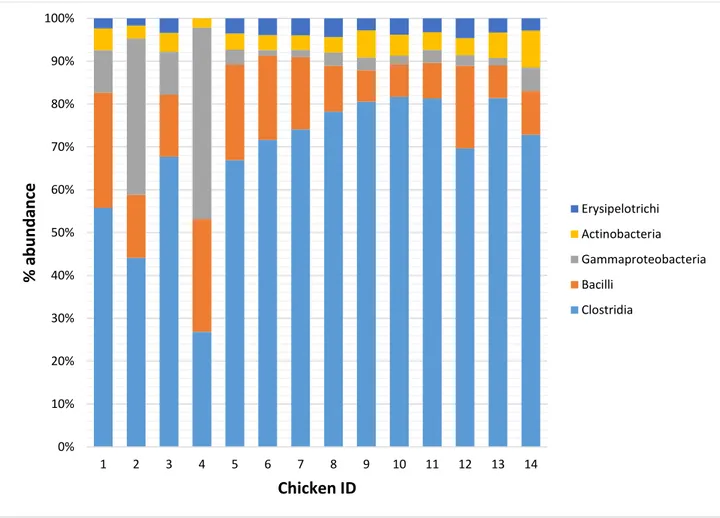
3.1.3.2 Caeca microbiota composition 41
3.1.3.3 Caeca metabolic genes composition 47
3.2 Trial on metagenomic investigation of caeca of chickens fed
with serine protease 53
2
3.2.2 Methodology 55
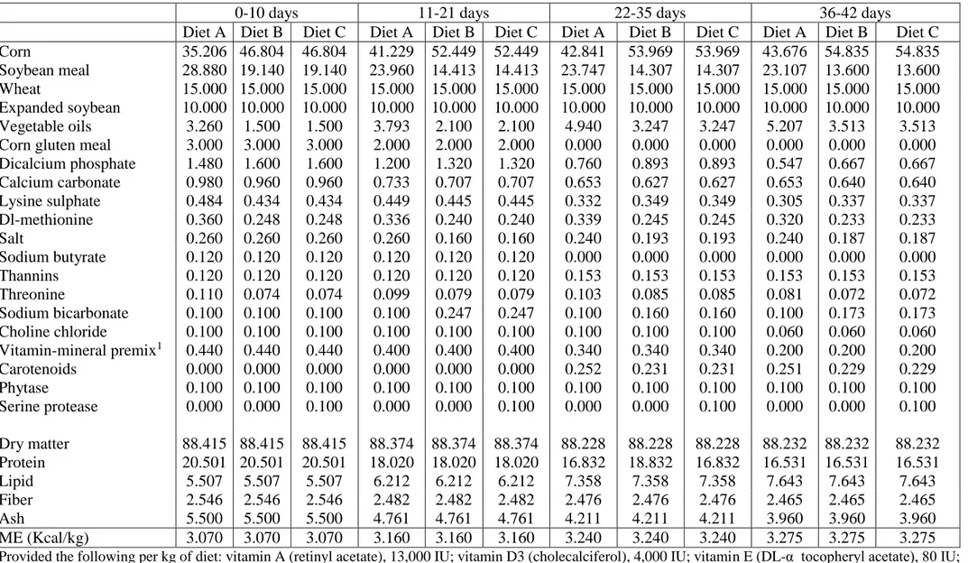
3.2.2.1 Animals and diet groups 55
3.2.2.2 Sample collection 57
3.2.2.3 DNA extraction from chicken caecum contents 57 3.2.2.4 Library preparation and metagenomic sequencing 57
3.2.2.5 Sequences analysis 58
3.2.2.6 Statistical analysis 59
3.2.3 Results 60
3.2.3.1 Sequences obtained 60
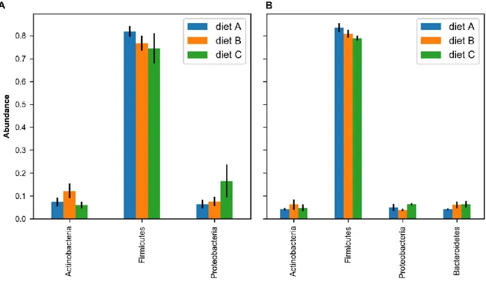
3.2.3.2 Caeca microbiota composition 62
3.2.3.3 Impact of diet, age and their interaction on microbiota
composition 69
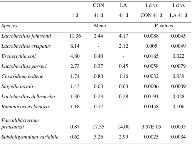
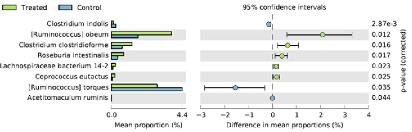
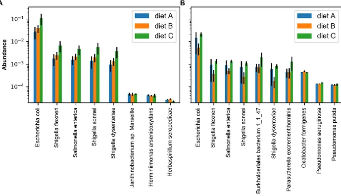
3.2.3.4 Identification of signature species 75
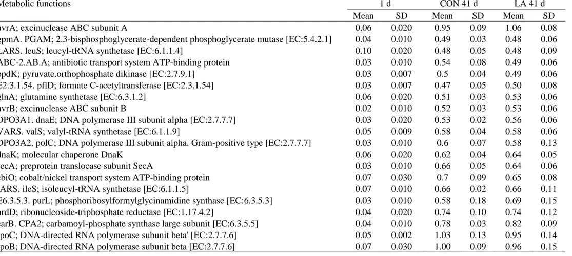
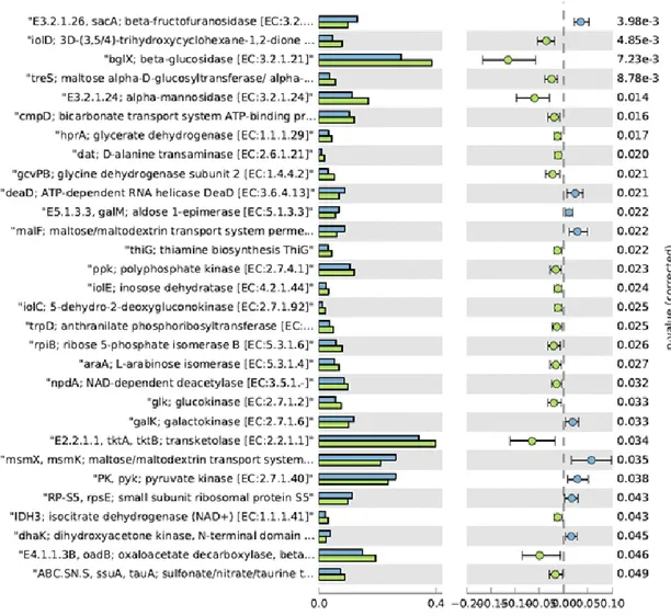
3.2.3.5 Caeca metabolic genes composition 77
3.3 Trial on metagenomic investigation of caeca of chickens fed with
phytase and corresponding carcasses 87
3.3.1 Background 87
3.3.2 Methodology 89
3.3.2.1 Animals and diet groups 89
3.3.2.2 Sample collection 91
3.3.2.3 DNA extraction from chicken caecum contents 91 3.3.2.4 DNA extraction from chicken carcass skin and
washing water 92
3.3.2.5 Library preparation and metagenomic sequencing 92
3.3.2.6 Sequences analysis 93
3.3.2.7 Statistical analysis 94
3.3.3 Results 94
3.3.3.1 Sequences obtained 94
3.3.3.2 Caeca microbiota composition 97
3.3.3.3 Caeca metabolic genes composition 106
3.3.3.4 Chicken carcass microbiota composition 108
4. DISCUSSION AND CONCLUSIONS 116
4.1 Impact of feed supplemented with Lactobacillus acidophilus D2/CSL
3
4.2 Impact of feed supplemented with serine protease on chicken
gastrointestinal tract 118
4.3 Impact of feed supplemented with phytase on chicken
gastrointestinal tract 121
4.4 Impact of feed supplemented with phytase on chicken carcass microbiota 122
4.5 Future prospectives 123
5. REFERENCES 126
6. LIST OF PUBBLICATIONS AND ATTENDANCE TO CONFERENCES 149
4
1. INTRODUCTION
1.1 The chicken gut microbiota: composition and development
Domestic chickens are the most common avian species and valuable sources of proteins for humans. The chicken gastrointestinal tract is densely populated by microorganisms, which closely and intensively interact with the host, the diet and the ingested feed. A better understanding of these interactions, impacting on animal nutrition and health, is required to further enhance poultry productions, safety of poultry products and host growth performance (Rinttilä and Apajalahti, 2013; Pan and Yu, 2014). The total DNA that can be extracted from the chicken gut is the metagenome and is the aggregate of the DNA of the host and the microbiota. Microbiota is the collective microbial community inhabiting the chicken gut. Metagenome and microbiome are often used interchangeably but the microbiome is the collective genomic content of a microbiota and indicates the total genetic capacity of the community (Tremaroli and Bäckhed, 2012).
Between all body sites the gastrointestinal tract (GIT) is the most densely colonized organ by different microbial cells representing the GIT microbiota (Scott et al., 2013; Doré and Blottière, 2015; Jandhyala et al., 2015; Yoon et al. 2015). The microbiota consists of trillions of symbiotic microbial cells harbored by each host, primarily bacteria in the gut; furthermore, the microbiome consists of the genes these cells harbor. In fact, the microbiome is defined as the combined genetic material of the microorganisms in a particular environment (Ursell et al., 2013). All animals coexist with their microbiota establishing a symbiotic equilibrium that confers them a variety of physiologic benefits. The composition of the microbiota is host specific, evolve during the animal's lifetime and is susceptible to both exogenous and endogenous modifications (Neish, 2009; Sekirov et al., 2010). Rawls et al. (2006) showed that the transplantation of microbial communities between different host species results in the transplanted community transforming to resemble the native microbiota of the recipient host. The symbiotic equilibrium between host and microbiota is established soon after the animal birth. Later on, changes in its composition are influenced by the exposure to the microorganisms present in the surrounding environment by diet (Pan and Yu, 2014; Blottière Doré, 2015; Wang et al., 2016). The loss of balance settled between the various populations that compose each microbiota may lead to serious repercussions on the host, such as the onset of a large number of metabolic, immune-mediated, allergic and inflammatory pathologies (Cogen et al., 2008; Yoon et al. 2015).
Compared with mammals, chicken and other poultry, as turkey and duck, have a shorter gastrointestinal tract causing a faster digesta transit, a relatively short retention time for ingested food and consequently selecting a deeply different intestinal microbiota compared to other food animals.
5
The average transit time through the whole chicken gastrointestinal tract is less than 3.5 h (Hughes, 2008; Pan and Yu, 2014). The chicken gastrointestinal apparatus is divided into histologically and anatomically distinct structure, named esophagus, stomach, small intestine (duodenum, jejunum and ileum) and large intestine (caecum, colon and rectum). The ingested food is initially stored in the crop, an extension of the esophagus, which inner surface is covered with non-secretory stratified squamous epithelium, then it passes to the stomach (Van de Graaff, 1986; Grist, 2004). The food begins the digestion process in the crop where it is fermented by bacteria, especially Lactobacillus genus, since crop is the election site for bacteria colonization. From the crop, food passes into the proventriculus and successively into the gizzard, that constitute the glandular and muscular parts of the avian stomach. From the gizzard, the digesta passes into the small intestine which is comprised of the duodenum, jejunum and ileum, where it is mixed with bile salts and proteinases, amylases and lipases secreted in enzyme secretions from the pancreas and others digestive enzyme produced by the secretory mucosa of the small intestine. The small intestine is the major site of chemical digestion and nutrient absorption due, other than presence of pancreatic and small intestine-produced enzymes, to villi and microvilli. The digesta then passes into the large intestine where two caeca branch out forming two separate blind ended pouches (Fuller and Brooker, 1974; Barnes et al., 1980; McLelland, 1989; Mead, 1997) that are filled trough retrograde peristalsis from the colon. The caeca are thought to be involved in the breakdown of plant material indigestible for the host and the absorption of water, glucose and volatile fatty acids. From the ileo-caecal junction the digesta enters the colon where there is very little absorption or digestion processes and then finally the feces pass into the cloaca and expelled mixed with uric acid (McLelland, 1989).
The short digesta retention time, during chicken digestion process, selects bacteria that can adhere to the mucosal layer and/or grow fast. The regions which have less tolerable conditions and faster passage of contents have lower numbers of bacteria. However, abundance and diversity of microbiota are different along the gastrointestinal tract and the ceca. When young chicks are delivered from the hatchery to a chicken house (typically at the age of 1–2 days), their initial gastrointestinal microbiota is very simple, containing a very small number of bacteria belonging to a few species but, after housing, chicks are exposed to several sources of bacteria that can enter in the immature gut. Since the chicks in this stage of life have little colonization resistance, the bacteria coming from litter materials, feed, water, and ambient air readily colonize their gastrointestinal tract. As young chicks grow, their gastrointestinal microbiota undergoes through a series of changes becoming increasingly diverse and complex (Wang et al.,2016; Wei et al., 2013b).
6
Several studies have been conducted trying to characterize chicken gastrointestinal microbiota and microbiome composition, beginning with early cultivation-based studies that revealed low abundances of Lactobacilli (>104/g CFUs) and Clostridia (102–104/g CFUs) in the small intestines and high abundance (1010‒1011/g microscope counts) of anaerobic bacteria in the cecum. Peptostreptococcus, Propionibacterium, Eubacterium, Bacteroides, and Clostridium were the major genera recovered from cecum by cultivation but it was possible to cultivate jusy between 20–60% of the total cecal bacteria (Pan and Yu, 2014). The technologies used for 16S rRNA gene based metataxonomic analyses make it possible to comprehensively characterize the chicken intestinal microbiota and to obtain sequence information, expanding our knowledge on bacterial diversity present in the intestinal tract, particularly the cecum, of chickens and turkeys (Wei et al., 2013b). Wei et al., (2013), identified in chicken gastrointestinal microbiota 117 established genera of bacteria, represented by the sequence collection, with most genera belonging to the phyla Firmicutes, Proteobacteria, and Bacteroidetes. Within phylum Firmicutes, genera Clostridium, Ruminococcus, Lactobacillus, Eubacterium, Fecalibacterium, Butyrivibrio, Ethanoligenens, Alkaliphillus, Butyricicoccus, Blautia, Hespellia, Roseburia, and Megamonas were represented by more than 1% of the total bacterial sequences. Within phylum Proteobacteria, genus Desulfohalobium was represented by the most sequences, and within phylum Bacteroidetes most of the sequences were classified into order Bacteroidales, and only genera Bacteroides, Prevotella, Parabacteroides, and Alistipes were each represented by >1% of the bacterial sequences. Of the minor phyla, Actinobacteria was the most predominant, but only the genus Bifidobacterium was represented by >1% of sequences within this phylum. The most predominant phyla in cecal microbiota included Firmicutes and Bacteroidetes, accounting for approximately 78 and 11% of the total cecal sequences, respectively. Firmicutes alone contained 31 genera, but only Ruminococcus, Clostridium, and Eubacterium each represented ≥5%, of the sequences classified to this phylum. Other genera that contained more than 1% of the total cecal bacterial sequences included Fecalibacterium, Blautia, Butyrivibrio, Lactobacillus, Megamonas Roseburia, Ethanoligenes, Hespellia, Veillonella, and Anaerostipes. Bacteroides was the most predominant genus in the phylum Bacteroidetes, accounting for 40% of the cecal sequences in this phylum. Other relatively predominant genera in this phylum included Prevotella and Paraprevotella, Tannerella and Riemerella. Within phylum Proteobacteria, Desulfohalobium, Escherichia/Shigella, and Neissenia were the most predominant genera.
Comparison of data obtained with different methods shows discrepancy that might reflect the bias of individual studies that could hinder a comprehensive knowledge of composition of the intestinal microbiome (Wei et al., 2013b). Furthermore, the composition of the gut microbiota is strongly
7
influenced by a large range of factors that include the microbial species acquired at birth, host genetics, immunological factors, antibiotic usage and dietary effects (Scott et al., 2013).
1.2 Interactions between populations belonging to the chicken gut microbiota
It has to be taken into account that, aside from the characterization of the composition of the microbiota is essential to understand the interactions between the different bacterial populations that compose this complex ecosystem. Other than between the microbiota and its host, there are extensive interactions among avian gut microbes. In fact, the avian gastrointestinal tract is an ideal habitat for microorganisms but it does not support unlimited microbial proliferation due to the limited availability of nutrient and niches. Therefore, different bacterial populations have different interactions, such as competition, cooperation, and antagonism. Competition for attachment sites and nutrients among bacteria is a common phenomenon in intestinal ecosystem (Soler et al., 2010). The lack of available attachment sites and niches, due to competition could represent a valid strategy to inhibit the pathogen colonization and proliferation in the host gastrointestinal tract. In fact, in order to cause infections in birds, enteric pathogens need to first attach to the intestinal mucosa and then to break through the epithelial barrier, but the commensal bacterial populations of healthy birds colonize intestinal mucosa forming a protecting layer that cover the mucosal surface. This layer of dense and complex microbial communities occupying the adhering niches can prevent the attachment and the subsequent colonization of most enteric pathogens trough the phenomenon so called “competitive exclusion” (Lan et al., 2005; Gabriel et al., 2006; Lawley and Walker, 2013). For this reason, a critical stage for pathogen colonization could be the post hatching period, when the gastrointestinal tract of chicks is still not colonized by the microbiota and consequently more susceptible to pathogens. Newly hatched chick’s gastrointestinal tract is sterile, but is immediately colonized by microorganisms present in the surrounding environment. In nature, these microorganisms would belong to the mother’s feces microbiota, but in poultry productions, the chicks are hatched in incubators, away from the hens. Since the incubators are relatively clean, there is a delay in normal colonization and succession of intestinal microbiota. The prolonged absence of a normal gut microbiota offers to enteric pathogens in the environment a greater opportunity to colonize gastrointestinal tract and to cause infection in new hatchlings, making them more susceptible to enteric infections, in particular to necrotic enteritis and to the colonization of potential human pathogens, such as Salmonella enteritidis (Lan et al., 2005; Dahiya et al., 2006; Lutful Kabir, 2009). Varmuzova et al. confirmed this theory testing whether microbiota from donor hens of different age will protect chicks against Salmonella Enteritidis infection. They inoculated groups of newly hatched chicks with cecal extracts of 35-week-old hens either on day 1 of life followed by S. Enteritidis infection on day 2 or were
8
infected with S. Enteritidis infection on day 1 followed by therapeutic administration of the cecal extract on day 2 or were inoculated on day 1 of life with a mixture of the cecal extract and S. Enteritidis. In this experiment, terminated when the chickens were 5 days old, both Salmonella culture and chicken gene expression confirmed that inoculation of microbiota from 35-week-old hens protected chickens even 24 h after while simultaneous administration or therapeutic microbiota administration failed to protect chickens against S. Enteritidis infection.
Other than the competitive exclusion, another strategy used by some bacterial populations to gain competitive advantages is to produce bacteriostatic or bactericidal substances hostile to competitors. In fact, the term competitive exclusion generally does not refer only to the mechanism of the site’s physical occupation but it includes even other mechanisms as the direct physical or chemical insult to the potential colonist and the resource competition in a physical or chemical niche (Oakley et al., 2014). For example, lactic acid and some short chain fat acids produced by various commensal bacteria have an inhibitory action against certain pathogens. Furthermore, a great number of Gram positive and Gram negative bacteria produces, during their growth, substances of protein structure (either proteins or polypeptides) possessing antimicrobial activities, called bacteriocins. Lactic acid bacteria can inhibit the pathogens growth using both mechanisms. In fact, lactic acid bacteria fermenting the carbohydrates present in chickens’ feed produce lactic acid, which, lowering the pH in the surrounding environment, inhibits the growth of certain pathogens such as Escherichia coli, Salmonella Typhimurium and Clostridium perfringens through the production of bacteriocins as a natural barrier against pathogens.
Other than lactic acid that was proven to be effective against Escherichia coli, Salmonella Typhimurium and Clostridium perfringens, in an in vitro study conducted on chicken, Van der Wielen et al. (2000) showed that in ceca there is a negative correlation between some short chain fat acids concentrations (acetate, propionate, and butyrate) and Enterobacteriaceae abundance. This could happen because short chain fat acids in un-dissociated form, other than lowering extracellular pH, can diffuse across the bacterial cell membrane. Once into the cell their dissociation causes a lowering of the intracellular pH causing the inhibition of some essential enzymes or metabolism (Van der Wielen et al., 2000; Van Immerseel et al., 2004; Van Immerseel et al., 2006).
The resource competition in a physical or chemical niche mostly refers to the nutrient competition. A good example is the competition for zinc among microbiota’s microbes, since zinc is an essential element involved in several cellular functions, such as enzymatic reactions and gene expression. Under low-zinc conditions some pathogen bacteria as Campylobacter jejuni, Salmonella Typhimurium and Escherichia coli use the high affinity ZnuABC transporter mechanism to bring zinc
9
into cell (Patzer and Hantke, 2000; Campoy et al., 2002; Davis et al., 2009; Gielda and DiRita, 2012). Gielda and DiRita 2012, showed that both a wild-type C. jejuni strain and a znuABC- mutant strain of C. jejuni were able to colonize limited-mirobiota chicks at similar efficiencies, but only the wild-type C. jejuni strain was able to colonize conventional chicks. However, since the zinc level in cecal content was significantly lower in the conventional chicks than in the limited-microbiota chicks, they suggested that under low zinc conditions, C. jejuni lacking the high-affinity zinc uptake system was outcompeted by other bacteria present in the GI tract.
Another important interaction between microbes in the horizontal gene transfer that is ‘the non-genealogical transmission of genetic material from one organism to another (Goldenfeld and Woese, 2007). The commensal bacteria present in the gastrointestinal microbiota usually possess some characteristics which allow them to survive in the gastrointestinal tract and more importantly, to outcompete other adverse bacteria and the pathogens. However, the horizontal gene transfer make it possible for the pathogens to acquire these characteristic traits and became more competitive, and for commensal bacteria to acquire virulence factors from pathogens becoming pathogenic for chickens. Finally, poultry enteric pathogens can directly exchange the virulence traits increasing their pathogenicity (Johnson et al., 2010; Van Reenen and Dicks, 2011). The inappropriate or prolonged use of antibiotics can lead to the horizontal transfer of resistance genes and may contribute to spread of antimicrobial resistance among adverse and pathogenic bacteria. In particular, the litter, if used for multiple growth cycles, can represent the main source of antibiotic resistant bacteria in poultry allowing their recycle between litter and gastrointestinal tract of animals (Dhanarani et al., 2009).
1.3 Microbiota’s role in host physiology
In the past decades, most of the research on the impact of bacteria in the intestinal environment has focused on gastrointestinal pathogens and the way they cause disease, while there has recently been a considerable shift towards the study of the effect that commensal microbes exert on the host gut. In fact, the intestinal microbiota is an extremely dense and complex ecosystem, which plays a relevant role in the maintenance of the animal's well-being through the production of biologically relevant metabolites and the prevention of pathogenic microorganisms’ colonization, acting as an intestinal barrier. The microbiota is intimately involved in numerous aspects of normal host physiology. It can influence the usage of nutrients by the host and host’s stress and immune response. Furthermore, it contributes to the optimal development of its intestinal mucosa and immune system (Scott et al., 2013; Doré and Blottière, 2015; Jandhyala et al., 2015; Yoon et al. 2015). The mechanisms through which microbiota exerts its beneficial or detrimental influences remain largely undefined, but it includes
10
elaboration of signaling molecules and recognition of bacterial epitopes by both intestinal epithelial and mucosal immune cells (Sekirov et al., 2010).
1.3.1 Immunostimulation, immunomediation and mucosal development
The gastrointestinal microbiota contributes to gut immunomodulation in tandem with both the innate and adaptive immune systems and maintain gut homeostasis by protecting the host from infections stimulating the gut enteric system and keeping it always active inducing a base level of inflammation. The cells of the host immune system that cooperate with gut microbiota in the immunomodulatory process are the gut associated lymphoid tissues (GALT), effector and regulatory T cells, IgA producing B (plasma) cells, Group 3 innate lymphoid cells, and, resident macrophages and dendritic cells in the lamina propria (Cebra, 1999; Chung et al., 2012; Jandhyala et al., 2015). The implication of gut microbiota in shaping a normal GALT is implied by the reduced development of the Peyer’s patches and isolated lymphoid follicles that are marked by the abundance of IgE+ B cells instead of the normal IgA+ B cells, documented by Durkin et al. (1981), in germ free mice compared to conventionally raised mice. Gut microbiota is also associated to the normal development and function of Foxp3+, a protein regulator of regulatory T cells (Tregs) even if the mechanism by which this is mediated is still not clear. Furthermore, short chain fat acids (CFAs), especially butyrate, has also been implicated in the development and function of Tregs. In fact, they activate G-protein coupled receptors expressed by the IECs and regulate Treg by epigenetic regulation of the Foxp3 (Smith et al., 2013; Arpaia et al., 2013). Other roles played by the microbiota during the immunostimulation process regards the My-D88 signaling, the differentiation of innate lymphoid cells and the support of IL1β in response to pathogen. The production of IgA is induced by DCs. MyD88 signaling is the mediator of this function and its signaling process can be activated by the gut microbiota. Furthermore, the microbiota can stimulate directly DCs in the Peyer’s patches to secrete TGF-β, CXCL13, and B-cell activating protein leading to IgA production and class switching (Suzuki et al., 2010).
The microbiota’s composition can also affect the diverse differentiation in the innate lymphoid cells or in T helper Th17 cells of a common lymphoid precursor. Commensal flora induces MyD-88 dependent mechanisms, which are essential during the rapid production of the mature IL1β, from the pro-IL1β, in response to pathogen invasion (Spits and Cupedo, 2012). Other than an involvement in the immunostimolation, the gut microbiota plays a relevant role in maintaining the structure and function of the gastrointestinal tract. This theory is supported by the observation that germ free mice shows a lower intestinal surface area, a significant reduction of villus capillary network and a decreased nutrient digestion and absorption (Gordon and Bruckner-Kardoss, 1981). In fact, the gut
11
microbiota induces the transcription factor angiogenin-3, implied in the development of intestinal microvasculature. It can prevent cytokine induced apoptosis of the intestinal epithelial cells through the production by Lactobacillus rhamnosus GG strain of two soluble proteins (p40 and p75) and can increase the levels of endocannabinoids, trough Akkermansia muciniphilia strain action, that control gut barrier functions by decreasing metabolic endotoxemia (Jandhyala et al., 2015). Furthermore, the microbial cell wall peptidoglycan stimulates signaling path of TLR2, a mechanism that is necessary for the maintenance of the tight junctions (Cario et al., 2007).
Other than stimulating the immuneresponse, some bacteria populations produce butyrate, a short chain fatty acid, that it is the preferred energy source for the enterocytes in the lower intestinal tract and is known to regulate cellular differentiation and proliferation within the intestinal mucosa, thereby increasing intestinal tissue weight. So, it should be pointed out that, the contribution of butyrate, other than feeding the enterocytes, since the epithelium act as a highly selective barrier preventing the passage of toxic and proinflammatory molecules into the submucosa and systemic circulation, is indirectly essential in the maintenance of normal intestinal barrier functions (Waite and Taylor, 2014).
1.3. 2 Synthesis of dietary compounds
Microbe-host interactions are mutualistic. In fact, commensal intestinal bacteria help during the digestion and synthesis of dietary compounds, some of which could not be otherwise available, and energy metabolism. In return the host provides to the microbes a secure growth conditions and a constant stream of nutrients (Waite and Taylor, 2014). However, the amounts and types of compound produced trough bacterial fermentation depend on relative amounts of each substrate available and fermentation strategy of bacteria involved in the fermentation process (Waite and Taylor, 2014). Some of the principal end products of intestinal microbial fermentation are short-chain fatty acids, vitamins and protein degradation products.
Vitamins are critically involved in regular energy metabolism and enzymatic functions important for gene expression. Deficiency of one or more water-soluble vitamins can contributes to various diseases and dysfunctions. An adequate supply of vitamins obtained by dietary intake seems necessary to ensure sufficient vitamin status, but microbiota can also act as an important supplier of vitamins. (Biesalski, 2016). Some bifidobacterial species are claimed to convert a number of dietary compounds into health-promoting bioactive molecules, such as conjugated linoleic acid and certain vitamins. Such findings have been confirmed by in vivo studies: administration of high-producing folate strains was shown to cause an increased faecal level of folate in rats (Pompei et al., 2007a, Pompei et al., 2007b). Folate biosynthetic properties of bifidobacteria, though folate de-novo
12
biosynthesis, appear to be restricted only to certain species/strains, while other species are capable of folate biosynthesis just in the presence of para-aminobenzoic acid (pABA) (LeBlanc et al., 2013). In fact, Bifidobacterium adolescentis ATCC15703 and Bifidobacterium dentium Bd1 are the only strains in which genome possesses the genetical determinants for entire de novo-pathway for pABA biosynthesis. No complete pathways for the biosynthesis of biotin, panthothenate, pyridoxine, cobalamin and menaquinone are present in any of the so far sequenced bifidobacterial genomes. Lactobacilli do not appear to harbour the genetic determinants for de novo pABA synthesis, except for Lactobacillus plantarum WCFS1, suggesting that the vast majority of Lactobacilli are unable to synthesize folate in the absence of pABA (LeBlanc et al., 2013; Ventura et a., 2009).
Regarding instead riboflavin, the enzymes needed for the biosynthesis of this vitamin seem to be partially or completely absent from most of the currently available bifidobacterial genomes (Ventura et al., 2009). Cobalamin is the only vitamin that is exclusively produced by microorganisms, particularly by anaerobes, in fact the commercial vitamin B12 is bacterial produced. Lactobacillus reuteri CRL1098 is able to produce a cobalamin-like compound with an absorption spectrum closely resembling that of standard cobalamin, but with a different elution time. The asset of 30 genes, involved in the B12 vitamin biosynthesis of Lactobacillus reuteri CRL1098 is similar to those found in Salmonella enterica and Listeria innocua genome, with the exception of hem genes location on their genome that appear to be different. Propionibacteria and L. reuteri are normally present in the intestine and may thus (partially) fulfil the vitamin B12 requirement of the host (Santos et al., 2007; LeBlanc et al., 2013). Gut microbiome of poultry may also serve as a vitamin (especially B vitamins) supplier to its host. Similar as bacterial protein, most of the vitamins synthesized by gut bacteria are excreted with feces because they cannot be absorbed in the cecum. However, coprophagic birds may benefit from bacterial vitamin synthesis. This is evidenced by a greater vitamin requirement by chickens housed in wire cages, where coprophagy is prevented, than by chickens raised on hard floors (Pan and Yu, 2014).
Aside from vitamins, short-chain fatty acids (acetate, butyrate, propionate, succinate, and lactate) represent relevant end products of intestinal microbial carbohydrate fermentation that benefits the host. Short-chain fatty acids are mainly used by the host as source of energy but they can bring even other benefits as reducing pH of the intestinal environment in chicken cecum, potentially inhibiting acid-sensitive pathogenic bacteria, such as members of the family Enterobacteriaceae, by dissipating the proton motive force across the bacterial cell membrane (van Der Wielen et al., 2000). Butyrate is used from the host enterocytes as energy source. Furthermore, it contributes to the regulation of cellular differentiation and proliferation within the intestinal mucosa, consequently increasing
13
intestinal tissue weight. The contribution of butyrate and other SCFA to epithelial development is essential in the maintenance of normal intestinal barrier functions and, therefore, indirectly in the protection of the host from the pathogens (Rinttilä and Apajalahti, 2013). Although butyrate production is distributed across many Clostridial clusters like the IV and the XIV, belonging to phylum Firmicutes, it is mainly produced by members of Roseburia spp. and Eubacterium rectale, both members of the family Lachnospiraceae, and especially from Faecalibacterium prausnitzii of family Ruminococcaceae (Rinttilä and Apajalahti, 2013; Miquel et al., 2014).
The production of short chain fatty acids by bacteria’s fermentation can be observed in most part of the avian gut, but primarily takes place in the cecum, which is the most densely populated and diversified ecosystem in the gastrointestinal tract. However, fermentation increases as young birds grow. In fact, cecal acetate, propionate and butyrate are almost undetectable in 1-d-old broilers. As the cecal microbiota becomes established, these short chain fat acids reach high concentrations in 15-d-old broilers and remain stable afterwards. Chicken microbiome produce greater concentrations of short chain fatty acids than human microbiome (Rehman et al., 2007; Pan and Yu, 2014).
Lactic acid is another compound produced by gastrointestinal bacteria and particularly from lactic acids bacteria. This compound would tend to reduce residual pH more than other short chain fatty acids but it is normally absorbed from the intestine or used as a substrate for lactate-utilizing bacteria, such Eubacterium, Anaerostipes, Veillonella, and Megasphaera genera, quickly enough to not being able to pathologically acidify the gut environment (Harmsen et al., 2002; Belengueret al., 2007; Rinttilä and Apajalahti, 2013).
Another noteworthy microbiota’s metabolism is the protein and amino acid fermentation in the lower intestine. The real role of this metabolism lies, more than in the energy or compound production, in the fermentation, by putrefactive bacteria, of potential systemic toxins and carcinogens resulted from the protein catabolism. In fact, in broiler chicken cecum once carbohydrate sources are exhausted, sources of protein material are fermented and metabolized to salvage energy but at that gastrointestinal level, proteins and amino acids provide a less significant energy source. Common examples of undesired metabolic end products include phenols and indoles (as a result of anaerobic fermentation of the aromatic amino acids, tyrosine and tryptophan, by intestinal bacteria), ammonia (as a result of oxidative or reductive deamination of amino acids), and amines (as a result of amino acid decarboxylation in the gut). These compounds other than be toxic could result in an increase the pH of intestinal contents, but a low pH is beneficial for the suppression of the growth of the acid-sensitive pathogenic microorganisms. As the bacteria generally favor fermentable sources of carbohydrates, protective measures against excessive putrefactive activity and the undesired
14
metabolites in the hindgut can be achieved by adding dietary fiber in the diet or limiting the intake of poorly digested proteins (Apajalahti, 2005; Rinttilä and Apajalahti, 2013).
Gut bacteria also contribute to host nitrogen metabolism. Since in birds the intestinal and urogenital tracts meet in the cloaca where urine mixes with feces, some urine, may travel to the ceca due to the retrograde peristalsis in the rectum. Cecal bacteria can then catabolize uric acid to ammonia, which can be absorbed by the host and used to synthesize a few amino acids such as glutamine (Pan and Yu, 2014). Some of the dietary nitrogen is incorporated into bacterial cellular proteins. Therefore, gut bacteria themselves can be a source of amino acids. However, the majority of these bacterial proteins are lost by the host with the excretion of feces, because most of the intestinal bacteria in birds reside in the cecum which does not have the ability to digest and absorb proteins. Utilization of bacterial proteins is possible when chickens are housed on hard floors, where coprophagy (ingestion of feces) can occur and bacterial proteins can be digested and absorbed in proximal gut(Pan and Yu, 2014).
1.4 Factors affecting and modulating the chicken GI microbiota
The intestinal track of poultry harbors a complex and dynamic microbiota that has a symbiotic relationship with its host. The interaction between host and microbiota affects the physiological, immunological, and nutritional status of the host and consequently his growth performance and health status. The evidence for metabolic interactions is particularly strong, as many data support the conclusion that gut microbiota influences the energy harvest from dietary components, particularly complex carbohydrates, and that metabolites, such as the short-chain fatty acids produced by gut bacteria, can perturb metabolic traits. The gut microbiota communities are assembled each generation, since their composition is influenced by environmental factors, age and diet (Wei et al., 2013b; Org et al., 2015). In particular, the symbiotic equilibrium between host and microbiota is established soon after hatching and later on bacteria present in microbiota can be affected by a range of factors, such as host genetics and age, litter management, diet, and feed additives. Since the gastrointestinal microbiota is strongly related to several host functions, numerous efforts have been attempted on manipulation and control of the exogenous factors, especially dietary intervention and litter management, in order to modulate its composition to enhance feed conversion and gut health (Wei et al., 2013b; Pan and Yu, 2014; Blottière Doré, 2015; Wang et al., 2016).
1.4.1 Environment, age and diet
For commercial chickens, the environment is represented by the litter. Litter can have a significant effect on the initial composition and structure of the microbiota gastrointestinal tract of chickens, while later on the main role in the microbiota shaping process is played by the diet. In fact, when young chicks are delivered from the hatchery their gastrointestinal tract contains very small number
15
of bacteria belonging to a few species, and just when they begin pecking at and consuming litter materials they inoculate their young gastrointestinal tract from the litter (Wang et al., 2016). If hatching takes place in an environment where the microbial load has been minimized, individual chicks may pick up a random inoculum from their surroundings, which may lead to differences in the intestinal physiology of individual birds in a flock. It is worth noting that intentional inoculation of the chicks with a competitive exclusion culture at hatch might render a flock microbiota composition more uniform (Rinttilä and Apajalahti, 2013). Poultry litter is a mixture of bedding materials and in the USA, where the litter is changed every 6 cycles, even of chicken excreta that contain chicken GI bacteria, undigested feed, uric acid, and other substances of host origin. Several studies have documented that poultry litter contains a complex and dynamic microbiota, composed primarily of environmental bacteria and its composition can be affected by the bedding materials used (Lu et al., 2003; Torok et al., 2009). Repeated use of poultry litter and poor litter management can result in considerable changes in microbiological conditions leading to an increase of density and diversity of microbes. In addition, reused litter can serve as a driving force that shapes the chicken GI microbiota because exposing young chicks to different bacterial inocula can profoundly affect GI microbiota development (Cressman et al., 2010). It was shown that reused litter harbors less Salmonella and Clostridium perfringens but enables Campylobacter jejuni and C. coli to survive longer compared to fresh litter (Kassem et al., 2010; Roll et al., 2011; Wei et al., 2013a). Moreover, two recent studies have shown that reused litter can affect the immune system of chickens, which suggests that litter conditions can also affect the GI microbiota of chickens indirectly through their immune system (Lee et al., 2011; Shanmugasundaram et al., 2012).
Cressman et al., (2010) examined the microbiota both in the GI tract and in the poultry litter and their interaction revealing that the litter microbiota and the GI microbiota affected each other in a reciprocal manner. In fact, fresh litter resulted in increased diversity and predominance of environmental bacteria in the GI microbiota of young chicks, while reused litter increased the bacteria of gut origin. Another study conducted by Wang et al., (2016), investigating the effect of fresh and reused litter on chicken gastrointestinal microbiota at different ages, showed that the ileal mucosa and the cecal contents were affected by both litter management regimen and age of birds. At days 10 and 35, in the cecal luminal microbiota eight and three genera, respectively, differed significantly in relative abundance between the two litter management regimens. Compared to the fresh litter, reused litter increased predominance of halotolerant/alkaliphilic bacteria and Faecalibacterium prausnitzii, a gut butyrate-producing bacterium. This study suggests that litter management regimens affect the chicken GI microbiota, which may impact the host nutritional status and intestinal health.
16
Torok et al., (2009) investigated linkages between litter material, gut microbiota and chicken growth performance. Cecal microbial populations were investigated at 14 and 28 d of age and at both ages. The caeca microbiota of chickens raised on reused litter was significantly different from that of chickens raised on any of the other litter materials, except for softwood shavings at d 28. However, age had a significant influence on ceca microbiota composition regardless of litter material. We can conclude that the environment and consequently the type of litter material and management, can influence colonization and development of cecal microbiota in chickens. Litter-induced changes in the gut microbiota may be partially responsible for some of the significant differences observed in early rates of growth; therefore, litter choice may have an important role in poultry gut health particularly in the absence of in-feed antibiotics (Torok et al., 2009).
Gong et al. (2008) examined the effect of dietary bacitracin, bird age and access to range on the richness and microbiota community structure concluding that age had the most profound effect on microbiota composition. This is demonstrated clearly since most birds of the same age were grouped together and, regardless of access to range or dietary treatment, the richness of microbiota increased as the birds grew older. Furthermore, they showed that chickens at 42 days of age had a well-developed bacterial microbiota in both ileum and caeca and at 14 days the development of caecal bacterial microbiota was close to that of 42-day-old chickens, while the ileal microbiota did not appear to be fully developed, a stage which could be more sensitive to dietary treatments and other environmental factors. It is worth pointing out that, the abundance of Lactobacilli in the caecal microbiota was greatly higher in 3-day-old chickens than in 42-day-old chickens, suggesting the importance of Lactobacilli in the early development of caecal microbiota while, on the contrary, the abundance of Bifidobacteria population in the ileum and caeca were hardly detected in 3-day-old chicks and high in 42-day-old chickens.
Regarding the immature chicken microbiota, Lu et al. (2003b) reported that 3-day-old broiler chickens had a similar community structure of bacterial microbiota in ileum and caeca and the caecal microbiota was a subset of the ileal microbiota during the first 14 days of age based on the metataxonomic analysis of random clone libraries of partial 16S rRNA genes. On the contrary, Gong et al., 2008 in their study, reported that PCR–DGGE profiles of bacterial microbiota from the ileum and caeca of 3-day-old chicks were significantly different, suggesting two different bacterial communities in these two intestinal regions at that age.
The correlation between the advancing of age and the increase of microbiota composition richness were reported even by other authors, like Van der Wielen et al. (2002), Knarreborg et al. (2002), Van Wielen et al. (2002) and Hume et al. (2003). Van der Wielen et al., 2002 found that the PCR–DGGE
17
profiles of the microbiota were similar in crops, duodenum and ileum in 4-day-old chicks, but observed an increased diversity of bacterial microbiota in crops, duodenum, distal ileum and caeca when broiler chickens aged. All these studies highlight that, regardless the influence of other factors, microbiota changes and matures on time during the chicken’s life.
Another factor that can influence the microbiota composition is host’s genotype. The role of host genetics in shaping microbial communities’ composition is not clear but it could be speculated that the host may affect its microbiota composition either directly, through secretions into the gut, control of gut motility and modification of epithelial cell surfaces, or indirectly, through food and lifestyle preferences (Zhao et al., 2013). Zhao et al. (2013) conducted an experiment using next generation sequencing technology to investigate the effect of genetic on the gut microbiota’s population structure in two different lines (56-day high or low body weight) of adult chickens. The pattern of host genetic influence was different in adult males and females, demonstrating gender as a factor that impacts the composition of gut microbiota. Of 190 species, 68 were affected by genotype (line), gender and by genotype and gender interactions, where 15 of the 68-species belonged to Lactobacillus. In fact, of host-microbe interactions, Lactobacillus was the major influenced genus showing different abundances between low body weight males and high body weight females. Beside environment, age and genotype the chicken gut microbiota composition is mainly shaped by diet and feed additives.
18
Figure 1. Interactions among gut microbiome, avian host, diet, and litter microbiome (Pan and Yu,
2014)
1.4.2 Diet and feed additives
Diet is the most relevant factor impacting on intestinal microbiome in poultry as dietary components that escape host digestion and absorption serve as substrates for the growth of intestinal bacteria (Pan and Yu, 2014). In fact, several studies already demonstrated its potential to impact the chicken GI microbiota with respect to diversity, composition, and structure. It was also showed that the same action can be performed by feed additives (Amerah et al., 2011; Danzeisen et al., 2011; Rodriguez et al., 2012; Wang et al., 2016).The first studies focused on how feed and feed additives affect the prevalence of enteric pathogens, such as Salmonella (Santos et al., 2008), Clostridium perfringens (Si et al., 2009; Wei et al., 2013a), and Campylobacter jejuni (Ridley et al., 2011), while the prevalence of these pathogens could be decreased by the effect of a healthy GI microbiota. In fact, microbiota can perform colonization resistance and competitive exclusion to inhibit the pathogen growth (Wagner, 2006; Kerr et al., 2013). Furthermore, other than creating this positive barrier, the commensal bacteria can positively affect the efficiency of feed utilization by the chicken host. That’s why, now the interest of the researchers shifted towards the understanding of how diet and feed
19
additives, could modulate the GI microbiota of chickens instead of just focusing on their effect on pathogens bacteria (Gong et al., 2008; Santos et al., 2008; Danzeisen et al., 2011).
One of the most remarkable example of how diet can modulate the microbiota, is represented by the use of diets containing high levels of indigestible, water-soluble, non-starch polysaccharides as wheat-, barley-, or rye-based diets, that favor the proliferation of Clostridium perfringens and predispose young chicks to necrotic enteritis, while diets poor in non-starch polysaccharides, such as corn-based diets, do not. In fact, this proliferation can be due to increase of digesta viscosity, decrease of digesta passage rate and a decline in nutrient digestibility caused by high level of non-starch polysaccharides, supporting the growth of Clostidium perfringens. When compared with corn-based diet, wheat-based diets also affect a number of other bacteria (Annett et al., 2002; Pan and Yu, 2014).
In a study conducted in 2010, Hammons et al. showed that even a small variation in dietary cereal grain composition can potentially affect the intestinal bacteria at species and strain levels. In fact, they showed that a standard corn-soybean ration supported Lactobacillus agilis type R5, whereas a ration high in wheat favored L. agilis type R1. In order to see the effect of grain base on the microbial community profile Apajalahti et al., (2004), analyzed 256 caecal samples of broilers being fed either wheat or corn based diet from all around the world. The % G+C profiling method used to reveal the most significant sources of variation showed that the two grain bases favored different bacterial groups in the caecum. This analysis did not reveal the identity of the bacteria, but it was possible to establish that corn favors low G+C Clostridia, Enterococci and/or Lactobacilli and wheat improves higher %G+C Bifidobacteria.
Another dietary nutrient category that can affect gut microbiota can be the protein. In fact, the source and level of dietary protein have been demonstrated to stimulate the proliferation of different bacteria populations. Sun et al. (2013) noticed that the use of fermented cottonseed meal as protein source, instead of soybean meal which is widely used as a source of protein in poultry production, increases the population of Lactobacilli and decreases the number of coliforms in cecum of broiler chickens. Furthermore, other than water-soluble and non-starch polysaccharides, even diets containing high percentages of animal protein support the growth of Clostridium perfringens in the chicken gut and are considered as one of the predisposing factors of necrotic enteritis. In addition, it has been reported that C. perfringens proliferation can be improved even by dietary fat source, as it was more abundant in the ileum of broiler chickens fed diet with animal fat than chickens fed diet with soy oil (Pan and Yu, 2014).
However, not only the nutrients inside the diet affect the microbiota composition, even processing significantly affects the characteristics of the feed as a substrate for the bacterial community. If the
20
bacterial shifts following different set of feed processing conditions were understood, the manufacturing process itself could be used to partly control and manage the gastrointestinal microflora trough the identification of signature species. Apajalahti et al. (2004) investigated the changes caused by inclusion of whole wheat in the feed on the bacterial community structure in chicken caecum, giving an overview of the effect of processing procedure on microbial community compositions. They showed that grinding and processing of wheat affect wheat characteristics and act as a microbial modulator. In particular, they investigated the changes caused by of feed amendment with the addition of whole wheat, compared to the commercial feed with no amendments, on microbiota using the G+C% profiling. Effect of whole-wheat addition on microbial community structure was statistically significant and in particular bacteria with %G+C ranging from 35 to 54 were stimulated by whole wheat, while those with %G+C between 60 and 69 were suppressed (e.g., Bifidobacteria).
Another way to influence gut microbiota in poultry, reducing enteric pathogens and increasing growth performances, is using feed additives. In the past growth-promoting antibiotic (AGP), as feed additives, were added in the feed to gain effects on gastrointestinal microflora and performance. These effects were obtained because growth-promoting antibiotic reduced competition for nutrients in the small intestine, reduced local inflammation due to control of pathogens, and reduced intestinal thickening and length, as a result of improved digestibility and reduced pathogen loading (Thomke and Elwinger, 1998b; Thomke and Elwinger, 1998a). The latter two mechanisms result in a more efficient digestion and reduced maintenance energy requirement (Apajalahti et al., 1999; Apajalahti et al., 2004). However, it was hypothesized by Niewold (2007) that a different mechanism is behind the AGP positive effect on animals’ growth performances. According to the author, the different microbial compositions when using AGP are a consequence of an altered immune status of the host rather than of a direct effect on the microbiota. The changes in microflora are most likely the consequence of an altered condition of the intestinal wall due to the anti-inflammatory effect on intestine cells. Growth promoter’s antibiotic have long been supplemented to poultry feed to stabilize the intestinal microbial flora, improving the general performances and prevent some specific intestinal pathology. However, due to the growing concern about microbes resistant to antibiotics used to treat human and animal infections, the European Commission (EC) decided to phase out, and ultimately ban (1 January 2006), the marketing and use of antibiotics as growth promoters in feed
(EC Regulation No. 1831/2003;
21
Since the ban of AGP as feed additives for livestock production, the research of new molecules able to selectively promote the growth of microbial populations related with positive effect on animals’ growth performances and health has always be continuous (Butaye et al., 2003; Huyghebaert et al., 2011). Whatever the mechanism of action of AGPs, the main characteristic of a good alternative, from a practical point of view, should be that it must positively modulate microbiota composition improving good microbial populations and reducing pathogens. Good alternatives to AGPs to influence the intestinal microbiota population is using probiotic and feed enzymes.
Modulation of the intestinal bacteria by feeding probiotics is currently under active research (Garriga et al., 1998; Jin et al., 1998; Gusils et al., 1999; Samli et al., 2007; Gérard et al., 2008; Nakphaichit et al., 2011; Babot et al., 2014; Pedroso et al., 2016; Hu et al., 2017). The target of such nutraceutical products is to improve gastrointestinal health by selecting for beneficial microflora and suppressing known intestinal and food-borne pathogens. Direct-fed microbials (probiotics) are products which are targeted to improve the health of the gastrointestinal tract, but these are likely to be effective only if the requirements for their growth are fulfilled. Many of these benefits apply also to the use of feed enzymes. Dietary enzymes, such as xylanase and β-glucanase, has already been showed to increase intestinal lactic acid bacteria and decrease the population of adverse and pathogenic bacteria, such as E. coli (Rodríguez et al., 2012). Dietary supplementation with xylanase and β-glucanase can also offer chickens some protection against necrotic enteritis as the enzymes breakdown the non-starch polysaccharides in the diet and reduce the digesta viscosity. Furthermore, feed enzymes have the ability to remove fermentable substrate from the small intestine, that could be an optimal substratum for some pathogens growth requirements (Apajalahti et al.,2004;McDevitt et al., 2006; Owens et al., 2008).
The role of feed enzymes in improving the productive value of diets for monogastric animals has received extensive reviews, and several modes of action have been proposed. They include hydrolysis of specific chemical bonds in feedstuffs that are not sufficiently degraded or indeed not at all by the animal’s own enzymes (for example, mixed salts of phytic acid); the elimination of the nutrient-encapsulating effect of the cell wall polysaccharides and therefore increased availability of starches, amino acids and minerals; the breakdown of anti-nutritional factors that are present in many feed ingredients (for example, soluble NSP and phytic acid) and the complementation of the enzymes (for example, amylase, protease, lipase)produced by young animals where, because of the immaturity of their own digestive system, endogenous enzyme production may be inadequate. For example, the indigestibility of some protein contents could limit the inclusion of these nutrients into pig feed. However, the supplementation of proteases might allow high inclusion of such feedstuffs. However,
22
it must be taken into consideration that the supplementation of any feed enzyme does not just impact directly animal nutrition but, since the gastrointestinal tract is densely populated, it will impact also the microbiota. Since microbiota plays a critical role for animal nutrition, performance and safety of animal products, there is a clear need to understand the role of feed enzymes in influencing gut health through its modulation (Kiarie and Nyachoti, 2009; Kiarie et al., 2013). The feed additives whose modulating action on chicken gut microbiota will be analysed in this study are a probiotic (i.e., Lactobacillus acidophilus) and two feed enzymes, a commercial protease and a commercial phytase, alone and combined with inositol.
1.5 Analysis of chicken gut microbiota: from traditional techniques to metagenomic analysis
One of the most remarkable events in the field of microbial ecology in the past decade has been the advent and development of metagenomics. Metagenomics is the study of the metagenome (microbiome). Metagenomics can either be targeted (usually 16S ribosomal RNA) or untargeted (shotgun sequencing) (Tremaroli and Bäckhed, 2012). Metagenomics provides both access to the functional gene and the composition of microbial communities, within an environmental sample (Thomas et al., 2012). Metagenomic analysis has recently developed. Previously microbial communities were characterized culturing them on selective growth media and subsequently carrying out a range of biochemical tests to identify the bacteria that survived under the specific culture conditions employed. Such methods, other than being laborious and time consuming, were not suitable for extensive monitoring of the unknown microflora, because only a small fraction of bacteria composing the community could be found. In fact, up to 99% of the bacteria in many environments fail to grow under artificial conditions. This disadvantage is due to the growth requirements of most bacteria that are still unknown or cannot be mimicked under laboratory conditions, leading to an incomplete data recovery regarding the whole community. Since microbial communities have individual bacterial members specialized on different functions and providing elements to other bacterial members, conducting a metagenomic analysis investigating these communities’ dynamic and interactions with the omission of unculturable populations would have been impossible (Apajalahti et al., 1999; Apajalahti et al., 2001; Apajalahti et al., 2004).
That is why, in the late 1970s, Woese and Fox proposed the use of ribosomal RNA genes as molecular markers, revolutionizing the classification of microorganisms. Some decades later, advances in molecular techniques were applied to microbial diversity description and granted access to a “new uncultured world” of microbial communities. DNA-based culture-independent methods’ basic principle was the analysis of the bacterial DNA without harvesting it from in vitro isolated pure cultures. Total bacterial DNA is directly recovered from a sample derived from the site of interest,
23
extracted and then analysed. Some of these techniques were the polymerase chain reaction (PCR), fluorescent in situ hybridization (FISH), denaturing gradient gel electrophoresis (DGGE and TGGE), restriction-fragment length polymorphism, and terminal restriction-fragment length polymorphism (T-RFLP) (Woese and Fox, 1977; Apajalahti et al., 2001; Apajalahti et al., 1999; Apajalahti et al., 2004; Hiergeist et al., 2015; Escobar-Zepeda, 2015).
The target for many of the molecular profiling techniques is the 16S ribosome and its encoding gene. In bacteria, the three rRNA molecules are genetically organized in a ribosome operon and primarily transcribed as a single 30S rRNA precursor that is subsequently cleaved by RNase III into 16S, 23S, and 5S rRNA subunits. Operon size, sequences, and secondary structures of these three rRNA genes are conserved within a bacterial species. The 16S rRNA gene contains both variable regions and conserved regions, allowing the design of PCR primers which target all or specific bacterial DNA. The 16S rRNA gene is constituted of nine variable regions (V1-V9), where the V1 region was found to be the most variable, followed by V9 and then by V3 (Yu and Morrison, 2015; Hedgiest., 2015). For more than 30 years, culture-independent microbial profiling has been based on the 16S ribosomal rRNA gene (Olsen et al. 1986). By doing this, researchers received a key tool to species and phylogenetic trees identification by comparing these relatively stable parts of the genome. However, these 16S ribosomal rRNA gene based technologies, other than being low-throughput technologies, could not deliver exhaustive insight into microbial diversity and metabolic and ecological functions, making impossible to deduce the potential biological tasks carried out by a community as a whole (Woese and Fox, 1977; Escobar-Zepeda, 2015; Hiergeist., 2015). In fact, metagenomics provides access to the functional gene composition, e.g. metagenome, of microbial communities and therefore gives genetic information on potentially novel biocatalysts or enzymes, genomic linkages between function and phylogeny for uncultured organisms, and evolutionary profiles of community function and structure, a much broader description than phylogenetic surveys based on 16S rRNA gene (Thomas et al., 2012).
In 1990, for the first time, clone libraries of 16S rRNA genes from environmental bacteria were directly amplified and sequenced by the Sanger method (Giovannoni et al., 1990). This procedure represented a breakthrough that permanently changed the way prokaryotes in the environment were analyzed, leading to the advent of the metagenomic analysis era trough sequencing techniques (Hiergeist., 2015). Sanger and others introduced the concept of DNA sequencing called the chain-terminator method. This first-generation sequencing technology is based on incorporation of fluorescently labelled deoxynucleoside triphosphate and primers into a PCR that set the stage for automated high-throughput DNA sequencing. With the information obtained from the last terminator
24
base in the four individual base reaction tubes after size separation, the original sequence could be determined (Sanger and Coulson, 1975; Sanger et al., 1977). However, Sanger sequencing method presented a high number of limitations and disadvantages, mainly associated with the low throughput of DNA sequences obtained and the high cost. That’s why others sequencing technologies were developed during the following years (Schloss, 2008; Metzker, 2010).
Particularly relevant was the development of pyrosequencing technique, also called sequencing by synthesis, that permits the detection of pyrophosphate released when a nucleotide is incorporated in the chain resulting in detectable light in a real-time format. Improvements and development of this technology resulted in advances in the next-generation devices based on the same principle, leading to so-called next generation HT-NGS platforms produced by Roche, Illumina-Solexa, Life Technologies, Helicos, and other companies.
Illumina sequencing technology is based on reversible dye-terminators principle and can perform shotgun High throughout Whole Genome sequencing. In fact, in shotgun sequencing random DNA fragments are immobilizes on a surface and then a solid-surface PCR amplification is performed with the result of clusters of identical DNA fragments. These are then sequenced with four types of reversible terminator bases in a sequencing-by-synthesis process. After the incorporation of reversibly terminating nucleotides, a camera capture images of the fluorescence and the dye along with the terminal 3′ blocker is chemically removed from the DNA allowing the next cycle. Clustered fragments can be sequenced from both ends (pair-end mode) and the cluster density is enormous, with hundreds of millions of reads per surface channel. The read length can be different in relation to the Illumina instrument and the sequencing mode chosen but, in any case, it is relatively short compared to other sequencing technologies read length. Yields of ~60 Gbp can therefore be typically expected in a single channel. In fact, this technology can sequence the equivalent of one-third of the entire human genome in a single run (approx. 10 days), while the sequencing of the entire human genome with the sanger method lasted 10 years and costs 2 billion dollars. The lower costs of this technology and recent success in application to metagenomics, are currently making the Illumina technology an increasingly popular choice. The only limitation of Illumina technology is the read length. In fact, a limited read length means that a greater proportion of unassembled reads might be, after the quality clipping of the first bad quality sequences of the reads, too short for functional annotation. However, some current software packages (e.g. MG-RAST and Mg-Mapper) are designed to analyze unassembled Illumina reads of 75 bp and longer, bypassing this limit (Thomas et al., 2012; Diaz-Sanchez et al., 2015).
25
Next-generation sequencing platforms (NGS) have allowed the substantial researches into the diversity and functions of microbiota from the guts of various livestock animals. High-throughput NGS generates large volumes sequence data containing genetic information and this allows hypothesis-driven researches on chicken GIT microbiota, thereby highlighting the roles of previously unknown and rare microbial GIT species. Furthermore, metagenomic data have raised new questions such as how microbiota stability and ecological shifts in species diversity are influenced by nutrients and hosts (Metzker, 2010; Medinger et al., 2010; Choi et al., 2015).
The application of HT-NGS sequencing is emerging and moving toward the development and the improvement of the poultry industry raising the food safety measures and avoiding foodborne pathogens. Relative few studies have been conducted on chicken gut microbiota to determine any changes that affect health and disease and a detailed assessment of probiotics or/and other feed additive to control pathogenic growth and or shaping the gut microflora, trying to lead to the development of novel alternatives to antibiotic growth promoters. Frequently, the sequencing methods used to gain data regarding the microbiota composition, aside from the platforms, are the small-subunit ribosomal RNA (16S rRNA gene) amplicons or shotgun metagenomic sequencing (Diaz-Sanchez et al., 2013).
1.5.1 16S rRNA gene based metataxonomic analysis
Amplicon sequencing is the most widely used method for characterizing the diversity of microbiota, even in chicken. For bacteria and archaea classification, the small-subunit ribosomal RNA (16S rRNA gene) locus is targeted and amplified by PCR. The obtained amplicons are sequenced and characterized to determine microbial community composition and population relative abundance (Pace et al., 1986; Hugenholtz and Pace, 1996). Comparing 16S rRNA gene based metataxonomic analyses profiles across samples clarifies how microbial diversity is associated with environmental conditions generating insight into host–microbe interactions and yields hypotheses about microbiota-based disease mechanisms (Muegge et al., 2011; Sharpton, 2014). However, amplicon sequencing presents several limitations. In fact, it may fail to resolve a substantial fraction of diversity in a community because of various biases associated with PCR and can produce widely estimates of diversity (Hong et al., 2009; Sharpton et al., 2011; Sharpton, 2014; Jumpstart Consortium Human Microbiome Project Data Generation Working Group, 2012). Furthermore, amplicon sequencing only provides insight into the taxonomic composition of the microbial community, with taxa for which taxonomically informative genetic markers are known, and sometime result in an overestimation of community diversity, since the 16S rRNA gene locus can be transferred between distantly related taxa. Another limitation is the impossibility to provide information on the biological