Alma Mater Studiorum – Università di Bologna
DOTTORATO DI RICERCA IN
BIOLOGIA CELLULARE E MOLECOLARE
Ciclo XXIX
Settore Concorsuale di afferenza: 05/D1
Settore Scientifico disciplinare: SSD BIO/09 - Fisiologia
IDENTIFICATION OF MOLECULAR AND FUNCTIONAL
MECHANISMS BEHIND CELL VOLUME REGULATION IN
ASTROCYTES
Presentata da: Francesco Formaggio
Coordinatore Dottorato
Relatore
Giovanni Capranico
Barbara Monti
1
TABLE OF CONTENTS
I: INTRODUCTION ………... 3
1 CELL VOLUME REGULATION... 4
1.1 INORGANIC AND ORGANIC OSMOLYTES... 6
1.2 MOLECULAR MECHANISMS IN CELL VOLUME REGULATION... 7
1.2.1 VOLUME SENSORS……….……..7
1.2.2 CYTOSKELETON……….………..7
1.2.3 INTEGRINS……….….…....8
1.2.4 TRANSIENT RECEPTOR POTENTIALS……….…..8
1.3 EFFECTORS IN CELL VOLUME REGULATION ………... 10
1.3.1 REGOLATORY VOLUME DECREASE (RVD)………..……….……...…. 10
1.3.2 POTASSIUM CHANNELS... 10
1.3.3 VOLUME-REGULATED CL- CHANNELS ………...… 11
1.3.4 KCl SYMPORT………... 11
1.3.5 CELL VOLUME IN PHYSIOLOGICAL PROCESSES……….12
1.3.6 CELL VOLUME IN PHYSIOPATHOLOGICAL PROCESSES………....13
2 VOLUME REGULATION IN THE CENTRAL NERVOUS SYSTEM: ROLE OF ASTROCYTES AND ION CHANNELS ………...………... 14
2.1 GLIAL CELLS: PHYSIOLOGICAL ROLE WITHIN THE CENTRAL NERVOUS SYSTEM…. 14 2.1.1 ASTROCYTES ... 15
2.1.2 MICROGLIA... .... 15
2.1.3 OLIGODENDROCYTES... 16
2.1.4 EPENDYMAL CELLS... 17
2.2. ASTROCYTES AND VOLUME HOMEOSTASIS IN THE CNS... 18
2.3 ASTROCYTES PHYSIOLOGICAL FUNCTIONS IN HOMEOSTASIS ……….. 19
2.3.1 GLUTAMATE METABOLISM... 19
2.3.2 POTASSIUM BUFFERING………..………..……….… 19
2.4 BRAIN VOLUME REGULATION: ROLE OF ASTROCYTES AND ION CHANNELS.….…... 22
2. 5 ASTROGLIAL ION CHANNELS IN VOLUME REGULATION ………. 24
2.5.1 AQUAPORIN 4………..……..24
2.5.2 TRPV4………..…………25
2.5.3 VRAC………...26
2.6 INTERACTION BETWEEN ION CHANNELS AND AQUAPORINS IN VOLUME REGULATION ……….. 28
2.6.1 AQP4 AND KIR 4.1……….…………..28
2.6.2 AQP4 AND VRAC………...…………28
2
II AIM OF THE STUDY……….… 30
III MATERIAL AND METHODS...33
3.1 CELL CULTURE... 34
3.1.1 ASTROCYTES ... 34
3.2 COS-7 ... 35
3.2.1 TRANSFECTION OF COS-7 CELLS………..….35
3.3 ANTIBODIES……….…. 35
3.4 IMMUNOBLOT ANALYSIS IN COS-7………... 36
3.5 ELECTROPHYSIOLOGY... 36
3.6 SOLUTIONS AND CHEMICALS ………...……….. 38
3.7 CALCIUM MICROFLUORIMETRY……….……..38
3.8 BRAIN SECTIONS AND IMMUNOFLUORESCENCE……….……39
3.9 FIXATION AND TISSUE PROCESSING………....40
3.10 IMMUNOGOLD HISTOCHEMISTRY………...40
3.11 STATISTICAL ANALYSIS………...….41
IV RESULTS ... 42
4.1 TRPV4 MEDIATES INTRACELLULAR CALCIUM ELEVATION UPON HYPOTONIC STIMULUS IN COS-7... 43
4.2 TRPV4 AND AQP4 CO-EXPRESSION IS CRITICAL FOR VOLUME REGULATED ANION CHANNEL (VRAC) ACTIVITY……….…..……… 46
4.3 AQP4 AND TRPV4 PROTEIN-PROTEIN INTERACTION IN CO-TRANSFECTED COS-7 CELL LINE... 49
4.4 THE D184E MUTATION DRAMATICALLY AFFECT THE MOLECULAR INTERACTION BETWEEN TRPV4 AND AQP4-M1 ISOFORM... 50
4.5 D184E MUTATION INTERFERE WITH THE MEMBRANE LOCALIZATION OF AQP4 A ND TRPV4... 52
4.6 EXPRESSION OF VRAC IN COS-7 CELL LINE ………...54
4.7 VRAC IS EXPRESSED IN RAT CULTURED CORTICAL ASTROCYTES AND MEDIATES TYPICAL HYPOTONIC-INDUCED CHLORIDE CURRENTS………...…..56
4.8 VRAC IS EXPRESSED IN THE MOUSE BRAIN, ESPECIALLY IN ASTROCYTES……..….58
5 DISCUSSION AND CONCLUSIONS... 61
REFERENCES ... 68
3
PART I
4
1. Cell Volume Regulation
The capability of regulate the cell volume is a fundamental features of every cell in terms of structure and function. In physiological conditions, the ionic strength inside the cytoplasm is tightly regulated and in mammals, for instance, the total osmolytes concentration is maintained proximate to 300 mOsm.
For many years, cell volume regulation has been consider as a minor physiological process, since the plasma osmolytes concentration is strictly regulated. Recently, important varieties of studies have shown that in some tissue, for instance renal medullary cells and chondrocytes, cells are challenged with osmolarity stresses that desire volume readjustments. Nevertheless, in the central nervous system (CNS) cells are encased in a rigid surrounding (head bone), thus their function is dramatically dependent on cell volume, especially involved when non-physiological conditions occur.
Anisotonic conditions can also originate from pathological states like hypo- or hypernatraemia, the most common electrolytes disorder, where the homeostasis is impaired (75).
Nevertheless, volume changes are integrant part of many physiological and cell-autonomous processes, including cell migration, proliferation, differentiation signalling and apoptotic cell death (73, 55, 161). It is possible to find gradients in other physiological stages like neuronal firing in the CNS, in kidneys, intestine and liver where cell swelling increase the production of glycogen and protein synthesis and vice versa, and of course blood.
Every cell is constantly challenged with volume perturbation, originated from osmolyte microgradients, which may affect the concentration of molecules hypothetically involved in every cell function. In this way, the basic mechanisms involved in volume regulation have been preserved among all animal cells.
5
Cell volume changes subsequently to external challenges are based on a peculiar permeability of the cellular membrane. The lipid bilayer is permeable to water even though specialized proteins, which mediate the water diffusion across the plasma membrane, so-called water channels or aquaporins, can increase this permeability (82, 85).
Given a precise water permeability every cell disclose an “osmometric” behaviour in response to anisotonicity, thanks to passive movement of water inside and outside the cell until the osmotic gradient is reset in a novel chemical equilibria. However, the cell does not behave as a perfect osmometer, in the sense that cell volume changes do not follow a perfect proportionality, as predicted by the Boyle/Van’t Hoff law.
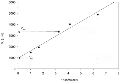
Apparently, the 20-40% of the cellular volume is included in the so-called osmotically inactive components, like solutes and membranes. However, using freezing experiments and calculating the water content of cells it has been demonstrate that one fraction of water is osmotically inactive. Indeed, the ratio between the total cell volume minus the osmotically inactive component and the total water content it is always less than one. This ratio is termed as Ponder R (114). This discrepancy is likely to be ascribe to hydration water around macromolecules and ion, which is not free to move under the force generated by osmotic pressure.
Figure 1.1: Chart where osmolality and cell volume are plotted. The interpolation serves to calculate
6
When a cell is exposed to hypertonic solution this cause cell shrinkage, whereas a hypotonic solution leads to cellular swelling. In the first case the initial volume is restore thanks to the process called regulatory volume increase (RVI), with the contribution of Na+-K+-2Cl− cotransport and Na+/H+exchange. In the latter, the process is termed as regulatory volume decrease (RVD) with a net efflux of KCl and organic osmolytes, followed by osmotic movement of water.
1.1 Inorganic and organic osmolytes
The most important inorganic osmolytes within the cell are two inorganic ions: potassium and chloride; they are indeed involved in small perturbation of the intracellular or extracellular environment; for many years research focused on these two ions even though in the recent years an increasing piece of evidence has shown the dramatic role of organic osmolytes in volume regulation.
Organic osmolytes are confined inside the cell, including amino acids (taurine, glutamin acid), polyhols like myo-inositol and sorbitol and methylamines like betaine (165, 166). The necessity of organic osmolytes beside ions seems to be ascribed to a couple of reasons. First, a potential higher inorganic salt concentration might be not compatible with the function of some enzymes or other proteins (100). In this case, we do not have an interference role for such organic osmolytes. Another important aspect is that the majority of osmolytes do not carry positive or negative charges and beyond that, they are electroneutral compared to inorganic salts which would excessively have an impact on membrane potential, for instance in neuronal excitability in the brain (63).
Organic osmolytes are especially required in renal medulla (14), chondrocytes (32) and in the brain due to limited distensibility (110).
7
1.2
MOLECULAR MECHANISMS IN CELL VOLUME REGULATION
1.2.1 Volume sensors
Cell volume regulation origins from a variation of environmental parameters which lead to an adaptive response. When one cell perform this task, we are able to classify the mechanism in three different phases: sensing, transduction and effectuation.
The sensing process is a complicate process that involved various mechanisms and actors. Despite the strong effort put in the recent years, the machinery involved in sensing volume perturbation remains unclear.
Three types of osmotic stresses can be identified: chancing in ionic strength, mechanical membrane stretches (also related to extracellular matrix and cytoskeleton) and macromolecular concentration (altered metabolism) (55, 24, 111). These mechanisms are part of the osmosensing tool case of the cell and can act together and contribute to a specific response.
Interestingly, in fungi, a histidine kinase complex set up the adaptive response (12), whereas in higher eukaryote the lack of this structure led to others osmosensory pathways.
1.2.2 Cytoskeleton
During osmosensing, a rapid and broadly activation e reorganization of actin cytoskeleton is involved in cell volume regulation. Moreover, the cytoskeleton plays a protective role in volume changes, reducing localized damages and increasing an intrinsic resistance of the cell (27).
The impact of cytoskeleton in osmosensing is particularly clear when it comes to the dramatic alteration of polymerized actin in swelling (decrease) and shrinking (increase). (49, 76, 36, 123). The mechanism behind this organization in rather complex and involves several protein group and families, including Rho family G proteins (78), cortactin (33), which is
8
activated during cell shrinkage, and ezrin/radixin/moesin proteins (118, 143). Most evident, when F-actin is disrupted, the osmo-response is overwhelmed, indicating the major role of cytoskeleton in these processes.
Noteworthy, the activation of some ion channels in these conditions is linked to the cytoskeleton. For instance, the activation of the osmosensor TRPV4 (transient receptor potential vanilloid 4) shows a F-actin dependent mechanism (55). Another example is the Na+/K+/2Cl- transporter (NKCC1), which contributes to the regulatory volume increase (84).
1.2.3 Integrins
Integrins are protein receptor localized at the plasmamembrane level that are involved in cell adhesion, connecting the extracellular matrix (ECM) to the actin cytoskeleton (28). They carry out important functions in cell migration, proliferation and beside that, they have been implicated in cell volume regulation as osmosensors, although a direct evidence is still elusive. What we know is that integrins are activated after osmotic perturbations, shrinkage or swelling. Moreover, they clearly activate volume regulated anion channels (VRAC) (23) and regulate an assortment of volume-sensitive potassium channels (77).
1.2.4 Transient Receptor Potentials
TRP ion channels are cation channels that can be highly selective or non-selective for calcium and response to a variegate physical stimuli. Compose of various subgroups, the vanilloid family (TRPV) is the most investigated group in osmotransduction. However, TRP can be classified into six subgroups, beside the vanilloid family, which are canonical (TRPC), mucolipin (TRPM), ankyrin (TRPA), melastatin (TRPML) and polycystin (TRPP).
TRPV1 is deeply involved in volume regulation and it probably responses to membrane stretch, even though is experimentally challenging to separate a stretching signal from that of swelling (80). Nevertheless, TRPV4 KO mice exhibit impaired RVD and a close relation with mechanosensitivity, since it mediates swelling-activated calcium influx (106). Other channels seems to be involved in mechanosensitivity and cell swelling like TRPM7 (101), TRPC1 (83)
9
and TRPC6 (134). In the past few years two protein from the same family has been cloned and characterize as mechanically activated channels. They are called Piezo 1 and Piezo 2, which resemble the non-selective cation currents stretch-activated (SACs) found in bacteria.
10
1.3 EFFECTORS IN CELL VOLUME REGULATION
1.3.1 Regolatory Volume Decrease (RVD)
After cell swelling, instant mechanisms take place leading to extrusion of intracellular organic osmolytes together with net efflux of KCl, via independent pathways or coupled to each other.
1.3.2 Potassium Channels
Many potassium channels exhibit a volume-dependence. Thanks to the gradient occurring in every cell, an increase of K+ conductance would augment the exit of K+. This in turns will hyperpolarize the cell membrane, favouring the efflux of Cl-. Basically, the most important and effective mechanism in regulatory volume decrease, is the parallel activation of mechanisms which lead to the efflux of K+ and Cl-, known to be almost electroneutral. BKCa or maxi-potassium channels are activated in swelling conditions after membrane depolarization and an increase of cytosolic calcium. They are voltage-dependent and this could be related to a coupling mechanisms with chloride channels activation. The activity under hypotonic conditions has been demonstrated in several studies (see reference 161 for an extensive description).
IKCa constitute another family which exhibit a voltage independence while activated by cytosolic Ca++. Many reports showed and identify this channels as mediator of potassium conductance in hypotonicty conditions (147). Several mechanism have been proposed to explain the activation of these channels, including membrane stretch, actin cytoskeleton reorganization, intracellular calcium increase and activation by kinase such as PKC (72).
11
1.3.3 Volume-regulated Cl- channels
The most important ion channel involved in the increased conductance of chloride upon cell swelling is the so called Volume-Regulated Anion Channel (VRAC). After 30 years being on the run the essential structure of VRAC has been solved (150).
VRAC is a moderately outward rectifying channel, with inactivation at positive potentials. The most selective inhibitors of VRAC is DCPIB (an indanone compound). The activity of this channel is absent in resting conditions, when there is not any osmotic gradient. In hypotonic conditions VRAC is deeply activated and several mechanisms has been proposed including a reduction of intracellular strength, F-actin organization and changes in the membrane lipid composition (72). Interestingly, it shows an intrinsic permeability to organic osmolytes such as taurine, and amino acids (aspartate and glutamate). Hence, taurine efflux during RVD is a prominent mechanism by which every cell responds to swelling. During RVD the passive loss of taurine, following its chemical gradient, is around 30-50%.
1.3.4 KCl symport
KCl release from the symport channel plays an important role in RVD (56). Four isoforms have been cloned so far (KCC1-KCC4) and three of them (KCC1, KCC3,KCC4) are activated in hypotonic conditions. KCC2 is not sensitive to this kind of stimuli and it is exclusively expressed in the central nervous system where it decrease the chloride concentration below its equilibrium in order to facilitate hyperpolarization from GABA channels (160).
12
1.3.5 Cell Volume in physiological processes
Easy to imagine, cell volume regulations involved in many physiological processes and influences cell functions. Two of the most important of these functions are cell migration and apoptosis.
In cell migration, volume changes have a profound impact in epithelial cells (129) while in neutrophils migration, cell swelling is pivotal (122). Recently, it became clear that ion channels and transporter involved in cell volume regulation, are evidently important actors in cell migration. For instance, this role has been elucidated for NHE1 (sodium/hydrogen exchanger) which is present to the leading edge of migrating cells (136), for volume-regulated anion channels (127) and for transient receptor potential channels like TRPV4 and TRPV1 (157). Particularly, imagine a cell in the process of migration, the shrinkage-related channels are localized at the leading edge, while the swelling-activated channels are found at the lagging edge, resulting in a forward movement (128).
13
Apoptotic volume decrease (AVD) is an important stage of programmed cell death.
Important ion channels involved in this process are for instance VRAC, which are essential in AVD and nevertheless are involved in cancer resistance (72). Moreover VRAC are activated by cisplatin (Ise et al., 2005). In the first stage of AVD, a reduction of intracellular concentration of K+, Cl- and Na+ is observed, followed by a 30% loss of water. In the second stage, due to inhibition of the Na+,K+-ATPase, a reduction of potassium and an increase of intracellular sodium take place.(72)
1.3.6 Cell Volume in physiopathological processes
Cell volume and extracellular tonicity is usually maintained in a narrow window by homeostatic control. In any case, major disturbances occurring to biological fluids are common features in some pathological disorders. These states includes hyponatremia, hypernatremia (74) and malaria (131). Other pathological conditions like hypercatabolism and burn injuries lead to cell volume dysregulation (154, 10).
Osmotic disturbance can also occur due to changing in the chemical composition of extracellular fluids, for instance during liver or kidney insufficiency where urea and NH3 accumulate in the extracellular space. In diabetes mellitus, sorbitol accumulation can also challenge the correct volume homeostasis (153, 74).
It is well known that in liver cirrhosis, ammonia augmentation leads to astrocytic swelling in the brain, process which is partially involved in hepatic encephalopathy (51). In the central nervous system, moreover, osmotic changes can have tremendous consequences as the given space is confined and the extracellular space is very little compared to other tissues. During ischemia and reperfusion insults, energy depletion leads to insufficient activity of the Na+,K+-ATPase and consequent cell swelling due to impaired ability to maintain ions gradients (112).
14
2. Volume regulation in the central nervous
system: role of astrocytes and ion channels
2.1 Glial cells: physiological role within the central nervous system
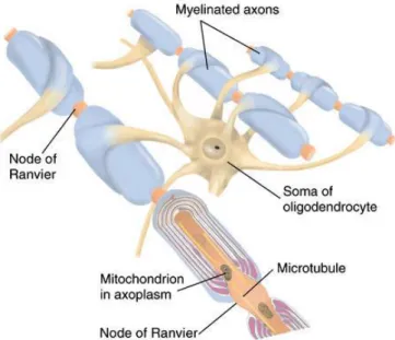
Glial cells, commonly termed as glia, form the nervous system together with neurons. The Greek etymology suggest glia as the “glue of the nervous system”; however, this is not completely correct. Glia are non-neuronal cells that significantly contribute neuronal physiology as they maintain neuronal homeostasis, modulate synaptic genesis and fiber myelination, and provide neurons of trophic support and immune protection within the brain and central nervous system (CNS) as well as in peripheral nervous system (SNP) (Fig. 2.1).
Furthermore they destroy pathogens and remove dead neurons in order to maintain the appropriate neuronal function. In addition, the recognition of their action on the immune system led glia to become an integral part of a new discipline: neuroimmunology.
It is also important to remind that we can distinguish between different types of glia: in the CNS we find astrocytes, oligodendrocytes, microglia and ependymal; in the (SNP) we can distinguish between Schwann cell, that form myelin sheaths, and satellite cells, which surround the bodies of ganglion neurons.
Figure 2.1: Anatomical and functional organization in neuron-glia interactions: glial cells interacting with neurons, blood vessels and the micro-environment. Oligodendrocytes cover axons with myelin sheath to accelerate neuronal impulses. Astrocytes take contacts with synapses and vessels. Microglia monitor the brain parenchyma as immunitary cell type of the CNS (3).
15
2.1.1 Astrocytes
Astrocytes are derived from neuronal stem cells and populate all regions of CNS, where they represent the most widely distributed cell type (52). Astrocytes (Fig. 2.2) are stellate cells with multiple fine processes that contact blood vessels, pial surface, and surround neurons. In gray matter, astrocytic processes unsheathe virtually every synapse. Astrocytes serve various functions including structural roles (they forms the blood-brain barrier), metabolic support of neuronal system supplying nutrients and neurotransmitter precursors, regulation of ion concentrations. Furthermore, they express several receptorial systems (neurotransmitter receptors) and immune signalling (cytokines and chemokines), providing a critical role in neuronal functions and affecting all neighbouring cell types.
Figure 2.2: This immunofluorescent light micrograph shows mammalian brain astroglial cells, stained in green. Nuclei are stained in blue. Scientists are uncovering evidence that astrocytes do more than simply support neurons (60).
2.1.2 Microglia
Believed to be the most reactive and mobile cells of the central nervous system, microglia are the specialized phagocytic cells and they behave as central macrophages (45). Adult microglial cells originate from primitive myeloid precursors and show different
16
characteristics as compared to other macrophages: microglia, indeed, have highly motile processes by which they examine the brain surrounding (Fig. 2.3) (31).
Thanks to a variety of signalling pathways microglia communicate with macroglial cells (astrocytes, oligodendrocytes and other glial cells), and neurons. These cells have the capacity to release a number of soluble factors that determine either beneficial or detrimental for the vicinal cells. Activated microglia can migrate to the site of injury, proliferate, and phagocytose cells and cellular compartments.
Figure 2.3: Example of microglia cells around the red blood vessels in the retina. Both microglia and blood vessels are surrounded by nerve cells, not stained in this picture (57).
2.1.3 Oligodendrocytes
Oligodendrocytes are glial cells populating the CNS and producing myelin (80% lipid and 20% protein), an insulating sheath on axons and nerve fibers. A single oligodendrocyte is able to extend its processes to 50 axons, wrapping approximately 1 μm of myelin sheath around every axon (Fig. 2.4). Between the different segments of myelin-isolated neurons, it is possible to identify uncovered spots called nodes of Ranvier, which are crucial in the electrical transmission system. Indeed, the speed up of impulses occurs as propagation of
17
action potentials follows a salutatory fashion at the nodes of Ranvier, in which ionic flows is permitted.
In the peripheral nervous system, the equivalent cell present is termed as Schwann cells: these cells, by contrast, can wrap around only one axon.
Figure 2.4: Oligodendrocyte and myelin sheath: oligodendrocytes insulate neuronal axons, speeding-up electrical transmission (58)
2.1.4 Ependymal cells
Termed as ependymocytes, they are specialized cells that produce and secrete cerebrospinal fluid (CSF) and beat their cilia to permit the CSF fluid circulation. Situated in the cavities of the CNS where build up the walls of the ventricles as well as the blood brain barrier in the interface with blood circulation. They are thought to act as neural stem cells.
18
2.2 Astrocytes and volume homeostasis in the CNS
Astrocytes constitute the most abundant cells within the brain; even though they are considered “star-like cells” they do exhibit a morphological heterogeneity. Indeed not all the astrocytes express the classic astrocytic marker GFAP (glial fibrillary acidic protein). This astrocytic marker stains all cultured astrocytes but in situ the expression is rather heterogenous; for instance is virtually expressed in every cell of the Bergmann glia, while in adult animals cortical astrocytes the expression of this protein is confined to a 15-20 percent. Astrocytes are both present in the gray matter and in the white matter. In the former, we find protoplasmatic astrocytes, which have fine and elaborate processes about 50 µm long. This type of astrocytes are capable to cover the entire three-dimensional space, virtually covering all the neuronal membranes and every synapse. The also take contact with blood vessel, constitute the so-called perivascular end feet. Moreover, they project towards the pial surface, isolating the brain parenchyma from the vascular and subarachnoid space, forming the glia limitants. Fibrous astrocytes are present in the white matter. They have different features including longer and less elaborate processes (300 µm) which also cover the nodes of Ranvier (perinodal astrocytes).
Astrocytes play major role in brain homeostasis, morphological definition of the central nervous system, synaptic maintenance and brain defence. (94)
Figure 2.5 Protoplasmatic astrocytes from mouse
neocortex. Noteworthy, the complexity of astroglial processes virtually covering all the CNS synapses and perivascular interfaces (53).
19
2.3 Astrocytes physiological functions in homeostasis
2.3.1 Glutamate metabolism
Glutamate uptake from extracellular environment is a major task for astrocytes. The extracellular concentration of glutamate needs to be maintained under 1 µM. Hence, astrocytes uptake glutamate in order to terminate glutamatergic neurotransmission and prevents glutamate to reach excitotoxic levels (glutamate is the most important and bountiful excitatory neurotransmitter in the brain) (132).
Specific glutamate transporters are expressed and enriched in astrocytic processes, especially where they take contact with synapses. GLT1 and GLAST 1 are the most specific glutamate transporters (119, 125).
Glutamate transport is an active process and require energy from ATP breakdown; briefly glutamate is cotransported with three Na+, while one hydroxide ion and one K+ are extruded from the cell (124). This leads to an increase of intracellular sodium concentration beside a Ph reduction. Glutamate is subsequently converted in glutamine and recycled by neurons after extracellular release by astrocytes.
Glutamate receptors, included mGluR, are present in the plasma membrane of astrocytes. During neuronal firing, release of glutamate in the synaptic space lead to astrocytic calcium intracellular store activation, leading to a calcium-dependent discharge of glutamate in the extracellular environment, eventually modulating synaptic activity (52)
2.3.2 Potassium buffering
In the central nervous system, neuronal action potential is followed by a repolarization phase, driven by potassium movements across the membrane, towards the extracellular space. The potassium increase is rapidly buffered in the interspace narrowing synapses, by
20
astrocytes. This important task accomplish the maintenance of the resting membrane potential of neurons, since a compromised K+ regulation leads or is related to severe pathologies, such as epilepsy (130).
The basic mechanism proposed to address the role of astrocytes in potassium buffering implies the involvement of the astrocytic syncytium, which spatially buffers local high potassium concentration towards extracellular compartments where K+ is lower and eventually blood circulation. Among many potassium channels, the Kir 4.1 (Inwardly rectifying potassium channel 4.1) is mainly involved in the astrocytic potassium conductance (26). As a weakly-rectifying is capable to drive K+ inside and outside the cell, in a bidirectional fashion. Kir 4.1 expression is highly polarized in astrocytes, particularly in the processes enwrapping synapses and the processes facing capillaries. In this context, it is possible to term this process as “potassium spatial buffering” where K+ is cleared from high concentration areas (synapses) and released in the blood circulation, through the astrocytic syncytium. Gap junctions, connecting astroglial cells to each other and favouring cell-cell communication, play an important role in this mechanism (18).
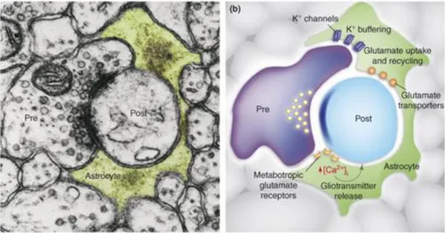
Figure 2.6 Astrocytes modulate neuronal activity by releasing different transmitters. (A) The tripartite
synapses in this electron microscopy micrograph (A). The astrocytic process (green) enwraps both the presynaptic (pre) and postsynaptic (post) terminals. (B) Astrocytes release glutamate that modulate synaptic plasticity, neuronal excitability, and synchrony due to specific activation of the NMDA receptor. Astrocytes also release ATP, which is degraded to adenosine and strongly suppresses synaptic transmission by binding to the receptor adenosine A1 (48).
21
Astroglial cells are also involved in other physiological processes. For instance, astrocytes regulate synapse arrangement (otherwise formation of synapses would be greatly depressed), maintenance and elimination. Synaptic formation strictly depends on cholesterol, which is synthetized and secrete by astroglial cells, which can control for instance receptor density in the post and presynaptic membranes of neurons. Other signalling molecules, including tumour necrosis factor alpha (TNF-alpha), can affect synaptic maturation. TNF-alpha has been demonstrated to regulates the expression of glutamate receptors into postsynaptic membranes neurons (94).
Moreover astrocyte, covering virtually all the surface facing the basal lamina of blood vessels, are capable to control and regulate local blood flow through calcium-dependent mechanisms which in turns cause vasodilatation or vasoconstriction. Regarding metabolic support, astrocytes seems to be correlated to the glucose-lactate shuttle, taking up glucose from the general circulation and supply neurons with new metabolic fuel. Indeed, astrocytes are the only cellular entities that can synthesize glycogen from glucose, operatic as an energy storage (94).
22
2.4 Brain volume regulation: role of astrocytes and ion channels
An action potential is very fast increase of the membrane potential from −70 mV, which is a typical resting potential, to a peak value of about +40 mV. This elevation occurs in some milliseconds and it is followed by a less rapid decrease back to the resting potential value. In an action potential, voltage-gated sodium and potassium channels open and close in a very coordinate manner to permit the flow of ionic currents through the plasma membrane, according to the Nernst equation for each ionic entity. Hence, sodium enters in the cell and potassium is directed towards the extracellular space. These transient variations in the membrane potentials triggers not only voltage-gated channels but also allow electro-diffusion of ions through passive channels, particularly chloride which exhibits in most excitable ells, an high chloride permeability in resting Conditions. Minor changes in the spatial distributions of sodium, potassium and chloride occurs after every action potential and require fine adjustments through active ionic transportation. If the resetting of this perturbations are not fully complete before the following action potential, a progression of these ionic gradients is observed, which may induce volume changes (8, 13, 54). In the central nervous system the interstitial spaces are reduced and cell swelling upon repeated action potentials can further increase extracellular potassium concentration and in turns affecting the membrane resting potential to increased values toward the firing threshold and an augmented excitability, which is linked to a number of disease including epilepsy and spreading depression. (9, 67).
Astrocytes, involved as main actors in central homeostasis and volume maintenance and regulation, are deeply studied in order to unravelled the mechanisms beside volume regulation. Primary, volume regulation is a cell-cell mechanisms and ion channel together with aquaporin are crucial in addressing this task. Astrocytes indeed in physiological state, show a moderate and transient swelling upon neuronal activity, especially nearby synapses, as the extracellular space between pre- and post-synaptic membranes decreases (138). Astrocytes cultured in vitro and exposed to hypotonicity, rapidly swell through an osmotic driven water flux, mediated by aquaporin 4. At this moment, cellular components sense the
23
volume change in order to ignite the response and restore the initial volume. Elevation of intracellular calcium and cytoskeletal rearrangements seem to be the most important mechanisms in astrocytes (102, 88). [Ca2+]
i rising in RVD is still controversial, as it is not clear whether calcium per se is able to trigger this adaptive response, or a biochemical calcium-dependent pathways are activated. Involvement of the intracellular kinases such as P38, MAP-kinase and JUNK has been demonstrated, together with phospholipase A2 (PLA2), even though the exact contribution is still unclear. (109). Mechanosensitive receptors, such as transient receptor potential (TRP) might be also implicated in calcium transduction.
Concerning effectors, VRAC channels play a pivotal role in RVD (155, 107, 69). Other anion channels might be also involved, particularly from the CLC family (39). Potassium conductance is as well involved in RVD even though the K+ channels have not been discovered yet. In this perspective, Kir 4.1 channel is also essential for volume regulation in the spinal cord, even though evidence in other regions of the CNS are still lacking. (34).
2.5 Astroglial ion channels in volume regulation
2.5.1 Aquaporin 4
Aquaporin 4 is a water channel included in the aquaglyceroporin family, mainly expressed in the brain and constituting the most expressed aquaporin of the central nervous system. Aqp4 monomer is a 323 amino acids protein called isoform M1, since a shorter isoform that lacks the first 22 amino acids (M23, 301 AA) due to a shift in the initiation site on the same gene, has been also identified. (91)
The two classical isoforms cooperate together to form primarily tetramers with a predicted central pore, and larger arrays called orthogonal array of particles (OAPs), which cover the astrocytic membrane where AQP4 is highly enriched, at the perivascular endfeet of astroglial cells. This clustering seems to be the active from of AQP4 even though the real role of these particles needs to be clarified.
AQP4 is highly concentrated in astrocytic end feet (42), attesting to the polarization nature of astrocytes. AQP4 allows bidirectional flux of water following osmotic driven force.
24
Subsequently the AQP4 role has been deeply investigated in pathologies included ischemia and edema. After 23 years from AQP4 discovery, the role of this aquaporin is still not clear in these pathological states. The level of AQP4 in astrocytic end feet is depressed following transient ischemia (43) and this loss is more prominent in the regions where there was a vascular damage, delimiting the ischemic area. (41). Noteworthy, the polarized distribution of AQP4 is lost in reactive astrocytes (163).
Downregulation of AQP4 were also found in other pathologies such as epilepsy (37), brain injury (121) and one model of Alzheimer (167), while increasing of AQP4 has been found in some inflammatory processes (from LPS treatment, 145) and central haemorrhage (156) and brain tumours (35, 99).
Robust data indicate that AQP4 is involved in water and waste clearance from the CNS, becoming rate limiting in the water movement between the blood brain barrier. This was indicated from a recent study where studying fluorescent tracers in a living mouse brain were used (61). Cerebrospinal fluid (CSF) recycles inside the brain parenchyma along
perivascular spaces. The tracer, following distribution in the brain parenchyma is accumulated nearby capillaries and veins, suggesting a paravenous drainage process. AQP4 -/- mice shows reduced tracer flow and delayed clearance in parenchyma. AQP4 may be relevant, not only form interstitial water, but also soluble factors, proteins and waste, included such neurodegenerative disease, where protein accumulation is an important factor in the etiopathogenesis. (167)
2.5.2 TRPV4
Cell-cell communication in astroglial syncytia and astroglia signaling is mediated by Ca2+ waves elicited by extracellular soluble factors and a variety of physical and chemical stimuli, included cellular swelling. Transient receptor potential cation channel subfamily V member 4 (TRPV4) is a poly-modal cation channel and can be activated by modest heat (47, 152) and endogenous ligands including arachidonic acid and anandamide (159). Cell swelling
25
activation of TRPV4 in anisotonic conditions seems to be linked to PLA2 stimulation and arachidonic acid action (152).
Several studies demonstrated that TRPV4 is critically involved in cell volume regulation and regulatory volume decrease in various cell types (11, 15). Our group recently showed that TRPV4 is expressed in rat cultured cortical astrocytes and in situ observation in the mouse brain, (16). Indeed activation of TRPV4 by a selective activator like 4-alpha-phorbol 12,13-didecanoate (4αPDD), induced [Ca2+]
i signals in cortical astrocytes in vitro, resembling the currents detected endogenously in other cell types (158, 120). Hypotonicity-induced [Ca2+]
i elevation was dependent on extracellular calcium concentration and was blocked by ruthenium red, a non-specific TRPV4 inhibitor (151). In situ studies revealed that TRPV4 highly enriched in the astrocytic endfeet membranes facing endothelial cells of blood vessels, as well as in ependymocytes in the ventricles. Interestingly, in this two domains, AQP4 is also very abundant. (96).
2.5.3 VRAC
The volume-regulated anion channel (VRAC) is probably the most important anion channel involved in cell volume regulation. This concept and VRAC expression has been demonstrated mostly in all types of vertebrate cells, including those of the CNS (135, 97, 104).
In many studies, pharmacological inhibition of VRAC, blocked or seriously impaired RVD (55, 97). This pivotal role of VRAC in volume regulation may partially ascribed to the ability to mediate efflux of organic osmolytes. In astrocytes, the release of massive amounts of excitatory amino acids, glutamate and aspartate, upon brain injury, causes excitotoxic neuronal death. In this context, VRAC has been demonstrated to be permeable to glutamate involved in this excitotoxicity (68, 110). Astrocytes are more susceptible to ischemic swelling than neurons (89) and VRAC might be the main source of volume-dependent release of toxic amino acids in the brain.
26
After 30 years being on the run, the molecular identity of VRAC has been partially solved. In 2014, two independent groups established that an essential sub-unit of VRAC is the leucine rich repeat containing 8 family member A (LRRC8A). (115, 150)
The LRRC8 family includes five members. LRRC8A member is a protein of about 800 amino acid residues with a molecular weight of 95 kD. The C-terminal contains 17 leucine-rich repeats. (1). Notably, LRRC8 proteins share a common ancestor with pannexins, beside a weak homology in their transmembrane domain. Inlight of this, a hexameric conformation similarly to pannexins, has been suggested (1). This speculation might also include that multimers of LRRC8 are capable to form a structured channel-pore. Notably, it was reported that carbenoxolone, a VRAC blocker, inhibits connexin hemichannels (22).
Swelling-induced VRAC activation
VRAC activation induced by cell swelling has been extensively demonstrated, even though the precise mechanisms is still elusive. Many different types of signaling cascade might contribute to the swelling-induced VRAC activation but their contribution seems to be partial or only permissive. Since cell swelling is usually accompanied by calcium signals, the contribution of calcium has been investigated in VRAC activation. VRAC is not a calcium dependent ion channel, as its activity does not directly reflects calcium dynamics (139). In any case, many studies reported that a minimal calcium concentration is required or some calcium-dependent enzymes are crucial for swelling-induced anionic current activation, like protein kinase C or Ca2+/calmodulin-dependent kinases (97). In cultured astrocytes, the mechanism of swelling-induced activation of VRAC clearly consists of Ca++-dependent and independent components. This [Ca++]
i increase contributes to 30–40% of the total VRAC activation. Concerning the remaining 70-60%, the mechanisms in not yet determined, and it seems to be multicomponent and evidently calcium-independent (2). Remodeling of actin cytoskeleton has long been suggested to relate in some way to VRAC activation in many types of cells. In general, swelling-induced VRAC current reflects disassembly of submembrane cortical F-actin networks. (72)
27
Cell volume regulation is an essential process of every cell, and it occurs to not only restore the original volume when a cell is exposed to anisotonic condition, but also participate in such physiological processes in which change of shape is required to address such as apoptosis, differentiation, migration or proliferation. In this context, VRAC activation in has been well demonstrated in apoptosis, where VRAC is stimulate despite isovolumetric conditions. Moreover, in several cancer cell types, VRAC is downregulated and this correlate with a decreased propensity to apoptosis. Notably, this effect is more pronounced in drug-resistant cancer cells (see Review 116). Thus, VRAC currents seems to be highly regulated throughout the cell cycle, and consistent with its specific role in volume regulation, VRAC inhibition impairs cell proliferation. VRAC may also play an important role angiogenesis since specific blockers impaired vessel formation in many models (81, 117). In cell migration, the inhibition of VRAC activity limited cell movements, presumably reflecting the close involvement of local cell volume changes in cell motility (116).
2.6 Interaction between ion channels and aquaporins in volume regulation
2.6.1 AQP4 and Kir 4.1Potassium spatial buffering through the astroglial syncytium is accompanied by an osmotic driven water flux. Thus, a molecular partner for Kir 4.1 has been rapidly proposed. AQP4, the most important and expressed water channel in the brain has been extensively studied, since both proteins are enriched and co-localize in astrocytic endfeet in situ (90). This hypothesis has been subsequently tested in co-immunoprecipitation experiments, where Kir4.1 form a complex with APQ4 in Muller cells (29). In this study DAPC proteins were shown to selective anchoring both AQP4 and Kir 4.1, as indeed shown by Amiry-Moghaddam group, which deeply studied APQ4 polarization and molecular anchoring in
situ, reporting an AQP4/alpha-syntrophin interaction (alpha- syntrophin is part of the DAPC
complex) (4, 5). At a functional level, it has been demonstrated that genetic knockout of AQP4 led to slowed dynamics in potassium buffering and seizures predisposition (20, 21). In
28
more physiological experimental paradigms, the role of this interaction is still unclear. An explanation for some discrepancy might be that the two proteins belong to different micro-domains at the plasma membrane level, but able to cross talk to accomplish the same functions (18)
2.6.2 AQP4 and VRAC
Molecular interaction between AQP4 and VRAC has not been investigated yet. However, our group provided in vitro evidence that functional interaction between AQP4 and VRAC may occur in cortical rat astrocytes, as genetic knockout of aquaporin 4, reduce VRAC currents (19). Moreover, the interaction might be ATP-dependent, since the supplement of ATP in the internal solution in the patch-clamp experiment, restored the hypotonic-induced VRAC current (19).
2.6.3 AQP4 and TRPV4
Hypotonic stress in astroglial cells produce an increase in intracellular calcium. Our group demonstrated the presence of TRPV4 in rat cortical astrocytes in vitro and in situ (16). Later our group provided evidence of molecular and functional interaction between AQP4 and TRPV4, in co-immunoprecipitation experiments and functional assay, showing that an increase of intracellular calcium upon hypotonic stimuli require the presence of AQP4, since this response is absent in AQP4 Knockout mice (17)
29
Fig 2.8 Astroglial cells and ionic homeostasis. Noteworthy, the high polarized expression of astrocytic
30
PART II
31
Aquaporin 4 (AQP4) is a highly conserved protein in mammals, since point mutation usually lead to a reduce water permeability (133). A recent study investigated the effect of a mutation in AQP4 gene in AQP4 membrane protein expression and water permeability (95). In this study, a novel point mutation of AQP4 origin an Asp/Glu switch, was observed in a Spanish sporadic case of deafness. Aspartate is position 184 is conserved across species. Moreover, the study of Nicchia e co-workers shows a reduce permeability of this mutant, heterologously expressed in cellular systems, probably affecting the mobility of the D loop of the protein.
The group where I carried out my PhD program demonstrated the role of protein-protein interactions between AQP4 and the channel Transient receptor potential channel vanilloid subfamily 4 (TRPV4), in the regulation of cell volume of cultured rat cortical astrocytes (17). Interestingly, TRPV4-KO mice develop delayed-onset hearing loss together with a pronounced vulnerability of the cochlea to acoustic insults (140). Chloride channels are also involved in the maintenance of homeostasis in the brain and in particular, volume-regulated anion channels (VRAC) mainly address this task, possibly in every cell of vertebrate organisms (65) The current mediated by VRAC has been deeply studied over the last 30 years. The molecular identity, partially solved in 2014 by two independent groups (115, 150), consist of an essential sub-unit termed as leucine rich repeat containing 8 family member A (LRRC8A). Notably, a functional interaction between AQP4 and VRAC has been demonstrated in astrocytes (17).
On this basis, in the context of a project supported by MIUR FIRB-Futuro in Ricerca (project protocol BFR12SJA8_002 "Studio del ruolo fisiopatologico della mutazione D184E nel gene dell'Acquaporina-4" to Prof. Nicchia and Dr. Benfenati) we hypothesized that this mutation in the aquaporin 4 gene may affect the dynamics of AQP4 ion channel interaction. Indeed, ion channels and aquaporin play a pivotal role in maintaining the homeostasis in the auditory system, especially for the cochlear fluid homeostasis and generation of the endocochlear potential (64).
Hence, the aim of this study was to investigate the molecular and functional interactions between the mutant D184E of aquaporin 4 protein, with the ion channels TRPV4 and VRAC.
32
Moreover, since molecular tools, such as antibodies, are still not commercially available for VRAC, three antibody raised against LRRC8A antigenic polypeptides have been produced to investigate the expression of VRAC underpinning subunits in cortical astrocytes and mouse brain.
33
PART III
34
3.1 Cell Culture
All primary cell culture were prepared at the Department of Pharmacy and Biotechnology (FABIT) of the University of Bologna. The experiments were performed according to the Italian and European council law on protection of laboratory animals, with the approval of a local bioethical committee and under the supervision of a veterinary commission of the University of Bologna. Every effort was made to minimize the number of animals used and their sufferings. Adult male C57BL/6 mice (Jackson Laboratories, Boulder, CO) were used in Medicine department of the University of Oslo, according to the European Council law on protectionn of laboratory animals.
3.1.1 Astrocyte
Cortical rat astrocytes we obtained from new born animals (Sprague Dawley) as described elsewhere (40). Briefly, neonatal cerebral occipital cortices devoid of meninges, were mechanically triturated, using micropipettes until the tissue was completely dissociated and filtered with 70 µm nylon cell strainer (Falcon, BD Bioscience Bedford, MA, USA). The filtrate was transferred into a 25 cm2 culture flasks containing Dulbecco’s Modified Eagle supplemented with 15% fetal bovine serum and penicillin/streptomycin (100 U/ml and 100 µg/ml, respectively), purchased from Gibco-Invitrogen, Milano, Italy. Culture flasks were maintained for 2–5 weeks in an incubator at controlled temperature and pH (37° C and 5% COs). After 2-3 weeks, astrocytes were re-plated onto the respective substrates by enzymatically dispersion, using 0,05% trypsin-EDTA (Gibco-Invitrogen), seeded at the desired concentration depending on the experiment.
35
3.2 COS-7 cells
The COS-7 cells belong to an immortalized line derived from the African green monkey kidney. COS-7 cells were cultured in DMEM-glutamax, 10% FBS and penicillin (100-200 U/ml) - streptomycin (100 µg /ml) (Gibco-BRL). Cells cultured in cell flask were incubated at 37 °C and 5% CO2.
3.2.1 Transfection of COS-7 cells
The day before transfection, COS-7 cells were seeded in 60-mm Petri dishes at a density of 5-8*105 per dish. Lipofectamine 2000 were used as transfection reagent (Invitrogen). COS-7 were transfected with the following constructs, depending on the experiment: human TRPV4/pEGFP-N1 or AQP4 constructs, including wild type and carrying the D184E mutation; AQP4-M1/pTarget, AQP4-M23/pTarget, AQP4-M1-D184E/pTarget, AQP4-M23-D184E/pTarget). Co-transfection experiments were also performed to evaluate the interaction between TRPV4 and AQP4.
3.3 Antibodies:
The following primary antibodies were used:
Target Protein Dilution Application Purchased
Anti-TRPV4 1:200 Western Blot Alomone
Anti-AQP4 1:500 Western Blot Santa Cruz
Anti-AQP4 7µg/500 µg lysate Immunoprecipitation Chemicon
Anti-LRRC8A-3 1:500 Western Blot Custom
Anti-LRRC8A-2 1:200 Immunofluorescence and Immunoelectron Microscopy
Custom
Anti-GFAP 1:500 Imunofluorescence Chemicon
36
Three custom polyclonal antibodies against LRRC8A were produced (Twin Helix, Milan). Two have been used in this experimental set up. One polyclonal antibodies was raised in rabbits against the peptide sequence QRTKSRIEQGIVDRSE, coupled to KLH through an N-terminally added cysteine. This sequence belong to the intracellular loop between TMD2 and TMD3. Another polyclonal antibody against the C-terminus of LRRC8A was raised in rabbits against the peptide NLTQIELRGNRLE following the same set up as the previous Ab. Sera were affinity-purified against the respective peptides. Peptides and serum negative control were also included in the project.
3.4 Immunoblot analysis in COS-7
COS-7 cells and 3–4 weeks cultured astrocytes were used for immunoblotting experiments. Cells were washed and scraped off in lysis buffer (50 mM Tris–HCl pH 7.4, 100 mM NaCl, 1 mM EGTA pH 7.4, 0.5% sodium deoxycholate, 1% Triton) with supplemented protease inhibitors. The lysate was spun at 14,000 g for 30 min at 4 °C and protein concentration was determined in the supernatant using Bradford quantification (Invitrogen). For membrane protein isolation, biotinylation of cell membrane protein was performed and biotinylated protein were separated with immobilized streptavidin as previously described (38).
Required amount of total protein lysate were separated into a polyacrylamide gel, electro-transferred onto a PVDF membrane (Invitrogen), blocked in 5% BSA and incubated with the primary Ab at 4° C for 12-16 hours.
Membranes were then washed three time with PBS-T and probed with the required IgG horse radish peroxidase–conjugated secondary antibodies (Sigma), and developed with the enhancing chemiluminescence detection system (Santa Cruz Biotechnology, Inc).
Mouse brain tissue was dissected (n=4) and processed for protein analysis. Brain regions were homogenized at 4 °C using 2 mL Lysing matrix D (MP Biomedicals) Eppendorf tubes containing Lysis buffer supplemented with protease inhibitor cocktail (Roche). Homogenization was performed in a FastPrep FP120 Cell Disrupter (MP Biomedicals),
37
shaken at intervals and intermittently cooled on ice. Supernatants were subsequently spun at high centrifuge speed and stored at -80 °C until downstream application as described in the previous paragraph.
3.5 Electrophysiology
Electrophysiology is a branch of physiology that studies the function of the organism from an electrical point of view, in normal conditions and under the influence of an external electrical potential. In 1991 the Nobel Prize in Physiology or Medicine was awarded to two German biophysical: Erwin Neher and Bert Sakmann with the following motivation: "for their discoveries concerning the function of single ion channel cell". They were the inventors of the patch-clamp technique ("block of the aria") that makes it possible to record the currents that flow through single ion channels in many types of biological membranes. The physiology of ion channels has Always Been a main topic of interest in neuroscience research. The patch-clamp technique is currently the gold standard for real-time investigation of ion channel conductance. It allows the investigation of a small set of ion channels down to single-channel recording. The technique can be applied on single cells in culture as well as on freshly prepared brain.
To electrically isolated membrane patches, a thin glass pipettes that, after having been polished by the flame generated by an instrument called puller, appear to have a tip diameter of about 1 micrometer and a resistance of 1-10 ohm, they were sealed onto the membrane; with a suction it is established a high-resistance seal in the gigaohm range (cell-attached configuration). The pipette tip once resting on the plasma membrane, was capable of electrically isolating the small flap (patch) membrane and allowed to record ionic currents mediated by the small population of channels within it until you get to possibly record the activities of a single channel. Thereby all ions passing through this patch flow into the membrane and current can be recorded by a chlorinated silver electrode connected to an electronic amplifier.
38
Small petri dishes were mounted on an inverted microscope (Nikon Diaphot, Nikon Italy, Firenze, Italy), equipped with epifluorescence filters to detect fluorescence from transfected cells, labeled with GFP (green). Currents were recorded with the patch clamp technique, in whole cell configuration (50). Patch pipettes were prepared from thin-walled borosilicate glass capillaries to obtain a tip resistance of 2–4 MΩ. Membrane currents were amplified with an EPC-7 amplifier (List ElectronicDarmstadt, Germany), and low-pass filtered at 2 kHz (3 dB) and data were acquired with a sample rate of 5 kHz. Traces were analyzed with offline with pClamp 6 software (Axon Instrument, Foster City, CA, USA) and Origin 6.0, (MicroCal, Northampton, MA, USA). Experiments were performed at room temperature (22–24 °C).
3.6 Solutions and chemicals
Saline solutions for patch clamp experiments were prepared with salts (Sigma-Aldrich) of the highest purity grade, and de-ionized and sterilized water.
For electrophysiological recordings the standard isotonic and control saline solution was (mM) 140 NaCl, 4 KCl, 2 MgCl2, 2 CaCl2, 10Hepes, 5 glucose, adjusted pH (7.4 with NaOH and osmolarity (310 mOsm with mannitol). In order to eliminate cation currents in some electrophysiological experiments, cations were substituted with the impermeant cation N-methyl-D-glucamine (NMDG 140 nM). For microfluorometric experiments the control bath saline was (mM) 130 NaCl, 4 KCl, 2 MgCl2, 5 CaCl2, 10 TES, 5 glucose, adjusted pH (7.4 with NaOH and osmolarity (310 mOsm with mannitol). The Ca2+ free extracellular saline was prepared by omitting CaCl2 salt and adding a calcium-chelating agent (EGTA 0.5 mM). The hypotonic saline (260 mOsm) was prepared without adding mannitol.
3.7 Calcium microfluorimetry
[Ca2+]
i intracellular concentration was calculated by ratiometric microfluorometry, using the ratiometric probe Fura 2-AM (Molecular Probes, Invitrogen, Milano, Italy), which is one of the most used dyes in calcium studies. The binding of Ca2+ leads to a shift in the excitation wavelength, because of different conformation of the dye after calcium binding. 24 hours
39
before the measurements, low-density COS-7 cells were seeded on coverslips. The day of the experiment, the cells were incubated in 10 µM Fura-2-AM diluted in standard control solution, for 45 min at room temperature (22-25°C). For microfluorimetric analysis, cells seeded on coverslips were mounted on a perfusion chamber and perfused at a rate of 0.5 ml/min as described elsewhere (152).
Measurements of [Ca2+]i in single cells were performed by using an inverted fluorescence microscope (Nikon EclipseTE2000U, Nikon Italy) equipped with long-distance dry objective(40X) and appropriate filters. The emission fluorescence was selected through a 510-nm narrow-band filter and acquired with a digital charge-coupled device camera (VTi, VisiTech International Ltd., Sunderland, UK). Chopper frequency and settings were controlled by QuantiCell 2000 software (VisiTech). The fluorescence intensity measured at 340 nm to 380 nm with a sampling rate of 0.5 Hz., was plotted as a ratio between 340 nm and 380 nm. This ratio is directly proportional to calcium concentration and can be use as a direct indicator of [Ca2+]
i. The calibration of the 340/380 reflecting the free [Ca2+]i was achieved as previously described (46).
3.8 Brain sections and immunofluorescence
The animal was transcardially perfusion-fixed for 15 min with 4 % formaldehyde and post-fixed for 24 hours. The sections (thickness 15–20 nm) after a two-step washing in PBS, were blocked in 2% BSA, 1% Triton X-100 in PBS and incubated overnight with primary antibodies at the desired dilution, as explained in the paragraph 3.3. Brain sections were then incubated with a secondary antibody conjugated with different fluorescent probes, depending on the experiment. After washing, nuclei were stained with 4′,6-Diamidine-2′-phenylindole (DAPI) and mounted on the coverslips with Fluoromount (Molecular Probes).
Images of brain sections were captured using a Zeiss SP1/MP (Oberkochen, Germany) confocal microscope and processed using Zeiss Imaging software. The microscope was equipped with a 400 nm diode, 488 nm Ar+ and 543 nm He-Ne lasers as exciting sources.
40
For COS-7 and astrocytes immunofluorescence experiments, cells were seeded on coverslips, after treatment with poly-D-lysine; after 48 hours, cells were fixed and probed with the primary Abs as described above. The next day cells were incubated with a secondary antibody, stained with DAPI and after mounting, analyzed using a Zeiss SP1/M confocal microscope
In experiments conducted with control antigen, antibody was pre-adsorbed by incubation with immunizing peptide 1 h at room temperature (3 µg peptide/1 µg antibody).
3.9 Fixation and tissue processing
In immunoelectron experiment, the animals were perfusion-fixed as previously described (7). The specimens were plunged in subsequently higher glycerol concentrations (10%, 20%, 30%) in order to cryoprotect them. The samples were then immersed into liquid propane (170 °C) in a cryofixation unit (Reichert KF80, Wien, Austria), incubated 0.5% uranyl acetate dissolved in anhydrous methanol (90 °C). The temperature was increased from to 45 °C, in 4°C/h steps. Specimens were washed with anhydrous methanol and infiltrated with Lowicry lHM20 resin at 45 °C with a progressive increase in the ratio of resin to methanol. Polymerization was performed with UV light (λ 360 nm) for 48 h.
3.10 Immunogold histochemistry
The brain tissue ultrathin sections (90 nm) were incubated overnight with an anti-LRRC8A primary antibody at a proper concentration at room temperature, overnight. The following day, tissue sections were then probed with a secondary antibody, coupled to 15nm gold particles and after washing, contrasted with uranyl acetate and lead citrate. Labeled sections were analyzed with a Philips CM 10 electron microscope (Eindhoven, Netherlands) at 60 kV (5, 6). Data were compared, as control, with pre-immune serum and immunizing peptide negative controls.
41
3.11 Statistical analysis
Data are expressed as the mean standard error (SE) from at least three independent experiments. The statistical analysis was performed with two-tailed Student’s t-test (paired or unpaired depending on the experiments) and a statistically significant difference P was reported if p<0.05 or less. Analysis of variance was also performed (one-way ANOVA), followed by Newman-Keuls post hoc test for multiple comparisons. p<0.05 was considered statistically significant.
42
PART IV
43
4.1 TRPV4 mediates intracellular calcium elevation upon hypotonic stimulus in
COS-7
Acquaporins and ion channels cooperate to maintain homeostasis; in the central nervous system, AQP4 and TRPV4 interact in order to serve this task. The former facilitating the osmotic-driven water movement and letter possibly sensing the mechanical stretch induced by osmotic cell swelling. The laboratory where I carried out my PhD program recently demonstrated a molecular and functional interaction between these two proteins. In order to study the physiophatological role of the D184E mutation in the AQP4 gene, we set up a heterologous system, able to express AQP4 and TRPV4 in the COS-7 cell line (African green monkey kidney fibroblast-like cell line suitable for transfection). This system provide a straightforward methodology to study protein-protein interactions in cellular models. This cell line derives from monkeys kidney does not endogenously express AQP4 nor TRPV4. COS-7 were transiently transfected with plasmid constructs, as indicated in the material and methods , in order to selectively express AQP4 M1 or M23 isoforms and TRPV4-GFP in a wild type genotype. Moreover, co-transfection with TRPV4 and AQP4 M1 or TRPV4 and AQP4 M23 was performed to investigate the interaction between the two proteins, in respect of the two AQP4 isoforms as well.
On these cells we investigated the calcium dynamics upon hypotonic stimulus. The effect of exposure of the cells to hypotonic solution (Δ 60 mOsm) on intracellular calcium concentration [Ca2+]
i was evaluated by calcium imaging, using the calcium fluorescent probe Fura-2. COS-7 cells, previously transfected with TRPV4, AQP4 M1, AQP4 M23 or co-transfected with TRPV4/AQP4 M1 or TRPV4/AQP4 M23 were perfused with a control solution for 5 min, followed by a challenge with hypotonic solution in presence of extracellular calcium followed by the omission of Ca2+ from the external solution (Fig. 4). COS-7, which does not endogenously express TRPV4 nor AQP4, did not respond to the hypotonic stimulus in terms of intracellular [Ca2+] increase. The transfection of AQP4 M1 or M23 constructs did not affect calcium mobilization upon hypotonic challenge (Fig. 4,B-C), whereas transfection with TRPV4/EGFP induced a [Ca2+]
44
resting condition when extracellular calcium was omitted from the hypotonic perfusion (fig 4 D). COS-7 co-transfected with TRPV4 + AQP4-M1 or TRPV4 + AQP4-M23 and stimulated with the same experimental paradigm showed a similar calcium dynamics to those observed in the TRPV4/EGFP transfected cells (Fig. 4.1 D-F). The peak of the hypotonic induced increase in [Ca2+]
I was significantly higher in TRPV4/EGFP transfected and co-transfected cells, when compared with the untrasfected and AQP4-M1 and AQP4-M23 ones. (Fig. 4.1, G).
45
Figure 4.1 Hypotonic challenge induces [Ca2+]
I increase in Cos-7, when transfected with TRPV4. A)
Representative trace of [Ca2+]
I in native COS-7 cell line, perfused with control solution followed by hypotonic
stimulus (Δ 60 mOsm), in the presence or lack of extracellular calcium. B) C) COS-7 transfected respectively with AQP4-M1 or AQP4-M23 did not affect calcium dynamics upon hypotonic stimulus. D) E) F) COS-7 transfected or co-transfected with TRPV4 and AQP4-M1 or AQP4-M23 showed intracellular calcium raising when challenged with hypotonic solution. G) Graph of Fura-2 fluorescent ratio showing intracellular calcium peaks amplitude within the experimental group. Statistical analyses revealed a difference between the groups where TRPV4 was not transfected vs the groups were TRPV4 were transfected alone or co-transfected with AQP4 isoforms (n=10).