Current Gene Therapy, 2017, 17, 000-000 1
RESEARCH ARTICLE
1566-5232/17 $58.00+.00 © 2017 Bentham Science Publishers
Molecular Adjuvants Based on Plasmids Encoding Protein Aggregation
Domains Affect Bone Marrow Niche Homeostasis
Maria Giovanna Sabbieti
1, Giovanna Lacava
1, Andrea Amaroli
2, Luigi Marchetti
1, Roberta Censi
3,
Piera Di Martino
3and Dimitrios Agas
11School of Biosciences and Veterinary Medicine, University of Camerino, 62032
Camerino (MC), Italy;
2Department of
Surgical and Diagnostic Sciences, University of Genova, Italy;
3School of Pharmacy, University of Camerino,
Camerino, (MC), Italy A R T I C L E H I S T O R Y Received: November 07, 2017 Revised: December 12, 2017 Accepted: December 29, 2017 DOI: 10.2174/1566523218666180105122626
Abstract:
Background: During last years, DNA vaccine immunogenicity has been optimized by the employment of co-stimulatory molecules and molecular adjuvants. It has been reported that plasmid (pATRex), encompassing the DNA sequence for the von Willebrand A (vWA/A) domain of the An-thrax Toxin Receptor-1 (ANTXR-1, alias TEM8, Tumor Endothelial Marker 8), acts as strong im-mune adjuvant by inducing formation of insoluble intracellular aggregates. Markedly, we faced with upsetting findings regarding the safety of pATRex as adjuvant since the aggregosome formation prompted to osteopenia in mice.
Objective: The present study provides additional evidences about the proteinaceous adjuvants action within bone marrow and questioned regarding the self-aggregation protein adjuvants immunotoxicity on marrow niches.
Methods & Results: Using histological, biochemical and proteomic assays we shed light on pATRex effects within bone marrow niche and specifically we evidenced an aplastic-like bone marrow with disrupted cytokine/chemokine production.
Conclusion: The above findings provide compelling support to the thesis that adjuvants based on plasmids encoding protein aggregation domains disrupt the physiological features of the bone marrow elements.
Keywords: Bone marrow, Bone marrow niche, Osteopenia, DNA vaccines, Adjuvants, Protein aggregates.
1. INTRODUCTION
The central dogma behind DNA vaccines is to induce immune responses against recombinant antigens encoded by genetically engineered DNA plasmids expressed in vivo. After immunization, host cellular apparatus enables the ex-pression of plasmid-encoded genes, and consequently the activation of both major histocompatibility complex class I and class II molecules [1]. DNA vaccines provide high toler-ability and safety and can be rapidly produced, but are typi-cally hampered by reduced immunogenicity. An approach that has been effective in increasing DNA vaccine immuno-genicity is the use of “vaccine cocktails” containing the DNA vaccine as well as plasmids encoding immunomodula-tory proteins. Molecular adjuvant plasmids expressing cyto-kines, chemokines, or stimulatory molecules may be co-administered with the antigenic DNA vaccine plasmid [2]. *Address correspondence to this author at the School of Biosciences and Veterinary Medicine, University of Camerino, 62032 Camerino (MC), Italy; Tel:+39-0737-40 2715; E-mail: [email protected]
Previously, we have demonstrated that plasmids encoding aggregation-promoting domains act as genetic adjuvants. These plasmids induce formation of insoluble intracellular aggregates (aggregosomes) that trigger caspase activation and apoptotic cell death leading to innate immune system activation. Of importance, we reported that a plasmid vector (pATRex) encompassing the DNA sequence for the von Willebrand I/A domain (VWA) of tumor endothelial marker-8 (TEMmarker-8, alias Anthrax Toxin Receptor-1) when given in combination with DNA encoding tumor associated antigens (TAA) enhanced immune protection against various tumors (e.g. breast cancer, melanoma) and infectious disease (e.g. malaria) [3, 4]. Moreover, we addressed the question of whether there is any considerable immunotoxicity associated with the use of self-aggregating proteins as genetic adju-vants. Remarkably, we uncovered a “hidden” reactogenicity of pATRex-derived aggregates, which reflected in os-teopenia. Indeed, we focused on long bones examination due to their peculiar feature as read-out system for immunotoxic-ity [5-8]. Namely, it has been observed a cortical and trabe-cular bone loss with concomitant disruption of bone mineral
content and a decrease of bone forming markers in pATRex treated mice. The osteo-disruptive scenario observed after pATRex administration was also accompanied with bone remodeling failure and fatty infiltration within bone marrow reservoir [9]. The above observations clearly depict the un-comfortable hypothesis to employ these proteinaceous “sticky” adjuvants into practical vaccination protocols be-cause of their deleterious skeletal effects.
Of importance, the study of the proteotoxic aggregates that chronically activate the innate immune system in amy-loid and aggregosome associated disorders remains complex and under concurrent investigation.
In this view, we extended our analyses on pATRex ef-fects on bone marrow landscape, a nest for function, homing, migration and selective retainment of innate and adaptive progenitor and mature immune cells [8, 10]. The bone mar-row dynamic operational microareas (referred as niches) are depicted by interdependence and interconnectedness and establish numerous physical, autocrine or paracrine type of interactions involving cytokines/chemokines, growth factors and other signaling molecules, which guaranty the steady state functions of this composite machinery [8]. The bone marrow multi niche concept (some authors have coined the niches as endosteal and vascular in relation of their topogra-phy into bones) is thus characterized by an elegant poise between the hematopoietic and stromal components. Some reports have already described the ability of intramuscular injected plasmid to reach the bone marrow and to be assimi-lated by resident immune-components such as neutrophils and monocytes [11, 12]. Hence, in this study, we assessed whether plasmid encoding ATRex administration in mice could perturb bone marrow homeostasis and modify niche functional features. These results could exert a peculiar sig-nificance considering also the direct link between aggre-gosome (amyloid peptides) formation and neurodegenerative diseases.
2. MATERIALS & METHODS
2.1. DNA Plasmids
For DNA immunizations, large scale preparation of the plasmids was routinely performed by alkaline lysis using Qiagen Plasmid Maxi kits (Qiagen). To assure endotoxin free products, DNA plasmids were purified using either Endo Free Plasmid Kit (Qiagen) or Gen Elute HPSelect Plasmid Giga Prep columns (Sigma # NA0800).
2.2. Animals Trials
All the experimental protocols implicated in this study were achieved from our previously work [9] and thus, for animals, treatments and tissue collection the reader must refer to above cited article. This manuscript represents a se-quential part of the initial one.
Briefly, female and male FVB and Balb/c mice (Harlan Italy SrL, Correzzana Milano, Italy) were used. Mice were kept in laminar-flow cage in a standardized environmental condition. Mice (6-8 weeks old) were randomized in four groups (6 mice for each group). The groups were intramuscu- larly (i.m.) injected into the hind limb on time per week for 3 weeks as follows: one group of mice (n = 6) received 100 µg
of ATRex DNA (experimental) in saline; the second group of mice (n = 6) received 100 µg of ANTXR-1 DNA (control, coding for the parental ANTXR-1, alias TEM8) in saline; the third group of mice (n = 6) received 100 µg pcDNA3.1 (scaf-fold plasmid) in saline; the fourth group of mice (n = 6) re-ceived only saline.
Mice from all groups were sacrificed at 90th days after
the first injection by CO2 narcosis according to the
recom-mendation of the Italian Ethical Committee and under the supervision of authorized investigators. To evaluate the sys-temic effects of the injection sites, plasmids were injected either into the left or right quadriceps, and counter-lateral bone collected.
2.3. Histological Bone Analysis and
Immunohistochemi-cal Staining
Femurs, dissected from adhering tissues, were fixed in 4% PFA and decalcified as previously described [13]. Sam-ples were embedded with Tissue-Tek OCT compound. Then, 12 µm thick sections of femurs were obtained by a rotatory -30°C air-dried microtome cryostat (Ames Cryostat Miles). Sections were stained with toluidine blue or with hematox-ylin and eosin (H&E) stains.
Other sections, after rinsing with PBS and incubation
with 0,3% H2O2 were incubated with blocking buffer (0.3%
Triton X-100 and 1% BSA in PBS) containing 10% normal serum for 30-60 min at RT in a humidified chamber. Then, sections were incubated 1 h a RT with the following primary antibodies diluted 1:80 in blocking buffer: rabbit anti-Osterix (Santa Cruz Biotechnology, DBA, Milano, Italy); rabbit anti-CXCL12 (Abcam; Prodotti Gianni, Milano, Italy); mouse anti-nestin (Abcam; Prodotti Gianni, Milano, Italy). After washing in PBS, sections were incubated for 30 min at RT with a biotinylated goat anti-mouse IgG (Vector Laborato-ries, DBA Milano, Italy) or with a biotinylated goat anti-rabbit IgG (Bethyl Laboratories, Aurogene s.r.l., Roma, It-aly) both diluted 1:200 in blocking buffer. Control experi-ments were performed by omitting the primary antibody. Slides were imaged using a Leica DM 2500 microscope.
2.4. Quantitative Analysis of Immunohistochemistry
High-resolution, 36-megapixel, digital micrographs were taken of defined bone marrow using a Leica DM 2500 opti-cal microscope. Freely available software ImageJ software [version 1.34j, National Institutes of Health (NIH)] quanti-fied pixel intensity.
2.5. Total Bone Marrow Cell (BMCs) Derivation and
Cultures
Long bones (femurs, tibiae and humeri) from the mice groups were dissected and the marrow cavity was flushed as previously described [13]. Total bone marrow cells (BMCs) were cultured for 3 days in DMEM containing 10% heat-inactivated-fetal calf serum (HIFCS), penicillin and strepto-mycin (Invitrogen, Milano, Italy). Then, the culture medium was collected to study the BMCs release of cytokines and chemokines while cells were used to perform western blot-ting analysis.
2.6. Western Blotting
Proteins from BMCs were extracted in cell lysis buffer (Cell Signaling, EuroClone, Milano, Italy) after 3 days of culture, and the concentration was determined by the BCA protein assay reagent (Pierce, EuroClone). Western blotting was performed as previously described [14]. Membranes were immunoblotted in blocking buffer with rabbit anti-CXCL12 or with rabbit anti-CXCR4 (Abcam, Prodotti Gianni, Milano, Italy) both diluted 1:600. After washing with PBS-T, blots were incubated with horseradish peroxi-dase (HRP)-conjugated donkey anti-rabbit IgG IgG (Cell Signaling, EuroClone) diluted 1:100,000.
Immunoreactive bands were visualized using LiteAblot Turbo luminol reagents (EuroClone) and Hyperfilm- ECL film (EuroClone) according to the manufacturer’s instruc-tions. To normalize the bands, filters were stripped and re-probed with a monoclonal anti-α-tubulin (Sigma-Aldrich). Band density was quantified densitometrically.
2.7. Cytokines and Chemokines Assay
The cytokine/chemokine profiles in supernatants of 3-day cultured BMCs population were assessed by using Mouse Cytokine Array Panel A kit (R&D Systems, Milano, Italy) accordingly to the manufacturer’s instructions. Briefly, after
3 days in culture BMCs supernatant (600 µl) from each
ex-perimental group was incubated overnight on nitrocellulose membranes spotted with specific antibodies. Chemilumines-cence detection produced signals (blots) directly proportional to the amount of cytokine bound. Immunoreactive dots were visualized using LiteAblot Turbo luminol reagents (Euro-Clone, Milano, Italy) and Hyperfilm-ECL film (Euro(Euro-Clone, Milano, Italy) and quantitated densitometrically using Image J software.
2.8. Statistical Analysis
GraphPad Prism 5.0 for Macintosh was used for drawing graphs and for statistical analysis (GraphPad Software, San Diego, CA, USA). All experiments were repeated at least three times. t- student test was used to test for significant differences (*p< 0.05) between two groups.
3. RESULTS & DISCUSSION
Specialized cellular compartments within the bone cavi-ties establish dynamic operational microareas designated as niches. The niche milieu encompasses a panorama of stem and progenitor elements as well as mature cells characterized by a complex symbiotic type of relationship [8, 15].
In this study, we showed the bone marrow niche modifi-cation of mice administered with plasmids encoding protein aggregation domains. Bearing in mind that these genetic adjuvants form intracellular protein aggregates and thus en-hance the immunogenic host response, we faced with ques-tionable results regarding the safety of the “aggregosome adjuvant”.
Likewise, with biochemical, western blotting and histo- logical methods, we shed light on pATRex effects on bone marrow reservoir due to the peculiarity of this multi-niche
environment as the principal hematopoietic and osteogenic source [8].
As a first approach, histological sections of mice femurs were stained with toluidine blue and hematoxilin/eosin stain-ing for the evaluation of the bone marrow structural details and bone marrow cellularity. Of note, pATRex-treated fe-murs evidenced an ectopic invasion of adipocytes within bone marrow, mainly located in the sub-metaphyseal region and gradually decreased towards the femur diaphyseal area (Fig. 1A, B). Previous data demonstrated that adipocyte infil-tration within bone marrow compartment could modify the clonal bias of certain HSC subpopulation, could disrupt the MSC and HSC commitment due to impaired cell-cell inter-action and could alter production of key role niche elements such as cytokines and chemokines [16]. In this view, bio-chemical studies were performed to evaluate the cytoki-nes/chemokines release in the culture medium of BMCs re-trieved from pATRex-treated and ANTXR1-administrated group of mice. Specifically, an increase of the interleukin (IL)-1a, IL-1b, IL-6, IL-7, IL-17, IL-27, was observed in BMCs from pATRex-treated group (Fig. 2A). Indeed, such cytokines are well known participants on osteoclast differen-tiation and function, myeloid differendifferen-tiation, HSC prolifera-tion and in full-blown inflammatory plateau [8, 17, 18]. On the contrary, cytokines devised as anti-inflammatory such as IL-1ra, IL-5 and IL-10 [8, 17, 19] were found slightly re-duced in BMCs from pATRex-injected mice (Fig. 2B). Also, given that cytokines exert a watchdog role in physiological conditions within bone marrow and control stromal compo-nents and HSCs homing and egression, we showed that pA-TRex-induced protein aggregates, provoke perturbations in the fine-tuned intramural cytokine trafficking. Thus, conven-tional wisdom would suggest that pATRex administration scattered pro-inflammatory signaling cascades, disrupting the HSC homeostatic niche framework.
Besides, niche ontogeny is characterized by interdepen-dency of the various niche inhabitants, and explicitly (i) HSC functional attitudes are MSC/osteoblast-dependent [20], (ii) HSC and MSC pool play reciprocal regulation [8, 21, 22]. In this circumstantial, we questioned whereas aggregosomes could direct influence pre-osteoblast/MSCs population.
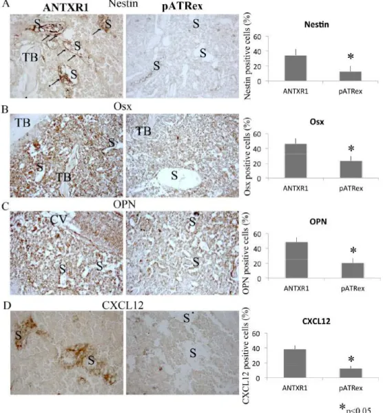
Par-ticularly, Nestin+ cells have been identified as MSC
sub-population within bone marrow responsible for HSC
regula-tion [23]. Moreover, Osterix+ (Osx+) osteolineage cells
regu-late the maturation of early B lymphoid precursors creating microniches for B cell development [24]. In addition, osteo-pontin (OPN), matrix glycoprotein expressed by the os-teoblasts, supports the migration and the adhesion of HSC toward to the osteoblastic niche and negatively regulates HSC proliferation, contributing to the maintenance of a qui-escent state [25, 26]. In line, using immunohistochemical techniques we observed that MSC and osteoblast pool was reduced. Particularly, the decrease of Nestin+ and Osx+ pool, found in pATRex group compared with the control group, predicted variations of the endosteal niche cellular inhabitants (Fig. 3A, B). In addition, the reduction of OPN in pATRex-treated mice (Fig. 3C) could be ascribed to the di-rect consequence of the reduction of the bone progeni-tor/mature forming components.
Fig. (1). Representative sections H&E (A) and toluidine bleu (B) stained of femour sub-metaphyseal area after ANTXR1 or pATRex
admini-stration. Note the fat cells infiltration near the endosteal niche (red dashed line). Magnification 40x. N= 6 per group.
Fig. (2). Pro-inflammatory (A) and anti-inflammatory (B) cytokines and chemokines production from BMC cultures obtained by ANTXR1
and pATRex treated mice was analyzed using Mouse Cytokine Array Panel A kit (R&D Systems, Milano, Italy) accordingly to the manufac-turer’s instructions as specified in the M&M section. Immunoreactive spots were quantitated densitometrically using Image J software. (p<0.05) N= 6 per group.
The concurrent decrease in osteolineage cells with paral-lel increase of fat cells in pATRex-treated mice group might predict a deregulation of MSC fate toward adipo-genesis than osteoadipo-genesis. Nonetheless, previous our
re-sults sustained the above posture. Namely, in BMSCs of pATRex treated mice, we found a decrease of Runx2 and Osx, both widely recognized as the main transcriptional factors of osteoblast differentiation [9].
Fig. (4). Representative Western blotting of CXCL12 and its
recep-tor CXCR4 of total bone marrow population from femours of ANTXR1 and pATRex-treated mice. (p<0.05). N= 6 per group.
Osteoblasts and MSCs express the soluble stromal-cell-derived factor 1 (also termed as CXCL12), that binds to its specific receptor C-X-C chemokine receptor type 4 (CXCR4) and prevents HSC mobilization supporting HSC maintenance [27, 28]. Interestingly, our findings revealed a reduced expression and a diminished perivascular
distribu-tion of CXCL12 in pATRex-treated mice in respect with control group (Fig. 3D). As further evidence, western blot-ting analysis showed a reduce expression of CXCL12 and its receptor CXCR4 (Fig. 4). Since CXCL12/CXCR4 signaling regulates osteoclast recruitment, bone resorption, hema-topoietic chemotaxis, fusion and cell survival [29, 30], we speculated that peculiar vascular niche metabolic feature, attributed to CXCL12/CXCR4 signaling, has been modified in the bone marrow of pATRex-treated mice. This instance is toughly buttressed by the observation of bone marrow hypocellularity in pATRex treated mice. Surprisingly we observed morphological modifications, which reflects in architectural niche changes. The vascular sinuses (Fig. 5A,
C, E) were interspersed among adipocytes (Fig. 5A, C) and
the endosteal niche cellularity were decreased with disrupted bone lining cell micro-architecture (Fig. 5B, D, F). The fact that there was no apparent change in appearance or distribu-tion of the remaining hematopoietic tissue, predicting a de-creased number of all hematopoietic cell lines in the bone
Fig. (3). Immunohistochemistry and quantification of Nestin (A), Osx (B), OPN (C) and Cxcl12 (D) expression in pATRex-treated and
ANTXR1-treated mice. Note the disrupted distribution of Nestin (A) and Cxcl12 (D) immunostaining in pATRex-treated cells; on the con-trary ANTXR1-treated samples exhibit a localization pattern, primarily around the perivascular mesenchymal and endothelial cells lining the sinusoid throughout the bone marrow (arrows). TB: trabecular bone; S: sinusoid. Magnification 40x. N= 6 per group.
Fig. (5). Histological sections illustrate the bone marrow cellularity after ANTXR1 treatment with HSCs residing in close contact with
vascu-lar and perivascuvascu-lar cells but also megakaryocytes and other stromal cells (Aa, Ca); note also the distribution of the endosteal lining bone cells and the HSCs in close proximity (arrows) (Ba, Da). pATRex-treated group revealed a decrease in bone marrow hematopoietic cellular density at the perivascular (Ab, Cb) or endosteal (Bb, Db) compartments, probably due to adipocytes infiltration. Notably, after pATRex ad-ministration the hematopoietic and stromal cells were found scattered among the fat cells and the sinus micro-architecture toughly disturbed by the adipocyte pool (E, F). The distinct, brown-colored, closely packed granules observed referred as hemosiderin granules particularly evident in pATRex-treated group. Hs: hemosiderin; S: sinusoid; A: adipocyte. Magnification A, C 40x; B, D, 60x. Reconstruction E, F. N= 6 per group.
marrow. In this scenario, H&E sections revealed an aplastic marrow, which appears devoid of hematopoietic cells, con-sisting primarily of vascular sinuses and adipose tissue. The aplastic-like marrow was primarily observed at the sub-metaphyseal level with a progressive reduction moving to-wards the diaphysis of the mice femour. Considering that approximately 70 to 80% of the marrow being hematopoietic tissue in mice [31] and that changes in bone marrow cellula- rity can be an indicator of systemic toxicity affecting multi-ple cell lineages [32], here we underscored the unseen reac-togenicity of the aggregosome-induced adjuvants transduced in a pancytopenia-like condition.
CONCLUSION
In this report, we extended our knowledge concerning the safety of genetic adjuvants, which promote intracellular pro-tein aggregates and sequentially induce frustrated autophagy and enhanced vaccine immunogenicity. Fully consistent with our previous observation in bone tissue, here we uncovered that protein aggregate formation, also seen in aggregosome disorders within a neurodegenerative proteinopathy scenario, disrupt bone marrow homeostatic features and drives to en-feebled physical, autocrine and/or paracrine progeni-tor/mature cell communication.
Considering that preceding studies evidenced that pA-TRex DNA administration did not trigger histopathological changes in a wide array of soft tissues [3], we undoubtedly
sustain that bones [9] and bone marrow remain the preferen-tial target tissues of these injectable adjuvants in mice. This hypothesis is also consistent with other findings underlining
that amyloid β peptides from Alzheimer patients have been
found stored in osteoporotic bone tissue with concomitant increase of osteoclastic activity [33].
Collectively, our data point on the fact that, albeit the op-tions for gene-based immunomodulatory proteins are consis-tently developed, the real challenge resides in finding the optimal choice for each adjuvant that will lead to protective outcomes in clinical trials.
ETHICS APPROVAL AND CONSENT TO PARTICI-PATE
Ethical approval is according to the recommendation of the Italian Ethical Committee and under the supervision of authorized investigators.
HUMAN AND ANIMAL RIGHTS
All the bone tissues implicated in this study were
achie-ved from our previous work (CGT, Agas et al. 2016). The
institutional and national guidelines for the care and use of laboratory animals was followed.
CONSENT FOR PUBLICATION
CONFLICT OF INTEREST
The authors declare no conflict of interest, financial or otherwise.
ACKNOWLEDGEMENTS
This work was partially supported by University Re-search Projects (Unicam- FAR) grant to MGS. Authors ac-knowledge receipt of funding from the European Commis-sion of a H2020-MSCA-ITN-2015 award through the ISPIC project (grant number 675743) and an H2020-MSCA-RISE-2016 award through the CHARMED project (grant number 734684).
BIBLIOGRAPHY
[1] Flingai S, Czerwonko M, Goodman J et al. Synthetic DNA vac-cines: improved vaccine potency by electroporation and co-delivered genetic adjuvants. Front Immunol 2013; 4: 354.
[2] Suschak JJ, Schmaljohn CS. Future Approaches to DNA
Vaccina-tion Against Hemorrhagic Fever Viruses. Methods Mol Biol 2018; 1604: 339-48.
[3] Felicetti P, Mennecozzi M, Barucca A et al. Tumor endothelial marker 8 enhances tumor immunity in conjunction with immuniza-tion against differentiaimmuniza-tion. Ag Cytotherapy 2007; 9: 23-34.
[4] Capitani M, Saade F, Havas KM et al. Plasmids encoding protein
aggregation domains act as molecular adjuvants for DNA vaccines. Curr Gene Ther 2014; 14: 161-9.
[5] Hermesh T, Moltedo B, Moran TM, López CB. Antiviral instruc-tion of bone marrow leukocytes during respiratory viral infecinstruc-tions. Cell Host Microbe 2010; 7: 343-53.
[6] Raggatt LJ, Partridge NC. Cellular and molecular mechanisms of bone remodeling. J Biol Chem 2010; 285: 25103-08.
[7] Redlich K, Smolen JS. Inflammatory bone loss: pathogenesis and therapeutic intervention. Nat Rev Drug Discov 2012; 11: 234-50.
[8] Agas D, Marchetti L, Douni E, Sabbieti MG. The unbearable
light-ness of bone marrow homeostasis. Cytokine Growth Factor Rev 2015; 26: 347-59.
[9] Agas D, Concetti F, Capitani M et al. Administration of DNA Plasmid Coding Protein Aggregating Domain Induces Inflamma-tory Bone Loss. Curr Gene Ther 2016; 16: 144-52.
[10] Zhao E, Xu H, Wang L et al. Bone marrow and the control of im-munity. Cell Mol Immunol 2012; 9: 11-9.
[11] Coelho-Castelo AM, Trombone AP, Rosada RS et al. Tissue distri-bution of a plasmid DNA encoding Hsp65 gene is dependent on the dose administered through intramuscular delivery. Genetic Vac-cines and Therapy 2006; 4: 1-10.
[12] Rush CM, Mitchell TJ, Garside P. A detailed characterisation of the distribution and presentation of DNA vaccine encoded antigen. Vaccine 2010; 28: 1620-34.
[13] Agas D, Gusmão Silva G, Laus F et al. INF-γ encoding plasmid administration triggers bone loss and disrupts bone marrow micro-environment. J Endocrinol 2017; 232: 309-21.
[14] Sabbieti MG, Agas D, Marchetti L et al. Signaling pathways impli-cated in PGF2alpha effects on Fgf2+/+ and Fgf2-/- osteoblasts. J Cell Physiol 2010; 224: 465-74.
[15] Sabbieti MG, Marchetti L, Censi R, Lacava G, Agas D. Role of PTH in Bone Marrow Niche and HSC Regulation. Curr Stem Cell Rep 2017; 3: 210.
[16] Adler BJ, Kaushansky K, Rubin CT. Obesity-driven disruption of haematopoiesis and the bone marrow niche. Nat Rev Endocrinol 2014; 10: 737-48.
[17] Takayanagi H. Osteoimmunology: shared mechanisms and cros-stalk between the immune and bone systems. Nat Rev Immunol 2007; 7: 292–304.
[18] Herman S, Kronke G, Schett G. Molecular mechanisms of inflam-matory bone damage: emerging targets for therapy. Trends Mol Med 2008; 14: 245–53.
[19] Datta HK, Ng WF, Walker JA, Tuck SP, Varanasi SS. The cell biology of bone metabolism. J Clin Pathol 2008; 61: 577–87.
[20] Calvi LM, Adams GB, Weibrecht KW et al. Osteoblastic cells
regulate the haematopoietic stem cell niche. Nature 2003; 425: 841–6.
[21] Bowers M, Zhang B, Ho Y et al. Osteoblast ablation reduces nor-mal long-term hematopoietic stem cell self-renewal but accelerates leukemia development. Blood 2015; 125: 2678–88.
[22] Zhao M, Li L. Osteoblast ablation burns out functional stem cells. Blood 2015; 125: 2590–1.
[23] Mendez-Ferrer S, Michurina TV, Ferraro F et al. Mesenchymal and haematopoietic stem cells form a unique bone marrow niche. Na-ture 2010; 466: 829–34.
[24] Yu VW, Scadden DT. Heterogeneity of the bone marrow niche. Curr Opin Hematol 2016; 23: 331-8.
[25] Nilsson SK, Johnston HM, Whitty GA et al. Osteopontin, a key component of the hematopoietic stem cell niche and regulator of primitive hematopoietic progenitor cells. Blood 2005; 106: 1232–9.
[26] Guerrouahen BS, Al-Hijji I, Tabrizi AR. Osteoblastic and vascular endothelial niches, their control on normal hematopoietic stem cells, and their consequences on the development of leukemia. Stem Cells Int 2011; 2011: 375857.
[27] Tzeng YS, Li H, Kang YL et al. Loss of Cxcl12/Sdf-1 in adult mice decreases the quiescent state of hematopoietic stem/progenitor cells and alters the pattern of hematopoietic regen-eration after myelosuppression. Blood 2011; 117: 429–39.
[28] Kirito K, Fox N, Kaushansky K. Thrombopoietin induces HOXA9
nuclear transport in immature hematopoietic cells: potential mechanism by which the hormone favorably affects hematopoietic stem cells. Mol Cell Biol 2004; 24: 6751–62.
[29] Zhu W, Boachie-Adjei O, Rawlins BA et al. A novel regulatory role for stromal-derived factor-1 signaling in bone morphogenic protein-2 osteogenic differentiation of mesenchymal C2C12 cells. J Biol Chem 2007; 282: 18676-85.
[30] Shahnazari M, Chu V, Wronski TJ, Nissenson RA, Halloran BP. CXCL12/CXCR4 signaling in the osteoblast regulates the mesen-chymal stem cell and osteoclast lineage populations. FASEB J 2013; 27: 3505-13.
[31] Valli VE, McGrath JP, Chu I. Hematopoietic system. In: Haschek WM, Rousseaux CG, Wallig MA, Eds. Handbook of Toxicologic Pathology, 2nd ed. Academic Press: San Diego, CA 2002; pp. 647– 79.
[32] Elmore SA. Enhanced histopathology of the bone marrow. Toxicol Pathol 2006; 34: 666-86.
[33] Li S, Liu B, Zhang L, Rong L. Amyloid β peptide is elevated in osteoporotic bone tissues and enhances osteoclast function. Bone 2014; 61: 164-75.