SCUOLA NORMALE SUPERIORE
Pisa
Classe di Scienze Matematiche, Fisiche e Naturali
Corso di Perfezionamento in Neurobiologia
Triennio 2003-2005
Tesi di Perfezionamento
PLASTICITY OF THE MAMMALIAN RETINA
DURING DEVELOPMENT
Candidata:
Silvia Landi
Relatori:
Prof.ssa Nicoletta Berardi
Prof. Lamberto Maffei
INDEX
INTRODUCTION
The Rodent visual system p.4
The target of this study: the retina p.5
Architecture of the mature retina p.5 Photoreceptors
Bipolar cells Horizontal cells Amacrine cells
Retinal ganglion cells
Mammalian retina and its development p.11 Intrinsic and environmental cues shaping retinal neurons during development p.16
Cell-cell interactions Neurotransmission Neurotrophins
Neural plasticity p.22
Visual cortical plasticity p.23
NMDA receptors Neurotrophins
Intracortical inhibition Intracellular signalling Extracellular environment
Is the retina plastic? p.30
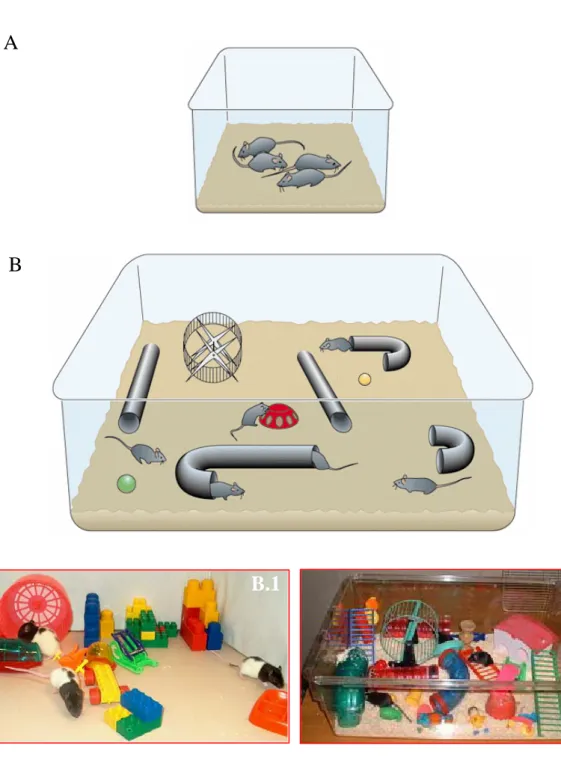
Environmental enrichment as a tool to unmask retinal plasticity p.31
The experimental protocol p.31 Environmental enrichment and its influence on adult brain p.34 Environmental enrichment and visual system development p.36 Enrichment and maternal care p.38
Aim of this work p.40
MATERIALS AND METHODS
Animal handing and treatments: p.41 Mice
Rats
Animal treatments
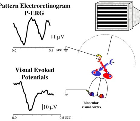
Immunohistochemistry p.45 Analysis of dendritic RGC arborizations p.45 Analysis of BDNF expression in the retina p.46 Electrophysiological assessment of retinal and cortical acuity p.46 Pattern electroretinogram (P-ERG)
Visual Evoked Potentials (VEPs)
RESULTS
PART I: RGC developmental stratification is influenced by environmental enrichment
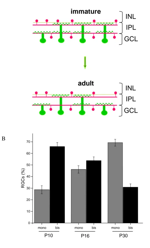
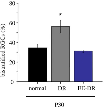
Segregation of RGC dendrite stratification p.49 RGCs stratify during postnatal development p.51 EE from birth counteracts dark rearing effects promoting RGC dendritic maturation p.51 EE early in life affects the maturational refinement of RGC dendrites p.55 The acceleration of RGC dendrite segregation induced by EE is dependent on the enhanced levels of BDNF p.57
PART II: Retinal functional development is affected by environmental enrichment
Development of retinal acuity is accelerated by EE p.60 Precocious eye opening is not responsible for the effects of EE on retinal acuity development p.65 Accelerated retinal development in EE animals is induced during early phase of
Development of BDNF protein level in RGC layer is affected by EE p.68 BDNF mediates EE effects on retinal functional development p.71 IGF-I is capable to enhance retinal acuity in standard reared rats p.74
DISCUSSION
Retinal ganglion cell developmental stratification is influenced by environmental enrichment p.78 Sensitivity of retinal functional development to environmental enrichment p.80 A possible link between RGC anatomical development and P-ERG acuity maturation Eye opening is a trigger or a consequence of EE effects? p.83 EE, maternal care and retinal development p.85
CONCLUSIONS p.86
REFERENCES p.89
INTRODUCTION
Understanding the effects of experience on neural circuitry development and the underlying mechanisms is an important issue in neurobiology. A large literature documents the plasticity of nervous structures such as the cortex or hippocampus in response to experience during development (reviewed in Berardi et al., 2003; Mangina and Sokolov, 2005); the retina on the contrary has generally been thought of as unresponsive to experience. It was well known, indeed that while the visual cortex responds to paradigms of visual deprivations, such as monocular deprivation (MD) or dark rearing (DR) with dramatic functional and anatomical alterations, retinal development was not substantially modified in cats, rats and humans by these rearing conditions (Baro et al., 1990; Fagiolini et al., 1994; Fine et al., 2003). Recently, there has been the first indication of retinal response to experience in mice by Tian and Copenhagen (2001 and 2003). Their studies have shown that retinal development is sensitive to DR; in particular, retinal ganglion cell (RGC) development is affected by DR both at electrophysiological and anatomical level. In this work, we have chosen to investigate the actual sensitivity of the developing retina to experience; in particular, we have used a paradigm of complex sensory-motor stimulation as that provided by environmental enrichment (EE). EE seemed to us a valid tool to probe retinal plasticity, since we have recently found that it affects visual cortical development and plasticity (Cancedda et al., 2004) and prevents DR effects (Bartoletti et al., 2004). We have studied the anatomical and functional maturation of Rodent retina, analyzing the development of RGC dendritic stratification and of retinal acuity, and the molecular factors activated by EE and capable to trigger these changes.
We find a remarkable response of RGC developmental remodelling of dendritic stratification to EE, both in normal and in DR animals and a response of functional retinal development to EE as robust as shown by the developing visual cortex. Both effects of EE requires retinal BDNF action.
The Rodent visual system
In the Mammalian visual system, visual information are processed in the retina and sent to different structures of the CNS through RGC axons, which represent the output of the retina. RGCs project to the visual centres of the brain that are located in the midbrain and in the thalamus. The pattern of retinal projections varies considerably from species to species. In Rodents, the vast majority of RGCs project to the superior colliculus (SC) and the
pretectal nuclei, with about 30% of them sending collaterals to the dorsal-lateral geniculate nucleus (dLGN) in the thalamus (Sefton and Dreher, 1985). RGC axons from each eye project to both sides of the brain, however the major afferents to the SC and dLGN arise from the contralateral eye and only 5% of optic axons project ipsilaterally. Within the dLGN ganglion cell axons are not intermixed; in cats, ferrets and primates they terminate in a set of separate, alternate eye-specific layers that are strictly monocular (Hickley and Guillery, 1974). In Rodents there is not a proper lamination of the dLGN; however, even if the rat dLGN is not clearly laminated, ipsilateral and contralateral retinal fibers are segregated in a patchy fashion originating two eye-specific territories in the dLGN: the ipsilateral patch or inner core and the contralateral patch or outer shell (Reese and Jeffery, 1983; Reese, 1988). The geniculate body provides ascending input to the visual cortex via thalamo-cortical connections that terminate in the layer IV of the primary visual cortex. In carnivores and primates, afferents from the dLGN segregate by eye within the cortical layer IV into alternating, equal-sized stripes called ocular dominance columns (Hubel and Wiesel, 1963; Shatz and Stryker, 1978). This functional and structural organization has been found also in Rodents, although at a very primitive state; Thurlow and Cooper (1988) observed hints of patchy organization of ipsilateral and contralateral inputs in the visual cortex of hooded rats, using a functional marker as deoxiglucose and this has been confirmed with electrophysiological techniques by Caleo et al., 1999.
The target of this study: the retina Architecture of the mature retina
Unlike other sensory structures, such as cochlea or somatic receptors in the skin, retina is not a peripheral organ but part of the CNS and its synaptic organization is similar to that of other central neural structures. The identification and classification of retinal neurons begun more than 100 years ago by Santiago Ramon y Cajal and is nearing completion (Masland et al., 2001).
Briefly, light is focused by the cornea and the lens, onto the photoreceptor layer in the retina. The retina lies in front of the pigmented epithelium that lines the back of the eye; these cells contain melatonin that adsorbs any light not captured by the retina and this prevents the light from being reflected off the back of the eye to the retina again -which would degrade the visual image. To allow light reach the photoreceptors without being adsorbed or greatly scattered, the axons of neurons in the proximal layers of the retina are unmyelinated so that these layers of cells are relatively transparent. Moreover, in one
region of the retina, the fovea, the cell bodies of the proximal retinal neurons are shifted to the side, enabling the photoreceptors there to receive the visual image in the least distorted form; the shifting is more pronounced in the foveola, the center of the fovea. Fovea, as a specialized retinal region, is not present in the retina of Rodents.
Although much remains to be learned, the fundamental structural principles are now becoming clear giving a bottom-up view of the strategies used in the retina’s processing of visual information.
Retina consists of many parallel, anatomically equipotent microcircuits; it presents an intricate pattern of connections in spite of a layered anatomical rearrangement. Mammalian retina contains a huge diversity of neuronal types; it is composed approximately by 55 distinct cell types, each with a different function.
In particular, five different layers can be identified: ONL -outer nuclear layer- with cell bodies of photoreceptors, OPL -outer plexiform layer- with cone and rod axons, horizontal cell dendrites, bipolar dendrites, INL -inner nuclear layer- with nuclei of horizontal cells, bipolar cells, amacrine cells and Müller cells, IPL -inner plexiform layer- with axons of bipolar cells and amacrine cells, dendrites of ganglion cells and GCL -ganglion cell layer- with the soma of ganglion cells and displaced amacrine cells.
Photoreceptors
In nocturnal Rodents the number of rods is many-fold that of cones; in particular, in mice cones are about 3% of photoreceptors (Jeon et al., 1998). A typical mammalian retina contains 9-11 types of cone-driven bipolar cells. Everyone of these assortments of pathways from cones to the inner retina is responsible for a different type of information and this characteristic initially attributed to the diverse cell structure and to the different proteins expressed, is now known also at a functional level.
First, the output of the cone photoreceptors is separated into ON and OFF signals. All cone synapses release glutamate, but bipolar cell types respond to glutamate differently. Some bipolar cells have ionotropic glutamate receptors: glutamate opens a cation channel, and the cell depolarizes. Other bipolar cells have a sign-inverting synapse mediated by metabotropic glutamate receptors, mainly mGluR6; these bipolar cells hyperpolarize in response to glutamate (Nawi et al., 1991). Photoreceptor cells hyperpolarize after light absorption, causing their synapses to release less glutamate. When the retina is stimulated by light, one type of bipolar cell hyperpolarizes and the other type depolarizes. OFF and ON bipolar cells occur in approximately equal numbers. The distinction, created at the first retinal synapse, is propagated throughout the visual system.
Bipolar cells
The classes of ON and OFF bipolars are each further subdivided; there are three to five distinct types of ON and three to five types of OFF bipolars. The purpose of the subdivision is, at least in part, to provide separate channels for high-frequency (transient) and low-frequency (sustained) information. Thus, there are separate ON-transient, ON-sustained, OFF-transient and OFF sustained bipolar cells (Kaneko et al., 1970; Awatramani et al., 2000). An elegant series of experiments shows that the distinction is caused by different glutamate receptors on the respective OFF bipolar cells; they recover from desensitization quickly in the transient cells and more slowly in the sustained cells (DeVries et al., 2000). An important point is that there are no dedicated cones -cones that provide input, say, only to ON bipolars or only to OFF bipolars. Instead, the output of each cone is tapped by several bipolar cell types to provide many parallel channels, each communicating a different version of the cone’s output to the inner retina.
Most amacrine cells and all ganglion cells receive their main bipolar cell synapses from cone bipolars, but retinas work in starlight as well as daylight, and this range is created by a division of labor between cones (for bright light) and rods (for dim light). Signals originating in rod photoreceptors reach the RGCs via an indirect route using as its final path the axon terminals of the cone bipolar cells (Famiglietti and Kolb, 1975; Strettoi et al., 1990 and 1992). That a single set of ganglion cells is used for both starlight and sunlight represents an obvious efficiency, long known from electrophysiological findings. However, it was not obvious a priori that rod-driven information would reach the ganglion cells by an indirect path. Furthermore, rod photoreceptors far outnumber cones in most mammalian retinas; it was a surprise to learn, when quantitative methods became available, that cone bipolars outnumber rod bipolars in all but a few mammalian retinas (Strettoi et al., 1995). The reason is that more rods converge onto a single rod bipolar than cones onto cone bipolars; the rod system trades acuity for sensitivity, and the circuitry associated with rods is simpler than that of cones. Because rods evolved after cones, the likely scenario is that the rod circuitry was grafted onto the cone pathways.
Only one kind of rod photoreceptor exists, and rods drive only a single type of bipolar cell. It synapses on a specialized amacrine cell, termed AII, which then transmits the output of rod bipolar cells to ganglion cells. This occurs largely via synapses (chemical or gap junctional) by AII onto axon terminals of cone bipolar cells, which then excite the ganglion cells. It may seem strange that rod bipolar cells would not simply drive retinal ganglion cells directly, but seems less strange when one appreciates the complexity of the
pre-existing inner retinal circuitry of the cone pathways. By synapsing on the axon of the cone bipolar cell, the rod pathway gains access to the elaborate circuitry of the cone pathway, including its associated amacrine circuitry. For example, the directionally selective type of ganglion cell retains its function in very dim light, even though it receives no direct synapses from the rod bipolar cells. The rod system piggybacks on the cone circuitry rather than re-inventing it (Masland et al., 2001).
Horizontal cells
All rods and cones receive feedback from horizontal cells, but these cells are a numerically small proportion of the retina’s interneurons, generally less than 5% of cells of the inner nuclear layer (Jeon et al., 1998). In most mammals, there are two morphologically distinct types of horizontal cells, while Rodents have only one. In monkeys, these have different numbers of synapses with different types of cones. The reason for this biasing is not yet certain; it may involve chromatic opponency in the red–green system. Traditionally, horizontal cells are said to enhance contrast between adjacent light and dark regions. Excitation of a central cone causes feedback inhibition of both the excited cone and a ring of neighbouring ones. Because each cone -both the central one and its neighbours- transmits a signal to the inner retina, the upshot is that a small stimulus excites those ganglion cells that lie directly under the stimulus, but inhibits neighbouring ganglion cells. This is the classical ‘center–surround’ organization, in which a ganglion cell is excited or inhibited by stimuli falling in its receptive field center, whereas stimulation of the surrounding region has an opposite effect. An alternate formulation of the same facts is that horizontal cells adjust the system’s response to the overall level of illumination; they measure illumination across a broad region and subtract it from the signal that is transmitted to the inner retina about a local image. In effect, this reduces redundancy in the signal transmitted to the inner retina. The mean luminance across a large region of retina is shared by many cones and contains little information.
Amacrine cells
All retinal ganglion cells receive input from cone bipolar cells, but direct synapses from bipolar cells are a minority of all synapses on the ganglion cells; most are from amacrine cells (Freed et al., 1988; Calkins et al., 1994). The exact fraction varies among different functional types of ganglion cells, ranging from roughly 70% for alpha cells (large, movement-sensitive ganglion cells found in most mammals) to 50% for the midget ganglion cells located in the monkey central fovea. Amacrine cells also make inhibitory synapses on the axon terminals of bipolar cells, thus controlling their output to ganglion
cells. In contrast to horizontal cells, which have a single broad role, amacrine cells have dedicated functions since they carry out narrow tasks concerned with shaping and control of ganglion cell responses. Amacrine cells outnumber horizontal cells by amounts that range from 4:1 to 10:1 (depending on the species) and can outnumber ganglion cells by 15 to 1. The different amacrine cells have distinct pre- and postsynaptic partners, contain a variety of neurotransmitters, survey narrow areas of the visual scene or broad ones, branch within one level of the inner synaptic layer or communicate among many. Both the specific molecules expressed and their morphology point to diverse functions.
Those amacrine cells with functions that are more precisely understood do remarkably specific jobs. The dopaminergic amacrine cells globally adjust the retina’s responsiveness under bright or dim light. Dopamine affects many elements of the retina’s circuitry; it alters the gap-junctional conductance between horizontal cells and between amacrine cells (Hampson et al., 1992), potentiates the responses of ionotropic glutamate receptors on bipolar cells, and ultimately affects the center–surround balance of ganglion cells. Remarkably, retinal dopamine can even cause pigment migration in cells of the retinal pigment epithelium, a neighbouring non-neural tissue. In the latter case (and very likely some of the former as well), this is mediated non-synaptically, via a diffuse, paracrine release of the neurotransmitter. In contrast, the starburst amacrine cells seem to be narrowly associated with a particular computational circuit. They arborize in thin (2-4 μm) strata within the IPL, where they make excitatory cholinergic synapses on certain RGCs, notably those particularly sensitive to moving stimuli. By feed forward excitation and/or inhibition (these neurons release both acetylcholine and GABA), starbust cells are important for direction selectivity (Masland et al., 1986).
Retinal ganglion cells
RGCs process and convey information from the retina to visual centres in the brain. These output neurons comprise subpopulations with distinct structure and function (Sernagor et al., 2001). The morphology of RGCs is highly disparate; their somata and dendritic field vary in size, and they exhibit strikingly varied dendritic architecture (Cajal, 1893; Wassle and Boycott, 1991; Rodieck, 1998) and axonal projection patterns (Garraghty and Sur, 1993; Yamagata and Sanes, 1995a,b). Functionally, RGCs differ in their response to light in a variety of ways (reviewed by Wassle and Boycott, 1991; Rodieck, 1998; Dacey, 1999). Their response to light may be transient or sustained, brisk or sluggish, tonic or phasic. Some RGCs are good motion detectors and may prefer a specific direction of stimulus movement, whereas others are sensitive to the orientation of the stimulus but not
to its direction. In addition, RGCs show different contrast sensitivity, visual acuity, and color-coding. Despite the enormous diversity in structure and function, combined anatomical and electrophysiological studies have revealed a close correlation between the morphology and function of RGCs in vertebrates (Saito, 1983; Stanford and Sherman, 1984; Amthor et al., 1984, 1989a, b; Dacey, 1999).
Within a species, structure and function studies have enabled classification of RGCs into broad subclasses (Rockhill et al., 2002). In Primate retina, RGCs fall into two functional classes, M (for magno or large) or midget cells and P (for parvo or small) or parasol cells. Each class includes both on-center and off-center cells. M cells have large receptive fields (reflected in their large dendritic arbors) and respond relatively transiently to sustained illumination. They respond optimally to large objects and are able to follow rapid changes in the stimulus; on the contrary, the smaller P cells, which are numerous, have small receptive fields, respond specifically to certain wavelengths and are involved in the perception of form and color. P cells are thought to be responsible for the analysis of fine details in the visual image, although some M cells may also be involved in this function. In the well-studied cat retina, small-field beta RGCs are the anatomical correlate of physiologically identified brisk-sustained or X-RGCs, and large-field alpha RGCs are correlated with brisk-sustained or Y-RGCs (reviewed by Wassle and Boycott, 1991). Major subclasses of RGCs, such as the alpha and beta cells in cat, can be further divided into subtypes, notably those which are depolarized (ON RGCs), or hyperpolarized (OFF RGCs), by light. In general, within a species, each subtype of RGC shares key features: (i) their dendritic branching patterns and arbor size are similar at any fixed retinal location; (ii) their dendritic fields overlap forming mosaics that cover the retinal surface effectively (Wassle et al., 1983; Cook and Chalupa, 2000); (iii) they receive the same complement of presynaptic inputs; (iv) they project to common regions within targets in the brain. But not all RGC subclasses defined within one species are present in all species. However, in all species studied thus far, the IPL, the plexus within which RGCs form intraretinal connections, is organized into structurally and functionally distinct sublaminae. Irrespective of RGC subclass, ON RGCs have dendritic arbors that stratify in the inner region (sublamina b) of the IPL, whereas OFF RGCs stratify in the outer sublamina (sublamina a) of the IPL (Famiglietti and Kolb, 1976; Nelson et al., 1978). Cells with arbors in both sublaminae have ON and OFF responses (e.g. Amthor et al., 1984). The diverse morphological and physiological properties of RGCs have presented an enormous challenge to investigators seeking to understand how the visual image is encoded and relayed to the brain. For
developmental neurobiologists, the rich diversity of RGC structure and function make these neurons ideal for studies of cell-fate determination (reviewed by Cepko et al., 1996; Harris,1997; Rapaport and Dorsky, 1998) and axonal and dendritic development (Goodman and Shatz, 1993; Wong and Wong, 2000).
Mammalian retina and its development
The eye originates as a bilateral organ from a single field in the anterior neural plate. The primordial eye field is separated into two regions by anterior migration of diencephalic precursor cells along the midline. Proliferation and evagination give rise to the optic vesicles. Their infolding into optic cups and their progressive determination originates the optic stalk, the neural retina and the retinal pigment epithelium. In the retina, cell differentiation from retinal precursors is initiated in the inner layer of the central portion of the optic cup to progress concentrically in a wave-like fashion towards the peripheral edges of the retina (Isenmann et al., 2003).
Neurones seems to be generated in the same sequence during the first phase of ventricular cytogenesis in all species analyzed apart from minor differences: RGC, displaced amacrine cells, horizontal cells and cone photoreceptors. Progenitor cells in the neurepithelium lining the surface of the neural tube, later become the ventricular zone of the optic vesicles, optic cup and early retina. Postmitotic cells leave the ventricular zone to migrate to one of three cell layers in the retina remaining attached radially from one side of the retina to the other, as noted by Cajal a century ago. The neural cells lie at different levels in the retina and when in correct position lose their anchoring radial connections. Then, polarity of the differentiating cells occurs and dendrites and axons grow out appropriately. Actually, ultrastructural studies (Olney, 1968; Fisher, 1979a; Blanks et al., 1974) suggest that synaptogenesis between the major neuronal classes of the vertebrate retina occurs in three major steps. Retinal ganglion cells and amacrine cells are the first cell classes to differentiate and form the earliest functional circuits in the IPL of the developing retina. Shortly after, horizontal cells and photoreceptors differentiate and contact each other in the outer retina, forming the OPL. The vertical networks in the inner and outer retina are later interconnected when bipolar cells are born and connections with ganglion cells are established. This sequential pattern of retinal circuit development is common across species, although the separation in time between inner and outer retinal circuit development varies, ranging from a few hours in animals such as the zebrafish to many days and weeks in Mammals.
Finally, bipolar cells, rod photoreceptors and Muller cells are generated throughout the second phase. Therefore, Muller cells, so important in guiding optic nerve formation and in organizing plexiform and nuclear layers, are evident in two different waves of propagation by using different types of labelling. After the cytogenesis in the ventricular zone, cell proliferation continues in the sub-ventricular zone with the generation of macroglia (Ichikawa et al., 1983), microglia (Kitamura et al., 1984) and certain classes of intrinsic neurons. A fundamental process in retinal development is cell loss by apoptosis; 54-74% of axons initially present in the mammalian optic nerve are eliminated during development and so a corresponding number of RGCs and amacrine cells undergo this fate (Dreher and Robinson, 1988).
There is good evidence that neurotransmitters can be found at the earliest stages of retinal development and these neurotransmitters can function in the absence of traditional synapses (Redburn and Rowe-Rendleman, 1996). For instance, cholinergic neurons can be observed by means of antibodies against choline acetyltransferase and acetylcholine esterase in the neuroblastic layer as early as embryonic day 3 in chick and P0 in the ferret and mouse (Feller et al., 1996; Bansal et al., 2000), the developmental period during which amacrine cells are being generated. These cells are presumably starburst amacrine cells, the only source of acetylcholine (ACh) in the adult retina. By monitoring intracellular calcium concentrations using fluorescence imaging, Wong (1995) showed that during these initial stages of retinal development muscarinic acetylcholine receptor (mAChR) agonists cause substantial increases in intracellular calcium of many cells in the neuroblastic layer.
M-AChRs are cGMP gated channels that lead to increases in intracellular calcium by causing a release of calcium from internal stores, as opposed to influx through ligand- or voltage-activated channels. After the cells were postmitotic and began to migrate out of the ventricular zone, this responsiveness to mAChR agonists was reduced. Amacrine and ganglion cells still had responses to cholinergic agonists, but they were mediated via nicotinic receptors, as they are in the adult. Hence, it seems possible that even before cholinergic neurons have left the ventricular zone, and long before these neurons have formed synaptic connections, they could be inducing signalling that is important for early phases of neurogenesis and also cell migration. Another neurotransmitter, γ−aminobutyric acid- GABA is expressed in more cells during development than during adulthood. This exuberance of GABA positive neurons suggests that like ACh, GABA may play a transient role in circuit formation (for review, see Sandell, 1998). For instance, GABA is thought to play a role in synaptogenesis between cones and horizontal cells early in postnatal
development of the OPL in rabbit retina (Messersmith and Redburn, 1993). GABA has a particularly high and transient expression in the ganglion cell layer during the first few postnatal days of rabbit. In addition, markers for enzymes involved in the synthesis of GABA can be found on either side of the IPL early in development in ferret retina (Karne et al., 1997).
The first synaptically connected circuits that appear in the developing IPL are between amacrine and ganglion cells (Greiner and Weidman, 1981; Karne et al., 1997). Prior to photoreceptor maturation and eye opening, RGCs periodically fire bursts of action potentials. This spontaneous rhythmic activity was first measured in fetal rat pups. This activity was found to be highly correlated among neighbouring ganglion cells (Galli and Maffei, 1988). Both extracellular recording using a multielectrode array (Meister et al., 1991) and imaging of calcium transients associated with bursts of action potentials (Feller et al., 1997; Wong et al., 1995) have revealed that these spontaneous bursts propagate from one cell to the next in a wave-like manner. Recent experiments demonstrate that blockade of spontaneous retinal activity disrupts the normal pattern of retinal ganglion cell axons in its primary target, the lateral geniculate nucleus of the thalamus (Penn et al, 1998), indicating that spontaneous activity in the retina plays a critical role in the normal development of the adult visual system.
Retinal waves are a characteristic phenomenon, observed in a large variety of vertebrate species, including chick, turtle, mouse, rabbit, rat, ferret and cat (Wong, 1999). Though wave periodicity and velocity in all species are comparable, the circuitry underlying the propagation may be substantially different. Waves are first seen around the time that neurons residing in the inner retina are starting to form circuits while the outer retinal neurons have not made synaptic connections, and photoreceptors are not yet functional (Greiner and Weidman, 1981; Mey and Thanos, 1992). At this stage of development, ganglion cells have migrated into the ganglion cell layer and their axons have reached their primary targets, the lateral geniculate nucleus in mammals, and the tectum in chick.
In postnatal ferret and mice retinas, chemical synaptic transmission is a prerequisite for wave propagation, as indicated by several experimental results. First, simultaneous whole cell voltage clamp recordings from ganglion cells demonstrate that increases in [Ca2+]i correlated across cells are driven by compound synaptic inputs (Feller et al., 1996). Second, the compound postsynaptic currents measured from ganglion cells are blocked by bath application of Cd2+, a blocker of voltage-activated calcium channels, including those associated with transmitter release (Feller et al., 1996). Third, the periodic Ca2+ increases,
action potential, and compound postsynaptic currents associated with waves can all be blocked by a variety of nAChRs antagonists (Feller et al., 1996; Penn et al., 1994). Although in the adult retina, acetylcholine acts as a modulator of ganglion cell firing, while glutamate is the primary excitatory transmitter at the earliest ages studied, glutamatergic blockers do not affect wave generation (Wong, 1995).
The synaptic circuitry that drives retinal waves changes postnatally. Though cholinergic neurotransmission is required and GABA contributes to the depolarization of cells during retinal waves early in development in the ferret, recent studies in older ferrets indicate that waves are insensitive to cholinergic antagonists and can be blocked by glutamate receptor antagonists (Wong, 1999). This switch in the requisite transmitter occurs at the age that bipolar cells are making their initial synaptic connections with ganglion cells and when conventional synapses between amacrine and ganglion cells become morphologically mature and numerous.
GABA has a modulatory role in retinal waves. Imaging of the spontaneous increases in Ca2+ associated with waves has shown that GABA-A receptor antagonists can dramatically alter the amount of wave-induced depolarization (Fischer et al., 1998). GABA is the primary transmitter of most amacrine cells in the retina, and, at the youngest ages studied, it provides excitatory input for ganglion cells (Fischer et al., 1998). Unlike ACh, however, GABA does not influence wave periodicity, since GABA blockers do not alter either the frequency of the cholinergic barrages that are associated with waves measured in ganglion cells (Feller et al., 1996) or other properties of wave propagation at ages less than P10 (Fischer et al., 1998). Waves persist after GABA becomes inhibitory (Fischer et al., 1998). However, these changes in the circuitry mediating waves with development leads to changes in the frequency of events occurring in different subsets of ganglion cells.
Until few years ago, it was largely assumed that retinal function was mature by the time of eye opening (Tian et al., 2001). Consistent with this idea, most morphological and neurochemical systems of the retina as the number of cells, the number of ribbons and conventional synapses, the expression of synthetizing enzymes, of transporters and receptors for neurotransmitters have reached adult levels at the age of eye opening (Fisher, 1979a; Greiner and Weidman, 1981; Pow and Barnett, 2000; Sassoe-Pognetto and Wassle, 1997). Nonetheless, some studies show a continued maturation of visual responsiveness in retina after eye opening. In RGCs of cat and rabbit, the centers of the receptive fields shrink and the surrounds become much more prominent with age (Bowe-Anders et al., 1975; Rusoff and Dubin, 1977). Moreover, light-evoked response amplitudes of RGCs are larger
and the latencies are shorter after eye opening in cat (Tootle, 1993) and ferret (Wang et al., 2001).
Light responses emerge as the photoreceptor-bipolar pathway begins to mature shortly before eye opening (Dacheux and miller, 1981a, 1981b; McArdle et al., 1977; Tootle, 1993). Electrophysiological recordings from retinal ganglion cells show several major trends in the maturation of their responses to light. The early response of RGCs to light stimulation is weak and the cells adapt rapidly (Masland, 1977; Tootle, 1993). But when robust responses to light become detectable a few days later, the concentric center-surround organization of the receptive fields, as well as ON and OF center responses, are already present (Bowe-Anders et al., 1975; Masland, 1977; Tootle, 1993). Whether the connectivity that underlies these physiological properties is established before photoreceptors are present is unknown. Determining how surround inhibition appears in the RGCs has not been straightforward. Some immature rabbit RGCs have silent surrounds, that when stimulated, can suppress the response to center stimulation, but direct stimulation of the surround does not evoke a response (Masland, 1977). In the cat, however, the strength of the antagonistic surround relative to that of the center does not seem to change with postnatal maturation (Tootle, 1993). Recordings from ferrets, however, clearly demonstrate that connectivity in the inner retina is remodelled with maturation. In the postnatal ferret, α and β−like RGCs have convergent ON and OFF inputs prior to maturity (Wang et al., 2001). In these cells, maturation of the receptive field center responses thus involve the loss of one type of input. Specialized receptive fields properties such as direction selectivity also develop before eye opening (Masland, 1977; Sernagor and Grzywacz, 1995), although the synaptic basis for this property remains to be determined. How is the RGC receptive field established during development? Visual experience after eye opening does not appear to alter the receptive field properties of Mammals that were raised in an environment with unidirectionally moving stimuli (Daw and Wyatt, 1974). But this may be because, in rabbits, ganglion cell receptive fields are fairly mature by the time of eye opening (Masland, 1977). In contrast, the peak firing rate of RGCs in response to light stimulation is decreased in DR mice (Tian and Copenaghen, 2001). Cells responds more sluggishly in DR animals. The spatial organizations of the receptive fields have not yet been assessed after DR of mice. In turtles, which become light responsive prior to hatching, DR causes an increase in receptive field size (Sernagor and Grzywacz, 1996). However, this study suggests that spontaneous activity rather than visual stimulation regulates the receptive field size. Clearly, much
remains to be done to fill our knowledge gaps concerning how the light responses of RGCs are established in ways that are characteristic of each cell type.
Little is known of structural plasticity in the mature mammalian retina. Light adaptation is an archetypal plasticity that effects a functional transition from scotopic to photopic vision. In fishes and amphibians, both graphic structural and subtle molecular events attend light adaptation, including photomechanical movements of the RPE and photoreceptors, neurite extension and retraction by horizontal cells, and alterations in bipolar cell synaptic terminal structure. More subtly, but perhaps more physiologically evident, several molecular switches are invoked by light-adaptation, e.g. reduction of homologous coupling between horizontal cells (reviewed in Witkovsky, 1991 and Dearry, 1991; Weiler et al., 2000) and reduction of spike firing frequency and truncation of firing episodes in ganglion cells (Vaquero et al., 2001). Many adaptive processes are apparently gated by dopamine, presumably released by amacrine-like cells driven by cone-dominated circuits (Marc, 1995, 2003). Light adaptation attenuates coupling between cone horizontal cells, gated at least by dopamine in most vertebrates, including mammals (He et al., 2000; Weiler et al., 2000) and is presumed effected through a D1-type PKA-dependent pathway. Other adaptive mechanisms are gated by nitric oxide signaling, which is more complex, but nevertheless potent (Blute et al., 2000). Both dopamine and nitric oxide appear involved in mammalian network adaptation of glycinergic AII amacrine cells. Dopamine selectively attenuates homologous AII-AII gap junctional coupling while exogenous nitric oxide donors attenuate heterologous AII-cone bipolar cell coupling (Mills and Massey, 1995). These network plasticity will become unregulated when photoreceptor drive is removed in retinal degenerations and, glossing the details, retinal degenerations should effectively convert the retina to a perpetually or at least sporadically photopic network.
Intrinsic and environmental cues shaping retinal neurons during development
There are at least two features of the structure of retinal neurons that impact their connectivity and therefore the circuits that they form. The first relates to the lateral branching patterns of their axons and dendrites. The lateral organization of the pre- and postsynaptic arbors of retinal neurons determines the spatial coverage of the cell, and possibly the density of input and output that they form. Second, as mentioned earlier, the pre- and postsynaptic arbors of retinal neurons are highly restricted to laminae in the inner and outer retina. This laminar organization in structure reflects the connectivity between specific subsets of cells. Thus, one way to gain a better understanding of how retinal
circuits form appropriately, is to determine the mechanisms that regulate the lateral and vertical organization of the axonal and dendritic arbors of retinal neurons. As aforementioned, there are many types of RGCs, each with characteristic branching patterns, arbor size and amount of overlap with neighbours of the same subtype. Both intrinsic and environmental cues appear to shape the branching patterns of retinal neurons, at least for ganglion cells (reviewed in Sernagor et al., 2001).
Cell-cell interactions
When RGCs are isolated in culture without contact with other cells, they elaborate a dendritic arbor that is complex in branching and resembles that in vivo. Stereotypic organization of the branching patterns of the various cell types (for example, alpha cells and beta cells in the cat retina) also argue that cell intrinsic mechanisms help to define their branching patterns. However, the shape and size of the ganglion cell dendritic arbor changes when the density of neighbouring RGCs is altered during development. Laser ablation or axon section results in a local lesion depleted of RGCs (Eysel et al., 1985). This manipulation results in cells at the edge of the lesion projecting their dendrites into the ganglion cell-free region. Experimental manipulation of eye size that causes an increase in the spacing between cells is paralleled by an increase in their arbor size (Troilo et al., 1996). However, for certain subtypes of ganglion cells in the mouse retina, a reduction in ganglion cell density does not affect the size and patterning of their dendritic arbors (Lin et al., 2004). These differences raise the possibility that the balance between cell-intrinsic and cell-extrinsic cues in shaping the lateral organization of ganglion cell dendritic arbors may vary from one cellular subtype to another, or perhaps even across species. It’s important to ask what factors shape dendritic stratification of retinal ganglion cells during development. In Mammals, retinal ganglion cells initially project dendrites throughout the depth of the IPL. Stratification occurs progressively and becomes precise by maturity. How immature amacrine cells elaborate their processes after neuronal differentiation has been described by Golgi technique (Prada et al., 1987) and electron microscope (Hinds and Hinds, 1978; 1983) studies a few decades ago. From observations of retinas fixed at different ages, it is thought that cells in the differentiating inner nuclear layer that are multipolar in shape are immature amacrine cells. Such cells extend neurites in many different directions prior to reaching the border of the INL and IPL. Thereafter, amacrine cells elaborate processes within the IPL to form their arbors. Time-lapse studies of amacrine cells labeled by expression of fluorescent protein in stable transgenic zebrafish lines have provided some
insight into the lamination of amacrine cell neurites (Kay et al., 2004). In a transgenic line in which subpopulations of ON and OFF amacrine cells express GFP, it is possible to visualize the emergence of a plexus of processes from these amacrine cells. Initially, it appears that GFP-positive amacrine cell processes contributing to the nascent IPL are diffusely arranged, as no sublamination is observed. However, over time, two distinct bands of bright fluorescence resolve, corresponding in depth to sublamina a (OFF) and sublamina b (ON) of the IPL. This is because it is impossible to differentiate between closely apposed, but segregated, arbors in the IPL from arbors that are intermingled in depth. It is evident that even after amacrine cell neurites have attained their correct sublamination, their arbors still undergo significant reorganization in the lateral dimension prior to achieving their mature form. Starburst amacrine cells in the mammalian retina show a distinctive radial morphology early in development, but their detailed branching pattern alters with maturation. Like RGCs, the morphological changes primarily involve the loss of small protrusions, and the emergence of bouton-like structures at the distal branches (Wong and Collin, 1989) that in the mature cell, are sites of presynaptic transmitter. RGCs, although a major postsynaptic target of amacrine cells, are not required for lamination of amacrine cell arbors. Optic nerve section in Rodents that leads to ganglion cell death, does not alter the stratification of starburst amacrine cells (reviewed in Chalupa and Gunhan, 2004). In mutant animals in which ganglion cells do not differentiate (Math5 knockout mice as in Brown et al., 2001 and in zebrafish in Wang et al., 2001a), amacrine cell lamination also occurs. Since bipolar cells differentiate only after amacrine cell lamination has taken place, it is unlikely that these neurons influence the initial stratification of amacrine cells. It is possible that interactions between amacrine cells that are specific to each subtype result in the formation of their separate laminae. Alternatively, lamination cues may arise from Muller glial cells or their precursors. Such cues may be diffusible, creating a gradient of permissive or repulsive factors with retinal depth, or are contact-mediated. Because amacrine cells express neurotransmitters even before their cell bodies reach the INL/IPL border, it also remains plausible that communication between these cells via secretion of transmitters influences their arborization. Golgi studies of the chick retina suggest that the axonal and dendritic arbors of developing bipolar cells elaborate from vertical processes terminating in the inner and outer limiting membranes (Quesada et al., 1981). Stratification of the axonal arbors of bipolar cells may be influenced by interactions with amacrine cells. AII amacrine cells, which are the postsynaptic targets of rod bipolar cells, have a bistratified arbor, each with stereotypic morphology. The outer arbor (OFF sublamina)
comprises lobular appendages whereas the inner arbor (ON) is finely branched. AII amacrine cells express Disabled1, an adaptor protein involved in the reelin pathway (Rice et al., 2001). In the reeler knock-out mouse in which reelin is absent, the distribution of lobular and non-lobular appendages of the AII amacrine cells are imprecise. Likewise, some rod bipolar cells are found to have axonal stratification defects. Also in the lakritz mutant, bipolar axonal terminals are similarly perturbed in their organization in local regions of disrupted amacrine cell lamination. ON and OFF bipolar cell lamination persists in the absence of ganglion cells (Gunhan-Agar et al., 2000; Wang et al., 2001; Brown et al., 2001). Together, these observations raise the possibility that amacrine cells, rather than ganglion cells, provide lamination cues for bipolar axon terminals. Early Golgi studies and immunolabeling for GAD67 (Schnitzer and Rusoff, 1984), one of the two synthetic enzymes for GABA, suggest that horizontal cell processes undergo dramatic progression from a radial to lateral organization during development. Horizontal cells achieve this lateral organization in the absence of cone photoreceptors (Reese et al., 2005). However, abnormal lamination patterns in horizontal cells are observed in the Rb mouse during development in regions where rods are absent (Donovan and Dyer, 2004). In the mature retina, horizontal cells do not contact cones uniformly within their dendritic fields. The number of dendritic terminals that contact cones decreases as a function of distance from the cell body. Interestingly, in developing horizontal cells, the number of terminals does not vary greatly with distance from the cell body. Because the overlap of dendritic fields of neighbouring horizontal cells is relatively unchanged after birth in the mouse, horizontal cells appear to lose peripheral terminals but gain central terminals with maturation. The terminal branching pattern of horizontal cell dendrites is altered in the coneless mouse (see Reese et al., 2005). This suggests that contact with cones is either important in the initial organization, or in the maintenance, of these structures. Structural changes in the morphology and axonal targeting of photoreceptors have been assessed using immunolabeling methods. In a variety of species, these studies reveal that the terminals of a large number of rods (80% of the population) and some cones project beyond the forming outer plexiform layer, reaching the IPL (Johnson et al., 1999; reviewed by Reese, 2004). With maturation, all photoreceptor terminals become restricted to the OPL. The transient projection of photoreceptor terminals to the IPL is not perturbed when RGCs are ablated. However, pharmacological deletion of amacrine cells, for example with VAChT-saporin, results in these photoreceptor terminals ending at various depths of the IPL, and even
extending into the GCL. This implicates the amacrine cells as targets of the immature photoreceptor terminals (see Johnson et al., 2001).
Neurotransmission
It has long been known that neurotransmitters such as acetylcholine affect neurite outgrowth of RGCs in culture (Lipton et al., 1988); the blockade of cholinergic transmission in vivo results in RGCs with reduced total dendritic length and branch numbers. Likewise, dendritic arbors of the embryonic chick retina simplify in the absence of glutamatergic transmission during the period of bipolar synaptogenesis. Moreover, neurotransmission influences dendritic filopodial motility and remodelling (Wong et al., 2000); antagonists to NMDA and non-NMDA receptors affect the rate and extent of filopodial movements. These movements are not activity-dependent and TTX injections in the kitten eye don’t prevent the normal loss of dendritic filopodia with maturation (Wong et al., 1991). Early studies with intraocular injections of 2-amino-4-phosphonobutyrate, APB (a mGluR6 receptor agonist) prevented the emergence of stratified alpha and beta ganglion cell dendritic arbors in cat (Bodnarenko and Chalupa, 1993; Bodnarenko et al., 1995) and ferret (Bodnarenko et al., 1999) retina. This is supported by pharmacological studies in turtles and chick whereby blockade of cholinergic or glutamatergic transmission, respectively, resulted in ganglion cells with relatively smaller, and less branched arbors (Wong et al., 2000; Sernagor and Mehta, 2001). If direct bipolar to RGC transmission is important, the segregation of arbors into ON and OFF laminae may be the result of competition between ON and OFF bipolar cells for synaptic targets. Studies by Wang et al. (1999b) demonstrate that during development, alpha and beta RGCs initially receive convergent ON and OFF inputs. RGC dendritic restructuring occurs during the time when bipolar cells make synaptic contacts with RGCs (Maslim and Stone, 1986 and 1988). The blockade of glutamate release by ON-center bipolar cells without affecting OFF-bipolar cell properties by means of APB can suggest that the restriction of multistratified RGC dendritic processes and the establishment of bipolar cell RGC synaptic contacts may be causally related (Chalupa and Gunham, 2004). Indeed, the reorganization of ON and OFF inputs onto RGCs may require little restructuring of the bipolar axon terminals because these terminals are stratified even before ribbon synapses appear in the IPL (Miller et al., 1999; Gunhan-Agar et al., 2000). Interestingly, bipolar terminals are stratified even in the absence of RGCs (Gunhan-Agar et al., 2000). As proposed by Bodnarenko et al., 1995, regions of the dendritic tree that receive little innervation by bipolar cells may eventually be
to elaborate. We can’t forget that also cholinergic amacrine cells are important in contributing in RGC stratification; in vivo blockade of cholinergic transmission with curare results in relatively smaller dendritic arbors and ON-OFF stratification of RGCs is perturbed in animals lacking the β2 nicotinic receptor subunit (Bansal et al., 2000).
Neurotrophins
Neurotrophins and their receptors partecipate in the development of visual connectivity at multiple levels, from guiding the morphological differentiation of neurons to controlling the functional plasticity of visual circuits (Huang and Reichardt, 2001; Cohen-Cory and Lom, 2004). During development, RGCs are characterized by two coincident events: the axon extension, growth cone pathfinding and target recognition at one side and growth of dendritic arborisation on the other (Holt, 1989). Many environmental factors modulate these different aspects of RGC development as neurotrophins, ephrin ligands, ephrin receptors, semaphorins, cell adhesion molecules; neurotrophins especially can retrogradely influence the development of presynaptic neurons and anterogradely the development of presynaptic cells (von Bartheld et al., 2001; Caleo et al., 2000 and 2004). In particular, studies in vitro and in vivo have repeatedly shown that BDNF is an important neurotrophic signal that influences multiple phases of vertebrate RGC development including survival, morphological differentiation of axons and dendrites, synapse formation and regeneration (Bahr, 2000; Frost et al., 2001; von Bartheld et al., 1998). BDNF and its receptor Trk B are highly expressed in the visual system of most vertebrate species examined, from fish to Mammals (Cellerino and Kolher, 1997, Cohen-Cory et al., 1996; Frost et al., 2001; Herzog and von Bartheld, 1998). It was first characterized for its ability to promote survival of cultured RGCs (Di Polo et al., 1998). BDNF is expressed by target neurons in the tectum of Xenopus tadpoles, where RGC axons arborize and locally by retinal neurons in regions where RGC dendrites arborize. Indeed, most but not all neurons in the RGC layer express BDNF and TrkB, indicating that these cells are capable to produce and respond to BDNF (Cohen-Cory et al., 1996; Cohen-Cory and Fraser, 1994; Hallbook et al., 1995; Perez and Caminos, 1995). As shown by Cohen-Cory and coworkers (1996), BDNF released by RGC stimulates also a subset of amacrine and bipolar cells, that express TrkB receptor. Branching and refinement of RGC axon terminals seems to be controlled by activity (Cohen-Cory et al., 1999).
Neural plasticity
In the malleable young brain, neurons readily adapt to new experiences by changing which cells they connect to and how to communicate with those partners. This situation is present during a ‘critical period’ after which brain loses most part of its plasticity. This phenomenon is phylogenetically conserved, as it is present in mice (Gordon and Stryker, 1996), rats (Fagiolini et al., 1994), ferrets (Issa et al., 1999), cats (Hubel and Wiesel, 1998), monkeys (Blakemore et al., 1978) and humans (Ellemberg et al., 2000). Sensory experience during the postnatal critical period is essential for the normal maturation of visual cortical circuits and function. Since Hubel and Wiesel’s studies demonstrating the influence of visual experience on ocular dominance columns, much effort has been focused on determining how experience shapes neuronal architecture and connectivity in ways that impact their physiology and behavior. Technical advances in live-imaging studies and molecular approaches have contributed significantly to our current understanding of developmental plasticity. Visual and somatosensory systems of Mammals represent an elective model to understand the mechanisms of plastic changes in CNS because of the detailed knowledge of their anatomical and physiological organization, as well as to the ease of manipulating the visual/external environment (Berardi et al., 2000). Then, rats and mice, initially neglected in this type of studies, are nowadays the preferred model for their simplified CNS and the possibility to study single gene function by using transgenic models (Hubener et al., 2003).
In particular, the role of visual experience has typically been studied by raising animals in the dark or by depriving one (monocular deprivation, MD) or both (binocular deprivation, BD) eyes of patterned vision by lids suturing. MD is accompanied by a dramatic degradation of spatial vision through the deprived eye if vision is not restored before critical period closure, a phenomenon known as amblyopia (reviewed by Mitchell and MacKinnon, 2002). The developmental decline of plasticity is evident when attempting the rescue from sensory deficits established during infancy. Indeed, recovery from the loss of visual acuity that occurs when one eye is deprived of patterned vision during infancy is extremely limited in adults (Mitchell and MacKinnon, 2002; Fine et al., 2003). Similarly, deep neural deafness can be effectively cured by cochlear implants only when surgery is performed in the first years of life (Rauschecker and Shannon, 2002).
It’s clear that there are experience-independent and experience-dependent processes; these roughly correspond to two stages of development: the initial formation of anatomical and physiological maps and the subsequent maturation or refinement, respectively, of these
maps to produce a mature visual system. Examples of intrinsic, experience-independent processes include the formation of layers in the LGN and of ocular dominance bands in layer 4 of the primary visual cortex (reviewed by Sengpiel and Kind, 2002). Although these features form prior to the onset of visually evoked activity, they could require spontaneously generated activity.
Visual cortical plasticity
The cellular and molecular mechanisms that control the developmental plasticity of visual cortical connections and restrict experience-dependent plasticity to short critical periods are still little known, though intensely investigated. In general, the first steps of neural plasticity, which are changes in synaptic efficacy, that do not require new protein synthesis, are followed by long-lasting changes in neuronal circuitry that require gene expression and protein synthesis. These molecular determinants have been summed up in a review by Berardi and colleagues (2003).
• NMDA receptors
The first modifications induced by experience in visual cortical circuits are likely to be changes in synaptic efficacy. Ever since the discovery of NMDA receptors, these synaptic receptors have been implicated in experience-dependent plasticity. Their characteristic of being both transmitter and voltage-dependent, and their coupling via Ca2+ influx to plasticity-related intracellular signalling, has led to the notion that they might be a neural implementation of Hebbian synapses.
Involvement of NMDA receptors in developmental visual cortical plasticity has been initially suggested by the observation that block of NMDA receptors blocks the effects of MD (Bear et al., 1990). A difficulty with pharmacological block of NMDA receptors can be that it significantly affects visually driven activity, but the use of different NMDA receptor antagonists (Daw et al., 1999) or antisense oligonucleotides to reduce expression of the NMDAR1 subunit has overcome this problem, showing that it is possible to block the effects of MD without affecting visual responses (Roberts et al., 1998) and confirming NMDA-receptor involvement in visual cortical plasticity.
NMDA receptors are developmentally regulated and their expression is modified by electrical activity. In particular, their subunit composition varies in the visual cortex, from a dominant presence of receptors containing the subunit 2B to a high presence of receptors containing the subunit 2A, with a time course paralleling that of functional visual cortical
development and the critical period. Expression of the 2A subunit correlates with the progressive shortening of NMDA current. DR, which delays critical period closure and impairs development of functional properties of the visual cortex and of visual acuity, delays the developmental shortening of NMDA-receptor currents and of subunit 2A expression, suggesting that the 2B-to-2A switch is related to visual cortical development and, possibly, to the closure of the critical period (Berardi et al., 2000). However, recent results have shown that in mice with deletion of the NMDA-receptor 2A subunit, the sensitivity to monocular deprivation is restricted to the normal critical period, thus suggesting that expression of the 2A subunit is not essential to delineate the time course of the critical period of ocular-dominance plasticity (Fagiolini et al., 2003) and might be related to other features of visual cortical plasticity.
• Neurotrophins
Several observations have suggested that neurotrophins control visual cortical plasticity during the critical period. Initially, it was shown that exogenous supply of neurotrophins in the visual cortex strongly affects the ocular dominance plasticity induced by MD. In these studies, the effects of neurotrophins on ocular dominance plasticity were sometimes accompanied by alteration of other properties of visual cortical neurons, such as their pattern of discharge and orientation selectivity (Gillespie et al., 2000; Lodovichi et al., 2000), possibly owing to the high concentration of exogenous neurotrophins. Other studies, which followed the opposite course of antagonizing the action of endogenous neurotrophins, have clearly shown that neurotrophins are important for normal visual cortical development and plasticity (Berardi et al., 1994; Cabelli et al., 1997; Patz and Wahle, 2004; Wirth et al., 2005). Then, Huang et al. (1999) generated a mouse overexpressing brain-derived neurotrophic factor (BDNF) in the visual cortex, maintaining a normal cellular pattern of BDNF expression and release. In this mouse, BDNF overexpression accelerates both the development of visual acuity and the time course of ocular dominance and synaptic plasticity. Neurotrophin production and release depend on electrical activity and, in particular, depend on visual activity (Berardi et al., 2003). In turn, neurotrophins can modulate electrical activity and synaptic transmission at both presynaptic and postsynaptic levels (Poo, 2001). They can have both fast actions, for instance by increasing transmitter release (Sala et al., 1998) or by directly depolarizing neurons (Kafitz et al., 1999), and slow actions, by modulating gene expression. BDNF also enhances visual cortical synaptic plasticity (Berardi et al., 2003). This reciprocal regulation between
neurotrophins and neural activity might provide a means by which active neuronal connections are selectively strengthened.
Indeed, neurotrophins seem to require the presence of electrical activity to exert their actions. In fact, it has been demonstrated that the coincidence between weak synaptic activity and localized BDNF application, which by themselves do not lead to long lasting changes in synaptic efficacy, induces long-lasting potentiation of synaptic transmission, suggesting that neurotrophins operate in synergy with electrical activity in promoting synaptic plasticity (Kovalchuk et al., 2002). It is interesting to note that, although BDNF can promote the phosphorylation of the transcription factor cAMP response-element-binding protein (CREB), it evokes only weak CREB-mediated gene expression unless it is coupled with electrical activity (Hu et al., 1999). Several studies on neurotrophin-receptor expression and on the effects of neurotrophins on visual cortical neurons or afferents to the visual cortex have then indicated that different neurotrophins act on different neuronal targets. Therefore, the synergy between neurotrophins and activity has to be considered to be specific for each neurotrophin and the neuronal populations that are its targets. For example, a strong link between BDNF and intracortical inhibition has been recently suggested by the finding that development of intracortical GABA-mediated inhibition is accelerated in BDNF-overexpressing mice, suggesting that BDNF controls the time course of the critical period by accelerating the maturation of GABA-mediated inhibition.
• Intracortical inhibition
Inhibition is not only a ‘brake’ for excitation but also has an important role in sculpting the pattern of electrical activity. This action contributes to the detection of imbalance of activity between the afferents to a cortical neuron. A failure of the postsynaptic neuron to evaluate the timing of arrival of its synaptic inputs is bound to be a failure in plasticity. Indeed, Hensch and colleagues have shown that inhibitory interactions are necessary for the manifestation of experience-dependent plasticity (for a review see Hensch., 2005). In transgenic mice lacking the 65-kDa isoform of the GABA-synthesizing enzyme GAD (GAD65), experience-dependent plasticity in response to monocular deprivation is deficient. Normal plasticity in these animals can be rescued if GABA transmission is enhanced in the visual cortex by means of benzodiazepines (Hensch et al., 1998). Development of inhibition seems also to be a determinant of the critical period (Hanover et al., 1999). The results obtained in mice with precocious BDNF expression clearly show that accelerated development of GABA mediated inhibition results in an early opening and
closure of the critical period. This point is further strengthened by the work of Fagiolini and Hensch (2000) showing that precocious enhancement of inhibitory tone by early administration of diazepam to the visual cortex accelerates opening of the critical period.
• Intracellular signalling
Another important question is how central neurons integrate electrical activity and neurotrophin signalling to control plasticity of cortical circuitry. A flurry of recent experiments has identified three kinases that are necessary for shift of ocular dominance during monocular deprivation: cAMP-dependent protein kinase (PKA), extracellular-signal-regulated kinase (ERK) and αCa2+/calmodulin-dependent protein kinase II (αCaMKII) (Taha et al., 2002; Di Cristo et al., 2001). Each kinase is activated by a specific pattern of extracellular signals and is tightly woven within a network of mutual interactions. The possible targets of PKA, ERK and αCaMKII after visually driven activation are at two different levels: the cytoplasm and the nucleus. In the first case, we can envisage a local and rapid action of these kinases and that, upon their activation, they phosphorylate substrates that are crucial for synaptic transmission, neuronal excitability and morphological stabilization. The list of possible targets is continuously expanding, underlining the complexity of the action of these kinases on neuronal function. Because the PKA, ERK and αCaMKII pathways vary in the signal integration that leads to their activation and in their downstream targets, it is somewhat surprising that interfering with the activation of any of these pathways causes the same end result: the suppression of the ocular dominance shift after MD. This could be due to the extensive overlap and cross talk of these pathways, so that the blockade of a single kinase reverberates on the entire network. It is easy to see how the block of any of these kinases can lead to a depression spreading through the entire signalling network. It is now clear that this is true also for ocular-dominance plasticity in the visual cortex (Mower et al., 2002).
Thus, the pattern of kinase activation has to be translated into a pattern of gene expression, probably through the activation of transcription factors. How can the crucial kinase -transcription-factor interactions be individuated? Several transcription factors, such as early-growth-response 1(egr1/zif 268), are regulated by visual activity (Caleo et al., 1999). However, the condition of being visual-activity-dependent does not necessarily imply that the activation of a specific transcription factor is necessary for ocular-dominance plasticity, as exemplified by egr1/zif 268: mice with this factor knocked-out exhibit a normal response to monocular deprivation (Mataga et al., 2001). An important hint leading
to the molecular identity of the transcription factors necessary for plasticity is offered by the recent finding that the activation of CREB is necessary for ocular-dominance plasticity (Liao et al., 2002; Pham et al., 1999). To cause CREB phosphorylation, activated kinases must translocate to the nucleus, where they start the expression of genes under the cAMP-response-element (CRE) promoter, with the consequent production of gene transcripts essential for establishment and maintenance of plastic changes (Silva et al., 1998). Both PKA and ERK are well-characterized activators of CREB (Impey et al., 1996), although the ability of αCaMKII to translocate into the nucleus and directly activate CREB is far less certain. Another activator of CREB is Ca2+/calmodulin-dependent protein kinase IV (CaMKIV) but the role of this factor in the visual system is unknown. What is the pathway responsible for CRE-mediated gene expression activated by visual stimulation? This question can only be answered by in vivo studies on behaving animals because the details of the PKA–ERK interaction depend strongly on the cellular context (Grewal et al., 1999). Recently, it has been shown that patterned vision is a powerful activator of ERK in neurons of the visual cortex. Visually induced ERK activation relies, at least partially, on the cAMP-PKA system, and pharmacological block of ERK phosphorylation completely suppresses CRE-mediated gene expression after visual stimulation (Cancedda et al., 2003). This is a strong indication that ERK is the final effector linking extracellular signals with gene expression in the visual system during the critical period. A rough scheme could be designed with the activation of αCaMKII by means of NMDA action, possibly helped by the co-occurring activation of PKA and the consequent inhibition of the αCaMKII phosphatase, protein phosphatase 1 (PP1). Locally activated αCaMKII acts on local targets, such as AMPA receptors (Benke et al., 1998), contributing to further depolarization. Finally, ERK detects the simultaneous and stabilized activation of PKA and αCaMKII, integrates these signals with those of the neurotrophin signalling cascades, and controls CRE-mediated gene expression and the induction of long-lasting modification of cortical circuitry.
A recent paper by Majdan and Shatz (2006) points out about the effects of visual experience on activity-dependent gene regulation in cortex trough DNA microarray technique. In particular, these authors suggest that sensory experience is needed for the sequential acquisition of age-specific, but not ‘common’ gene sets (present throughout development and common to all age groups), comparing the different level of gene expression both before, during and after the critical period for MD. Between the different signalling pathways identified as visually experience regulated, it emerges still MAP
(Mitogen-Activated Protein) kinase signalling pathway; visual deprivation leads to a sustained, rather than transient, downregulation of the MAP kinase pathway and these observation expands on earlier findings that visual stimulation enhances MAP kinase activity (Cancedda et al., 2003; Di Cristo et al., 2001) and that MEK1/2 is required for OD shifts induced by MD during the critical period (Di Cristo et al., 2001).
Thus, a dynamic interplay between experience and gene expression drives activity-dependent circuit maturation.
• Extracellular environment
Downstream effectors that implement the program initiated by the signalling mechanisms described in the preceding section are largely unknown; however, recent results indicate that removal of factors present in the extracellular environment is necessary for the experience-dependent modification of visual cortical circuits. The extracellular protease tissue plasminogen activator (tPA) is induced by electrical activity as an immediate-early gene (Qian et al., 1993) and its proteolytic activity in the visual cortex is increased during monocular deprivation (Mataga et al., 2002). The first investigations on the role of tPA in visual cortical plasticity indicated that its pharmacological inhibition attenuates the ocular dominance shift induced by MD (Mataga et al., 1996) and prevents the effects of reverse suture in kittens (Muller et al., 1998). The implications of these pharmacological studies have been deepened by analysis of the effects of MD on tPA-knockout mice. These mice displayed an impaired ocular-dominance shift that could be rescued by exogenous tPA. tPA has a wide spectrum of possible molecular targets, including extracellular-matrix proteins (Wu et al., 1999), growth factors (Yuan et al., 2002), membrane receptors (Nicole et al., 2001) and cell adhesion molecules (Endo et al., 1999), and the available information is not sufficient to dissect which of these actions of tPA are relevant for inhibition of plasticity. tPA has been recently implicated in the regulation of dendritic spine dynamic after brief periods of MD in two converging work (Oray et al., 2004 and Mataga et al., 2004). Oray and colleagues applied tPA on visual cortical slices and observed a dramatic increase of spine motility in all cortical layers. Then, tPA was applied to slices obtained from MD animals and it was found that the effects of tPA were not additive with the effects of MD, suggesting that tPA is a mediator of MD action on spine motility. Moreover, Mataga et al. have shown that tPA is needed for MD-induced changes in spine density. Counting spines on dendrites of layer III pyramids, the authors find that the decrease of spine density caused