DIPARTIMENTO DI SCIENZE BIOMOLECOLARI
CORSO DI DOTTORATO DI RICERCA IN
SCIENZE DELLA VITA, SALUTE E BIOTECNOLOGIE CICLO XXXII
Epigenomic profiling of archived FFPE tissues by
enhanced PAT-ChIP technology and in vivo decoding
of the aging-associated epigenetic drift and possible
role of caloric restriction
SSD: MED/46
RELATORE DOTTORANDO
Chiar.mo Prof. Fanelli Mirco Persico Giuseppe
CO-RELATORE
Chiar.mo Prof. Amatori Stefa
no
Table of Contents
Abstract ... 3 1. Epigenetics ... 5 1.1 DNA methylation ... 7 1.2 Histone modification ... 9 Histone acetylation ... 10 Histone methylation ... 12 1.3 Histone code and chromatin states ... 14 2. Chromatin immunoprecipitation ... 18 3. Pathology-tissue chromatin immunoprecipitation (PAT-ChIP) ... 21 3.1 Limitation of the original PAT-ChIP protocol ... 22 4. Next Generation Sequencing (NGS) and bioinformatics analysis ... 24 5. Theories of aging ... 29 5.2 Stochastic theories of aging ... 30 Wear and tear theory ... 31 Free radical theory of aging (FRTA) ... 31 5.3 Non-stochastic theories of aging ... 33 6. Epigenetics of aging ... 39 7. Epigenetic, aging and caloric restriction ... 42 7.1 Caloric restriction ... 45 8. Aims of the project ... 47 9. Epigenomic profiling of archived FFPE tissues by enhanced PAT-ChIP (EPAT-ChIP) technology ... 48 9.1 Materials and methods ... 49 Materials ... 49 Preparation of FFPE tissues ... 50 Different attempts to increase chromatin extraction efficiency ... 51 Chromatin immunoprecipitation ... 53 DNA isolation and locus-specific analysis by real-time PCR ... 53 Library preparation and sequencing ... 54 Pipeline of ChIP-Seq analysis ... 55 Immunofuorescence ... 55 9.2 Results ... 57 Chromatin extraction ... 57 Immunofluorescence ... 59 Chromatin immunoprecipitation using colon FFPE tissues fixed for different time ... 60 Validation of the EPAT-ChIP protocol using real archival FFPE samples ... 62 EPAT-ChIP coupled with NGS for the epigenomic profiling of archival samples 65 Application of EPAT-ChIP to investigate the genome-wide distribution of other histone marks (H3K27me3 and H3K27ac) in archival samples ... 68 9.3 Discussion ... 72 10. Epigenomic profiling of aging in murine models: the possible role of caloric restriction ... 75 10.1 Material and methods ... 76 Mice colonies and samples collection ... 76Pathology-tissue chromatin immunoprecipitation (PAT-ChIP). ... 79 Chromatin preparation ... 79 Immunoselection ... 79 Library preparation and Hi-seq 2000 Illumina sequencing ... 80 Bioinformatic analysis ... 80 10.2 Results ... 83 Chromatin extraction ... 83 Immunoprecipitation and purification of “bound” fraction ... 84 Locus-specific analysis by real-time PCR ... 84 Global analysis of histone mark profiles identified age and diet groups. ... 86 H3K4me3 and H3K27me3 signals around the TSS change with time. ... 89 Time-dependent chromatin state transitions ... 93 10.3 Discussion ... 96 Conclusion and future perspective ... 99 Bibliography ... 100
Abstract
In the last 25 years, chromatin immunoprecipitation (ChIP) has become a powerful experimental approach to better understand the role of epigenetic modifications. Developed in the 1980s by Gilmour and Lis, chromatin immunoprecipitation makes possible the study of histone post-translational modifications (HPTMs) or other chromatin-associated proteins (e.g., transcription factors) to better understand their crucial role in reversible chromatin remodelling and regulation of gene expression.
Coupled with next-generation sequencing, chromatin-
immunoprecipitation (ChIP-sep) allows the mapping of HPTMs over the entire genome unrevealing the so-called “epigenome”.
Recent studies indicate that alterations of the epigenome is one of the most important hallmark of aging process, attracting much interest due the potential reversibility of epigenetic marks that makes them promising therapeutic targets to delay or minimize age-related diseases and potentially extend lifespan.
Although the majority of ChIP studies have been conducted on cultured cells with several limitations, the main being the alteration of the epigenetic profile in consequence of the adaptation of cells to tissue culture conditions. However, in 2010 Fanelli and colleagues introduced a modified version of ChIP, named pathology tissue-chromatin immunoprecipitation (PAT-ChIP), that allows chromatin extraction and immunoprecipitation from formalin-fixed and paraffin-embedded (FFPE) tissues. Formalin fixation followed by embedding in paraffin is the most cost-effective and simple method used to storage biopsy specimens in different therapeutics areas such as oncology. By extending the application of chromatin studies to clinical patient samples, PAT-ChIP makes possible epigenomic studies in a vast number of clinically
annotated tissues stored in pathology archives, providing an unprecedented opportunity to understand the epigenetic mechanisms underlying genome activity in progression of several human pathologies including aging-associated diseases.
However, due to the lack of standardization in the formalin fixation procedure, many FFPE tissue specimens stored in hospital archives or in tissue banks result heavily crosslinked and thus not suitable for genome-wide chromatin immunoprecipitation studies.
To overcome this problem, part of my Ph.D activity was dedicated to the improvement of PAT-ChIP to allow histone epigenomic studies also using “complex” biological samples. Thanks to these studies, a new procedure, called EPAT-ChIP, has been developed and recently published in “Clinical Epigenetics” journal (Amatori-Persico et al., 2018). At the same time, in collaboration with the European Institute of Oncology of Milan, we exploited the already set PAT-ChIP-seq protocol to characterize four different epigenomic landscapes during aging process in mice livers and the possible effects of caloric restriction on them. The first part of this work provides a detailed overview of all the procedures used to improve the original PAT-ChIP protocol, while the second part describes the global characterization of aging-associated epigenetic landscapes and the effects induced by CR.
1. Epigenetics
Conrad Waddington introduced the term epigenetics in the early 1940s defining it as “the branch of biology which studies the causal interactions between genes and their products which bring the phenotype into being”. In the original sense of this definition, epigenetics referred to all molecular pathways that modulate the expression of a genotype into a particular phenotype. Today the term epigenetics is generally accepted as “the study of heritable and reversible changes in genomic function that do not entail a change in DNA sequence”.
Unlike what happens with prokaryotic organisms, the organization and control of eukaryotic gene expression cells is very complex.
In eukaryotes, DNA is associated with protein complexes and packed into a highly organized and dynamic structure called chromatin, whose state of condensation regulates the accessibility of transcription factors to the DNA molecule. Chromatin is not a mere depository of the genomic content but rather a signal transduction platform for extracellular or intracellular signal that regulates all genomic function, including gene expression, DNA replication, cell division and genome stability. Upstream signals are translated into either transient or long-lasting changes of chromatin, thereby allowing chromatin to serve the double function of adapting cell to the environment changes while maintaining their lineage or identity. As mentioned before, these epigenetics modifications do not change the DNA sequence, but consist in the transfer of chemical groups to DNA (DNA methylation) or to a specific histone protein (histone modifications). These modifications (Figure 1) interfere with chromatin structure changing it in two distinct forms, that were originally defined by morphology as darkly stained constitutive
cell cycle, and as lightly stained euchromatin, a de-condensed chromatin conformation much more accessible than heterochromatin and containing the majority of actively expressed genes (Felsenfeld and Groudine - 2003).
Figure 1. Epigenetic modification and chromatin structure. Each histone tail can undergo
numerous post-translational modifications. In mammals the most common forms are acetylation and methylation of lysine. DNA can be chemically modified by methylation of cytosine of CpG dinucleotide. Epigenetic modifications through a remodelling of chromatin structure regulate the gene expression.
With the advancement in scientific research, the key players underlying these changes have been identified as epigenetic modifiers of chromatin. These epigenetic players are categorized as writers, readers and erasers.
Writers are defined as those activities that introduce various chemical
modifications on DNA and histones, readers as specialized domain containing proteins that identify and interpret those modifications, while
erasers are the enzymes proficient in removing these chemical tags
(LaSalle et al., 2013).
Increasing evidence shows that environmental and lifestyle factors (understood as “typical way of life or manner of living characteristic of
an individual or group”) may influence epigenetic mechanisms with the potential to change the health status.
1.1 DNA methylation
The most widely studied epigenetic modification in mammals, including humans, is DNA methylation.
It is worthy to note that, unlike histone modifications, DNA methylation is present in both prokaryotic and eukaryotic organisms. In prokaryotes DNA methylation involves both cytosine and adenine bases, whereas in eukaryotes it occurs manly on cytosine localized in CpG reach regions, known as CpG islands.
In general, DNA methylation acts as a stable and heritable epigenetic mark generally associated with repressed chromatin states and inhibition of transcriptional initiation, playing a key role in genomic imprinting, where hyper-methylation at one of the two parental alleles leads to monoallelic expression. A similar gene-dosage reduction is observed in X-chromosome inactivation in females.
DNA methylation can inhibit gene expression by various mechanisms: i) it can promote the recruitment of methyl-CpG-binding domain (MBD) proteins. MBD family members in turn recruit histone- modifying and chromatin-remodelling complexes (Ghosh et al., 2001), ii) can also directly inhibit transcription by precluding the recruitment of DNA binding proteins such as transcription factors (TF) from their target sites. DNA methylation is mediated by DNA methyltrasferases (DNMTs) that catalyze the transfer of a methyl group from S-adenosyl methionine (SAM) to the cytosine (Lyko - 2018). In mammals, five members of the DNMT family have been reported: DNMT1, DNMT2, DNMT3a, DNMT3b and DNMT3L, but only DNMT1, DNMT3a and DNMT3b
possess methyltransferase activity.
DNMTs families are classified into de novo DNMTs (DNMT3A and DNMT3B) and maintenance DNMTs (DNMT1). DNMT3A and DNMT3B are thought to be responsible for establishing the pattern of methylation during embryonic development while de novo DNMTs are highly expressed in embryonic stem (ES) cells and downregulated in differentiated cells.
Figure 2. DNA methyltrasferase enzyme family. DNMTs families are classified into de
novo DNMTs (DNMT3A and DNMT3B) and maintenance DNMTs (DNMT1). These
enzymes catalysed the transfer of a methyl group from S-adenosyl methionine (SAM) to the cytosine localized in CpG reach regions.
1.2 Histone modification
Histones and their post-translational modifications (PTMs) play a crucial role in the organization of chromatin structure and regulation of gene transcription. Generally, histone classifications comprise the main histones or their variants H1, H2A, H2B, H3, and H4.
The fundamental building block of chromatin is know as nucleosome and consists of DNA wrapped around an octamer of histones (Strah and Allis - 2000). Each octamer contains two units of each variant histone H2A, H2B, H3, and H4 forming the histone core, while DNA linker connecting nucleosomes associates with the main form or variants of the linker histone H1. The four histones (H2A, H2B, H3, H4) are relatively similar in structure and are highly conserved through evolution; their N-terminal tail, which protrude from the center of the nucleosome core, are the main – and most characterized, protein regions in which post-translation modifications occur. HPTMs include acetylation, methylation, phosphorylation, ubiquitination, and SUMOylation, modifications that occur on several amino acid residues of histones tails. Each combination of HPTM/a.a. residues, determines a specific epigenetic effect (Figure 2).
Figure 3. The main histone post-translational modifications (HPTMs). Different HPTMs
As mentioned before, these modifications, through the switch from euchromatin to heterochromatin state and vice versa, regulate several biological processes such as gene regulation, DNA repair, chromosome condensation, and spermatogenesis.
In mammals, high levels of methylation of lysine 9 and lysine 27 of histone H3 (H3K9, H3K27) (Sharakhov and Sharakhova - 2015) and concomitant low levels of acetylation of histone tails are typically associated with heterochromatin while trimethylation of lysine 4 or acetylation of lysine 27 of H3 (H3K4me3 or H3K27ac) are associated with euchromatin.
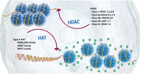
Histone acetylation
Allfrey and colleagues described for first histone acetylation in 1964. Now we know that the acetylation of lysines is highly dynamic and regulated by the opposing action of two families of enzymes, histone acetyltransferases (HATs) and histone deacetylases (HDACs).
HATs utilize acetyl-CoA as cofactor and catalyse the transfer of an acetyl group to the ε-amino group of lysine side chains. This covalent modification neutralizes the lysine's positive charge and this action has the potential to weaken the interactions between histones and DNA. There are two major classes of HATs: type-A and type-B.
The type-A HATs are located in the nucleus and can be classified into at least three separate groups depending on amino acid sequence homology and conformational structure: GNAT, MYST and CBP/p300 families (Hodawadekar and Marmorstein - 2007).
These proteins are often found associated in large multiprotein complexes (Yang et al., 2007) that play important roles in controlling enzyme recruitment, activity and substrate specificity
The type-B HATs are predominantly cytoplasmic, and acetylate free histones but not those already deposited into chromatin. Type-B HATs acetylate newly synthesized histone H4 at K5 and K12 (as well as certain sites within H3), and this pattern of acetylation is important for deposition of the histones, after which the marks are removed (Parthun
et al., 2007).
HDAC enzymes oppose the effects of HATs by removing lysine acetylation, an action that restores the positive charge of lysine. This potentially stabilizes the local chromatin architecture and is consistent with HDACs being predominantly transcriptional repressors. There are four classes of HDAC: Classes I and II contain enzymes that are most closely related to yeast scRpd3 and scHda1, respectively; class IV has only a single member, HDAC11, while class III (referred to as sirtuins) are homologous to yeast scSir2. This latter class, in contrast to the other three classes, requires NAD+ as specific cofactor for its activity (Seto and Yoshida - 2014).
In general, HDACs have relatively low substrate specificity; their specificity is indeed modulated by the incorporation of HATs in large multi-subunit protein complex. For instance, HDAC1 is found together with HDAC2 within the NuRD, Sin3a and Co-REST complexes (Yang et
al., 2007). Thus, it is difficult to determine which activity (specific
Figure 4: Histone acetylation. Histone acetylation is regulated by two families of enzymes:
histone acetyltransferases (HATs) and histone deacetylases (HDACs). The HATs utilize
acetyl-CoA as cofactor and catalyse the transfer of an acetyl group to the ε-amino group of lysine side chains while HDAC enzymes by removing lysine acetylation restore the positive charge of lysine.
Histone methylation
Unlike acetylation, histone methylation exerts its effect without altering the charge of the histone proteins. Moreover, in this case, the lysine ε-amino group of proteins can accept up to three methyl groups, resulting in either mono-, di-, or trimethyl lysine, (me1, me2, or me3) exerting distinct functions. Histone methylation is catalysed by enzymes named histone methyltransferases (HMT) that can be divided in two main categories: lysine-specific (KMT) and arginine-specific (PRMT) (Bannister and Kouzarides - 2005).
The histone methylation reaction, which is catalysed by carrying a methyl group (-CH3) derived from S-adenosylmethionine (SAM) on a lysine or arginine residue, can trigger the formation of either transcriptionally active euchromatin or transcriptionally inactive heterochromatin,
depending on the specific methylated residue, the number of methyl groups added and the availability of factors that remodel chromatin.
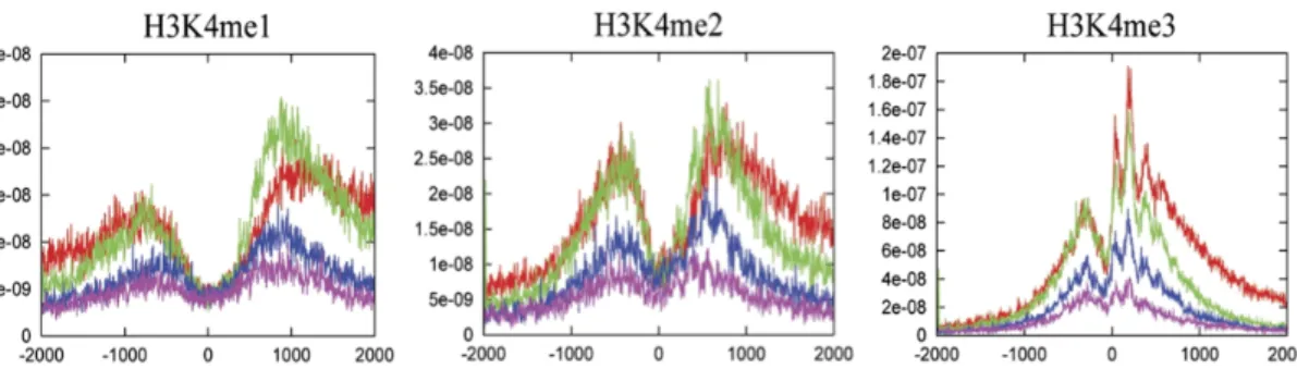
For example, lysine 4 could be subjected to three different methylation events, whose signals are progressively localized closer to TSSs as the modification moves from mono- to di- to trimethylation (Barski et al., 2007).
Figure 5. H3K4 methylation status. Different H3K4 methylation profiles near the
transcription start sites of highly active (red), two stages of intermediately active (green and blue) and silent genes (purple) - Barski et al., 2007.
Two major regions of enrichment are normally detected for each modification: −900 and +1000 from TSS for H3K4me1, −500 and +700 from TSS for H3K4me2, and −300 and +100 from TSS for H3K4me3. On the other hand, trimethylation of lysine 27 of histone H3 (H3K27me3) trigger heterochromatin assembly being related to gene silencing and transcriptional repression (Boyer et al., 2006). The H3K27me3 signals are modestly elevated on silent genes and reduced when genes are expressed. H3K27me3 can be bind by Polycomb Group proteins (PcG proteins), known to mediate chromatin repression, and to be implicated in inactivation of the X chromosome and cell proliferation control. Like trimethylation of lysine 27, also trimenthylation of lysine 9 on histone H3 (H3K9me3) is associated with costitutive heterochromatin and therefore
inactivation of the X chromosome and is associated with highly condensed centromeric and pericentromeric regions (Sullivan et al., 2004).
In general, methyl groups are believed to turnover more slowly than many other PTMs and histone methylation was originally thought to be irreversible, at least until the discovery of histone H3K4 demethylase
LSD1 (Lysine Specific Demethylase 1, also known as KDM1A) by
Byvoet et al., in 1972. Frequently, LSD1 is frequently found to be associated with a transcriptional co-repressor protein (CoREST) and histone deacetylase (HDAC) 1/2 to form a complex able to demethylate H3K4me1/2, but when LSD1 is complexed with the androgen receptor, it demethylates H3K9. This has the effect of switching the activity of LSD1 from a repressor function to that of a coactivator (Klose and Zhang - 2007).
In 2006 JMJD2, the first trimethyl lysine demethylase that demethylates H3K9me3 and H3K36me3, was also described (Whetstine et al., 2006).
1.3 Histone code and chromatin states
Form the discovery of histone modifications scientists have tried to attribute a specific function at each modification. As mentioned before, modifications such as H3K4me3 or H3K27ac are known to be associated with active genes, while H3K27me3 is associated with repressive genes. However, is know that the histones can be modified at different sites and that core histones forming the nucleosome can carry several modifications at same time, giving rise to cross-talk among the different
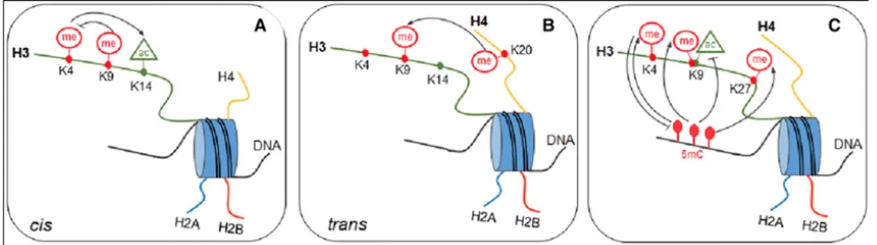
marks. Communication among histone modifications (Fig. 6) can occur within the same site, in the same histone tail and among different histone tails. The relationships between DNA methylation and histone modifications, has been called “epigenetic code” (Jenuwein and Allis - 2001) and seems to be much more complex than believed because act through different mechanisms:
• First, communication at the level of a single histone tail (cis effect); for example, methylation on H3K9, can inhibit acetylation of the H3 tail and methylation of H3K4 (Fischle et al., 2003);
• Second, interactions at the level of nucleosomes mean that the modifications on different histones can affect each other (trans
effect). For example, trimethylation on H3K9 is required for the
induction of H4K20 trimethylation (An - 2007).
• Third, DNA methylation and histone modification pathways can influence each other and establish the epigenetic landscape important for development, somatic cell reprogramming, and tumorigenesis. Relationships between DNA methylation and histone H3 methylation, particularly H3K4, H3K9, and H3K27, have been observed. In addition, there is also a strong anti-correlation between different histone methylations; for example, it is known that the presence of the H3K4me mark prevents de novo methylation of CpG islands in the embryo (Cedar and Bergman 2009).
Figure 6. Epigenetic code: The Cross-talk between chromatin marks and DNA methylation. (A) Communication at the level of a single histone tail is defined cis effect; (B)
different histones can affect each other (the trans effect); (C) DNA methylation and histone modification cooperate to enstablish the epigenetic landscape.
Understanding this code is one of the most compelling challenges in epigenomic studies field. Several bioinformatical tools have been developed in the intent of analyse the epigenomic landscape of several HPTMs at same time, to identify the so-called “chromatin states” (Baker – 2011). Identification of these states, characterized by different HPTMs levels at same locus (Fig. 7), allows to distinguish regions with different functions such as promoters, enhancers, transcribed, repressed, and repetitive regions, providing a systematic annotation of DNA elements and regulatory control regions.
For example, H3K4me1 alone marks primed enhancers, while H3K4me1 combined with H3K27ac mark active enhancers (Creyghton et al., 2010). H3K4me3 is predominant feature of active promoter, whereas poised (bivalent) promoter state is characterized by the occupancy of H3K4me3 and H3K27me3 modifications (Harikumar and Meshorer – 2015).
Figura 7. Chromatin states. The integrative analysis of histone modifications, TF and RNA pol II binding site allows to distinguish combinations of these features (chromatin states) associated with different functional regions such as promoters, enhancers, transcribed, repressed, and repetitive regions, providing a systematic annotation of DNA elements and regulatory control regions.
2. Chromatin immunoprecipitation
The biological significance of interactions of nuclear proteins with DNA in the context of gene expression, cell differentiation, or disease has
immensely been enhanced by the advent of chromatin
immunoprecipitation (ChIP). The technique involves the extraction and fragmentation of chromatin from a biological matrix, its selection through antibodies that specifically recognize a chromatin-associated protein (e.g., transcription factor) or histone post-translational modification, and the study of the DNA sequences present in the isolated chromatin.
Coupled with Next-Generation Sequencing approach (ChIP-seq), the technique make possible to know virtually all the binding sites of different transcription factors, RNA polymerase II or to study the histone post-translational modification at genome-wide level.
The original chromatin immunooprecipitation protocol involved the use of UV light to cross-linking proteins and DNA; later Solomon et al., (1988) introduced formaldehyde as a cross-linking agent able to form covalent bonds between DNA and proteins distant from each within 2Å. Today there are two main methodologies for carrying out ChIP experiments. The first one, called X-ChIP, is based on chromatin fixation with formaldehyde and subsequent extraction and fragmentation by sonication. This variant minimizes the chances of chromatin rearrangements during preparation and precipitation and allows to study also non-histone proteins (e.g. transcription factors) which interact with DNA in a less stable way than histones.
Another variant called native chromatin immunoprecipitation (N-ChIP) is performed using native (not fixed) chromatin, sheared by micrococcal
nuclease (Mnase) digestion, which digests linker DNA, leaving nucleosomes intact (Turner - 2001).
The standard ChIP protocol methods (Fig. 8) involves the following steps:
• Crosslinking of DNA and associated protein performed in living cells or tissues (this step is omitted in N-ChIP); When ChIP is performed on tissue cultured cells, this step is generally carried out using formaldheyde at final concentration of 1% incubated for 10– 15 min at 37 °C. If tissue specimens are used, the crosslinking condition step must be adjusted in terms of time of incubation in formaldehyde, considering both size and complexity of tissue. • After cell lysis step through incubation in a solution containing
detergents, chromatin fragmentation and subsequent extraction take place through physical processes (sonication) and/or through an enzymatic digestion with micrococcal nuclease.
• Using a specific antibody, DNA fragments associated with the protein of interest (or associated with the HPTMs of interest) are immune-selected. This is a critical step, that may determines the feasibility of the entire procedure.
• Immunoprecipitated complexes are washed to remove non-specifically bound chromatin and then, if fixation was performed, the cross-link is reverted through incubation at high temperatures in the presence of high salt concentration.
• The DNA from the isolated chromatin is purified and analysed by quantitative PCR (qPCR) for single locus analysis, or by Next-Generation Sequencing (NGS) approach – for for genome-wide studies.
Figure 8. Chromatin immunoprecipitation steps and various methods of analysis. Fixed
cells are lysated and chromatin is fragmented and extracted. Then, chromatin is immunoselected using specific antibodies directed against the protein of interest and the DNA associated with that protein is analysed through different methods.
3. Pathology-tissue chromatin
immunoprecipitation (PAT-ChIP)
As discussed above, since its introduction, chromatin
immunoprecipitation coupled with next-generation sequencing has become the most powerful approach to investigate specific chromatin-associated proteins (e.g., transcription factors, HPTMs).
Chromatin immunoprecipitation is conducted on chromatin extracted from cultured cells or from fresh/frozen tissues. However, in the first case it has been demonstrated that cultured cells change thair epigenome in function of the culture conditions, while fresh samples are always not available, especially in clinical practice. This is because clinical samples immediately after excision are normally formalin-fixed by immersion in a solution of neutral-buffered formalin for about 16-24 hours and embedded in paraffin (the so-called formaldehyde fixed and paraffin-embedded - FFPE - tissues). Fixation with formalin and subsequent paraffin-embedding are routinely used to preserve biopsies and maintain intact their cellular structure, including cross-linked DNA and proteins as well. However, due to the method of preservation, obtaining biomolecules from these samples has been an hard challenge for many years and the use of this biological resource has been limited mainly to immunohistochemistry and in situ hybridization-based techniques, allowing to study only few targets at the same time. In addition, these approaches require a prior knowledge of the target, allowing hypothesis-driven rather than discovery-based studies.
In 2010 Fanelli and colleagues have developed a new technique named pathology tissue chromatin immunoprecipitation (PAT-ChIP), a technique allowing extraction, immunoselection and high-throughput
analysis of chromatin derived from FFPE samples making possible epigenomic studies using innumerable FFPE tissues stored in hospital archives and tissue banks around the world, providing an unprecedented opportunity to understand the epigenetic mechanisms underlying genome activity in disease etiology and progression.
The original ChIP protocol was modified through introduction of initial steps designed to eliminate the presence of paraffin and to progressively rehydrate the samples. Being FFPE tissues usually heavily crosslinked samples, due long incubation times (usually 16–24 h and more at room temperature) and the high concentrations of formaldehyde (around 4%), the chromatin isolation required a combination of enzymatic digestion and physical approach (sonication).
3.1 Limitation of the original PAT-ChIP protocol
During years PAT-ChIP was applied by several research groups, (Sharma
et al., 2013 – Fang et al., 2015 – Serra et al., 2013) giving new impetus to
chromatin studies to the identification of new potential epimarkers in function of the clinical information of patients. However, some limitations of protocol have emerged. One of these was given by the heterogeneity of FFPE tissue slices, this problem was successfully circumvented by Amatori and colleagues in 2014 using laser capture microdissection (LCM) to increase the purity and homogeneity of the celllular populations under investigation. Nevertheless, the main problem and limitation of PAT-ChIP, is related to fixation status of tissues. Indeed, we have experience that the performances of the technique can be hindered when highly-fixed samples are used. Due to the lack of
standardization in the formalin fixation procedure in clinical practice, some users leave tissue biopsies in formalin solutions for a longer period (up to 72h) with a relatively low impact on standard analysis (e.g.
immunohistochemistry) but compromising any further
molecular analysis, in particular chromatin immunoprecipitation.
FA is a tight (2 Å) crosslinking agent that efficiently produces both protein–nucleic acid and protein–protein crosslinks. Amino and imino groups of amino acids (lysines, arginines, and histidines) and of DNA (primarily adenines and cytosines) are involved in formalin fixation forming a Schiff’s base that can participate in a second linkage with an additional amino group and condense to give the final DNA–protein complex.
Extensive crosslinking to which FFPE archival samples can be exposed produces a dense network of crosslinked cellular biomolecules that can render chromatin extraction extremely challenging.
Additionaly, less efficiency in chromatin extraction is not the only problem caused by formalin fixation, since fixation has a strong impact on lysine residues, one of the most studied target where occurs several PTMs occur, determining the masking of the epitope and hampering subsequent immunoselection.
4. Next Generation Sequencing (NGS) and
bioinformatics analysis
Genome-wide mapping of binding sites for transcription factors, or other DNA-binding proteins, cofactors and histone post-translational modifications, is essential for deciphering the gene regulatory networks that underlie various biological processes. The main tool for investigating these mechanisms is chromatin immunoprecipitation coupled with next-generation sequencing, also known as ChIP-seq. The ability to sequence tens or hundreds of millions of short DNA fragments in a single run is enabling studies that were considered impossible only ten years ago. There are diverse NGS techniques developed over the last few years although the majority share the same operating principle based on the sequencing by synthesis (SBS) technology (e.g Illumina platform – Fig. 9).
First of all, the DNA from ChIP is ligated to specific and know oligonucleotide called adapters and amplified to prepare a library (see library preparation in material and method section).
Then sequencing takes place by polymerization, adding one by one the four nucleotides (labelled or not, and chemically blocked at 3’-OH) whose progressive incorporation is detected and registered.
Figure 9. Illumina sequencing and data processing workflow.
A) Denaturated NGS library fragments are hybridize on a chip bearing millions of oligonucleotides covalently linked and complementary to the adapters. Bridge amplification is performed to amplify DNA and to generate clusters of the same fragment. Reverse filament is then cut and removed by washing to obtain clusters that consist of only forward filaments.
B) Fragments are primed and sequenced. C) Raw data is demultiplexed into individual
libraries and assessed for quality. Removing adapter reads reduces technical noise. Finally reads are aligned onto the assembly of interest.
The raw data coming from the sequencer are in FASTQ files, a text-based format for storing both a biological sequence and its corresponding quality score
After quality check and filtering of low quality reads, the remaining reads are aligned against an organism-specific reference genome using mapping programs such as Bowtie2 or BWA generating a Binary Alignment Map (BAM) file, a file type easier to read and process for later bioinformatics analysis. Once obtained a BAM file, read densities can be visualized on UCSC genome browser (or other software auch as Integrative Genomics Viewer – IGV) where it is possible to visualize the distribution of the sequenced reads over the entire genome. If the antibody selection was specific, a considerable proportion of the mapped reads will be dispersed by chance throughout the genome (the so-called background), while the others will cluster together constituting reads-enriched regions termed “peaks”. Peaks can differ substantially depending on the protein or histone modification investigated. For example, the majority of transcription factors and many histone modifications like H3K4me3 and H3K27ac tend to have narrow peaks, with a size ranging from several hundred to a few thousand base pairs while other histone modifications such as H3K27me3 or H3K9me3 tend to form broad genomic domains with diffusive ChIP-seq signals, which can span up to thousands kilo base pairs.
Peaks of enrichment are identified using different softwares, the most used being model-based analysis of ChIP-seq (MACS and MACS2) which assign they a p-value reflecting their significance. Moreover, when an input control data is present, is possible to filter the peaks that are called and to assign each peak a false discovery rate (FDR) score, which is the likelihood that the peak is not valid.
Figure 10. ChIP-seq data analysis workflow. Reads are filtered basing on a quality score
and high-quality reads are mapped on a reference genome. Reads density can be visualized along the genome and enriched regions (peaks) can be identified and used for comparative analysis.
Other tools are then used downstream to annotate these enriched regions. Analysis can include (but is not limited to) peak comparison among samples to observe presence/absence of specific peaks, global peaks distribution around transcription start site (TSS), or to annotate the genomic features of peak, gene ontology (GO) or pathway analysis, and recurrent motif search, checking for quantitative significant changes in binding levels.
However, most experiments nowadays are designed to study several histone modifications at once and to this end various computational algorithms are developed.
ChromHMM and Segway, for example, allows to identify the specific combination patterns of histone modifications and classify the genome into a preselected number of chromatin states.
Using a multivariate Hidden Markov Models (HMM), ChromHMM split the genome in 200-nucleotide (or more) intervals, called bins. For each bins the tools determines the presence or absence (1 or 0) of each mark and use it to learn a chromatin-state model, and create an annotation of state occurrences across the genome.
5. Theories of aging
The epigenetics field has rapidly developed into one of the most influential areas of scientific research due to involvement in the regulation of several and essential biological processes such as embryo development, cellular differentiation and tumorigenesis. The number of pathologies linked to the dysregulation of epigenetic systems continues to grow and with it, the list of potential targets for epigenetic-based therapeutics.
By the last century, one of the most intriguing challenges for scientific community is to understand the aging process. Recent studies indicate that epigenetic alterations represent one of the most important hallmark of it, attracting much interest due potential reversibility of epigenetic marks makes them promise therapeutic targets to delay or minimize age-related diseases and potentially extend lifespan.
Denham Harman (1950s) describes aging as the result of the progressive accumulation of changes in the body which occurs over the time and which that increase in the chance of getting sick or dying.
In the past couple of centuries, scientists proposed several aging theories but none of them is sufficiently able to explain the aging process. Some of these theories indeed match with others, while others are completely different.
In a frequently cited paper published in 1990, Zhores Medvedev had attempted to make a rational classification of theories of aging, but over the years, gerontologists have resigned to the futility of formulating a unified theory of aging because the large body of descriptive data underlines the multifaceted, different and complex nature of aging.
The rates of aging progression are highly variable in different species, in organism within a species, in organs and tissues within an organism, leading to the conclusion that aging has a no universal cause or phenotype, except death.
Today aging is defined as complex multifactorial process shared by all living organisms, characterized by progressive decline in intrinsic physiological functions, leading to an increase of susceptibility to many diseases, including cancer.
Aging theories can be fall into two main categories: stochastic and non-stochastic theories. The first group, also called “error theories”, consider aging as the result of environmental insults to living organisms that induce progressive damage at various levels (e.g., mitochondrial DNA damage, proteins or lipids oxidation).
In the second one, aging is thought to be depends on biological clocks regulating the timetable of the lifespan through the stages of growth, development, maturity, and old age: this regulation would depend on sequentially switching ON and OFF signals of genes expressed in nervous, endocrine, and immune systems responsible for maintenance of homeostasis and for activation of defense responses.
5.2 Stochastic theories of aging
Stochastic theories suggest that aging is the result of random accumulating changes that negatively affect biological systems. Aging could be the result of the accumulation of toxic by-products, damage due to nuclear radiation or other gradual deteriorative process. At this category belong many theories proposed such as “wear and tear theory”, “free radicals theory” and “somatic DNA damage theory”.
Wear and tear theory
The so-valled “wear and tear theory”, asserts that the organism like a machine become damaged and eventually break down when utilized for a certain period. This theory propose that aging is simply the result of wear and tear, due to the deteriorating effect of processes such as oxidation, or other molecular damage due to ionizing radiation and toxic element uptake, or again other unavoidable natural processes. Aging is view as result of the accumulation of toxic by-products and, believing in this, the aging process is, theoretically impossible to revert. However, there are only few gerontologists that currently support the “wear and tear theories”, mainly because this theory fails to explain enormous differences in lifespans between biochemically similar species. If aging is the result of fundamental limitations that presumably affect all organisms, why are lifespans of even very similar organisms so different (e.g naked mole-rats – 28.3 years vs nommon rock rats – 4.2 years)?
Free radical theory of aging (FRTA)
Originally described by Denham Harman et al., the error theory of aging is one of the most prominent theories to explain aging. This theory proposes that the free radicals - continuously generated during the life of the cell – aren’t counterbalance (in aged biological system) by cell antioxidant system causing damage. There is a great deal of experimental evidence in support of this theory. Old animals show a higher index of oxidation than young ones and indeed they accumulate oxidized proteins, DNA forms, and lipid (Stadtman - 1992).
Other experimental evidences support this theory, for instance, increasing antioxidant defense results in an extended life span, as demonstrated in experiments using flies. In fact, Tower (2000) show an increased life span
of transgenic flies expressing superoxide dismutase (SOD) indicating that free radical scavenging enzymes are sufficient to delay aging in
Drosophila.
Extension of C. elegans life span, by synthetic small molecules that mimic catalase and/or SOD, demonstrates that antioxidant compounds can delay aging in worms (Melov et al., 2000).
These results indicates that free radical damages in short-lived organism opposes longevity, but in long-lived organisms such as mice models, a antioxidant diet can effectively reduce the accumulation of oxidized molecules but fail to extend lifespan. Calorie restriction is an intervention that prolongs the life span of nearly every organism to which it has been applied. In rodents, calorie restriction reduces generation of ROS from isolated mitochondrial preparations and attenuates the accumulation of oxidative damage.
The free radical theory argues that mutations in mitochondrial DNA accelerate free radical damage by introducing altered enzyme components into the electron transport chain. Faulty electron transport results in elevated free radical leakage and ultimately more mitochondrial DNA mutation and exacerbated oxidant production. This “vicious cycle” of mutation and oxidant production eventually leads to cellular catastrophe, organ failure and senescence. Another hypothesis also belonging to FRTA argue that radicals cause aging when oxidized proteins accumulate in cells. An age-dependent reduction in the ability to degrade oxidative proteins may contribute to the build-up of damaged and dysfunctional molecules in the cell. The free radical theory of aging could “interact” with Somatic Mutation Theory of aging where accumulation of genetic mutations in somatic cells are thought to be responsible of decrease in cellular functions and ROS damage may be an important source of these somatic mutations.
5.3 Non-stochastic theories of aging
Theories of aging based on accumulation of stochastic damage to macromolecules were widely support by gerontologists, even if it does not provide a convincing explanation for vastly different longevity of different cell types within the same individual, or individuals from different species living in the same environment. A series of pioneering experiment carried out in yeast, worm, flies, and mice have showed that lifespan could be strongly influenced by molecular pathway that have been conserved during evolution, leading new theories where aging is thought to be a “deterministic” programmed process.
In the early ’80 years, Klass et al., (1983) observed that C. elegans lifespan could be altered, but only in 1988 Friedman and Johnson showed that life extension of worm up to 65% was due to a mutation of gene that called age-1. This is the first gerontogene (refers to any such genetic elements that are involved in the regulation of aging and life span) discovered.
In the 1993, Kenyon C. showed that mutation in gene daf-2 gene cause fertile, active, adult C. elegans hermaphrodites to live more than twice as long as wild type.
Since then, dozens of genes were identified influencing lifespan supporting the “longevity programmed theory” where the presence of these genes is thought to be crucial to allow the organism survival in presence of environmental stresses.
Under stress condition such as long exposure at cold temperature or reducing calorie intake, getontogenes are silenced to allow to exceed the normal lifespan of organism by entering in “maintenance mode” (reverted with improvement of environmental conditions) characterized by
biological processes changes such as hypometabolism, high stress-resistance and low or no fertility. Notewhorty, studies in C. elegans show that genetic program actively promote longevity when animal models are expose at cold temperatures (Xiao et al., 2013).
Many mutations that extend life perturb endocrine signalling. In fact,
daf-2 gene encodes the only insulin/IGF-1 receptor expressed in worms (Fig.
11), while age-1gene encodes a homologue of mammalin
phosphoinositide 3-kinase (PI3K) catalytic subunit (Morris et al., 1996;
Kimura et al., 1997). Both proteins are involved in insulin/IGF-1
pathway (IIS pathway), a well conserved evolutionarily pathway across
organism. The IIS pathway acts as a food and stress sensor during development. When food is abundant, worms develop rapidly and uninterrupted through four larval stages to reach adulthood. If worms develop in hot, food-limited or overcrowded conditions (stress conditions), they enter in an alternative long-lived larval state called
dauer in which reproductive maturity is delayed and stress resistance
increases.
IIS pathway is one of the best-understood molecular mechanisms able to influence the lifespan. Briefly, signalling through daf-2 (IGF-R in human) activates age-1 (PI3K in humans), which leads to the phosphorylation of
daf-16 and its inactivation by nuclear exclusion. In the absence of IIS
activity or in daf-2 or age-1 mutants, daf-16 enters the nucleus and enacts a transcriptional program that is thougth doubles worm life span (Lin et
Figure 11. Longevity pathway in yeast, worms, flies mice (and human?). A series of
pioneering experiment carried out in yeast, worm, flies, and mice have showed that lifespan can be strongly influenced by molecular pathway that have been conserved during evolution.
As mentioned before IIS is an evolutionarily conserved pathway that regulates life span across many organisms. Drosophila melanogaster, like
C. elegans, has a single insulin-like receptor (dInR) that, when mutated,
extends life span in a manner that is dependent on dFOXO, its daf-16 homolog, (Tatar et al., 2001). In mammals are present several daf-2 homologs (IGF-1R), insulin receptor A and B (IR-A and IR-B) that display even greater complexity due their ability to form multiple homodimer and heterodimer pair. Despite these differences in insulin receptor expression, the functional consequences are similar; in fact, reduced IIS pathway activity results in extends life span in multiple mammalian species. Heterozygous IGF-1R knockout mice are long-lived (Holzenberger et al., 2003) in addition IGF-1R mutations are highly represented in populations of centenarians (Suh et al., 2008). Through
ChIP-seq and gene expression approach, genome wide analysis of daf-16 in C.elegans was performed to extend our knowledge of how life extension is achieved in the worm. However, no single DAF-16 target that has been tested can recapitulate the long-lived phenotype of daf-2 worms, suggesting that many disparate cellular pathways are regulated in concert to extend life span (Murphy et al., 2003; McElwee et al., 2004). Similarly to IIS pathway, also the role of the TOR (target of rapamycin) pathway on aging is remarkably conserved. TOR is an evolutionarily conserved nutrient sensing protein kinase that regulates growth and metabolism in all eukaryotic cells. This kinase is inhibited by rapamycin, a by-product of the soil bacterium Streptomyces hygroscopicus (Vezina et
al., 1975).
Rapamycin through formyl peptide receptor 1 (FPR1), a peptidyl-prolyl cis-trans isomerase, regulate activities of two proteins identified as TOR1 and TOR2 in S. cerevisae (Heitman et al., 1991). However, in mammals there is only one gene encoding mammalian TOR (mTOR) and protein exists in two complexes:
• TOR complex I (TORC1 in yeast and mTORC1 in mammals), a rapamycin sensitive complex that controls temporal aspects of cellular growth mediated mostly through S6 kinase 1 (S6K1);
• TORC2 is rapamycin-insensitive and controls spatial aspects of growth within the cell and the effects are mostly mediated through
Figure 12. The TOR pathway and aging. mTOR is a key component of cellular metabolism
that integrates nutrient sensing with cellular process that fuel cell growth and proliferation.
TORC1 is involved in many human diseases, including diabetes, obesity,
heart disease, and cancer (Inoki and Guan, 2006). This pathway is also involved in lifespan control in multiple model systems; indeed, inhibition of TORC1 (hereafter referred to as TOR) extends lifespan in yeast, worms, flies and mice (Kapahi et al., 2010).
Large amounts of data show that signalling of TOR pathway can act both in parallel to but also interact with the insulin/IGF-1 pathway in flies (Kapahi and Zid, 2004). Combination of rapamycin and caloric restriction cause some additional longevity effects when compared to each single treatment (Bjedov et al., 2010) indicating that the effects of rapamycin
may not be identical to the mechanisms involved in caloric restriction-mediated extention of lifespan.
As mentioned before, the aging process is characterized by an increase of vulnerability to many diseases, due to an impairment of the different physiological systems, including the immune function.
In fact, it is well known that over the time there is a decrease in the resistance to infections and an increased incidence of autoimmune processes and cancer, indicating the presence of a less competent immune system.
6. Epigenetics of aging
Epigenetics is emerged as a major, nongenetic mechanism strongly related to the aging process is epigenetics. Epigenetic alterations affect all types of cells and tissues throughout life and have been linked to cancer development and are retained one of the major hallmarks of aging. During the aging process, the epigenome undergoes a progressive loss of its configuration that results in a significant change in genome integrity, chromatin architecture and gene expression pattern. This phenomenon is called “epigenetic drift”.
However, to date the relationship between aging and epigenetic drift is controversial. Indeed, is not yet unclear whether changes in the activity of epigenetic enzyme (and thus the epigenome) influence the expression of critical longevity genes or whether alterations in the longevity genes are able to drive large-scale epigenetic changes in the genome.
As shown above, the impact of the genome on aging is well established and seems to account for only 25% of an individual’s lifespan, while current studies indicate that epigenetic changes comprise a significant component of aging (Jones et al., - 2015).
One of the well-studied epigenetic mechanisms in relation to aging is DNA methylation.
In general, aging-associated changes in DNA methylation include global hypomethylation and region-specific hypermethylation (Xiao et al., - 2016). DNA hypomethylation also takes place in transposable DNA repetitive elements including Alu sequence and long interspersed nuclear elements (LINE-1), resulting in increased transposition activity and genomic instability (Wilson et al., 2007).
On the other hand, promoter hyper-methylation affects the expression of certain transcription regulatory genes (Gentilini, et al., 2013), apoptotic genes, development or differentiation regulatory genes. Promoter hyper-methylation has been also observed in several tumour suppressor genes such as CDKN2A, LOX, RUNX3, and TIG1 (Waki et al., 2003) as well as, on estrogen receptor (ER) and insulin-like growth factor II (IGF2).
To date, evidence has shown that reduced global methylation can be caused by the different regulation in the expression of DNMTs as reported by Casillas and colleagues in 2003 where DNMT1 (the predominant maintenance methyltrasferase) expression was reduced contributing to the reduction of genomic methylation of cells. On the other hands, DNMT3b expression steadily increased with aging and has been proposed to be responsible of a paradoxical sporadic gene hypermethylation in aging cells. In accordance with age-dependent hypomethylation, several age-related diseases, including neurodegenerative disease, cardiovascular disease, and cancer, show close association with marked global methylation decrease (Baccarelli et al., 2013 – Choulirias et al., 2013).
In the last few years, the role of histone modifications in aging has emerged, providing insights into epigenetic mechanisms of aging and lifespan regulation. The H3K4me3 histone modification, known to be a marker of active gene expression, is intimately involved in aging across model organisms. In C. elegans, deletion of the genes encoding any of the three Trithorax group proteins (WDR-5, SET-2 and ASH-2), that facilitate H3K4 trimethylation, results in decreased global levels of H3K4me3 and increased lifespan (Greer et al., 2010). Similarly, in flies, deletion or RNA interference (RNAi) knockdown of drosophila little imaginal discs (Lid), a recently described histone demethylases, resulted in increased levels of H3K4me3 and reduced lifespan (Li et al., 2010).
Lifespan regulation is influenced also by H3K27 methylation or acetylation, but in this case it seems to operate quite differently in different species.
In flies, loss of function mutations in genes encoding components of Polycomb Repressive Complex 2 (PRC2) - an H3K27me3 specific methyltransferase complex - result in decreased levels of H3K27me3 and increased lifespan (Siebold et al., 2010). In worms, however, the opposite effects are observed (increased levels of H3K27me3 through knockdown of its demethylase UTX-1) and are associated with increased lifespan in an insulin dependent manner (Maures et al., 2011).
In 2013 Liu and colleagues have shown that the repressive mark H3K27me3 increases in aged muscle quiescent stem cells and thus suggested to suppress stem functions in mice, while accumulation of H3K4me3 has also been observed in aged hematopoietic and muscle stem cells (Sun et al., 2014). In the mouse liver, nucleosome occupancy (Bochkis et al., 2014) and the overall content of different histone marks (Kawakami et al, 2009) were reported to change with age but genome-wide profiles of histone modifications during aging are not available. In humans, H3K4me3 distribution in prefrontal neurons from 11 individuals was the only genome-wide histone mark study reported throughout aging up to now.
7. Epigenetic, aging and caloric restriction
Environmental factors such as exercise, circadian rhythms and even sexual stimuli are shown to influence gene expression and longevity in different organisms. Nutrient availability and diet are to date the most thoroughly studied environmental factor to affect longevity; diet is known to significantly impact on the epigenome. This has brought the scientific community to focalize the attention on nutrition, to determine whether diet can affect chromatin structure and gene transcription through epigenetic changes. The viable yellow agouti mouse model (Avy/a), where coat’s colour variation and obesity is correlated to epigenetic marks established in early development, provided the earliest model for studying epigenetic inheritance in mammals. Wild-type murine Agouti gene encodes for agouti signalling protein (ASIP), a paracrine signalling molecule, whose binding to the melanocortin-1 receptor (MCR-1) prevents α-melanocyte-stimulating hormone signalling, thereby down-regulating synthesis of brown/black pigments (eumelanin) and increasing synthesis of yellow/red (pheomelanin) pigments during the mid-stage of the hair growth cycle.
In Avy /a mice is present an inserted retrotransposon (an intracisternal A
particle or IAP) is present upstream of the customary transcriptional start
site of the wild type A gene. This insertion contains a cryptic promoter in its 5’ long-term repeat (LTR) region, capable of driving agouti gene expression and overriding the control mechanisms that normally limit the production of ASIP to certain stages of the hair follicle cycle. In such mice, the fur is completely brown (pseudoagouti) when the cryptic promoter within the IAP is silent, but yellow when the promoter is fully active. This range of phenotypes (Fig. 13) is correlated with the degree of cytosine methylation of the 5′-LTR of the IAP, with CpG island
hypermethylation associated with the pseudoagouti phenotype and hypomethylation with yellow fur and broad ectopic expression (Morgan
et al., 1999). Moreover, Avy mice are larger, obese, hyperinsulinemic, more susceptible to cancer and, on average, shorter lived than their non-yellow siblings. This phenotype is due to ectopic expression of ASIP outside hair follicle, most critically in the hypothalamus, where it antagonizes the melanocortin 4 receptor (MC4R).
Waterland and Jirtle (2003) have showed that diet methy-donor supplementation such as folic acid, vitamin B12, choline, and betaine alter the phenotype of their Avy/a offspring via increased CpG methylation at
the Avy locus. The observation that the coat colour of these mice and their
associated degree of metabolic disease, can be modulated by diet provided to the dam during early development of her offspring, has also raised considerable medical interest, as it is becoming clear that both the quantity and quality of the food that a pregnant woman consumes during her pregnancy can either enhance or reduce the risks of her infant to develop diseases in adult life.
yellow agouti (Avy) mice representing the five coat color phenotypes. Yellow mice are hypomethylated at the transposable element upstream of the Agouti gene allowing maximal ectopic expression, whereas hypermethylation of this site silences ectopic agouti expression in the pseudoagouti animals. Mice that are predominately yellow are also clearly more obese than brown mice.
Other evidences come from by epidemiology studies. Heijmans and colleagues studied individuals exposed to the “Dutch Hunger Winter”, food rationing period in German-occupied Netherlands between ’45 and ’46, and have demonstrated, studying the descendants, that those who were exposed to famine in utero had very different methylation patterns in genes involved in growth and metabolic disease compared with controls. The studies reported a minor, but significant, decrease in DNA methylation at a differentially methylated region in the imprinted insulin-like growth factor 2 (IGF2) gene (Heijmans et al., 2008).
Further research identified persistent, small alterations in DNA methylation at other imprinted genes such as insulin (INS), guanine nucleotide binding protein a-stimulating (GNAS) and maternally expressed gene 3 (MEG3) (Tobi et al., 2009). Such alterations were also found at some loci that are involved in growth and metabolic disease, such as interleukin 10 (IL10), leptin (LEP) and ATP-binding cassette A1 (ABCA1). Moreover, our methylome can be influenced also by a modulation of caloric intake.
Different studies have shown that caloric restriction defined as moderate (normally, 20-40%) reduction in caloric intake - as compared with an ad
libitum diet - without compromising the maintenance of all essential
7.1 Caloric restriction
The remarkable effects of caloric restriction (CR) on aging have been first described in 1935 in experimental animal models by McCay and colleagues. Since then, numerous research findings have revealed effects of CR on lifespan among diverse species, including yeast, worms, flies, fish and even mammals. CR has also been shown to delay a wide range of aging-associated diseases, such as cancer, diabetes, atherosclerosis, cardiovascular diseases and neurodegenerative diseases in higher mammals, such as nonhuman primates and humans (Omodei and Fontana 2011). Further evidence coming from epidemiological studies conducted in Japan found that urban Japanese peoples consumed more calories and have a higher incidence of cancer than peoples of rural Okinawa, where a regime of CR was adopted.
Sirtuins are probably the best-studied family of enzymes implicated in changing of the epigenome in response to caloric restriction.
Sirtuins are present in a variety of organisms, from yeast to mammals, and have been involved in the organization of global chromatin structure and in the epigenetic regulation of specific genes. Activation of sirtuins is observed in individuals subjected to CR, in relation with the elevated NAD+ levels induced by the intervention (Kane and Sinclair – 2018). Among sirtuin, SIRT1 has been the most extensively studied. More than a dozen substrates have already been described for SIRT1 including members of the FOXO family (mammalian homologs of DAF-16 of C.
elegans), transcription factors that regulate the expression of genes
involved in cell growth and in other cellular processes. In mammals these transcription factors, undergoing post-translational modifications, such as acetylation, de-acetylation, methylation, phosphorylation (Webb and Brunet - 2014) and can access the nucleus regulating the transcription of
genes implicated in various cellular processes, such as cell cycle arrest, resistance to oxidative stress and apoptosis. Kops et al., 2002 suppose that FOXO3A, by increasing the antioxidant capacity of the cell through the expression of MnSOD and catalase (Kops et al., 2002), can influence longevity avoiding the accumulation of ROS (reactive oxygen species) or highly reactive molecules capable of causing damage to biological structures.
Moreover, SIRT1 has been shown to deacetylate FOXO3A allowing this protein to remain inside the nucleus and regulate the transcription of genes that permits to the stem cell to maintain an undifferentiated and quiescent state (phase G0), a fundamental condition for the own capacity for self-regeneration (Miyamoto et al., 2007). Vaquero and colleagues in 2007 have shown that SIRT1 could suppress the expression of inflammatory genes by enhancing the activities of histone methyltrasferase SUV39H1, resulting in increased levels of H3K9me3 (Vaquero et al., 2007).
8. Aims of the project
PAT-ChIP procedure is a powerful approach to investigate the epigenomic landscape in FFPE tissues although some limitations are still present. During my Ph.D. activity, I focused my research activity mainly on two objectives:
• The improvement of PAT-ChIP technology to allow epigenomic
studies of “critical” biological models;
• The in vivo investigation of the epigenomic basis of aging focusing
on the possible role of caloric restriction in the slowing down / revertion of the phenomenon.
The first part of this project provides a detailed overview of all the procedure used to improve the original PAT-ChIP protocl while the second part describes the global characterization of aging-associated epigenetic landscapes and the effects induced by CR.
9. Epigenomic profiling of archived FFPE
tissues by enhanced PAT-ChIP
(EPAT-ChIP) technology
As already mentioned, chromatin immunoprecipitation coupled with massive parallel sequencing approach is a powerful technique to investigate the epigenomic landscape with an enormous potential to extend our knowledge on the influence of epigenetic alterations in several biological processes. Thanks to the introduction of PAT-ChIP, epigenomic studies have been extended to innumerable formalin-fixed paraffin embedded (FFPE) tissues stored in hospital archives and tissue bank around the world. However, due to the lack of standardization in FFPE tissues production, extensive tissues fixation is a frequent technical problem that could hinder the application of PAT-ChIP. In the light of these evidence, I have investigated the possibility to improve chromatin extraction efficiency from FFPE tissues, to facilitate the combination of the original PAT-ChIP protocol with NGS technology, allowing genome-wide studies also in clinical “complex” archival samples.
First attempts allowed to identify the best condition to increase the efficiency of chromatin extraction using normal colon tissues (chosen due to the availability of high quantities of this human material as scrap from colorectal surgeries). Once identified the best condition, antigen integrity was evaluated through immunofluorescence approach and then the new
procedure, called Enhanced Pathology-Tissue Chromatin
Immunoprecipitation (EPAT-ChIP), was applied on real archive breast human FFPE tissues.