RESEARCH ARTICLE
Longitudinal quantitative electroencephalographic
study in mono-hemispheric stroke patients
*Correspondence to: Giovanni Assenza, MD, PhD, [email protected]. orcid: 0000-0002-6160-4348 (Giovanni Assenza) doi: 10.4103/1673-5374.251331 Received: August 11, 2018 Accepted: December 12, 2018
Filippo Zappasodi1, Franca Tecchio2, Laura Marzetti1, Vittorio Pizzella1, Vincenzo Di Lazzaro3, Giovanni Assenza3, *
1 Department of Neuroscience, Imaging and Clinical Sciences and Institute for Advanced Biomedical Imaging, “G. D’Annunzio” University, Chieti, Italy
2 Laboratory of Electrophysiology for Translational NeuroScience (LET’S), ISTC-CNR, and Fondazione Policlinico Gemelli IRCCS, Rome, Italy 3 Unit of Neurology, Neurophysiology, Neurobiology, Department of Medicine, Università Campus Bio-Medico di Roma, Rome, Italy
Funding: FZ and FT obtained financial support from the Italian Ministry of Health Cod. GR-2008-1138642 “Promoting recovery from Stroke:
indi-vidually enriched therapeutic intervention in Acute phase”, and PNR-CNR Aging Program.
Abstract
The identification of individual factors modulating clinical recovery after a stroke is fundamental to personalize the therapeutic intervention to enhance the final clinical outcome. In this framework, elec-trophysiological factors are promising since are more directly related to neuroplasticity, which supports recovery in stroke patients, than neurovascular factors. In this retrospective observational study, we in-vestigated brain neuronal activity assessed via spectral features and Higuchi’s fractal dimension (HFD) of electroencephalographic signals in acute phase (2–10 days from symptom onset, T0) and sub-acute phase (2.5 months, T1) in 24 patients affected by unilateral middle cerebral artery stroke. Longitudinal assess-ment of the clinical deficits was performed using the National Institutes of Health Stroke Scale (NIHSS), together with the effective recovery calculated as the ratio between difference of NIHSS at T0 and T1 over the NIHSS value at T0. We observed that delta and alpha band electroencephalographic signal power changed between the two phases in both the hemispheres ipsilateral (ILH) and contralateral (CHL) to the lesion. Moreover, at T0, bilateral higher delta band power correlated with worse clinical conditions (Spearman’s rs = 0.460, P = 0.027 for ILH and rs = 0.508, P = 0.013 for CLH), whereas at T1 this occurred only for delta
power in ILH (rs = 0.411, P = 0.046) and not for CHL. Inter-hemispheric difference (ILH vs. CLH) of alpha
power in patients was lower at T0 than at T1 (P = 0.020). HFD at T0 was lower than at T1 (P = 0.005), and at both phases, ILH HFD was lower than CLH HFD (P = 0.020). These data suggest that inter-hemispheric low band asymmetry and fractal dimension changes from the acute to the sub-acute phase are sensitive to neuroplasticity processes which subtend clinical recovery. The study protocol was approved by the Bioethi-cal Committee of Ospedale San Giovanni Calibita Fatebenefretelli (No. 40/2011) on July 14, 2011.
Key Words: mono-hemispheric stroke; delta band; fractal dimension; inter-hemispheric asymmetries; EEG;
plasticity
Chinese Library Classification No. R448; R741
Introduction
Ischemic stroke is a leading cause of death and the primary cause of chronic disability in Western countries, mostly in the elderly (Crichton et al., 2016). Care in the hyper-acute and acute period after a stroke has improved over the last decades, with a remarkable increase of stroke survival rate (http://www.rcplondon.ac.uk/resources/stroke-guidelines). However, the clinical outcome shows a huge inter-individ-ual variability, even with similar symptoms and features of the lesion (site/volume) at onset (Zeiler and Krakauer, 2013). Studies of global neural activity, neural circuitry and brain connectivity following an ischemic lesion in animals and humans represent the basis of our understanding of the biological mechanisms underpinning post-stroke recovery, and are important in orienting the clinical practice and to improve outcome. Indeed, a proper detection of neural markers of the neurological impairment in the acute phase, as well as of prognostic value and recovery abilities can pro-vide information about the selection of therapies, medical and/or rehabilitative, that enhances the individual ability for post-stroke recovery.
In humans, several neuroimaging techniques have been used in the last decades to broaden knowledge of short and long-term changes of neural activity after stroke (for a re-view see Rossini et al., 2007). Previous evidence strongly supports the notion that stroke patients show an uncoupling of their neuronal activations from their hemodynamics (Rossini et al., 2004), suggesting that electrophysiological features of neural activity are better candidates to describe the functional state of neurons surviving cerebral ischemia after a stroke and to characterize their reaction capability. Electroencephalography (EEG) is a sensitive, non-invasive, low-cost and widely available technique able to detect neu-ronal functional changes following a brain lesion. Indeed, EEG studies in humans (Nagata et al., 1982; Sainio et al., 1983; Ahmed, 1988; Niedermeyer, 1997; Makela et al., 1998; Murri et al., 1998) reported that slow-frequency activity and interhemispheric asymmetries of delta and alpha activity are signs of a cerebral lesion. We previously demonstrated, by means of magnetoencephalography (MEG) and EEG, that in acute unilateral stroke a higher cortical activity in the delta frequency band (1–4 Hz), together with a lower
ampli-tude of the somatosensory evoked fields in the ipsi-lesional hemisphere, is able to reliably predict most of the variability of the clinical status in the acute phase (Tecchio et al, 2005; Assenza et al., 2009).
In addition to features derived by the oscillatory nature of brain activity and extracted by spectral analysis, recent non-linear methods revealed neuronal dynamics dysfunc-tion following a stroke lesion. Indeed, brain processes are highly non-linear (He, 2011; Buzsaki and Mizuseki, 2014; Roberts et al., 2014; Ramon and Holmes, 2015) and mea-sures of system complexity have been proved to quantify critical aspects of the brain activity (Stam et al., 2005; Ro-dríguez-Bermúdez and García-Laencina 2015). Complexity has been associated to efficient processing and functional advantages (Garret et al., 2013). In particular, Higuchi’s fractal Dimension (HFD) of EEG at rest was found to be smaller in the first week after a mono-hemispheric stroke (Zappasodi et al., 2014) and this reduction was paired to a worse clinical status. It has been suggested that the HFD decrease in stroke patients describes the loss of complexi-ty subsequent to the global system dysfunction due to the structural damage. This picture is coherent with neuronal activity complexity decrease paired to a reduced repertoire of functional abilities.
The present study aimed to characterize the evolution of electrophysiological features in acute (within 10 days from ischemic attack) and sub-acute (after 2 months) phases of brain activity in unilateral ischemic stroke in the middle ce-rebral artery (MCA) territory. Moreover, links between the electrophysiological alterations and the clinical status were evidenced, at both acute and sub-acute phases, as well as possible associations between changes in neural activity in sub-acute phase and clinical recovery.
Participants and Methods
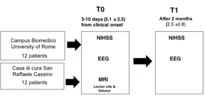
ParticipantsTwenty-four patients (72.0 ± 9.5 years, 9 women and 15 men), admitted to our clinical centers (San Raffaele Hospi-tal, Cassino; Università Campus Biomedico di Roma, Rome; Fatebenefratelli Isola Tiberina Hospital, Rome) from January 2010 to December 2011 were enrolled in this retrospective longitudinal observational study with a control group
(Fig-ure 1). The inclusion criteria were: first-ever
mono-hemi-spheric and mono-lesional ischemic stroke in MCA terri-tory; clinical evidence of motor/sensory deficit of the upper limb; neuroradiological diagnosis of MCA ischemia. The exclusion criteria were: a previous stroke revealed by clinical history; neuroradiological evidence of involvement of either hemispheres or brain hemorrhage; dementia or aphasia se-vere enough to impair patients’ compliance with the proce-dures; anti-epileptic and anti-psychotic treatments. Patients received a diagnostic/therapeutic approach following the Italian stroke guidelines (SPREAD – Stroke Prevention and Educational Awareness Diffusion. Ictus cerebrale: Linee gui-da italiane; www.spread.it). None of our patients received thrombolytic therapy. The rehabilitative treatment was or-ganized as follows: in the acute hospitalization in campus
Bio-Medico university of Rome, patients were examined by a medical doctor specialist in physical and rehabilitative medicine within 48 hours from the admission to the hospital to start active and passive therapy as soon as possible (but after 24 hour after stroke onset, Bernhardt et al, 2016) if not contraindicated. Once patient was admitted to the rehabil-itation institute, all our patients received intensive physical therapy for 3 hours/day 7/7 for 40–45 days. No robotic or neuromodulatory therapies were provided in these patients.
As a control group, 20 participants (8 females and 12 males) with age comparable to the patients (71.2 ± 6.2 years,
P = 0.736) were enrolled from patients’ relatives. All healthy
subjects had an unremarkable neurological examination and did not receive any pharmacological treatment at the time of recordings.
The experimental protocol was approved by the Bioethical Committee of Ospedale San Giovanni Calibita Fatebenefre-telli (No. 40/2011) on July 14, 2011 (Additional file 1), and all participants provided written informed consent
(Addi-tional file 2). Clinical evaluation
Patients underwent clinical status evaluation by National Health Institute Stroke Scale (NIHSS) between days 2 and 10 after stroke onset (acute phase: T0, mean 5.1 ± 2.5 days from symptoms onset), as well as EEG and magnetic resonance imaging (MRI).
Clinical evaluations by NIHSS and EEG recordings were repeated after 2 months (sub-acute phase: T1, 2.5 ± 0.8 months). The same clinician assessed the NIHSS scores both at T0 and at T1 in each patient. To quantify clinical fol-low-up, the effective recovery (ER) was then calculated as: ER = (NIHSS at T0 – NIHSS at T1)/NIHSS at T0 × 100%.
Neuroradiological evaluation
Brain MRI was taken at 1.5 T (MAGNETOM Avanto; Sie-mens, Erlangen, Germany), using Turbo Spin-Echo (TSE) and Spin-Echo (SE) T1- and T2-weighted sequences. All sequences provided contiguous 5 mm thick slices in sagittal, coronal and axial planes. These images allowed deriving a characterization of the ischemic lesion, which was classified according to lesion site as: ‘subcortical’ (S) when there was no visible cortical involvement and basal ganglia, thalamus,
Figure 1 Study flow chart.
NIHSS: National Institutes of Health Stroke Scale; EEG: electroenceph-alogram; MRI: magnetic resonance imaging.
caudate nucleus, nucleus lenticularis or internal capsule were affected (Dromerick and Reding, 1995); ‘cortical’ (CS), if also cortical structures were involved.
EEG recordings and data analysis
Subjects were sitting on a comfortable armchair during relaxed closed-eyes rest. Five minutes of EEG activity was acquired by means of 19 Ag-AgCl electrodes positioned according to the 10-20 International system (Fp2, F4, F8, C4, T4, P4, P8, O2, Fz, Cz, Pz, Fp1, F3, F7, C3, T3, P3, P7, and O1) in fronto-central reference by a Micromed EEG re-corder (Micromed, Mogliano Veneto, Italy). Two additional electrode pairs were used to monitor and record electro-oc-ulogram and electrocardiogram. Data were sampled at 256 Hz (analogical filter 0.1–70 Hz).
EEG data were off-line filtered between 1 and 45 Hz (sec-ond order forward-backward Butterworth filter) and visually inspected to remove epochs contaminated by movements. To identify and eliminate artefacts (i.e., eye movements, car-diac activity, scalp muscles contraction), we applied a pro-cedure based on Independent Component Analysis (Barbati et al., 2004). Scalp EEG recordings were re-referenced to a standardized reference by the Reference Electrode Standard-ization Technique (Marzetti et al., 2007). In this way, the reference is far from all possible neural sources and acts like a neutral virtual reference.
The power spectral density was estimated for each EEG channel via the Welch procedure (4-second epoch duration, resulting in a frequency resolution of 0.25 Hz, Hanning win-dowing, 60% overlap, about 180 artifacts free trials used). For each EEG electrode band power values were obtained as the mean of power spectral density in each frequency band. The investigated frequency bands were: 1–4 Hz (del-ta), 4.25–7.5 Hz (the(del-ta), 8–12.5 Hz (alpha), 13–25 Hz (beta). Spectral features were investigated in the classical frequency bands, instead of being settled on the basis of individual spectral characteristics (Klimesch, 1999), as spectral proper-ties are known to be affected by stroke.
Band power in both the hemispheres ipsilateral (ILH) and contralateral (CHL) to the lesion was calculated as the average of the band power at the electrodes laying over the area covered by the MCA (F4, F8, C4, T4, P4, P8 for right hemisphere and F3, F7, C3, T3, P3, P7 for left hemisphere,
Figure 2). Band power values were log-transformed to fit a
Gaussian distribution.
The interhemispheric asymmetry (IntAsy) between activi-ties of affected and unaffected hemispheres was obtained for each band power as follows:
IntAsy = (XILH – XCLH) / (XILH + XCLH)
Being X the power in delta, theta, alpha or beta band. HFD (Higuchi, 1988) was calculated for each EEG channel in time windows of 10 seconds (see Zappasodi et al., 2014, 2015 for details). A global measure of HFD was obtained by averag-ing HFD values over time and over all EEG channels. Separate values of HFD for the ILH and CLH were also calculated.
Statistical analysis
Statistical analysis was performed using the IBM SPSS Statistics for Windows (version 24.0; Armonk, NY, USA: IBM Corp., Released 2016). The significance level was set to alpha = 0.05. The first aim of the statistical analysis was to check for changes of electrophysiological features at rest in the sub-acute phase with respect to the acute phase of mono-hemispheric stroke. To evaluate possible differenc-es of spectral featurdifferenc-es between the two phasdifferenc-es, a repeated measure analysis of variance (ANOVA) was performed with
Band (delta, theta, alpha, and beta), Hemisphere (ILH, CLH)
and Time (T0, T1) as within-subject factors. Whenever an interaction Band × Time was found, reduced models were separately applied for each band with Hemisphere and Time as within-subject factors. Greenhouse-Geisser correction has been applied when the sphericity assumption was not valid. Significant main effect of Hemisphere, Time or interaction
Time × Hemisphere was followed up by unpaired t-test, to
compare ILH and CLH band power of patients with the values of the healthy control group, considered as the ref-erence. For the control group values, we arbitrarily aligned right hemisphere with ILH and left with CLH for half of the subjects, and the contrary for the other half. The opposite matching was also verified in order to exclude the presence of inter-hemispheric asymmetries.
Differences between values of global HFD at T0 and T1 were tested by two-tail paired samples t-test. Independent sample t-tests were also used to evidence difference between HFD values of patients and of healthy controls.
The second aim of the analysis was to evidence relationship between electrophysiological alteration and clinical status, both at T0 and T1, as well as relationship between changes in T1 and recovery. Thus, once differences between the two phases or between patients and healthy controls were as-sessed, Spearman’s correlation was calculated between spec-tral features or fractal dimension values and NIHSS scores at T0 or at T1 or ER values. A percentile-based bootstrap, with 5000 replicated samples, was applied to assess the 95% confidence interval of correlation coefficients. Moreover, the patients were split into two groups according to recovery lev-els (lower than and equal to 75%; higher than 75%) and elec-trophysiological features compared between the two groups.
Finally, to take into account the difference due to lesion site, spectral values and HFD values of patients with sub-cortical lesions and patients with sub-cortical lesions were com-pared with normative values of healthy control group. Since the number of patients in our study was not high enough to allow a finer classification in sub-groups, non-parametric Mann-Whitney U tests were used. Bonferroni correction was applied when needed.
Results
Clinical scoresStroke was localized in the left hemisphere in 14 patients and in the right hemisphere in 10 patients. Ten patients had a le-sion in subcortical areas, while 14 patients had a lele-sion with cortical involvement. NIHSS score in the acute phase (T0)
ranged from 1 to 17 (median: 4; 5–95 percentile: 1–14) and from 0 to 12 at T1 (median: 1.5; 5–95 percentile: 0–8). Dif-ferences between NIHSS at T0 and NIHSS at T1 and ER val-ues indicated that 7 patients (29% of patients; 3 with lesion in the right hemisphere, 4 with lesion in the left hemisphere) showed a complete recovery (ER = 100%), 13 patients (54%; 6 with right lesion, 7 with left lesion) showed some clinical recovery at T1, and 4 patients (17%; 1 with right lesion, 3 with left lesion) did not recovered (ER = –25%–0). No dif-ferences in NIHSS at T0 and T1, as well as in ER, were found between patients with right and left lesion (Mann-Whitney
U test, P > 0.200). No difference in age, NIHSS at T0 and T1
and in ER was found between patients with subcortical or cortical lesion (Mann-Whitney U test, P > 0.200).
Spectral features
Repeated measures ANOVA with Band (delta, theta, alpha, and beta), Hemisphere (ILH, CLH) and Time (T0, T1) as within-subject factors showed a significant Band ×
Hemi-sphere interaction effect (F(2.3,52.5) = 3.742, P = 0.025), as well
as a significant triple interaction Time × Band × Hemisphere (F(1.6, 38.2) = 3.644, P = 0.043). Reduced models applied on
the separate bands based on repeated measures ANOVA
Hemisphere (ILH, CLH) and Time (T0, T1) as
within-sub-ject factors were applied. Significant results were found only for delta and alpha bands, as we reported in the following. No differences were found for theta and beta bands between hemispheres, times and groups (both controls vs. patients and cortical vs. subcortical lesions).
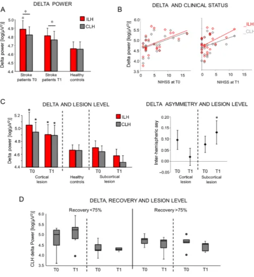
Delta band
In the delta band, a significant main effect of hemisphere was found (F(1,23) = 4.536, P = 0.044), indicating that in both
phases ILH delta power was greater than CLH delta power (Figure 3A). Neither Time main effect nor Time ×
Hemi-sphere interaction was present. Comparing delta power of
both hemispheres with the values of healthy controls, only ILH delta at T0 was significantly different from delta of healthy controls (unpaired t-test, P = 0.036 for ILH and P = 0.092 for CLH). At T1, no differences between the two groups were observed (unpaired t-test, P = 0.130 for ILH, P = 0.276 for CLH, Figure 3A).
Delta and clinical status
In the acute phase, delta band power of both hemispheres positively correlated with NIHSS scores (for ILH: Spear-man’s rs = 0.460, P = 0.027, confidence interval: 0.062, 0.722;
for CLH: rs = 0.508, P = 0.013, confidence interval: 0.128,
0.773; Figure 3B left), indicating a worse clinical status paired to an increase of delta power.
In the sub-acute phase, only ILH delta power positively correlated with NIHSS (rs = 0.411, P = 0.046, confidence
in-terval: 0.010, 0.672; Figure 3B). No significant correlations were found between clinical scores and CLH delta power (rs
= 0.346, P = 0.098, confidence interval: –0.041, 0.658). No correlation was found between ER and delta power in sub-acute phase (rs = –0.200, P = 0.348 and rs = –0.147, P = 0.493,
respectively for ILH and CLH).
Delta: dependence on lesion site (cortical – subcortical)
Both ILH and CLH delta power values at T0 of patients with cortical involvement were higher than healthy control values (Mann-Whitney U test, P < 0.008). No differences between values of healthy controls and subcortical patients were found (Figure 3C, left). No differences between patient groups and healthy controls were found at T1 (Mann-Whit-ney U test, P > 0.05).
For the delta band inter-hemispheric asymmetries, while no differences depending on the lesion site were observed in the acute phase (Mann-Whitney U test, P > 0.05), at T1 delta activity was more symmetrical in cortical patients (Mann-Whitney U test, P > 0.05) than in subcortical one (Mann-Whitney U test, P < 0.05) (Figure 3C, right).
Delta power and recovery in sub-acute phase depending on lesion site
To investigate whether CLH delta power values at T1 is linked to effective recovery, we split the group of patients in patients with effective recovery lower than 75% (13 patients) and higher than 75% (11 patients). This analysis revealed that CLH delta power at T1 was higher in the group with a lower recovery and cortical lesion (Figure 3D, Mann-Whit-ney U test between the two groups: P = 0.048 for cortical lesion; P = 0.662 for subcortical lesion).
Alpha band
In the alpha band, a significant interaction Time ×
Hemi-sphere was found (F(1,23) = 6.200, P = 0.020), but neither Time
nor Hemisphere main effects were present (Figure 4A). Indeed, the inter-hemispheric asymmetry in alpha power values in patients (ILH vs. CLH) was lower at T0 than at T1 (paired samples t-test: t(23) = –2.493, P = 0.020), indicating
that ILH alpha values were lower than CLH values at T0 with respect to T1 (Figure 4B).
The values of patients’ alpha power were not significantly different from the values of healthy controls in both hemi-spheres and in both phases (unpaired t-test, P > 0.05).
Alpha and clinical status
No correlation was found between clinical scores and alpha power in both phases (NIHSS and alpha power at T0: rs =
0.001, P = 0.995; rs = 0.126, P = 0.557, respectively for ILH
and CLH; NIHSS and alpha power at T1: rs = 0.008, P = 0.969;
rs = 0.086, P = 0.691, respectively for ILH and CLH).
Alpha: dependence on lesion site
No difference in alpha activity or asymmetries depending on the lesion site was found (P > 0.05).
Fractal dimension
In both patients and healthy controls, a similar HFD topog-raphy on the scalp was observed. HFD values were lower in parieto-occipital and fronto-polar areas and higher in fron-to-central and temporal areas (Figure 5A).
In patients, HFD at T0 was lower than at T1 (paired sam-ples t-test: t(23) = –3.093, P = 0.005, Figure 5B, left panel).
When comparing the HFD of stroke group with the refer-ence values of the healthy control group, we observed that in the acute phase HFD was lower in patients than in healthy controls (independent samples t-test: t(42) = 2.878, P = 0.006),
whereas no difference was observed in the sub-acute phase (independent sample t-test: t(42) = 1.585, P = 0.120). If we
consider only the patients with effective recovery lower than 75% (13 patients: 5 with lesion in the right hemisphere, 8 in the left hemisphere), HFD at T1 was significantly lower than the HFD values of healthy control group (1.395 ± 0.077 vs. 1.501 ± 0.109, t(31) = 2.137, P = 0.032, Figure 5C).
Considering the HFD values of the two hemispheres, an ANOVA design was applied with Hemisphere (ILH, CLH) and Time (T0, T1) as within-subject factors. Significant main effects of Time (F(1,22) = 8.566, P = 0.006) and
Hemi-sphere (F(1,22) = 6.311, P = 0.020) were found. The lack of
interaction Hemisphere × Time indicated that both at T0 and T1 the HFD values of both hemispheres were different. In particular, ILH HFD was lower than CLH FD (HFD ILH and HFD CLH values: 1.406 ± 0.089 and 1.441 ± 0.098 at T0; 1.416 ± 0.081 and 1.458 ± 0.086 at T1, respectively). More-over, in both hemispheres, HFD values in the acute phase were lower than those in the sub-acute phase.
ILH and CLH HFD values in patients were significantly lower at T0 than the corresponding values in healthy con-trols (unpaired t-test: t(42) = 3.337, P = 0.002 for ILH; t(42) =
3.247, P = 0.003 for CLH). In the sub-acute phase, this dif-ference was found only in ILH (t(42) = 2.128, P = 0.039),
al-though it did not survive Bonferroni correction (two hemi-spheres and two phases resulting in four comparisons), but not in CLH (t(42) = 1.733, P = 0.090). No correlation between
ILH and CLH HFD values with clinical scores (NIHSS) was found at both times (P > 0.200 consistently).
Discussion
Data from our cohort of mono-hemispheric stroke patients provided confirmative evidence that normalization of elec-tric neural activity at rest in both the ipsi- and contralesional hemisphere parallels clinical amelioration. Our study did not investigate the modifications occurring at cerebral orga-nization level depending on diverse rehabilitative approach-es; we investigated instead – as derived by EEG – the brain activity features associated with the recovery level. They can deepen understanding of recovery process, and provide an adjunctive indication prospectively to focus therapies in sus-taining brain adaptive reorganizations.
Delta power as an index of clinical damage
A power increase in low frequency band (1–4 Hz, delta
band) was the most evident hallmark of electrophysiologi-cal brain activity alteration at rest in stroke patients, clearly associated with the clinical status. Indeed, consistently with previous EEG literatures (Van der Drift and Kok, 1972; Sainio et al., 1983; Ahmed 1988; Jackel and Harner, 1989; Nagata et al., 1992; Niedermeyer 1997; Makela et al., 1998;
Murri et al., 1998; Fernandez-Bouzas et al., 2000; Tecchio et al., 2005, 2006a), delta power was higher in ILH that in CLH, in both acute and sub-acute phase. Nevertheless, in our patient cohort, we observed also an increment of delta power in the CLH with respect to healthy control values. Delta activity is believed to be the hallmark of two distinct phenomena, i.e. cortico-thalamocortical network dysfunc-tion and intracortical neuronal activity alteradysfunc-tion, as a result of deafferentation (Amzica and Steriade, 1995; Blatow et al., 2003). In animal models, it has been proven that delta activ-ity is cortical in origin, since it is still present after extensive ipsilateral thalamotomy (Steriade and Contreras, 1995) and cannot be recorded in the thalamus of decorticated animals or after cortical necrosis (Timofeev and Steriade, 1996). In humans, ILH delta activity in stroke has been described as a ‘perilesional’ rhythm (Butz et al., 2004; Machado et al., 2004) that increases in correspondence of neural tissue damage, necrosis or both structural or functional deafferentation. Additionally, ILH delta activity correlates with the lesion volume in acute phase (Harmony et al., 1995; Wu et al., 2016), as well as with a worse acute clinical status (Assenza et al., 2009; Wu et al., 2016). Our results are in line with these previous findings, as delta power in the ILH positive-ly correlates with NIHSS scores both in the acute and sub-acute phases. ILH delta activity is a sign of cortical rear-rangement accompanying a worse clinical status due to the damage. Thus, in patients, a reduction of ILH low-frequency activity at 2 months towards healthy normative values is a sign of a clinical amelioration. To be noted, a correlation of delta activity with the clinical status has been previously de-scribed also at six months from the ischemic attack (Tecchio et al., 2006a, b). However, delta activity, during both the sleep and the wakefulness, is a well-known sign of cortical plastic changes (Tononi and Cirelli, 2012; Assenza and Di Lazzaro, 2015, Giovanni et al., 2017), thus in stroke patients it can also be interpreted as a sign of ongoing cortical rear-rangement. Its correlation with a worse clinical status makes difficult to understand to which extent this delta activity is symptomatic of the severity of the lesion and to which ex-tent it marks an attempt to build a new cortical network to overcome the clinical deficits. We are aware that the present study cannot resolve this ambiguity. However, our observa-tion of a high delta activity in the sub-acute phase in patients with either a complete or a partial recovery suggests that a prolonged follow-up could reveal whether subacute chronic delta is a sign that cortical plasticity is still ongoing.
The presence of a delta power increase in patients with re-spect to healthy controls also in the hemisphere contra-later-al to the lesion suggests an involvement of the CLH in post-stroke recovery. Actually, not only previous EEG and MEG findings (Tecchio et al., 2005, 2007; Zappasodi et al., 2007; Finnigan et al., 2008; Assenza et al., 2013; Van Kaam et al., 2018), but also magnetic stimulation studies (Di Lazzaro et al., 2010) and positron emission tomography (Calautti et al., 2001) demonstrated that the CLH over-activation in the post-stroke phase is detrimental for clinical recovery. Nev-ertheless, it is still unclear whether this is a compensatory
mechanism activated by a severe lesion or an index of func-tional disconnection. As suggested in Assenza et al. (2013), from an electrophysiological point of view, the two main hy-potheses that may explain the presence of an enhanced con-tra-lesional delta activity are an interhemispheric spreading from the ipsi- to the contralesional hemisphere or a
break-down of interhemispheric communication due to transcal-losal diaschisis, i.e., a modification of functionality of a brain area remote from the lesion (Von Monakov, 1914). This modification is possibly mediated by cerebral blood flow and metabolism decrease, or by electrophysiological phe-nomena (Carrera and Tononi, 2014). Although in subacute
Figure 2 Electroencephalographic montage and power spectrum densities
Up: Electrode spatial positioning of the international 10–20 system used in this study. Electrodes covering the middle cerebral artery vascular ter-ritory used for the analysis are evidenced (in red for the left, in blue for the right hemisphere). Bottom: Examples of power spectrum densities in both hemispheres of one patient (female, 86 years old, lesion with cortical involvement in the left temporal area), in both acute (T0) and sub-acute phase (T1), and a healthy control (male, 73 years old).
Figure 3 Electroencephalographic delta band power.
(A) Mean (standard error) of band power values in delta band for ILH and CLH in patients in the acute and sub-acute phases. Reference values of controls are also shown. Stars indicate a significant difference in patients with respect to the corresponding value in the control group, as indicated by unpaired t-test (*P < 0.05). Horizon-tal bars indicate significant difference between ILH and CLH values. (B) Scat-terplot of the delta band power in ILH and CLH over NIHSS scores at both acute (T0) and sub-acute phase (T1). Regression lines are displayed. (C) Left: Mean (standard error) of band power values in delta band for ILH and CLH in patients in the acute and sub-acute phases, split in two groups on the basis of the lesion site: lesion with cortical involvement, subcortical lesion. Right: Mean (standard error) of inter-hemi-spheric asymmetries of delta activity at both phases in both groups of patients. Stars indicate a significant difference in patients with respect to the corre-sponding value in the control group (*P < 0.05), as assessed by Mann-Whitney U test (Bonferroni corrected). (D) Box-plot (median, 95% confidence interval) of CLH delta power in patients at both phases (T0 and T1), stratified in accor-dance to effective recovery (< 75% and > 75%) and lesion site (cortical involve-ment, subcortical lesion). ILH: Ipsile-sional hemisphere; CLH: contraleIpsile-sional hemisphere; NIHSS: National Institutes of Health Stroke Scale.
* *
stroke patients a decreased regional cerebral blood flow also in the CLH has been documented (Lagreze et al., 1987), to our knowledge no correlation between this reduction and low frequency activity increase has been found (Melamed et al., 1975).
Inter-hemispheric unbalance
Several studies in animal models and humans evidenced the functional role of interhemispheric unbalance in con-sequence of a brain lesion. In a rat model, interhemispheric connectivity and neurological improvement followed a paral-lel trend after cerebral ischemia, from acute to chronic stage (van Meer et al., 2010). In humans, both functional MRI and electrophysiological data in acute and chronic stroke patients demonstrated that the balancing is associated with a better clinical and functional picture (Carter et al., 2010; Pellegrino
et al., 2012; Graziadio et al., 2013; Baldassarre et al., 2016). In our patient cohort, we found changes in inter-hemispheric asymmetry in the alpha band between T0 and T1. In partic-ular, while alpha was lower in ILH than in CLH in the acute phase, in the sub-acute phase a restoration of ILH vs. CLH alpha activity balance was evidenced. Preservation of alpha activity in the acute phase has been associated to neuronal survival in ischemic regions (Juhász et al., 1997; Leon-Carri-on et al., 2009). Moreover, the prognostic value of the ratio between delta and alpha activities has been described, with a better clinical condition associated to an increment of alpha with respect to delta activity (Leon-Carrion et al., 2009; Fin-nigan et al., 2016; Aminov et al., 2017).
Dependence of the electrophysiological features on lesion site
Recent studies pointed out the need to consider a
stratifi-Figure 4 Electroencephalographic alpha band power.
(A) Mean (standard error) of band power values in alpha band for ILH and CLH in patients at both acute and sub-acute phases. Reference values of con-trols are also shown. (B) Mean (standard error) of inter-hemispheric asymmetries of alpha activity at both phases in both groups of patients. Star indicates signif-icant difference between acute (T0) and sub-acute phase (T1) values, as assessed by two-tailed paired samples t-test (*P < 0.05). ILH: Ipsilesional hemisphere; CLH: contralesional hemisphere.
Figure 5 Electroencephalographic fractal dimension
(A) Topographies of mean values of fractal dimension in patients at T0 (acute phase, left) and T1 (sub-acute phase, center) and in healthy age-matched controls (right). Black dots indicate the position of electroencephalographic electrodes in the montage. (B) Left: mean (standard error) of global fractal dimension (average over all sensors) in patients at T0 and T1 and in healthy controls. Right: mean (standard error) of Higuchi’s fractal dimension (HFD) values in ILH and CLH of patients at both T0 and T1. Stars indicate a sig-nificant difference between groups, as assessed by two-tailed paired samples t-test (*P < 0.05). (C) In the sub-acute phase, mean and standard deviation of global fractal dimension in patients with ER lower than 75% (left) and patients with ER higher than 75%. Stars indicate a significative difference with respect to values in healthy controls. ILH: Ipsile-sional hemisphere; CLH: contraleIpsile-sional hemisphere; ER: effective recovery [(NI-HSS at T0 – NI[(NI-HSS at T1)/NI[(NI-HSS at T0 × 100%]. patients T0 patients T1 * * * * * patientsT0 patientsT1
A
B
*cation of patients based on the lesion site when studying inter-hemispheric balance in stroke (Cramer 2010; Di Pino et al., 2014). For example, Fanciullacci et al. (2017) showed that a brain injury from unilateral stroke resulted in a bilat-eral enhancement of delta activity, and thus in an enhance-ment of delta power also in the CLH, only in patients with cortical involvement and not in subcortical stroke, in which an ‘asymmetrical’ delta activity was found. This is an inter-esting hypothesis that cannot be evaluated in the present study because of the limited number of patients that does not allow any stratification. Nevertheless, in our cohort of patients, while we found no differences in inter-hemisperic asymmetry in relation to lesion site in the acute phase, in the sub-acute phase we found a more symmetrical delta in patients with cortical involvement, with respect to pure subcortical patients. This result may suggest a cortical origin of delta activity, in line with previous evidence (Butz et al., 2004; van Wijngaarden et al., 2016). Moreover, enhanced delta in the CLH was found in patients with a poorer clin-ical recovery, in which brain reorganization processes are possibly still more heavily occurring. van Wijngaarden et al. (2016) recently developed a biologically constrained mod-el to explain the characteristic slowing of alpha peak and the increase of low frequency power in stroke, associated with a cortical lesion. They proposed that thalamic neurons switched from tonic spiking to a pathologic bursting regime in consequence of a de-inactivation of voltage-gated T-type Ca2+-channels. This inactivation derives from a thalamic
relay neuron hyperpolarization, secondary to a cortical le-sion. Bidirectional divergent connectivity between thalamic nuclei promotes propagation of bursting activity and further entrainment of thalamo-cortical circuits.
Fractal dimension
Our data confirmed previous results about fractal dimension of EEG signals in the acute phase of unilateral stroke (Zap-pasodi et al., 2014) and add evidence to the idea of the frac-tal nature of electrophysiological brain signals as a marker of brain complexity (Di Ieva et al., 2014), mirroring the system functionality. Indeed, in our sample the Higuchi FD globally decreased in the acute phase after the stroke as compared with healthy controls values. We can speculate that a loss of local neural activity, with a lesion induced damage in local functionality, reflects into a disruption of global brain pro-cessing and thus in a reduced complexity in spatially distrib-uted areas. In other words, HFD discloses the intimate na-ture of strucna-ture-function unity, where an anatomical lesion, even if local and discrete, destroys the whole brain multi-scale self-similar activity. In the sub-acute phase, a general increment of HFD with respect to the acute phase was found and no difference between patients and healthy controls was noticed. Such HFD increment was proportional to the clin-ical recovery since patients with more than 75% of effective clinical recovery showed higher fractal dimension values. These signs indicate that the recovery of EEG-derived brain activity complexity parallels a restored repertoire of func-tional abilities.
Limitations
This study has some limitations. First, the number of en-rolled patients is relatively low. However, our study has not the ambition to provide a new diagnostic tool for clinical stroke diagnostic and monitoring, rather it aims at strength-ening the use of EEG technique in the clinical model of the stroke pathology and recovery. We will devote future studies to confirm our results in a larger independent patient group. Furthermore, wider cohorts with homogeneous lesion sites and volumes, together with an accurate MRI assessment, will be the basis for the deepening the significance of elec-trophysiological features in stroke and their relationship with clinical status and recovery. Second, we evaluated the clinical status through NIHSS which is the typical scale used to evaluate the clinical status in the acute phase. We are aware that other scales can be more suitable to measure the functional outcome in the period following the acute phase. Nevertheless, since one of our primary aim was to assess electrophysiological changes in relation to the ‘recovery po-tential’ somehow independently from the clinical status at T0, we used the Effective Recovery, which partly normalizes for the acute phase conditions. The definition of the Effec-tive Recovery index required the use of the same scale for both phases (Tecchio et al., 2006b, 2007; Assenza et al., 2013; Squitti et al., 2018).
Conclusion
The present study offers an EEG longitudinal monitoring of cortical activity across the acute and subacute phases of stroke, describing electrophysiological rest activity changes paralleling the clinical recovery process. We confirmed the sensitivity of EEG signals to brain plasticity in stroke pa-tients and thus we encourage new studies paralleling EEG modifications with clinical assessment to find electrophysi-ological markers able to identify patients destined to an in-complete recovery. In turn, these electrophysiological mark-ers should lead new clinical trials of standard and enriched rehabilitation (e.g. robotic, neuromodulation) in stroke patients.
Acknowledgments: We would like to thank Fabrizio Emma e Rita Fini for their technical assistance in EEG acquisition, Salvatore Assenza and Federica Assenza for his and her contributions to the enrollment and follow-up of patients.
Author contributions: Collection, analysis and interpretation of data: FZ, FT, GA, and LM. Interpretation of data and critical revision of the manuscript: VP and VL. All authors approved the final version of the manuscript.
Conflicts of interest: The authors declare no conflicts of interest. Financial support: FZ and FT obtained financial support from the Italian Ministry of Health Cod. GR-2008-1138642 “Promoting recovery from Stroke: individually enriched therapeutic intervention in Acute phase”, and PNR-CNR Aging Program. The funding bodies played no role in the study design, in the collection, analysis and interpretation of data, in the writing of the paper, and in the decision to submit the paper for publication.
Institutional review board statement: This study protocol was ap-proved by the Bioethical Committee of Ospedale San Giovanni Calibita Fatebenefretelli (No. 40/2011) on July 14, 2011, and performed in accor-dance with the Declaration of Helsinki. All participants provided written informed consent.
Declaration of participant consent: The authors certify that they have obtain all appropriate participant consent forms. In the form the partic-ipants will give their consent for their images and other clinical informa-tion to be reported in the journal. The participants understand that their names and initials will not be published and due efforts will be made to conceal their identity.
Reporting statement: This study followed the STrengthening the Re-porting of OBservational studies in Epidemiology (STROBE) statement. Biostatistics statement: The statistical methods of this study were re-viewed by the biostatistician of Campus Bio-Medico of Rome.
Copyright license agreement: The Copyright License Agreement has been signed by all authors before publication.
Data sharing statement: The authors prefer to not share the data of this study.
Plagiarism check: Checked twice by iThenticate. Peer review: Externally peer reviewed.
Open access statement: This is an open access journal, and articles are distributed under the terms of the Creative Commons Attribution-Non-Commercial-ShareAlike 4.0 License, which allows others to remix, tweak, and build upon the work non-commercially, as long as appropri-ate credit is given and the new creations are licensed under the identical terms.
Open peer reviewer: Lijie Zhai, University of Illinois at Chicago, USA. Additional file:
Additional file 1: Ethical approval documentation (in Italian). Additional file 2: Model consent form (in Italian).
Additional file 3: Open peer review report 1.
References
Ahmed I (1988) Predictive value of the electroencephalogram in acute hemispheric lesions. Clin Electroencephalogr 19:205-209.
Aminov A, Rogers JM, Johnstone SJ, Middleton S, Wilson PH (2017) Acute single channel EEG predictors of cognitive function after stroke. PLoS One 12:e0185841.
Amzica F, Steriade M (1995) Disconnection of intracortical synaptic linkages disrupts synchronization of a slow oscillation. J Neurosi 15:4658-4677.
Assenza G, Zappasodi F, Squitti R, Altamura C, Ventriglia M, Ercolani M, Quattrocchi CC, Lupoi D, Passarelli F, Vernieri F, Rossini PM, Tecchio F (2009) Neuronal functionality assessed by magnetoen-cephalography is related to oxidative stress system in acute ischemic stroke. NeuroImage 44:1267-1273.
Assenza G, Zappasodi F, Pasqualetti P, Vernieri F, Tecchio F (2013) A contralesional EEG power increase mediated by interhemispheric disconnection provides negative prognosis in acute stroke. Restor Neurol Neurosci 31:177-188.
Assenza G, Di Lazzaro V (2015) A useful electroencephalography (EEG) marker of brain plasticity: delta waves. Neural Regen Res 10:1216-1217.
Baldassarre A, Ramsey LE, Siegel JS, Shulman GL, Corbetta M (2016) Brain connectivity and neurological disorders after stroke. Curr Opin Neurol 29:706-713.
Barbati G, Porcaro C, Zappasodi F, Rossini PM, Tecchio F (2004) Op-timization of ICA approach for artifact identification and removal in MEG signals. Clin Neurophys 115:1220-1232.
Bernhardt J, Dewey H, Collier J, Thrift A, Lindley R, Moodie M, Don-nan G (2006) A very early rehabilitation trial (AVERT). Int J Stroke 1:169-171.
Blatow M, Rozov A, Katona I, Hormuzdi SG, Meyer AH, Whittington MA, Caputi A, Monyer H (2003) A novel network of multipolar bursting interneurons generates theta frequency oscillations in neo-cortex. Neuron 38:805-817.
Butz M, Gross J, Timmermann L, Moll M, Freund HJ, Witte OW, Schnitzler A (2004) Perilesional pathological oscillatory activity in the magnetoencephalogram of patients with cortical brain lesions. Neurosci Lett 355:93-96.
Buzsaki G, Mizuseki K (2014) The log-dynamic brain: how skewed dis-tributions affect network operations. Nat Rev Neurosci 15:264-278. Calautti C, Leroy F, Guincestre JY, Marié RM, Baron JC (2001)
Se-quential activation brain mapping after subcortical stroke: changes in hemispheric balance and recovery. Neuroreport 12:3883-3886.
Carrera E, Tononi G (2014) Diaschisis: past, present, future. Brain 137:2408-2422.
Carter AR, Astafiev SV, Lang CE, Connor LT, Rengachary J, Strube MJ, Pope DL, Shulman GL, Corbetta M (2010) Resting interhemi-spheric functional magnetic resonance imaging connectivity pre-dicts performance after stroke. Ann Neurol 67:365-375.
Cramer SC (2010) Stratifying patients with stroke in trials that target brain repair. Stroke 41(10 Suppl):S114-116.
Crichton SL, Bray BD, McKevitt C, Rudd AG, Wolfe CDA (2016) Patient outcomes up to 15 years after stroke: survival, disability, quality of life, cognition and mental health. J Neurol Neurosurg Psychiatry 87:1091-1098.
Di Ieva A, Grizzi F, Jelinek H, Pellionisz AJ, Losa GA (2014) Fractals in the Neurosciences, Part I: General Principles and Basic Neurosci-ences. Neuroscientist 20:403-417.
Di Lazzaro V, Profice P, Pilato F, Capone F, Ranieri F, Pasqualetti P, Colosimo C, Pravatà E, Cianfoni A, Dileone M (2010) Motor cortex plasticity predicts recovery in acute stroke. Cereb Cortex 20:1523-1528.
Di Pino G, Pellegrino G, Assenza G, Capone F, Ferreri F, Formica D, Ranieri F, Tombini M, Ziemann U, Rothwell JC, Di Lazzaro V (2014) Modulation of brain plasticity in stroke: a novel model for neurore-habilitation. Nat Rev Neurol 10:597-608.
Dromerick AW, Reding MJ (1995) Functional outcome for patients with hemiparesis, hemihyposthesia and hemianopsia. Stroke 11:2023-2026.
Fanciullacci C, Bertolucci F, Lamola G, Panarese A, Artoni F, Micera S, Rossi B, Chisari C (2017) Delta power is higher and more symmet-rical in ischemic stroke patients with cortical involvement. Front Hum Neurosci 11:385.
Fernandez-Bouzas A, Harmony T, Fernandez T, Silva-Pereyra J, Valdes P, Bosch J, Aubert E, Casián G, Otero Ojeda G, Ricardo J, Hernán-dez-Ballesteros A, Santiago E (2000) Sources of abnormal EEG activ-ity in brain infarctions. Clin Electroencephalogr 31:165-169. Finnigan S, Wong A, Read S (2016) Defining abnormal slow EEG
activity in acute ischaemic stroke: Delta/alpha ratio as an optimal QEEG index. Clin Neurophysiol 127:1452-1459.
Finnigan SP, Rose SE, Chalk JB (2008) Contralateral hemisphere delta EEG in acute stroke precedes worsening of symptoms and death. Clin Neurophysiol 119:1690-1694.
Garrett DD, Samanez-Larkin GR, MacDonald SW, Lindenberger U, McIntosh AR, Grady CL (2013) Moment-to-moment brain signal variability: A next frontier in human brain mapping? Neurosci Biobehav Rev 37:610-624.
Giovanni A, Capone F, di Biase L, Ferreri F, Florio L, Guerra A, Ma-rano M, Paolucci M, Ranieri F, Salomone G, Tombini M, Thut G, Di Lazzaro V (2018) Oscillatory activities in neurological disorders of elderly: biomarkers to target for neuromodulation. Front Aging Neurosci 9:189.
Graziadio S, Tomasevic L, Assenza G, Tecchio F, Eyre JA (2013) The myth of the ‘unaffected’ side after unilateral stroke: Is reorganisation of the non-infarcted corticospinal system to re-establish balance the price for recovery? Exp Neurol 238:168-175.
Harmony T, Fernández-Bouzas A, Marosi E, Fernández T, Valdés P, Bosch J, Riera J, Jackel RA, Harner RN (1989) Computed EEG to-pography in acute stroke. Neurophysiol Clin 19:185-197.
He BJ (2011) Scale-free properties of the functional magnetic resonance imaging signal during rest and task. J Neurosci 31:13786-13795. Higuchi T (1988) Approach to an irregular time series on the basis of
the fractal theory. Physica D 31:277-283.
Jackel RA, Harner RN (1989) Computed EEG topography in caute stroke. Neurophysiol Clin 19:185-197.
Juhász C, Kamondi A, Szirmai I (1997) Spectral EEG analysis follow-ing hemispheric stroke: evidences of transhemispheric diaschisis. Acta Neurol Scand 96:397-400.
Lagreze HL, Levine RL, Pedula KL, Nickles RJ, Sunderland JS, Rowe BR (1987) Contralateral flow reduction in unilateral stroke: evidence for transhemispheric diaschisis. Stroke 18:882-886.
Leon-Carrion J, Martin-Rodriguez JF, Damas-Lopez J, Barroso y Mar-tin JM, Dominguez-Morales MR (2009) Delta-alpha ratio correlates with level of recovery after neurorehabilitation in patients with ac-quired brain injury. Clin Neurophysiol 120:1039-1045.
Machado C, Cuspineda E, Valdés P, Virues T, Llopis F, Bosch J, Aubert E, Hernández E, Pando A, Alvarez MA, Barroso E, Galán L, Avila Y (2004) Assessing acute middle cerebral artery ischemic stroke by quantitative electric tomography. Clin EEG Neurosci 35:116-124. Makela JP, Salmelin R, Kotila M, Salonen O, Laaksonen R, Hokkanen L,
Hari R (1998) Modification of neuromagnetic cortical signals by tha-lamic infarctions. Electroencephalogr Clin Neurophysiol 106:433-443.
Marzetti L, Nolte G, Perrucci MG, Romani GL, Del Gratta C (2007) The use of standardized infinity reference in EEG coherency studies. Neuroimage 15:48-63.
Melamed E, Lavy S, Portnoy Z, Sadan S, Carmon A (1975) Correlation between regional cerebral blood flow and EEG frequency in the con-tralateral hemisphere in acute cerebral infarction. J Neurol Sci 26:21-27.
Murri L, Gori S, Massetani R, Bonanni E, Marcella F, Milani S (1998) Evaluation of acute ischemic stroke using quantitative EEG: a com-parison with conventional EEG and CT scan. Neurophysiol Clin 28:249-257.
Nagata K, Mizukami M, Araki G, Kawase T, Hirano M (1982) Topo-graphic electroencephaloTopo-graphic study of cerebral infarction using computed mapping of the EEG. J Cereb Blood Flow Metab 2:79-88. National Institute of Health, National Institute of Neurological
Dis-orders and Stroke. Stroke Scale. https://www.ninds.nih.gov/sites/ default/files/NIH_Stroke_Scale_Booklet.pdf. Accessed December 2, 2018.
Niedermeyer E (1997) Cerebrovascular disorders and EEG. In: Electro-encephalography: Basic Principles, Clinical Applications and Related Fields, 4th ed (Niedermeyer E, Lopes Da Silva F, eds), pp320-321. Oxford, UK: Oxford University Press.
Pellegrino G, Tomasevic L, Tombini M, Assenza G, Bravi M, Sterzi S, Giacobbe V, Zollo L, Guglielmelli E, Cavallo G, Vernieri F, Tecchio F (2012) Inter-hemispheric coupling changes associate with motor improvements after robotic stroke rehabilitation. Restor Neurol Neurosci 30:497-510.
Ramon C, Holmes MD (2015). Spatiotemporal phase clusters and phase synchronization patterns derived from high density EEG and ECoG recordings. Curr Opin Neurobiol 31:127-132.
Roberts J, Iyer K, Finnigan S, Vanhatalo S, Breakspear M (2014) Scale-free bursting in human cortex following hypoxia at birth. J Neurosci 34:6557-6572.
Rodríguez-Bermúdez G, García-Laencina P (2015) Analysis of EEG signals using nonlinear dynamics and chaos: a review. Appl Math Inf Sci 9:2309-2321.
Rossini PM, Altamura C, Ferretti A, Vernieri F, Zappasodi F, Caulo M, Pizzella V, Del Gratta C, Romani GL, Tecchio F (2004) Does cerebrovascular disease affect the coupling between neuronal activity and local haemodynamics? Brain 127:99-110.
Rossini PM, Altamura C, Ferreri F, Melgari JM, Tecchio F, Tombini M, Pasqualetti P, Vernieri F (2007) Neuroimaging experimental studies on brain plasticity in recovery from stroke. Eura Medicophys 43:241-254.
Sainio K, Stenberg D, Keskimaki I, Muuronen A, Kaste M (1983) Vi-sual and spectral EEG analysis in the evaluation of the outcome in patients with ischemic brain infarction. Electroencephalogr Clin Neurophysiol 56:117-124.
Squitti R, Siotto M, Assenza G, Giannantoni NM, Rongioletti M, Zap-pasodi F, Tecchio F (2018) Prognostic value of serum copper for post-stroke clinical recovery: a pilot study. Front Neurol 9:333. Stam CJ (2005) Nonlinear dynamical analysis of EEG and MEG: review
of an emerging field. Clin Neurophysiol 116:2266-2301.
Steriade M, Contreras D (1998) Spike-wave complexes and fast com-ponents of cortically generated seizures. I. Role played by neocortex and thalamus. J Neurophysiol 80:1439-1455.
Tecchio F, Zappasodi F, Pasqualetti P, Tombini M, Salustri C, Oliviero A, Vittorio P, Vernieri F, Rossini PM (2005) Rhythmic brain activity at rest from rolandic areas in acute mono-hemispheric stroke: A magnetoencephalographic study. Neuroimage 28:72-83.
Tecchio F, Zappasodi F, Tombini M, Oliviero A, Pasqualetti P, Ver-nieri F, Ercolani M, Pizzella V, Rossini PM (2006a) Brain plasticity in recovery from stroke: an MEG assessment. Neuroimage 32:1326-1334.
Tecchio F, Zappasodi F, Pasqualetti P, Tombini M, Caulo M, Ercolani M, Rossini PM (2006b) Long-term effects of stroke on neuronal rest activity in rolandic cortical areas. J Neurosci Res 83:1077-1087. Tecchio F, Pasqualetti P, Zappasodi F, Tombini M, Lupoi D, Vernieri F,
Rossini PM (2007) Outcome prediction in acute monohemispheric stroke via magnetoencephalography. J Neurol 254:296-305. Timofeev I, Steriade M (1996) Low-frequency rhythms in the thalamus
of intact-cortex and decorticated cats. J Neurophysiol 76:4152-4168. Tononi G, Cirelli C (2014) Sleep and the price of plasticity: from
syn-aptic and cellular homeostasis to memory consolidation and integra-tion. Neuron 81:12-34.
Van der Drift JHA, Kok NKD (1972) The EEG in cerebrovascular dis-orders in relations to pathology. In: Handbook of Electroencepha-lography and Clinical Neuropysiology (Remond A, ed), pp12-30, 30, 47-64. Amsterdam, Netherlands: Elsevier.
Van Kaam RC, van Putten MJAM, Vermeer SE, Hofmeijer J (2018) Contralesional brain activity in acute ischemic stroke. Cerebrovasc Dis 45:85-92.
van Meer MP, van der Marel K, Wang K, Wang K, Otte WM, El Bouazati S, Roeling TA, Viergever MA, Berkelbach van der Sprenkel JW, Dijkhuizen RM (2010) Recovery of sensorimotor function after experimental stroke correlates with restoration of resting-state inter-hemispheric functional connectivity. J Neurosci 30:3964-3972. van Wijngaarden JB, Zucca R, Finnigan S, Verschure PF (2016) The
impact of cortical lesions on thalamo-cortical network dynamics af-ter acute ischaemic stroke: a combined experimental and theoretical study. PLoS Comput Biol 12:e1005048.
von Monakov C (1914) Die Lokalisation im Grosshirn und der Abbau der Funktion durch kortikale Herde. JAMA LXIII:797.
Wu J, Srinivasan R, Burke Quinlan E, Solodkin A, Small SL, Cramer SC (2016) Utility of EEG measures of brain function in patients with acute stroke. J Neurophysiol 115:2399-2405.
Zappasodi F, Tombini M, Milazzo D, Rossini PM, Tecchio F (2007) Delta dipole density and strength in acute monohemispheric stroke. Neurosci Lett 416:310-314.
Zappasodi F, Olejarczyk E, Marzetti L, Assenza G, Pizzella V, Tecchio F (2014) Fractal dimension of EEG activity senses neuronal impair-ment in acute stroke. PLoS One 9:e100199.
Zappasodi F, Marzetti L, Olejarczyk E, Tecchio F, Pizzella V (2015) Age-related changes in electroencephalographic signal complexity. PLoS One 10:e0141995.
Zeiler SR, Krakauer JW (2013) The interaction between training and plasticity in the poststroke brain. Curr Opin Neurol 26:609-616.