Ryanodine receptor point mutant E4032A reveals an
allosteric interaction with ryanodine
James D. Fessenden*†, Lili Chen*, Yaming Wang‡, Cecilia Paolini§, Clara Franzini-Armstrong§, Paul D. Allen‡, and Isaac N. Pessah*¶
*Department of Molecular Biosciences, School of Veterinary Medicine, University of California, Davis, CA 95616;‡Department of Anesthesia Research,
Brigham and Women’s Hospital, Boston, MA 02115; and§Department of Cellular and Developmental Biology, University of Pennsylvania
School of Medicine, Philadelphia, PA 19104
Contributed by Clara Franzini-Armstrong, December 20, 2000
The ryanodine receptor (RyR) family of proteins constitutes a unique type of calcium channel that mediates Ca2ⴙrelease from
endoplasmic reticulum兾sarcoplasmic reticulum stores. Ryanodine has been widely used to identify contributions made by the RyR to signaling in both muscle and nonmuscle cells. Ryanodine, through binding to high- and low-affinity sites, has been suggested to block the channel pore based on its ability to induce partial conductance states and irreversible inhibition. We examined the effect of ryanodine on an RyR type 1 (RyR1) point mutant (E4032A) that exhibits a severely compromised phenotype. When expressed in 1B5 (RyR null兾dyspedic) myotubes, E4032A is relatively unrespon-sive to stimulation by cell membrane depolarization or RyR ago-nists, although the full-length protein is correctly targeted to junctions and interacts with dihydropyridine receptors (DHPRs) inducing their arrangement into tetrads. However, treatment of E4032A-expressing cells with 200 –500M ryanodine, concentra-tions that rapidly activate and then inhibit wild-type (wt) RyR1, restores the responsiveness of E4032A-expressing myotubes to depolarization and RyR agonists. Moreover, the restored E4032A channels remain resistant to subsequent exposure to ryanodine. In single-channel studies, E4032A exhibits infrequent (channel-open probability, Po < 0.005) and brief (<250s) gating events and
insensitivity to Ca2ⴙ. Addition of ryanodine restores Ca2ⴙ -depen-dent channel activity exhibiting full, 3兾4, 1兾2, and 1兾4 substates. This evidence suggests that, whereas ryanodine does not occlude the RyR pore, it does bind to sites that allosterically induce substantial conformational changes in the RyR. In the case of E4032A, these changes overcome unfavorable energy barriers introduced by the E4032A mutation to restore channel function.
T
he plant alkaloid ryanodine has been broadly used toexamine the contribution of ryanodine receptors (RyR) to Ca2⫹signaling in many different cell types. The synthesis of [3H]ryanodine (1, 2) has enabled the purification and detailed characterization of all three RyR isoforms, first from muscle (3, 4), and subsequently from a variety of tissues (5). The specific binding of [3H]ryanodine to the RyR is typically used to study the influence of ligands on the activity of the Ca2⫹ channel, because ryanodine binding is generally correlated with channel-open probability (Po; refs. 6 and 7). Based on results obtained from the skeletal muscle isoform (RyR1), ryanodine has been proposed to bind to multiple sites via a sequential mechanism. What has remained unclear is the number of binding sites on the RyR for ryanodine, and the relationship between high- and low-affinity binding interac-tions. In one model, ryanodine binding to a single high-affinity site results in partial channel occlusion and is responsible for stable ⬇1兾2 subconductance transitions, whereas ryanodine binding to a single low-affinity site results in occlusion of the pore and subsequent channel blockade (8). In an alternative model, as many as four equivalent high-affinity binding sites reside on the RyR tetramer. Occupancy of these sites by ryanodine decreases the subsequent affinity of the remaining binding sites in an allosterically coupled sequential mechanism
coincident with several conformational subconductance states (9, 10). It is generally agreed, however, that ryanodine mod-ifies channel activity in a concentration- and time-dependent manner. Occupancy of high-affinity sites (Kd ⫽ 1–100 nM) activates the RyR, whereas binding to low-affinity sites (Kd⫽ 0.5–3.0M) initially locks the channel into a subconductance state, and then irreversibly inhibits the channel (11).
One model to account for the high- and low-affinity actions of ryanodine on channel conductance suggests that the alkaloid acts as a pore blocker. Thus, compounds that open the channel increase the affinity for ryanodine caused by the opening of the pore to which ryanodine binds; ryanodine bound to the pore can occlude passage of Ca2⫹ through the RyR. However, this hypothesis has not been tested directly, and recent molecular modeling studies have suggested that ryanodine may be acting at allosteric sites (12, 13).
Recently a class of RyR1 point mutants has been created that may contribute unique insights into the mechanism by which ryanodine modulates RyR function. These substitutions all reside at charged amino acids between positions 3987 and 4969 of RyR1, and are thought to lie within the putative transmembrane assembly (14, 15). When expressed in HEK-293 cells, several of these mutations severely decrease both the channel’s caffeine sensitivity and high-affinity [3H]ryanodine binding. One of these point mutations performed in an RyR3 context, E3885A (which is analogous to E4032A in RyR1), decreases sensitivity to calcium activation by 10,000-fold (14). It has been suggested that this glutamic acid residue, which is conserved in all three RyR isoforms, constitutes a portion of the calcium sensor, a region on the RyR necessary for trans-lating calcium binding into channel activation. An alternative hypothesis that could explain why these diverse mutations between positions 3987 and 4969 result in severe insensitivity to channel activation is that they all cause a localized folding error within the transmembrane assembly which is critical for the conformational transitions that are necessary for channel activation. Such a mechanism could introduce a large energy barrier that severely reduces channel-opening events. Further-more, these mutations might also be expected to affect ryan-odine sensitivity because this region encompasses the known ryanodine-binding domains (16, 17).
Abbreviations: Po, channel-open probability; RyR, ryanodine receptors; RyR1, skeletal muscle isoform; wtRyR1, wild-type RyR1; BLM, bilayer lipid membranes; IU, infectious units; wt, wild type; DHPR, dihydropyridine receptor; AMP-PCP, adenosine 5⬘-[,␥-methylene]-triphosphate; SR, sarcoplasmic reticulum.
†Present address: Department of Anesthesia Research, Brigham and Women’s Hospital,
Boston, MA 02115.
¶To whom reprint requests should be addressed at: Department of Molecular Biosciences,
School of Veterinary Medicine, University of California, 1 Shields Avenue, Davis, CA 95616. E-mail: [email protected].
The publication costs of this article were defrayed in part by page charge payment. This article must therefore be hereby marked “advertisement” in accordance with 18 U.S.C. §1734 solely to indicate this fact.
In the present study, we characterized E4032A RyR1 ex-pressed in the RyR null兾dyspedic 1B5 myogenic cell line (18). Our results indicate that this RyR1 mutant is largely unrespon-sive to RyR agonists and exhibits very rare and brief-lived open events (Po ⬍⬍ 0.1) upon reconstitution in bilayer lipid mem-branes (BLM). A finding which clarifies the molecular mecha-nism of ryanodine action reveals that dyspedic myotubes ex-pressing E4032A regain their ability to respond to depolarizing potentials, caffeine, and 4-chloro-m-cresol after the addition of micromolar ryanodine. Ryanodine also restores single channel activity, demonstrating that unlike wild-type RyR1 (wtRyR1), micromolar concentrations of this alkaloid do not persistently block the activity of the E4032A channels. These results indicate that ryanodine does not occlude the Ca2⫹permeation pore of RyR1 and reveals an allosteric mechanism for its action.
Materials and Methods
Cell Culture.1B5 cells were cultured as described (19). For Fura-2
ratio fluorescence imaging measurements, cells were grown on collagen-coated 72-well polystyrene plates (Nalge). For prepa-ration of sarcoplasmic reticulum (SR) vesicles for bilayer mea-surements and Western blot analyses, the cells were grown on collagen-coated 100-mm polystyrene dishes.
Construction of E4032A RyR1 cDNA.The point mutation E4032A
was introduced into RyR1 by the overlap extension method (20) using PCR. The outer two oligonucleotides used were as follows: forward, 5⬘-GTGTTCAACAGCCTCACCGA-3⬘ and reverse, 5⬘-GAACTGCTTCTGGCTGTCCA-3⬘. The oligonucleotides for the E4032A mutation are as follows: forward, GTCCCTACTGGCAGGGAACGTGGT-3⬘ and reverse, 5⬘-CCACGTTCCCTGCCAGTAGGGACA-3⬘. The sequence of the PCR product was confirmed by DNA sequencing. The XhoI (12018)-StuI (12224) fragment was removed from the PCR product and was used to replace the corresponding wt region to form the full-length E4032A-RyR1 cDNA.
Viral Infection.Herpes simplex virus-1 virions (21) containing the
cDNA encoding either wtRyR1 or E4032A-RyR1 were added to differentiated 1B5 myotubes at a concentration of 3 ⫻ 105 infectious units (IU)兾ml. Functional studies were performed 48 h after infection. In some experiments, ryanodine (10–500 M) was added either 30 min or 24 h before calcium imaging and was removed just before imaging the cells.
Calcium Imaging. Changes in cytosolic calcium levels in 1B5
myotubes were measured by using Fura-2 as described pre-viously (19).
Immunocytochemistry and Western Blot Analysis. 1B5 myotubes
were processed for RyR immunocytochemistry and Western blot analysis as described previously (19, 22) by using 34C primary antibody (Development Studies Hybridoma Bank, Iowa City, IA; ref. 23).
SR Membrane Preparations.Crude membrane homogenates from
myotubes infected with E4032A cDNA-containing virions were prepared as described previously (18, 19), and subsequently loaded onto a sucrose gradient consisting of layers of 10%, 27%, and 45% (wt兾wt) sucrose in 10 mM Hepes (pH 7.4). After sedimentation of the crude membranes on this gradient at 40,000⫻ g for 1 h at 4°C, the 27–45% interface containing heavy SR membranes was isolated and diluted in 10 mM Hepes (pH 7.4) and subsequently pelleted at 110,000⫻ g for 1 h. The pellet was resuspended in 10% (wt兾wt) sucrose and 10 mM Hepes (pH 7.4), divided into small aliquots, and stored at⫺80°C.
Single-Channel Studies.Measurements of single E4032A channels
reconstituted into BLM were performed by using Cs⫹as charge carrier as described previously (19). Membrane vesicles contain-ing RyR channels for reconstitution experiments were isolated from 1B5 myotubes expressing E4032A as described above, that had been either exposed to 200M ryanodine for 24 h or left untreated. In separate experiments, heavy SR membranes con-taining E4032A isolated from cells not exposed to ryanodine during culture were treated directly with 200 M ryanodine in the test tube for 30 min at 37°C before reconstitution in BLM.
Binding of [3H]Ryanodine.The binding of 10 nM [3H]ryanodine to
high-affinity sites on SR prepared from 1B5 myotubes expressing either wtRyR1 or E4032A was performed as previously de-scribed (18) in the absence or presence of 20 mM caffeine and 1 mM adenosine 5⬘-[,␥-methylene]triphosphate (AMP-PCP; a nonhydrolyzable ATP analog).
Electron Microscopy. Differentiated cultures of RyR1 and
E4032A-expressing 1B5 myotubes were fixed in 3.5% glutaral-dehyde in 0.1 M sodium phosphate buffer (pH 7.4) at room temperature. The cells were stored at 4°C (except during room temperature shipping between laboratories) and the cultures were cryoprotected in 30% glycerol, freeze-fractured, platinum shadowed, and carbon coated in a model BFA 400 Balzers apparatus. The replicas were examined in a Philips 410 electron microscope.
Results
The mutant E4032A RyR1 cDNA was introduced into 1B5 dyspedic myotubes by using the p-HSV amplicon system (21). 1B5 myotubes efficiently express E4032A RyR1 (which is the expected size by Western blot analysis; Fig. 1A) and properly target this protein to peripheral foci, representing sites of SR-surface junctions as detected by immunolabeling (Fig. 1 B and C). In freeze–fracture replicas, the surface membrane of E4032A-infected cells shows clusters of dihydropyridine recep-tor (DHPR) particles grouped into tetrads (Fig. 1D). The disposition of DHPR particles is indistinguishable from that observed in the membrane of wtRyR1-expressing cells (Fig. 1E and ref. 24), thus indicating that the E4032A mutant can associate with the DHPR in a manner similar to wtRyR1.
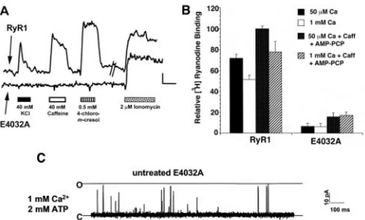
The functional phenotype of the E4032A RyR1 mutation was determined by expressing this mutated protein in 1B5 myotubes and examining functional responses by using Ca2⫹ imaging techniques with the Ca2⫹-sensitive dye Fura-2. The number of RyR-transduced 1B5 myotubes responding to RyR agonists was used as a semiquantitative measure of E4032A function. 1B5 myotubes expressing E4032A generally failed to exhibit excita-tion–contraction coupling (Fig. 2A) because only 4% (of n⫽ 158 myotubes examined) responded with a calcium transient to depolarizing medium containing 40 mM KCl. The lack of responsiveness of the E4032A mutant extended to direct cell-permeant modulators of wtRyR1, including 40 mM caffeine (13% responding) and 0.5 mM 4-chloro-m-cresol (6.4% respond-ing). In comparison, 45% and 60% of 1B5 cells infected with wtRyR1 responded to 40 mM KCl and 40 mM caffeine, respec-tively (of n⫽ 157 cells examined). Commensurate with the lack of E4032A function were very low, but clearly discernable, levels of high-affinity (10 nM) [3H]ryanodine-binding sites in mem-brane preparations isolated from E4032A-expressing 1B5 cells (Fig. 2B). Detection of specific high-affinity receptor occupancy required both 20 mM caffeine and 1 mM AMP-PCP, and was independent of free Ca2⫹ in the range of 50–1000 M. In contrast, specific Ca2⫹-dependent binding of 10 nM [3 H]ryano-dine was readily observed with identical membrane preparations from 1B5 cells expressing wtRyR1; this binding did not require the presence of caffeine and AMP-PCP (Fig. 2B).
Lack of RyR-dependent functional responses of myotubes expressing E4032A and low occupancy of nanomolar [3 H]ryano-dine suggested that E4032A channels were likely to exhibit inherently low open probability. To address this hypothesis, the same SR vesicles used for binding studies were fused with artificial BLM, and the single-channel characteristics of E4032A RyR were studied. E4032A channels displayed infrequent gating transitions and an extremely low open probability when com-pared with wtRyR1 (Fig. 2C). E4032A channels were also largely unresponsive to activation either by cis (cytoplasmic) calcium between 7M and 100 M in the presence of 1 mM ATP or the pharmacological agonist caffeine. The highest Po values were obtained in the presence of 1 to 2 mM Ca2⫹, 2 mM ATP, or 2 mM caffeine (a condition which significantly increased the Poof RyR3 E3885A channels; ref. 14), although E4032A RyR1 activ-ity remained low (Po⫽ 0.0045 ⫾ 0.0014, mean ⫾ SE, n ⫽ 11 channels, Fig. 2C). Mean open time histograms were best fit by a single time constant and were very brief (meano⫽ 0.24 ms, from n⫽ 10 channels) compared with wt channels, which were best fit by two time constants (o1 ⫽ 0.34 ms, o2 ⫽ 1.34 ms; ref. 25).
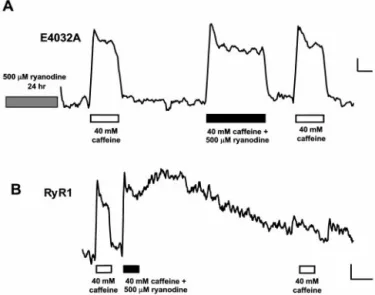
Although E4032A channels possess intact high-affinity bind-ing domains for ryanodine, the apparent affinity for alkaloid-induced modifications of function should be significantly re-duced as a direct result of the inherently low Pocontributed by the mutation. To test this hypothesis, we examined the effect of 500M ryanodine on 1B5 cells expressing the E4032A mutant protein. Interestingly, we encountered an unexpected result: treatment of E4032A-expressing 1B5 myotubes with 500 M ryanodine for 30 min restored the function of the mutated
E4032A RyR1 (Fig. 3). In these experiments, we examined caffeine responses of E4032A-transduced cells before and after ryanodine application and correlated the presence of a func-tional response in an individual cell with the expression of E4032A protein in the same cell. The cells were first tested functionally and subsequently identified by using immunocyto-chemistry to confirm that cells showing Ca2⫹ transients after ryanodine treatment expressed E4032A RyR1. Our results in-dicated that ryanodine treatment recruits E4032A-expressing cells to become responsive to RyR agonists (Fig. 3). In the representative field shown, only 2 of 25 cells identified as E4032A-expressing responded to an initial challenge with 40 mM caffeine (Fig. 3A, arrows). However, after a 30 min treat-ment with 500M ryanodine, 14 of 25 E4032A-expressing cells were responsive to a second 40 mM caffeine application (Fig. 3B, arrowheads). The ryanodine application by itself did not elicit any increase in [Ca2⫹]i in E4032A-expressing cells, although subsequent responses to 40 mM caffeine were robust (Fig. 3C). These findings were in contrast to similar experiments per-formed on 1B5 cells expressing wtRyR1 in which introduction of ryanodine led to a slow increase in cytosolic calcium, and responses to subsequently added caffeine were significantly diminished (Fig. 3D).
To confirm that micromolar ryanodine restores functional activity of E4032A, we tested the ability of the ryanodine-treated E4032A-expressing cells to support excitation–contraction cou-pling and respond to RyR agonists. After a 30 min to 24 h ryanodine incubation followed by removal of the alkaloid, the percentage of E4032A-infected cells responding to 40 mM KCl, 40 mM caffeine, and 0.5 mM chloro-m-cresol increased to 27%, 37%, and 38%, respectively (n⫽ 412 cells examined; Fig. 4A and Fig. 1. RyR1 carrying the E4032A mutation is properly expressed in 1B5
myotubes. 1B5 cells grown in collagen-coated 35-mm dishes were infected with either RyR1 or E4032A cDNA-containing herpes simplex virus amplicon virions at 3⫻ 105IU兾ml. (A) Western blot of 1B5 myotube preparations
expressing E4032A-RyR1 (lane 1, 20g), 1B5 null myotubes not transduced with cDNA (lane 2, 20g), and 1B5 myotubes expressing wtRyR1 (lane 4, 20 g). Lane 3 contains 0.5 g of rabbit junctional sarcoplasmic reticulum as a positive control. (B) The intracellular distribution of E4032A expressed in 1B5 myotubes was examined by using immunocytochemistry as described in
Ma-terials and Methods. E4032A was properly targeted to junctional domains at
the fiber periphery as indicated by the punctate appearance of the immuno-labeling pattern which was indistinguishable from the pattern obtained when wtRyR1 was expressed in 1B5 cells (C). (D and E) E4032A- and wtRyR1-expressing 1B5 myotubes were examined for DHPR tetrad formation by freeze–fracture electron microscopy. Both RyRs induced tetrad formation in the plasmalemma of 1B5 cells as indicated (arrows). The tetrads were similar in appearance and indicate the formation of a stereospecific link between four DHPRs and the four subunits of the RyR.
Fig. 2. E4032A channels are largely unresponsive to activation by RyR agonists. (A) 1B5 cells grown in collagen-coated 72-well microtiter plates (Terasaki format) were infected with either E4032A or wtRyR1 cDNA contain-ing herpes simplex virions at 3⫻ 105IU兾ml. Cells were examined for calcium
responses by using Fura-2 as described in Materials and Methods. The change in cytoplasmic calcium (as indicated by a change in F340兾F380ratio for Fura-2)
in response to 40 mM KCl, 40 mM caffeine, 0.5 mM 4-chloro-m-cresol, or 2M ionomycin for either a wtRyR1-expressing myotube (upper trace) or E4032A-expressing myotube (lower trace) is indicated. (Bar⫽ 0.1 340兾380 ratio units vs. 50 sec.) (B) E4032A RyR1 shows largely reduced high-affinity [3H]ryanodine
binding. wtRyR1 or E4032A RyR1-expressing 1B5 membrane preparations were incubated at 37°C for 3 h in a buffer containing 250 mM KCl, 15 mM NaCl, 20 mM Pipes (pH 7.4), and 10 nM [3H]ryanodine. Caffeine (20 mM) and 1 mM
AMP-PCP and兾or CaCl2was added as indicated in the graph. The relative
binding is calculated as the percentage of the binding of wtRyR1 in the presence of 50M CaCl2, 20 mM caffeine, and 1 mM AMP-PCP. The experiment
has been repeated at least twice in duplicate. (C) Single channel measure-ments of isolated E4032A channels reconstituted in BLM were conducted as described in Materials and Methods. Isolated E4032A channels give rise to infrequent gating transitions from the closed to fully open state in the presence of cis 1 mM calcium and 2 mM ATP. The open probability of this channel was 0.0022.
B). Ryanodine restored responsiveness of E4032A-expressing cells to caffeine in a dose–dependent manner with an EC50⫽ 165 M (Fig. 4B, Inset). In addition, ryanodine pretreatment restored skeletal excitation–contraction coupling because cal-cium transients elicited by 40 mM KCl were not dependent on extracellular calcium (Fig. 4C). This restoration of E4032A activity continued to be observed 90 min after the ryanodine was removed by extensive washing.
When E4032A-expressing 1B5 myotubes were pretreated with 200M ryanodine for 24 h, the reconstituted channels became significantly more active in the presence of 7M Ca2⫹(cis) with multiple substates, of which the 1兾4 state predominated (Fig. 4D, upper trace). Frequent channel transitions from the 1兾4 state to fully open were observed and the overall Poincreased to 0.7⫾ 0.07 (mean⫾ SE, n ⫽ 11 channels) as compared with wtRyR1, where Poaveraged 0.3⫾ 0.06 at 7M cis Ca2⫹. The substates of the ryanodine-treated E4032A channels were not observed in the control (untreated) E4032A channels. In the ryanodine-treated E4032A channels, gating behavior depended on the Ca2⫹
concentration on the cis face of the channel (Fig. 4D, lower trace). Lowering the cis side Ca2⫹ to 3.4 nM with EGTA completely inhibited each of the four channels tested. Recon-stitution of E4032A channel activity by ryanodine could also be achieved by treating E4032A-containing SR vesicles for 30 min with 200M ryanodine. These results demonstrate that ryano-dine treatment can reverse the phenotype of the E4032A mu-tation, thus enabling the channel to become responsive to activation.
To determine whether ryanodine can disrupt calcium release in 1B5 myotubes expressing E4032A, we tested the effect of caffeine added together with ryanodine (Fig. 5). Addition of 40 mM caffeine to a ryanodine-pretreated E4032A-expressing myo-tube produced a calcium transient. Subsequent application of caffeine supplemented with 500 M ryanodine produced a calcium transient with activation and deactivation kinetics sim-ilar to transients induced by caffeine alone. A final caffeine application yielded an identical Ca2⫹transient, thus indicating that channel function in these cells was unaffected by the additional application of ryanodine (Fig. 5A). In contrast, in Fig. 3. Ryanodine restores E4032A activity in 1B5 myotubes. 1B5 myotubes
expressing E4032A were initially examined for functional responses to RyR agonists. E4032A expression was determined by immunocytochemical analysis after methanol fixation. A and B show the same field of cells after immuno-staining using 34C antibody to reveal 25 cells expressing E4032A. (A) Only 2 of these E4032A-expressing cells responded to 40 mM caffeine (arrows). (B) After addition of 500M ryanodine for 30 min, 12 additional E4032A-expressing cells responded to a subsequent application of 40 mM caffeine (arrowheads). (C) Changes in intracellular calcium for the cell indicated by the asterisk in B are shown. This cell did not respond to the initial application of 40 mM caffeine (clear bar) or to the application of 500M ryanodine for 30 min (gray bar). After washout of the ryanodine, this cell responded to two consecutive applications of 40 mM caffeine. (D) A wtRyR1-expressing 1B5 cell tested with the same experimental paradigm as C responded to 40 mM caffeine and 500 M ryanodine. However, further responses to two consecutive applications of 40 mM caffeine were reduced. (Bar⫽ 0.05 340兾380 units vs. 125 sec.)
Fig. 4. Ryanodine-pretreated E4032A channels become responsive to RyR agonists. (A) In an E4032A-expressing 1B5 myotube pretreated with 500M ryanodine for 30 min, responses to 40 mM KCl, 40 mM caffeine and 0.5 mM 4-chloro-m-cresol are restored. (Bars⫽ 0.05 340兾380 units vs. 50 sec.) (B) The degree of restoration of E4032A activity by ryanodine is indicated. A small percentage of the total number of cells examined for changes in calcium respond to RyR agonists in dishes containing untreated E4032A-expresssing 1B5 cells (clear bar). Upon treatment with 500M ryanodine for 24 h, the percentage of total cells responding to each of the RyR agonists significantly increased (Black bars:*, P⬍ 0.001). (Inset): E4032A-expressing 1B5 myotubes were incubated with increasing concentrations of ryanodine for 24 h. The percentage of the total number of cells examined that responded to 40 mM caffeine is plotted vs. the ryanodine concentration used in the preincubation. (C) Ryanodine restores skeletal-type excitation– contraction coupling of E4032A. Addition of 40 mM KCl (black bar) to an E4032A-expressing cell pretreated with 500M ryanodine for 24 h produced calcium transients in both the presence and absence of extracellular calcium, indicating a func-tional interaction between RyR and DHPR. (Bar⫽ 0.05 340兾380 units vs. 60 sec.) (D) E4032A activity can be restored by ryanodine in single-channel studies. Single-channel measurements of E4032A channels reconstituted in BLM were conducted as described in Materials and Methods. E4032A channels isolated from 1B5 cells pretreated for 24 h with 200M ryanodine are active at 7 M calcium cis (upper trace). Of 15 channels reconstituted, all exhibited substate behavior approximating 3兾4, 1兾2, and 1兾4 transitions whose frequency of occurrence were approximately the same. Of these reconstitutions, 50% of the channels exhibited frequent transitions to full open similar to wt. For the channel shown, the open probability for transitions from closed to 1兾4 state (dashed line) and 1兾4 state to fully open were 0.83 and 0.028, respectively. In E4032A channels pretreated with ryanodine, channel activity depended on the level of calcium in the cis chamber of the bilayer, since lowering the level of calcium to 3.4 nM (⬇1 min before recording) fully inactivated the channel (lower trace).
wtRyR1-expressing 1B5 cells, addition of caffeine supplemented with ryanodine resulted in a long-lasting calcium transient that persisted after these compounds were removed (Fig. 5B). Sub-sequent addition of caffeine no longer affected cytoplasmic Ca2⫹ levels, thus indicating that wtRyR1-mediated Ca2⫹release was disrupted. Taken together, these results indicate that ryanodine does not alter Ca2⫹release through the E4032A channel, but instead restores its ability to respond to known stimuli of the RyR.
Discussion
Our results indicate that the E4032A mutation severely com-promises channel-gating activity. This finding is consistent with previous studies on E4032A expressed in HEK-293 cells (15) as well as studies on the corresponding mutation performed in RyR3 (E3885A; ref. 14). It is possible that this mutation may disrupt the calcium sensor (as was originally proposed by Chen et al., ref. 14), or the mutation may be in a membrane-spanning segment, as proposed by Du & MacLennan (15). It is difficult to distinguish between these two possibilities by using functional assays because most commonly used RyR agonists act by chang-ing the inherent calcium sensitivity of the channel, and also, channel gating requires a structurally intact transmembrane assembly. However, a general interpretation consistent with the observed effect of this mutation is that it causes a deleterious change in RyR conformation, rendering the channel insensitive to activation by RyR agonists, including Ca2⫹. In this regard, E4032A RyR1 may contain a localized conformational change that could stabilize the closed state, destabilize the open state, or affect both, leading to an energetically unfavorable closed-to-open channel transition (Fig. 6).
The E4032A mutation does not seem to affect RyR–DHPR structural interactions. Grouping of DHPRs into arrays of tetrads is indicative of a specific link between four DHPRs and
the four equal subunits of an RyR (26), and requires the simultaneous presence of the␣1s-subunit of DHPR and RyR1 (24). Interestingly, RyR1 carrying the E4032A mutation estab-lishes this link although functional responses to membrane depolarization are absent, thus indicating that this loss of function is not caused by disruption of the RyR-DHPR interaction.
The effects of this mutation are reversible because high concentrations of ryanodine restore both channel activity and responses to RyR activators. Our results would tend to suggest that the topology of the ryanodine binding site remains intact in E4032A RyR1 because specific occupancy with 10 nM [3 H]ry-anodine is clearly evident after a 3-h incubation in the presence of caffeine and AMP-PCP. The very high concentration (EC50 ⫽ 165M) of ryanodine needed to restore functional responses of E4032A RyR within myotubes and at the level of single channels can be explained by the extremely low occurrence of E4032A channel transitions to the open state. A likely mecha-nism is that E4032A introduces a large energy barrier associated with channel transitions from closed to open (Fig. 6). Ryanodine introduced in excess of⬇200,000-fold Kdfor high-affinity bind-ing is able to overcome the constraints of low Po in the time
Fig. 5. Calcium transients from ryanodine-pretreated E4032A-expressing 1B5 cells are not affected by ryanodine. 1B5 myotubes infected with viruses containing E4032A or wtRyR1 cDNA (3⫻ 105IU兾ml) were imaged for calcium
as described in Materials and Methods. (A) Pretreatment of E4032A with 500 M ryanodine for 24 h restored responsiveness to 40 mM caffeine (clear bar). Addition of 40 mM caffeine supplemented with 500M ryanodine produced a calcium transient whose activation and deactivation kinetics were similar to transients produced by 40 mM caffeine alone. Responses to subsequent applications of 40 mM caffeine were unaffected. (B) In an RyR1-expressing 1B5 myotube, addition of 40 mM caffeine兾500M ryanodine resulted in a long-lived rise in calcium that persisted after washout of these RyR agonists. Response to a subsequent application of caffeine was inhibited. (Bar⫽ 0.05 340兾380 ratio units vs. 30 (A) or 100 (B) sec.)
Fig. 6. Proposed model showing the interaction between ryanodine and either the wtRyR1 or the E4032A RyR mutant. With wtRyR, agents that increase channel Poare hypothesized to reduce the free energy (⌬G*C3O)
associated with the transition from closed to open conformations. A high concentration of ryanodine promotes sequential binding to allosterically coupled sites which bring the channel into a persistently inhibited state with a large energy barrier for transition to the open state (⌬G*I3O). By contrast,
E4032A exhibits a large energy barrier that is not affected by Ca2⫹, caffeine, and AMP-PCP, singly or in combination. A high concentration of ryanodine promotes sequential binding to allosterically coupled sites on E4032A, but the outcome is a dramatic decrease in free energy associated with channel gating between closed (C⬘) and open (O⬘) states in the ryanodine-modified E4032A RyR1.
frame of cell imaging and single-channel measurements, and allow closed-to-open transitions at a frequency near to that of wtRyR1 channels. An important finding is that once E4032A is occupied by ryanodine, possibly at low-affinity sites, function-ality is essentially restored. Considering that high concentrations of ryanodine produce complex changes in channel conformation ultimately leading to persistent changes in wtRyR1 function (8, 9), it is not unreasonable to suggest that the binding of this molecule to an RyR1 possessing an altered conformational topology could potentially reverse energy barriers to gating inflicted by the E4032A mutation (Fig. 6).
Our results suggest that ryanodine may not need to remain bound to restore E4032A activity because this activity persists even 90 min after ryanodine has been removed by extensive washing. Indeed, persistent effects of ryanodine on wtRyR1 have been observed even after ryanodine is removed from the recep-tor (10). However, the possibility still exists that ryanodine may remain bound to E4032A RyR1 because of the extremely slow off-rates of the ligand from high-affinity sites after low-affinity sites are occupied by ryanodine (9, 27).
The effects of ryanodine on E4032A are in contrast to its effects on wtRyR1. In single-channel studies, addition of low micromolar concentrations of ryanodine locks the channel into a persistent open state with a conductance roughly equal to half the conductance of the unmodified channel (11, 28). Elevation of ryanodine concentrations to high micromolar levels eventually irreversibly closes the channel (11). These irreversible changes in channel conductance attributed to ryanodine are thought to involve disruption of the calcium permeation pore in the RyR complex. Thus, it has been suggested that ryanodine can act as a molecular plug to block movement of calcium ions through the RyR. However, our work with the restored E4032A argues
against this hypothesis, because this channel is insensitive to occlusion of the pore by ryanodine in intact cells and displays frequent gating transitions from the 1兾4 state to the fully open state in BLM. Thus, irreversible effects generally attributed to ryanodine binding to low-affinity sites are not present for the restored E4032A channel. One possible interpretation is that low affinity interaction between ryanodine and E4032A relieves energetic barriers associated with channel gating by induced allosterism. This interpretation is consistent with the observed transitions between full and substrates. An alternative hypoth-esis—that the E4032A mutation directly disrupts the binding site of ryanodine necessary for channel occlusion—seems unlikely because photoaffinity and tryptic digest studies have localized high-affinity ryanodine binding between residues 4475 and the C terminus of the protein (16, 17). Our binding data, which show the discernable, although reduced, high-affinity binding of [3H]ryanodine to the E4032A mutant, also suggest that this site is intact. Our hypothesis that ryanodine binds to allosteric sites is supported by molecular modeling studies, indicating that steric and electrostatic components of ryanodine derivatives (which may be expected to alter RyR calcium permeability) are not correlated with their ability to affect native RyR function (7, 12, 13). Thus, our results with the E4032A mutant channel suggest that ryanodine does not act as a pore blocker but instead, that ryanodine binding sites reside outside of the permeation pore, and that ryanodine binding to these sites has allosteric effects on calcium permeability.
We thank Dr. S. R. W. Chen for providing us with the E4032A mutant cDNA and Dr. W. Feng for expert technical assistance in lipid bilayer measurements. This work is supported by National Institutes of Health Grants R01AR43640 (to I.N.P. and P.D.A.) and PO1AR17605 (to I.N.P., C.F.-A., and P.D.A.).
1. Waterhouse, A. L., Holden, I. & Casida, J. E. (1984) J. Chem. Soc. Chem.
Commun. 19, 1265–1266.
2. Sutko, J. L., Thompson, L. J., Schlatterer, R. G., Lattanzio, F. A., Fairhurst, A. S., Campbell, C., Martin, S. F., Deslongchamps, P., Ruest, L. & Taylor, D. R. (1986) J. Labelled Compd. Radiopharm. 23, 215–222.
3. Pessah, I. N., Waterhouse, A. L. & Casida, J. E. (1985) Biochem. Biophys. Res.
Commun. 128, 449–456.
4. Fleischer, S., Ogunbunmi, E. M., Dixon, M. C. & Fleer, E. A. (1985) Proc. Natl.
Acad. Sci. USA 82, 7256–7259.
5. McPherson, P. S. & Campbell, K. P. (1993) J. Biol. Chem. 268, 13765–13768. 6. Pessah, I. N., Francini, A. O., Scales, D. J., Waterhouse, A. L. & Casida, J. E.
(1986) J. Biol. Chem. 261, 8643–8648.
7. Tanna, B., Welch, W., Ruest, L., Sutko, J. L. & Williams, A. J. (1998) J. Gen.
Physiol. 112, 55–69.
8. Wang, J. P., Needleman, D. H. & Hamilton, S. L. (1993) J. Biol. Chem. 268, 20974–20982.
9. Pessah, I. N. & Zimanyi, I. (1991) Mol. Pharmacol. 39, 679–689.
10. Zimanyi, I., Buck, E., Abramson, J. J., Mack, M. M. & Pessah, I. N. (1992) Mol.
Pharmacol. 42, 1049–1057.
11. Buck, E., Zimanyi, I., Abramson, J. J. & Pessah, I. N. (1992) J. Biol. Chem. 267, 23560–23567.
12. Welch, W., Ahmad, S., Airey, J. A., Gerzon, K., Humerickhouse, R. A., Besch, H. R., Jr., Ruest, L., Deslongchamps, P. & Sutko, J. L. (1994) Biochemistry 33, 6074–6085.
13. Welch, W., Williams, A. J., Tinker, A., Mitchell, K. E., Deslongchamps, P., Lamothe, J., Gerzon, K., Bidasee, K. R., Besch, H. R., Jr., Airey, J. A., et al. (1997) Biochemistry 36, 2939–2950.
14. Chen, S. R. W., Ebisawa, K., Li, X. & Zhang, L. (1998) J. Biol. Chem. 273, 14675–14678.
15. Du, G. G. & MacLennan, D. H. (1998) J. Biol. Chem. 273, 31867–31872. 16. Callaway, C., Seryshev, A., Wang, J. P., Slavik, K. J., Needleman, D. H., Cantu,
C., 3rd, Wu, Y., Jayaraman, T., Marks, A. R. & Hamilton, S. L. (1994) J. Biol.
Chem. 269, 15876–15884.
17. Witcher, D. R., McPherson, P. S., Kahl, S. D., Lewis, T., Bentley, P., Mullinnix, M. J., Windass, J. D. & Campbell, K. P. (1994) J. Biol. Chem. 269, 13076–13079. 18. Moore, R. A., Nguyen, H., Galceran, J., Pessah, I. N. & Allen, P. D. (1998)
J. Cell Biol. 140, 843–851.
19. Fessenden, J. D., Wang, Y., Moore, R. A., Chen, S. R. W., Allen, P. D. & Pessah, I. N. (2000) Biophys. J. 79, 2509–2525.
20. Ho, S. N., Hunt, H. D., Horton, R. M., Pullen, J. K. & Pease, L. R. (1989) Gene
77,51–59.
21. Wang, Y., Fraefel, C., Protasi, F., Moore, R. A., Fessenden, J. D., Pessah, I. N., DiFrancesco, A., Breakefield, X. & Allen, P. D. (2000) Am. J. Physiol. Cell
Physiol. 278, C619–C626.
22. Protasi, F., Takekura, H., Wang, Y., Chen, S. R. W., Meissner, G., Allen, P. D. & Franzini-Armstrong, C. (2000) Biophys. J. 79, 2494–2508.
23. Airey, J. A., Beck, C. F., Murakami, K., Tanksley, S. J., Deerinck, T. J., Ellisman, M. H. & Sutko, J. L. (1990) J. Biol. Chem. 265, 14187–14194. 24. Protasi, F., Franzini-Armstrong, C. & Allen, P. D. (1998) J. Cell Biol. 140,
831–842.
25. Chen, L., Molinski, T. F. & Pessah, I. N. (1999) J. Biol. Chem. 274, 32603–32612. 26. Block, B. A., Imagawa, T., Campbell, K. P. & Franzini-Armstrong, C. (1988)
J. Cell Biol. 107, 2587–2600.
27. Chu, A., Diaz-Munoz, M., Hawkes, M. J., Brush, K. & Hamilton, S. L. (1990)
Mol. Pharmacol. 37, 735–741.