R E S E A R C H
Open Access
Loss of HER2 and decreased T-DM1 efficacy
in HER2 positive advanced breast cancer
treated with dual HER2 blockade: the
SePHER Study
Giulia Bon
1*†, Laura Pizzuti
2†, Valentina Laquintana
3, Rossella Loria
1, Manuela Porru
4, Caterina Marchiò
5,6,
Eriseld Krasniqi
2*, Maddalena Barba
2, Marcello Maugeri-Saccà
2, Teresa Gamucci
7, Rossana Berardi
8, Lorenzo Livi
9,
Corrado Ficorella
10, Clara Natoli
11, Enrico Cortesi
12, Daniele Generali
13, Nicla La Verde
14, Alessandra Cassano
15,
Emilio Bria
15,16, Luca Moscetti
17, Andrea Michelotti
18, Vincenzo Adamo
19, Claudio Zamagni
20, Giuseppe Tonini
21,
Giacomo Barchiesi
2, Marco Mazzotta
2, Daniele Marinelli
2,22, Silverio Tomao
23, Paolo Marchetti
12,22,
Maria Rosaria Valerio
24, Rosanna Mirabelli
25, Antonio Russo
24, Maria Agnese Fabbri
26, Nicola D
’Ostilio
27,
Enzo Veltri
28, Domenico Corsi
29, Ornella Garrone
30, Ida Paris
31, Giuseppina Sarobba
32, Francesco Giotta
33,
Carlo Garufi
34, Marina Cazzaniga
35, Pietro Del Medico
36, Mario Roselli
37, Giuseppe Sanguineti
38, Isabella Sperduti
39,
Anna Sapino
5,6, Ruggero De Maria
40,41, Carlo Leonetti
4, Angelo Di Leo
42, Gennaro Ciliberto
43, Rita Falcioni
1†and
Patrizia Vici
2†Abstract
Background: HER2-targeting agents have dramatically changed the therapeutic landscape of HER2+ advanced breast cancer (ABC). Within a short time frame, the rapid introduction of new therapeutics has led to the approval of pertuzumab combined with trastuzumab and a taxane in first-line, and trastuzumab emtansine (T-DM1) in second-line. Thereby, evidence of T-DM1 efficacy following trastuzumab/pertuzumab combination is limited, with data from some retrospective reports suggesting lower activity. The purpose of the present study is to investigate T-DM1 efficacy in pertuzumab-pretreated and pertuzumab naïve HER2 positive ABC patients. We also aimed to provide evidence on the exposure to different drugs sequences including pertuzumab and T-DM1 in HER2 positive cell lines.
(Continued on next page)
© The Author(s). 2020 Open Access This article is licensed under a Creative Commons Attribution 4.0 International License, which permits use, sharing, adaptation, distribution and reproduction in any medium or format, as long as you give appropriate credit to the original author(s) and the source, provide a link to the Creative Commons licence, and indicate if changes were made. The images or other third party material in this article are included in the article's Creative Commons licence, unless indicated otherwise in a credit line to the material. If material is not included in the article's Creative Commons licence and your intended use is not permitted by statutory regulation or exceeds the permitted use, you will need to obtain permission directly from the copyright holder. To view a copy of this licence, visithttp://creativecommons.org/licenses/by/4.0/. The Creative Commons Public Domain Dedication waiver (http://creativecommons.org/publicdomain/zero/1.0/) applies to the data made available in this article, unless otherwise stated in a credit line to the data.
* Correspondence:[email protected];[email protected]
†Giulia Bon, Laura Pizzuti, Rita Falcioni and Patrizia Vici contributed equally to
this work.
1
Cellular Network and Molecular Therapeutic Target Unit, IRCCS Regina Elena National Cancer Institute, Via Elio Chianesi 53, 00144 Rome, Italy
2Division of Medical Oncology 2, IRCCS Regina Elena National Cancer
Institute, Via Elio Chianesi 53, 00144 Rome, Italy
(Continued from previous page)
Methods: The biology of HER2 was investigated in vitro through sequential exposure of resistant HER2 + breast cancer cell lines to trastuzumab, pertuzumab, and their combination. In vitro experiments were paralleled by the analysis of data from 555 HER2 + ABC patients treated with T-DM1 and evaluation of T-DM1 efficacy in the 371 patients who received it in second line. Survival estimates were graphically displayed in Kaplan Meier curves, compared by log rank test and, when possibile, confirmed in multivariate models.
Results: We herein show evidence of lower activity of T-DM1 in two HER2+ breast cancer cell lines resistant to trastuzumab+pertuzumab, as compared to trastuzumab-resistant cells. Lower T-DM1 efficacy was associated with a marked reduction of HER2 expression on the cell membrane and its nuclear translocation. HER2 downregulation at the membrane level was confirmed in biopsies of four trastuzumab/pertuzumab-pretreated patients.
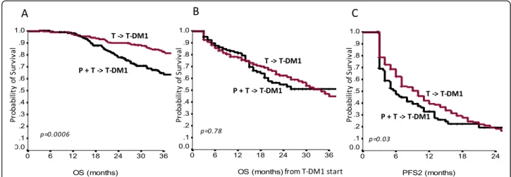
Among the 371 patients treated with second-line T-DM1, median overall survival (mOS) from diagnosis of advanced disease and median progression-free survival to second-line treatment (mPFS2) were 52 and 6 months in 177 patients who received trastuzumab/pertuzumab in first-line, and 74 and 10 months in 194 pertuzumab-naïve patients (p = 0.0006 and 0.03 for OS and PFS2, respectively).
Conclusions: Our data support the hypothesis that the addition of pertuzumab to trastuzumab reduces the amount of available plasma membrane HER2 receptor, limiting the binding of T-DM1 in cancer cells. This may help interpret the less favorable outcomes of second-line T-DM1 in trastuzumab/pertuzumab pre-treated patients compared to their pertuzumab-naïve counterpart.
Keywords: HER2+ breast cancer, Trastuzumab/pertuzumab blockade, T-DM1 efficacy Background
Human epidermal growth factor receptor 2 (HER2) is a member of the HER family of receptor tyrosine kinases, also including the epidermal growth factor receptor (EGFR), HER3, and HER4. Ligands binding to EGFR, HER3 and HER4 induce homo- and heterodimerization among the family members. Despite the lack of specific ligands, activated HER2 homodimerizes in HER2 posi-tive (HER2+) breast cancer (BC) cells and is then re-cruited as a preferred partner in heterodimers, resulting in the activation of cancer-driving pathways [1].
HER2 overexpression and/or gene amplification occurs in approximately 15–20% of BC, and is associated with a more aggressive behavior, with high rates of cell prolifer-ation and metastasis, and poor patient outcomes [2]. HER2+ advanced breast cancer (ABC) has significantly benefited from the approval of several HER2-targeting agents in the last decades. Trastuzumab, a monoclonal antibody targeting HER2, has revolutionized the thera-peutic landscape of HER2+ ABC [3]. Despite this, up to
40% of ABC patients show innate
trastuzumab-resistance, and most patients develop acquired resistance whithin the first year of trastuzumab treatment [4, 5]. The approval of three additional anti-HER2 agents, i.e., lapatinib, pertuzumab, and trastuzumab emtansine (T-DM1), has converted HER2+ ABC into a highly treatable disease, with more favorable outcomes [6–9]. Pertuzu-mab is a monoclonal antibody binding HER2 at a
differ-ent site compared with trastuzumab. A more
comprehensive signaling blockade underlies the notice-bly enhanced antitumor activity of trastuzumab and per-tuzumab combination treatment [10]. Results from the
CLEOPATRA trial showed an unprecedented median overall survival (mOS) advantage of 15.7 months in the pertuzumab arm, and the double-block combination has therefore become the new standard first-line treatment
in HER2+ ABC [7]. T-DM1 is an antibody-drug
conju-gate of trastuzumab with emtansine (DM1), an antimi-crotubule maytansine derivative [11, 12]. The activity of T-DM1 depends on both trastuzumab antitumor effects and intracellular DM1. Following T-DM1 binding to membrane HER2 receptor, the HER2-T-DM1 complex enters into the cell via receptor-mediated endocytosis [13]. Subsequently to the release from the lysosome, DM1-containing metabolites inhibit microtubule assem-bly, causing cell death [14].
In second-line treatment, after treatment with taxane and trastuzumab or as first-line in patients with rapid progression after adjuvant trastuzumab (≤6 months), TDM-1 has shown greater efficacy than lapatinib and capecitabine in the phase III EMILIA trial [8]. Conse-quently, TDM-1 has become the standard second-line treatment in HER2+ ABC patients.
To the aims of the present study, it is noteworthy that patients accrued in randomized trials of T-DM1 had not received prior pertuzumab. Therefore, we lack solid evi-dence on T-DM1 efficacy following trastuzumab/pertu-zumab-containing regimens. Data from observational trials are limited. We have previously shown evidence of lower T-DM1 efficacy in trastuzumab/pertuzumab-pre-treated patients providing data from a retrospective, multicentric study of 250 HER2+ ABC patients [15]. In a further retrospective evaluation of T-DM1 activity as second-line or later treatment from Dzimitrowicz and
colleagues [16], results in terms of tumor response rates and progression free survival (PFS) appeared less favor-able than those reported in randomized trials of T-DM1. These findings suggest the need to investigate the biol-ogy of HER2 through sequential treatments in order to define the molecular basis for the appropriate thera-peutic approach.
Based on the above reported evidence, we explored the effects of the exposure to trastuzumab and/or pertu-zumab on HER2 receptor expression and cellular localization in HER2+ BC cell lines, and their effects on T-DM1 activity. The pre-clinical experiments were par-alleled by the conduct of a large, multicentric, retro-spective observational study, i.e., the SePHER study,
Administration Sequence in Pertuzumab-pretreated
HER2 + ABC patients, aimed to explore the efficacy of T-DM1 in light of prior trastuzumab/pertuzumab treat-ment in the real-world setting.
Materials and methods Study approval
The SePHER study is a multicenter, observational trial with retrospective design including HER2+ ABC patients from 45 Italian cancer centers. This study was approved by the Institutional Review Board (IRB) of the Regina Elena National Cancer Institute, Rome, Italy [reference number: RS793/16(1815)]. The approval of the coordin-ating centre was tempestively notified to all the partici-pating partners for further consideration and approval by the respective IRBs. An ad hoc written informed con-sent was developed and implemented for this study participants.
Patients’ selection
Information on demographics, clinical, histopathological and immunohistochemical (IHC) features, anti-tumoral therapies and related outcomes were retrieved from pa-tients’ medical records by specifically trained research assistants. All included patients were treated for ad-vanced disease. Each patient was evaluated during treat-ment according to the follow-up strategies of each center. Clinical response was evaluated by response evaluation criteria in solid tumours (RECIST) criteria, version 1.1. Anonymized data were entered into a dedi-cated database with a SPSS operating interface. Median follow up was calculated starting from diagnosis of metastatic disease to death or date at last follow up. Endpoints for efficacy outcome included progression free survival (PFS) and overall survival (OS). Progression free survival for any specific line of treatment was calcu-lated from the time of treatment start to the time of dis-ease progression, interruption of treatment for toxicity, death or loss to follow-up. Overall survival was calcu-lated starting from diagnosis of metastatic disease to
death or last follow-up. Median PFS (mPFS) and OS (mOS) were calculated using the Kaplan-Meier product limit estimator method.
We first analyzed 371 patients treated with T-DM1 in second-line. We then split the whole cohort of 555 pa-tients into five subgroups, as follows: 1. Papa-tients treated in first-line without pertuzumab/second-line with T-DM1 (Number of patients, N, 194 patients); 2. Patients treated in first-line without pertuzumab/T-DM1 in third-line or beyond (N: 148); 3. Patients treated in first-line with pertuzumab/second-first-line with T-DM1 (N: 177); 4. Patients treated in first-line with pertuzumab/T-DM1 in third-line or beyond (N: 11); 5. Patients treated in first-line with T-DM1 (N: 25). The groups defined upon treatments’ sequence, as previously specified, were se-lected by hypothesizing a possible effect of the adminis-tration sequence on the main clinical outcomes, i.e., OS and PFS to first-line, second-line and third-line of treat-ment (PFS1, PFS2 and PFS3, respectively). Survival esti-mates were first computed for the whole cohort and then by treatments’ sequence. Subsequently, data on OS and PFS2 were also analyzed across strata defined by IHC tumor features, namely, estrogen receptor (ER) and progesterone receptor (PgR) positive (triple positive, TP), ER or PgR positive, and both hormonal receptors (HRs) negative.
Pathology assessment was performed in surgical speci-mens of primary tumors by dedicated pathologists at the participating centers as per national standards. Estrogen receptor and PgR status were determined at each center by IHC according to the local standards. Positivity was
considered at a cut-off of ≥1%. HER2 testing was
per-formed according to the 2013 ASCO/CAP guidelines on HER2 Testing in Breast Cancer. A positive HER2 status required an IHC score of 3+ or positive fluorescence in
situ hybridization/cromogenic in situ hybridization
(FISH/CISH).
Generation of drug-resistant cell lines
Drug-resistant cells were obtained by continuous expos-ure of HER2+ BT474 and SkBr3 cell lines, obtained from the American Type Culture Collection (ATCC), to
20μg/ml trastuzumab or pertuzumab, or 10 μg/ml
tras-tuzumab + 10μg/ml pertuzumab, for 2 months, followed
by 7 months of culture in medium supplemented with
50μg/ml trastuzumab or pertuzumab, or 25 μg/ml
tras-tuzumab + 25μg/ml pertuzumab. Further details are
re-ported in theSupplementary materials and methodsand
are available online. Statistical analysis
Within the overall cohort of the 555 patients, the associ-ations of interest were also evaluated in light of: a. T-DM1 administration in second- or subsequent line, and
b. molecular subgroups, with these latter being set based on the results of IHC analysis and according to the cri-teria fully reported in the patients’ selection paragraph. The covariates used in the Cox uni/multivariate models included the following categorical variables: “first-line pertuzumab” (yes vs. no) and “treatment sequence”, which both concurred to define the five categories de-scribed in detail in the methods section; age; IHC sub-type; Ki-67 (> 20% vs≤20%); metastasis at diagnosis (yes vs no); number of metastatic sites (> 1 vs 1); visceral me-tastasis (yes vs no); brain meme-tastasis (yes vs no); bone-only metastasis (yes vs no), lenght of PFS1 and disease free interval (DFI). This latter was calculated from the time of surgery to the time of metastatic disease diagno-sis. Variables testing significant in univariate analyses were further tested in multivariate models. The level of statistical significance was set at p≤ 0.05, with a 95% confidence interval (95%CI). The SPSS software (SPSS version 21.0, SPSS Inc., Chicago, IL) was used for all statistical evaluations.
Regarding the experiments in cell lines, all data were reported as mean +/− standard deviation. Differences were considered statistically significant when p≤ 0.05, with a 95%CI. Student’s t test was performed for the comparison of results from all different tests (*p < 0.05, **p < 0.001, ***p < 0.0001).
Results
Generation of trastuzumab, pertuzumab, and
trastuzumab+pertuzumab resistant SkBr3 and BT474 cells in vitro
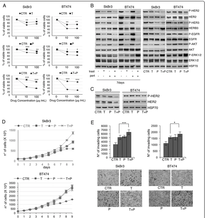
To investigate the mechanisms underlying lower T-DM1 efficacy following trastuzumab/pertuzumab in ABC pa-tients, we established trastuzumab (T), pertuzumab (P), and trastuzumab+pertuzumab (T + P)-resistant HER2+ SkBr3 and BT474 cell lines (Fig.1a). In agreement with previously reported data, Western Blot (WB) analysis re-vealed that short-term trastuzumab treatment inhibits the phosphorylation of HER family members and AKT, but not of ERKs (Fig. 1b, leftpanels) [17]. This effects were emphasized in trastuzmab+pertuzumab short term -treated cells. Wester Blot analysis of the generated re-sistant cell lines revealed reduced HER2 activity and ex-pression and marked induction of ERKs phosphorylation in T + P cells as compared to parental and both T and P cells, whereas HER3 and EGFR were upregulated despite the reduced phosphorylation, in both BT474 and SkBr3 (Fig. 1b, right most panels). T + P cells maintained de-creased HER2 phosphorylation and total expression, as confirmed by periodically testing resistant and control cell lines following their establishment (Fig. 1c). Resist-ant cell lines exhibited a higher proliferation rate (Fig.
1d) and invasive capability (Fig. 1e) compared to control
cells. The number of invading T + P cells was
significantly higher compared to T cells in both cell lines. Overall this data indicate that a more aggressive behavior is induced by the chronical exposure of HER2+ breast cancer cell lines to the combination trastuzumab/ pertuzumab rather than trastuzumab.
Dual HER2 blockade is associated with reduced T-DM1 efficacy due to HER2 downregulation
Following T-DM1 treatment, the percentage of re-sponsive T + P cells was significantly lower compared to T cells in BT474 (p < 0.0001), and in SkBr3, al-though at a lower level of statistical significance (p < 0.05) (Fig. 2a). Full T-DM1 dose-response curves of T and T + P cell lines are reported in Fig. 2b, that con-firm significantly lower T-DM1 efficacy in cells chronically exposed to the combination trastuzumab/
pertuzumab. T-DM1 induced a marked
down-regulation of HER2 as well as HER3 and EGFR, in parental, T and P BT474 cells, whereas T + P BT474 cells maintained unmodified levels of these receptors
(Fig. 2c). T-DM1 inhibited ERKs phosphorylation in
control cells, T and P cells, whereas it considerably induced ERKs activation in T + P cells, in both cell lines (Fig. 2c, data available upon request). The down-regulation of HER2 induced by trastuzumab/pertuzu-mab combination was confirmed in vivo, by the immunohistochemical assessment of bioptic specimens from four trastuzumab/pertuzumab-treated ABC pa-tients prior to and following exposure to double-block (Table 1). Representative images of eosin-hematossilin and HER2 staining from two patients are shown in Fig. 2d.
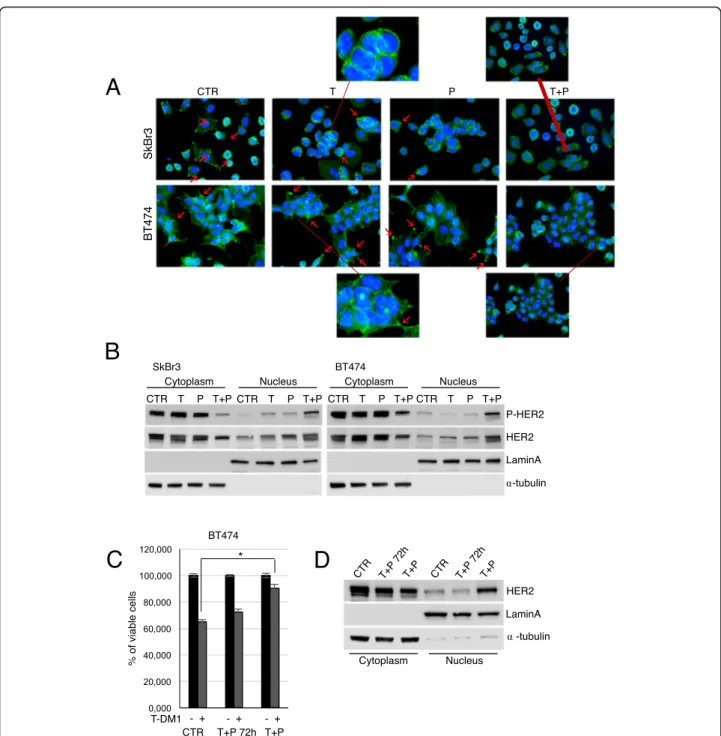
The combination trastuzumab+pertuzumab induces HER2 nuclear translocation
When administered in vitro as short-term treatments, trastuzumab and pertuzumab alone barely down-regulate HER2 [18–20], while their combination
in-duces a stronger HER2 downregulation [19, 20]. To
investigate the long-term effects of trastuzumab and pertuzumab on HER2 subcellular distribution, we per-formed immunofluorescence experiments. As shown
in Fig. 3a, in control cells, T and PBT474 and SkBr3
cells, HER2 was mainly localized at the plasma mem-brane level, and concentrated on cellular protrusions [17]. A diffuse cytoplasmic signal was also present. Conversely, T + P cells lost membrane-HER2 and
retained HER2 cytoplasmic distribution (Fig. 3a).
Western Blot analysis of the cytoplasmic and nuclear fractions of control and resistant cells showed a marked translocation of HER2 to the nucleus in T + P
BT474 and SkBr3 cells (Fig. 3b). Nuclear HER2 was
phosphorylated, suggesting its active involvement in transcriptional control mechanisms [21, 22]. A short
Fig. 1 Characterization of resistant cell lines. Cell viability of control (CTR), Trastuzumab (T), Pertuzumab (P), and T + P cell lines treated with 10 and 100μg/ml trastuzumab, pertuzumab and trastuzumab+pertuzumab for 5 days was evaluated by MTT assay (a). Western Blot (WB) analysis of phosphorylated and total HER2, HER3, EGFR, AKT and ERKs was performed on total cell lysates from untreated/treated parental cell lines for 7 days (b, leftmost panel). The basal levels of phosphorylated and total HER2, HER3, EGFR, AKT and ERKs were evaluated by WB in CTR, T, P, and T + P resistant cell lines (b, rightmost panel). HER2 total and phosphorylated levels evaluation by WB was repeated in CTR, T, P, and T + P cell lines following resistant cell lines establishment (c). The anti-HSP70 antibody was used to validate equivalent amount of loaded proteins in each lane. Proliferation curves (d) and Invasion assay (e, upper panel) of CTR, T, P, and T + P cell lines. Representative images of invaded cells are reported for each cell line (e, lower panel)
Fig. 2 Trastuzumab+pertuzumab combination is associated with HER2 downregulation. Cell viability of CTR, T, P, and T + P SkBr3 and BT474 cells treated with 0.1μg/ml and 1 μg/ml T-DM1, respectively, for 48 h was evaluated by Crystal Violet Assay (a). The results are expressed as percentage of T-DM1-responsive cells relative to control cells, as mean +/− standard deviation. T-DM1 dose-response curves of T and T + P cells are reported for both SkBr3 and BT474 (b). The expression of phosphorylated and total HER2, HER3, EGFR, AKT and ERKs was evaluated by Western Blot (WB) following exposure to 1μg/ml T-DM1 for 48 h (c). Representative pre- and post-therapy sections from 2 ABC patients, stained by immunohistochemistry with eosin-hematossilin and anti-HER2 antibody (d)
Table 1 Immunohistochemical expression of Estrogen Receptor (ER), Progesterone Receptor (PgR) and HER2 in pre- and post-treatment bioptical tissue samples from 4 HER2+, pertuzumab pretreated, advanced breast cancer patients
Pre-treatment Post-treatment
ER PgR HER2 score HER2% FISH ER PgR HER2
score
HER2% FISH
Patient 1 90% 90% 2+ 70% amplified 90% 90% 0 0% –
Patient 2 90% 40% 2+ 30% amplified 90% 60% 1+ 30% –
Patient 3 46% 0% 2+ 100% amplified 75% 18% 1+ 15% amplified
trastuzumab/pertuzumab pre-exposure of parental BT474 did not affect T-DM1 efficacy compared to control cells, and no HER2 nuclear translocation was observed (Fig.3c and d). Hence, prolonged exposure to trastuzumab/
pertuzumab induces the loss of membrane HER2 and its nuclear traslocation, that represent a mechanism of ac-quired resistance, which in turn reduces the T-DM1 tar-geting potential and efficacy.
Fig. 3 Prolonged trastuzumab+pertuzumab induces HER2 nuclear translocation. Control, T, P, and T + P cell lines were plated on poly-l lysine coated slides, and stained 24 hous later with anti-HER2 (green signal) (a). These cells were counterstained with Hoechst to highlight nuclei. Red arrows indicate HER2 localization on cellular protrusions. Cytoplasmic and nuclear fractions extracted from control, T, P, and T + P cells were analysed by Western Blot (WB) for the expression of phosphorylated and total HER2. Lamin A andα-tubulin were used to validate purity of nuclear and cytoplasmic extracts respectively (b). Following pre-treatment with 5μg/ml trastuzumab + 5 μg/ml pertuzumab for 72 h, cell viability of control cells, pre-treated and T + P BT474 exposed to1μg/ml T-DM1 for 72 h was evaluated by Crystal Violet Assay (c). Cytoplasmicand nuclear fractions of control, pre-treated and T + P BT474 cells were analysed by WB for the expression of total HER2 (d)
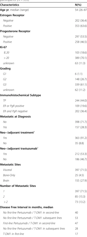
Patients’ cohort description
Overall, 555 patients were treated with T-DM1 for
ABC. Table 2 shows the main patient- and
tumor-related characteristics. Briefly, 25 patients (4.5%) re-ceived T-DM1 in first-line, following relapse while on/within 6 months from neo−/adjuvant treatment, 371 (66.8%) were treated in second-line, 96 (17.3%) in third-line, and 63 (11.4%) beyond the third-line. Details on treatments received in first-, second- and third-line for the whole population are available in
Supplementary Table 1. The median length of
follow-up for the whole population of 555 patients was 41 months.
Comparing clinical outcomes according to the T-DM1 line of treatment
Among the 371 patients who received T-DM1 in second-line, 177 had been treated with a trastuzu-mab/pertuzumab-based first-line, while the remaining 194 were pertuzumab naïve and had received a
first-line trastuzumab-based regimen. In Table 3, we show
the main baseline characteristics of pertuzumab naïve and pertuzumab-pretreated patients who received T-DM1 in second-line. Median OS from diagnosis of metastatic disease was 52 months in the pertuzumab pre-treated population vs 74 months in the
pertuzu-mab naïve patients (p = 0.0006; Fig. 4a). Median OS
calculated from the start of T-DM1 treatment was not significantly different, being not reached in the first group, and being 34 months in the second group (p = 0.78; Fig. 4b). Median PFS on first-line treatment (mPFS1) for patients who received a pertuzumab-trastuzumab-based regimen compared to those who received a trastuzumab-based regimen were 11 and 10 months, respectively (p = 0.22). Median PFS on second-line T-DM1 (mPFS2) was 6 months in patients who had received pertuzumab-trastuzumab in first-line compared to 10 months in those who were
pertu-zumab naïve (p = 0.03; Fig. 4c). Cox regression was
performed for significantly differing outcomes
Table 2 Clinicopathological characteristics of the study participants (N = 555)
Characteristics N(%)
Age yr, median (range) 54 (26–87)
Estrogen Receptor Negative 202 (36.4) Positive 353 (63.6) Progesterone Receptor Negative 297 (53.5) Positive 258 (46.5) Ki-67 ≤ 20 103 (18.6) > 20 389 (70.1) unknown 63 (11.3) Grading G1 6 (1.1) G2 148 (26.7) G3 339 (61.1) unknown 62 (11.2) Immunohistochemical Subtype TP 244 (44.0) ER or PgR positive 109 (19.6) ER and PgR negative 202 (36.4) Metastatic at Diagnosis No 398 (71.7) Yes 157 (28.3)
Neo−/adjuvant treatmenta
Yes 363 (91.2)
No 35 (8.8)
Neo−/adjuvant trastuzumaba
Yes 212 (53.3) No 186 (46.7) Metastatic Sites Visceral 397 (71.5) Bone-Only 25 (4.5) Brain 155 (27.9)
Number of Metastatic Sites
1 397 (71.5)
2 85 (15.3)
> 2 73 (13.2)
Disease Free Interval in months, median
No first-line Pertuzumab / T-DM1 in second-line 40 No first-line Pertuzumab / T-DM1 subsequent lines 53 First-line Pertuzumab / T-DM1 in second-line 47 No first-line Pertuzumab / T-DM1 in subsequent lines 28
T-DM1 in first-line 17
Table 2 Clinicopathological characteristics of the study participants (N = 555) (Continued) Characteristics N(%) T-DM1 treatment line First-line 25 (4.5) Second-line 371 (66.8%) Third-line 96 (17.3%) Subsequent lines 63 (11.4%) a
For patients with early disease at diagnosis (398 patients)
Abbreviations: N Number; yr Years; TP Triple positive; ER Estrogen receptor; PgR Progesterone receptor
between these two groups, namely OS and PFS2. Uni-variate analysis confirmed a negative impact of first-line pertuzumab for both OS [Hazard Ratio, HR 1.79; 95%CI 1.23–2.61; p = 0.002] and PFS2 [HR, 1.23; 95% CI 1.00–1.67; p = 0.049], which was exclusively con-firmed in multivariate analysis of OS [HR 1.89; 95%CI 1.24–2.89; p = 0.003]. Conversely, among 159 patients who received T-DM1 in third-line or beyond, no dif-ferences by pertuzumab-pretreatment emerged for OS, PFS1 and PFS2 (adjusted p-values were 0.99, 0.76 and 0.26, respectively; Table 4).
Comparing clinical outcomes according to the treatment sequences and immunohistochemical subtypes
Overall, with a median follow up of 41 months, mOS for the whole cohort of 555 patients treated with T-DM1 in any treatment lines was 73 months. Altogether, the ad-ministration sequence of anti-HER2 agents significantly
affected mOS (p < 0.0001) and mPFS2 (p = 0.001)
(Table4), with poorer outcomes in the group of patients having received pertuzumab-based first-line treatment followed by second-line T-DM1. No differences by tras-tuzumab/pertuzumab-pretreatment emerged for mOS, Table 3 Clinical-pathological characteristics of the study participants who received T-DM1 in second-line after a trastuzumab-based first-line (N = 194) and after a pertuzumab-trastuzumab-based first-line (N = 177)
Characteristics First-line trastuzumab/ Second-line T-DM1 [N (%)]
First-line pertuzumab-trastuzumab/Second-line T-DM1 [N (%)] p-value
Progesterone Receptor 0.83 Positive 91 (46.9%) 85 (48.0%) Negative 103 (53.1%) 92 (52.0)% Estrogen Receptor 0.46 Positive 132 (68%) 114 (64.4%) Negative 62 (32%) 63 (35.6%) Ki-67% 0.05 ≤ 20 45 (25.6%) 26 (16.6%) > 20 131 (74.4%) 131 (83.4%) Immunohistochemical Subtype 0.57 TP 87 (44.8%) 81 (45.8%) ER or PgR positive 45 (23.2%) 33 (18.6%) ER and PgR negative 62 (32.0%) 63 (35.6%) Metastatic at Diagnosis 0.05 No 147 (75.8%) 117 (66.1%) Yes 47 (24.2%) 60 (33.9%)
Number of Metastatic Sites 0.14
1 142 (73.2%) 117 (66.1%)
> 1 52 (26.8%) 60 (33.9%)
Neo−/adjuvant treatment * 0.38
Yes 128 (66.0%) 109 (61.6%)
No 66 (34.0%) 68 (38.4%)
Neo−/adjuvant trastuzumab * 0.48
Yes 76 (39.2%) 63 (35.6%)
No 118 (60.8%) 76 (39.2%)
Visceral Metastatic Site(s) 0.11
Yes 133 (68.9%) 135 (76.3%)
No 60 (31.1%) 42 (23.7%)
Disease Free Intervala 0.10
< 3 years 66 (46.8%) 47 (41.2%)
≥ 3 years 75 (53.2%) 67 (58.8%)
a
For patients with early disease at diagnosis
mPFS1 and mPFS2 in patients who received T-DM1 in third-line or beyond (Table4).
When the overall cohort of 555 patients was strati-fied by IHC subtype, a lower mOS in pertuzumab pre-treated patients was confirmed only in HRs-negative patients (p < 0.0001), while a shorter mPFS2 was observed in both TP (p = 0.04) and HRs-negative patients (p = 0.003). However, multivariate analysis showed no effect of the IHC-defined subtype on mOS, mPFS1 or mPFS2.
Discussion
Resistance to HER2 targeting agents is a challenging topic in BC. Several pre-clinical studies explored poten-tial resistance mechanisms to T-DM1, involving
reduc-tion of lysosomal proteolytic activity [23], STAT3
activation [24], HER2 genomic amplification [25], and sustained signaling from neuregulinβ1 [26]. Moreover, previous evidence has shown that trastuzumab-mediated engagement of immune effector cells induces HER2 downregulation in BC cells by STAT1 activation [27].
Fig. 4 Overall survival (OS) from diagnosis of metastatic disease (a), OS from T-DM1 start (b) and progression free survival to the second-line of treatment (PFS2) (c), in patients treated with trastuzumab-based first-line and T-DM1 in second-line (T - > T-DM1) and in patients treated with pertuzumab-trastuzumab-based first-line and T-DM1 in second-line (P + T - > T-DM1)
Table 4 Median OS, median PFS in first-line of treatment and median PFS in second-line of treatment by treatment sequences in the overall population and by subgroups defined upon immunohistochemical characterization of molecules features (N:5555)
To our knowledge, no previous study has specifically fo-cused on the potential cross-resistance mechanisms be-tween pertuzumab pre-treatment and T-DM1 at a molecular level, particularly if compared to previously documented cross-resistance mechanisms due to trastu-zumab alone.
We herein show first time evidence that dual HER2 blockade by trastuzumab/pertuzumab is asso-ciated with a marked inhibition of HER2 receptor expression on plasma membrane of cancer cells in vitro and in vivo, which limits the targetable HER2 receptor available to T-DM1. Our results also indicate that a significant amount of HER2 is trans-located to the nuclei of trastuzumab+pertuzumab-re-sistant BC cells in vitro. Conversely, this effect is not shown in trastuzumab-pretreated BC cell lines. In agreement, our data from BC cell lines indicate that T-DM1 treatment induces internalization of mem-brane HER2 receptor in trastuzumab-resistant and pertuzumab-resistant BC cells lines; on the contrary, the expression levels of HER2 are not affected by T-DM1 in trastuzumab+pertuzumab-resistant BC cells, since the amount of membrane HER2 is already re-duced by the previous double-block determining HER2 nuclear localization.
Nuclear HER2 has been reported to act as a transcrip-tional regulator and represents an independent prognos-tic factor of poor clinical outcome [21,22]. In preclinical trastuzumab-resistant BC models, the inhibition of clear HER2 suppresses cell growth, indicating that nu-clear HER2 is the major proliferation driver in trastuzumab-resistant BC. In this context, the inability of trastuzumab to disrupt the neregulinβ1-induced assem-bly of a nuclear HER2/HER3/STAT3 transcriptional complex has been demonstrated [28]. The lack of HER2 nuclear translocation following exposure to trastuzumab or pertuzumab alone highlights the synergy emerging from the combination of these two drugs.
These findings, together with the increased invasive-ness and marked activation of MAPK/ERK pathway of trastuzumab+pertuzumab-resistant BC cell lines, suggest that the administration of pertuzumab in combination with trastuzumab favors the selection of a more aggres-sive phenotype of HER2+ BC cells, as compared to tras-tuzumab alone.
We have a preliminary evidence of higher tumor growth in mice subcutaneously injected with T + P resistant cells, as compared to T cells. The validation of reduced T-DM1 efficacy following trastuzumab+ pertuzumab in a mouse model would strengthen our results, and experiments aimed at this goal are ongoing.
The selection of patients to be included in the present study was conditional in terms of considering
only those who received T-DM1 at some point during their clinical history for advanced BC, and the
analysis focused on trastuzumab/pertuzumab or
trastuzumab-based pretreatment and second-line T-DM1 efficacy.
Among patients treated with T-DM1 in second-line, mOS from the start of the first-line and mPFS2 resulted longer in patients who had received a trastuzumab-based first-line, with respect to the coun-terpart pretreated with a pertuzumab/trastuzumab-based-regimen. The mPFS2 advantage observed within the first patient group may be reconciled with a higher efficacy of T-DM1 in patients pre-treated with trastuzumab alone, compared to those having received the double-block. Conversely, when addressing the ad-vantage in mOS from the start of the first-line in the pertuzumab-naïve group, the link to differences in T-DM1 efficacy based on the administration sequence is less immediate. Indeed, we observed no difference in mOS from the start of T-DM1 between
trastuzumab-pretreated and pertuzumab/trastuzumab-pretreated
patients. In patients who did not received pertuzumab in first-line but for whom TDM-1 was instead already available at further disease progression, a quite long time to disease progression may be hypothesized, which may itself reflect a less aggressive biologic be-havior on behalf of cancer. In brief, for patients in the pertuzumab-naïve group, the longer mOS from the first-line treatment may be more closely related to a more indolent disease course due to intrinsic dis-ease characteristics than to the sequence of anti-HER2 agents administration.
The issue of a possible decrease in T-DM1 efficacy if given immediately after the double pertuzumab-based HER2 double-block has not been exhaustively addressed in previous studies [8, 9]. The EMILIA [8]
and TH3RESA [9] trials were the two pivotal
random-ized phase III clinical studies that brought T-DM1 as a standard of care in second-line or beyond for pa-tients with HER2+ advanced BC that progressed to standard treatments. TH3RESA trial showed clinical advantage by using T-DM1 compared to treatment of choice by the clinician also in patients that had re-ceived lapatinib and capecitabine, while EMILIA trial showed superiority of T-DM1 in second-line even when compared head to head with the lapatinib plus capecitabine regimen. Unfortunately, none of the pa-tients included in the EMILIA and TH3RESA trials had received pertuzumab before being treated with T-DM1. Data from prospective studies is lacking in this context. Evidence on the topic under debate comes from three recent retrospective studies showing lower response rate and shorter survival outcomes in pa-tients treated with T-DM1 following pertuzumab
administration [15, 16, 29] In a previous retrospective multicentric study from our group involving 250 pre-treated HER2 positive BC patients we showed lower efficacy of T-DM1 in the pertuzumab-pretreated co-hort in comparison with the trastuzumab pretreated
group [15]. In the Dzimitrowicz paper, authors
showed results that are congruent with our findings [16]. In this study, including only patients that had received a pertuzumab-trastuzumab-based first-line, a lower efficacy of T-DM1 in terms of response rate was reported when compared to the response rates observed in the randomized clinical trials, where pa-tients were only trastuzumab resistant. In the Noda-Narita et al. retrospective study enrolling 42 advanced HER2 positive patients, median PFS and objective re-sponses were lower in the group pretreated with
per-tuzumab/trastuzumab in comparison to the
trastuzumab subgroup [29].
The main limitation of the observational section of the present study is its retrospective, multicenter design, which per se represents a considerable source of data heterogeneity. Moreover, the lack of central assessment on IHC features of primary and metastatic lesions de-serves mentioning, although quality controls routinely performed at the pathology labs of the institutions in-volved increase our confidence in data quality. At the same time, the involvement of a relevant number of can-cer centres/oncologic divisions, i.e., N: 45, has allowed to collect and analyze the largest amount of data ever made available to investigate the efficacy of T-DM1 following trastuzumab/pertuzumab-based treatment. Beyond its intrinsic limitations and bias, this approach allowed us to confirm the reduced T-DM1 efficacy following dual HER2 blockade by trastuzumab/pertuzumab in HER2+ ABC patients.
Our choice of relying on an observational study with a retrospective approach has paved the way to confounding and bias, which we attempted to minimize in the phase of data analysis throughout stratification and statistical mod-eling. In more detail, concerning stratification, data were analyzed within strata defined upon a given pre-specified variable. In doing so, we managed the confounding effects of a given variable possibly acting as a confounding at the price of a reduction of the study power in detecting the as-sociation of interest in the case of small strata. Within the SePHER study, statistical modeling translated into the de-velopment of multivariable Cox models. These latter ap-proach allowed to simultaneously control for more than one confounder at the time, and help interpret the effect of each confounder in light of the others. In addition, data retrieving was performed by ad hoc trained research assis-tants who worked in strict collaboration with the oncolo-gists involved at the single centre level. This may have minimized the chances of residual confounding.
The main strength of the present study is that, to our knowledge, we first reported on HER2 downregulation as a key mechanism underlying lower T-DM1 efficacy observed in the clinical setting when this drug is admin-istered as second-line therapy in trastuzumab/pertuzu-mab-pretreated HER2+ ABC patients. Results from the experiments performed in bioptic specimens of trastuzu-mab/pertuzumab pretreated advanced breast cancer
pa-tients were further confirmative. Indeed, when
comparing the pre- and post-treatment HER2 scores for each of the 4 patients examined, the IHC assessment uniformly showed a score reduction. This evidence pro-vides support to our study hypothesis, in that the re-duced HER2 scores at the IHC evaluation of the post-treatment samples are in key with a lower availability of the HER2 at the membrane level, which may per sè at least partly account for less favorable outcomes in pa-tients exposed to pertuzumab.
As shown in Tables 1, 2 cases among the 4 assessed
resulted negative at the FISH in the post-treatment win-dow of evaluation. The co-existence of HER2-negative and HER2-positive clones is plausible from a biological standpoint. In addition, it may reflect the selective pres-sure applied by the prior administration of anti-HER2 agents. Indeed, HER2-negative clones may concur to less favorable outcomes in patients treated with anti-HER2
agents. The extent to which the “degree” of
HER2-negative clone selection has been driven by the specific sequence of anti-HER2 agents administration and/or this mechanism concurs with the mechanism we have origin-ally hypothesized deserves further investigation in more adequately sized samples.
Although hypothesis-generating, data from the study herein presented, as well as from prior similar studies within this same research pipeline, are limited in nature. Still, they provided an appropriate ground in terms of pre-liminary evidence, on which we designed a randomized clinical trial investigating the optimal treatment sequence in HER2-positive ABC patients. In more detail, our team at the IRCCS Regina Elena National Cancer Institute is the coordinating center of the STEP trial, an active randomized multicenter prospective trial exploring the optimal Se-quence TrEatment in HER2+ Pertuzumab-pretreated ABC patients. The STEP trial was granted formal approval and financial support by the Italian Ministry of Health (project code: GR-2018-12,367,431). Evidencefrom the STEP and similar ad hoc, prospective randomized trials are eagerly awaited to delineate the optimal treatment sequence in HER2 + ABC patients, in order to gain more favorable treatment outcomes in this patients’ population.
Conclusion
Overall, our findings suggested HER2 downregulation
pertuzumab as a key mechanism underlying lower T-DM1 efficacy as second-line therapy in HER2+ ABC pa-tients. Results from our retrospective study showed in-deed lower T-DM1 efficacy in terms of mOS and mPFS in 177 patients who received trastuzumab/pertuzumab in first-line, as compared to 194 pertuzumab-naïve pa-tients. In addition, we showed HER2 nuclear transloca-tion in trastuzumab+pertuzumab-resistant HER2+ BC cell lines in vitro.
The design of prospective randomized trials may lead to delineate the optimal treatment sequence in HER2 + ABC, and ultimately to more favorable outcomes in these patients’ population.
Supplementary Information
The online version contains supplementary material available athttps://doi. org/10.1186/s13046-020-01797-3.
Additional file 1. Additional file 2. Abbreviations
HER2:Human epidermal growth factor receptor 2; EGFR: Epidermal growth factor receptor; BC: Breast cancer; ABC: Advanced breast cancer; T-DM1: Trastuzumab emtansine; PFS: Progression free survival; OS: Overall survival; IHC: Immunohistochemistry; IRB: Institutional review board; RECI ST: Response evaluation criteria in solid tumours; ER: Estrogen receptor; PgR: Progesterone receptor; HRs: Hormonal receptors; FISH: Fluorescence in situ hybridization; CISH: Cromogenic in situ hybridization
Acknowledgements
We thank Anna Maria Edlisca and Alessandro Zennaro for technical support, and Letizia Perracchio and Anna Di Benedetto for Immunohistochemistry and FISH support, respectively.
Authors’ contributions
Conception or design of the study: GB, LP, RF and PV. Acquisition of data: RL, CM, EK, MB, MMS, TG, RB, LL, CF, CN, EC, DG, NLV, AC, EB, LM, AM, VA, CZ, GT, GB, MM, DM, ST, PM, MRV, RM, AR, MAF, NDO, EMV, DC, OG, IP, GS, FG, CG, MC, PDM, MR, GS, IS, AS. Immunohistochemistry: VL. In vivo mice experiments: MP and CL. All authors contributed equally to the analysis and interpretation of data. RDM, ADL, GC: Critical revision. GB, LP, RF and PV contributed equally to the preparation and final approval of the manuscript. Funding
This work was partially supported by an intramural grant to GB in 2018 [RC2018].
Availability of data and materials
The datasets used and/or analyzed during the current study are available from the corresponding author on reasonable request.
Ethics approval and consent to participate
The study was conducted in accordance with the Declaration of Helsinki. All patients provided written informed consent. The protocol was approved by institutional review boards at each study site.
Consent for publication
The datasets used and/or analysed during the current study are available from the corresponding author on reasonable request.
Competing interests
GB, RL, VL, EK, MB, MMS, RB, LL, CF, EC, DG, AC, VA, CZ, GT, GB, MM, DM, ST, PM, MRV, RM, MAF, NDO, EMV, DC, GS, FG, CG, PDM, MR, IS, AS, GC, RF, GS declare no conflicts of interest. LP received travel grants from Eisai, Roche,
Pfizer, Novartis; speaker fees from Roche, Pfizer, Novartis, Gentili. CM has received personal consultancy fees from Bayer, Roche, Daiichi Sankyo, MSD. TG received travel grants from Eisai, Roche, Pfizer, Novartis; speaker fees/ advisory boards from Roche, Pfizer, Novartis, Gentili, Lilly. CN received travel grants/personal fees from Pfizer, EISAI, Novartis, Merck Sharp &Dohme, AstraZeneca. NLV received personal fees from Eisai and Novartis; research funding from Eisai, travel grants from Pfizer, Roche, Gentili; consulting role from Celldex. Em.Br. is supported by the Italian Association for Cancer Research AIRC-IG 20583; he was supported by the International Association for Lung Cancer (IASLC), the LILT (LegaItaliana per la Lotta contro i Tumori) and Fondazione Cariverona; he received speakers’ and travels’ fee from MSD, Astra-Zeneca, Celgene, Pfizer, Helsinn, Eli-Lilly, BMS, Novartis and Roche; con-sultant’s fee from Roche, Pfizer; institutional research grants from Astra-Zeneca, Roche. Lu.Mos. received advisory board from Roche. An.Mic. received travel grants from Eisai, Celgene, Novartis, Ipsen; personal fees, advisory boards from EISAI, Novartis, Astra Zeneca, Teva, Pfizer, Celgene. A.R. received fees from Bristol, Pfizer, Bayer, Kyowa Kirin, Ambrosetti for advisory board ac-tivity; and speaker honorarium from Roche Diagnostics. OG received personal fees from Celgene, Novartis, Eisai; research funding from Eisai, consulting ac-tivities with Celgene, Eisai, Pfizer, Amgen. IP received personal fees/advisory boards from Roche, Pfizer, Novartis, Italfarmaco, Gentili, Pierre Fabre. MC re-ceived personal fees, advisory boards from Pierre Fabre, Astra Zeneca, Cel-gene, Eisai, Novartis, Lilly. RDM declares to be a scientific advisory board member at Exosomics SpA (Siena IT), Hibercell Inc. (New York, NY), Kiromic Inc. (Houston, TX) and at Exiris Inc. (Rome, IT). ADL received consulting fees from Novartis and Roche. PV received travel grants from Eisai, Roche, Pfizer, Novartis; speaker fees/advisory boards from Roche, Pfizer, Novartis, Gentili, Lilly.
Author details
1Cellular Network and Molecular Therapeutic Target Unit, IRCCS Regina Elena
National Cancer Institute, Via Elio Chianesi 53, 00144 Rome, Italy.2Division of Medical Oncology 2, IRCCS Regina Elena National Cancer Institute, Via Elio Chianesi 53, 00144 Rome, Italy.3Pathology Department, IRCCS Regina Elena
National CancerInstitute, Rome, Italy.4Area of Translational Research, IRCCS
Regina Elena National Cancer Institute, Rome, Italy.5Department of Medical Sciences, University of Turin, Turin, Italy.6Candiolo Cancer Institute, FPO-IRCC
S, Candiolo, Turin, Italy.7Medical Oncology, Sandro Pertini Hospital, Rome,
Italy.8Oncology Clinic,“Ospedali Riuniti di Ancona” Hospital, Ancona, Italy. 9
Radiotherapy Unit, Department of Oncology, Careggi University Hospital, Florence, Italy.10Medical Oncology Unit, St Salvatore Hospital, L’Aquila, Italy. 11Department of Medical, Oral and Biotechnological Sciences, University
Gabriele D’Annunzio, Chieti, Italy.12Department of Medical Oncology,
University La Sapienza, Rome, Italy.13Breast Cancer Unit, ASST Cremona, Cremona, Italy.14Oncology Unit, ASST Fatebenefratelli Sacco-PO
Fatebenefratelli, Milan, Italy.15Oncology Unit, IRCCS Foundation Polyclinic
University A. Gemelli, University Cattolica Del Sacro Cuore, Rome, Italy.
16
University of Verona, Verona, Italy.17Department of Oncology and Hematology, University Hospital, Modena, Italy.18UO Medical Oncology, S.
Chiara Hospital, Pisa, Italy.19Medical Oncology Unit, A.O. Papardo &
Department of Human Pathology, University of Messina, Messina, Italy.
20
Medical Oncology Unit, Addarii Institute of Oncology, S. Orsola-Malpighi Hospital, Bologna, Italy.21Department of Oncology, University Campus
Biomedico, Rome, Italy.22Medical Oncology Unit, Sant’Andrea University
Hospital, Rome, Italy.23Department of Radiological, Oncological and
Anatomo-Pathological Sciences, University La Sapienza, Umberto I University Hospital, Rome, Italy.24Medical Oncology, Paolo Giaccone University Hospital,
Palermo, Italy.25Department of Ematology & Oncology, Pugliese-Ciaccio
Hospital, Catanzaro, Italy.26Medical Oncology Unit, Belcolle Hospital, Viterbo,
Italy.27Medical Oncology Unit, Lanciano-Vasto, Chieti, Italy.28Medical Oncology Unit, Santa Maria Goretti Hospital, Latina, Italy.29Medical Oncology
Unit, Fatebenefratelli Hospital, Rome, Italy.30Medical Oncology AO S. Croce
and Carle Teaching Hospital, Cuneo, Italy.31Gynaecology– Oncology Unit,
University Cattolica del Sacro Cuore, Rome, Italy.32Department of Medical Oncology, ASL Nuro, Nuoro, Italy.33Department of Medical Oncology, IRCCS
Giovanni Paolo II, Bari, Italy.34Division of Medical Oncology, Pescara Hospital,
Pescara, Italy.35Research Unit Phase I Trials and Oncology Unit, ASST, Monza,
Italy.36Division of Medical Oncology, Reggio Calabria General Hospital, Reggio Calabria, Italy.37Department of Systems Medicine, Medical Oncology,
University Tor Vergata, Rome, Italy.38Radiotherapy Department, IRCCS Regina
Elena National Cancer Institute, Rome, Italy.40Institute of General Pathology,
University Cattolica del Sacro Cuore, Rome, Italy.41Department of Medical
Oncology, IRCCS Foundation University A. Gemelli, Rome, Italy.42Sandro
Pitigliani Medical Oncology Department, Hospital of Prato, Prato, Italy.
43Scientific Direction, IRCCS Regina Elena National Cancer Institute, Rome,
Italy.
Received: 4 September 2020 Accepted: 2 December 2020 References
1. Iqbal N, Iqbal N. Human epidermal growth factor receptor 2 (HER2) in cancers: overexpression and therapeutic implications. Mol Biol Int. 2014; 852748.
2. Dawood S, Broglio K, Buzdar AU, Hortobagyi GN, Giordano SH. Prognosis of women with metastatic breast cancer by HER2 status and trastuzumab treatment: an institutional-based review. J Clin Oncol. 2010;28:92–8. 3. Slamon DJ, Leyland-Jones B, Shak S, Fuchs H, Paton V, Bajamonde A, et al.
Use of chemotherapy plus a monoclonal antibody against HER2 for metastatic breast cancer that overexpress HER2. N Engl J Med. 2001;344: 783–92.
4. Marty M, Cognetti F, Maraninchi D, Snyder R, Mauriac L, Tubiana-Hulin M, et al. Randomized phase II trial of the efficacy and safety of trastuzumab combined with docetaxel in patients with human epidermal growth factor receptor 2-positive metastatic breast cancer administered as first-line treatment: the M77001 study group. J Clin Oncol. 2005;23:4265–74. 5. Brufsky A. Trastuzumab-based therapy for patients with HER2-positive breast
cancer: from early scientific development to foundation of care. Am J Clin Oncol. 2010;33:186–95.
6. Geyer CE, Forster J, Lindquist D, Chan S, Romieu CG, Pienkowski T, et al. Lapatinib plus capecitabine for HER2-positive advanced breast cancer. N Engl J Med. 2006;355:2733–43.
7. Swain SM, Kim SB, Cortés J, Ro J, Semiglazov V, Campone M, et al. Pertuzumab, trastuzumab, and docetaxel for HER2-positive metastatic breast cancer (CLEOPATRA study): overall survival results from a multicenter, double-blind, placebo-controlled, phase 3 study. Lancet Oncol. 2013;14:461– 71.
8. Verma S, Miles D, Gianni L, Krop IE, Welslau M, Baselga J, et al. Trastuzumab emtansine for HER2-positive advanced breast cancer. N Engl J Med. 2012; 367:1783–891.
9. Krop IE, Kim SB, Martin AG, LoRusso PM, Ferrero JM, Badovianc-Crnjevic T, et al. Trastuzumab emtansine versus treatment of physician's choice in patients with previously treated HER2-positive metastatic breast cancer (TH3RESA): final overall survival results from a randomised open-label phase 3 trial. Lancet Oncol. 2017;18:743–54.
10. Scheuer W, Friess T, Burtscher H, Bossenmaier B, Endl J, Hasmann M. Strongly enhanced antitumor activity of trastuzumab and pertuzumab combination treatment on HER2-positive human xenograft tumor models. Cancer Res. 2009;69:9330–6.
11. Lewis Phillips GD, Li G, Dugger DL, Crocker LM, Parsons KL, Mai E, et al. Targeting HER2-positive breast cancer with trastuzumab-DM1, an antibody-cytotoxic drug conjugate. Cancer Res. 2008;68:9280–90.
12. Junttila TT, Li G, Parsons K, Phillips GL, Sliwkowski MX. Trastuzumab-DM1 (T-DM1) retains all the mechanisms of action of trastuzumab and efficiently inhibits growth of lapatinib insensitive breast cancer. Breast Cancer Res Treat. 2011;128:347–56.
13. Barok M, Joensuu H, Isola J. Trastuzumab emtansine: mechanisms of action and drug resistance. Breast Cancer Res. 2014;16:209.
14. Chari RV. Targeted cancer therapy: conferring specificity to cytotoxic drugs. Acc Chem Res. 2008;41:98–107.
15. Vici P, Pizzuti L, Michelotti A, Sperduti I, Natoli C, Mentuccia L, et al. A retrospective multicentric observational study of trastuzumabemtansine in HER2 positive metastatic breast cancer: a real-world experience. Oncotarget. 2017;8:56921–31.
16. Dzimitrowicz H, Berger M, Vargo C, Hood A, Abdelghany O, Raghavendra AS, et al. T-DM1 activity in metastatic human epidermal growth factor receptor 2–positive breast cancers that received prior therapy with Trastuzumab and Pertuzumab. J Clin Oncol. 2016;34:3511–7.
17. Hommelgaard AM, Lerdrup M, van Deurs B. Association with membrane protrusions makes HER2 an internalization-resistant receptor. Mol Biol Cell. 2004;15:1557–67.
18. Zhu W, Okollie B, Artemov D. Controlled internalization of her-2/neu receptors by cross-linking for targeted delivery. Cancer Biol Ther. 2007;6: 1960–6.
19. Hughes JB, Rødland MS, Hasmann M, Madshus IH, Stang E. Pertuzumab increases 17-AAG-induced degradation of HER2, and this effect is further increased by combining Pertuzumab and Trastuzumab. Pharmaceuticals. 2012;5:674–89.
20. Nahta R, Hung MC, Esteva FJ. The HER2-targeting antibodies trastuzumab and pertuzumab synergistically inhibit the survival of breast cancer cells. Cancer Res. 2004;64:2343–6.
21. Wang SC, Lien HC, Xia W, Chen IF, Lo HW, Wang Z, et al. Binding at and transactivation of the COX-2 promoter by nuclear tyrosine kinase receptor ErbB-2. Cancer Cell. 2004;6:251–61.
22. Schillaci R, Guzmán P, Cayrol F, Beguelin W, Díaz Flaqué MC, Proietti CJ, et al. Clinical relevance of ErbB-2/HER2 nuclear expression in breast cancer. BMC Cancer. 2012;12:74.
23. Ríos-Luci C, García-Alonso S, Díaz-Rodríguez E, Nadal-Serrano M, Arribas J, Ocaña A, et al. Resistance to the antibody-drug conjugate T-DM1 is based in a reduction in Lysosomal Proteolytic activity. Cancer Res. 2017;77(17): 4639–51.
24. Wang L, Wang Q, Gao M, Fu L, Li Y, Quan H, et al. STAT3 activation confers trastuzumab-emtansine (T-DM1) resistance in HER2-positive breast cancer. Cancer Sci. 2018;109:3305–15.
25. Ricci F, Le Tourneau C. Trastuzumab emtansine in HER2-positive metastatic breast cancer: what is the best sequence? Chin Clin Oncol. 2018;7:3. 26. Phillips GD, Fields CT, Li G, Dowbenko D, Schaefer G, Miller K, et al. Dual
targeting of HER2-positive cancer with trastuzumab emtansine and pertuzumab: critical role for neuregulin blockade in antitumor response to combination therapy. Clin Cancer Res. 2014;20:456–68.
27. Shi Y, Fan X, Meng W, Deng H, Zhang N, An Z. Engagement of immune effector cells by Trastuzumab induces HER2/ERBB2 Downregulation in Cancer cells through STAT1 activation. Breast Cancer Res. 2014;16(2):R33. 28. Cordo Russo RI, Beguelin W, Díaz Flaqué MC, Proietti CJ, Venturutti L,
Galigniana N, et al. Targeting ErbB-2 nuclear localization and function inhibits breast cancer growth and overcomes trastuzumab resistance. Oncogene. 2015;34:3413–28.
29. Noda-Narita S, Shimomura A, Kawachi A, Sumiyoshi-Okuma H, Sudo K, Shimoi T, et al. Comparison of the efficacy of trastuzumab emtansine between patients with metastatic human epidermal growth factor receptor 2-positive breast cancers previously treated with combination trastuzumab and pertuzumab and with trastuzumab only in Japanese population. Breast Cancer. 2019;26:492–8.
Publisher’s Note
Springer Nature remains neutral with regard to jurisdictional claims in published maps and institutional affiliations.