Endocrine
IN VITRO ANTITUMOR ACTIVITY OF PROGESTERONE IN HUMAN
ADRENOCORTICAL CARCINOMA
--Manuscript
Draft--Manuscript Number: ENDO-D-18-00620R1
Full Title: IN VITRO ANTITUMOR ACTIVITY OF PROGESTERONE IN HUMAN
ADRENOCORTICAL CARCINOMA
Article Type: Original Article
Corresponding Author: Alfredo Berruti
Azienda Ospedaliera Spedali Civili di Brescia and University of Brescia ITALY
Corresponding Author Secondary Information:
Corresponding Author's Institution: Azienda Ospedaliera Spedali Civili di Brescia and University of Brescia Corresponding Author's Secondary
Institution:
First Author: Martina Fragni
First Author Secondary Information:
Order of Authors: Martina Fragni
Chiara Fiorentini Elisa Rossini Simona Fisogni Sara Vezzoli Sara A Bonini Cristina Dalmiglio Salvatore Grisanti Guido A.M. Tiberio Melanie Claps Deborah Cosentini Valentina Salvi Daniela Bosisio Massimo Terzolo Mariacristina Missale Fabio Facchetti Maurizio Memo Alfredo Berruti Sandra Sigala Order of Authors Secondary Information:
Funding Information: Associazione Italiana per la Ricerca sul Cancro
(IG17678)
Prof Massimo Terzolo
Associazione Italiana per la Ricerca sul
Fondazione Camillo Golgi
(not applicable) Prof. Alfredo Berruti
Private Donations
(not applicable) Prof. Alfredo Berruti
Abstract: Purpose. The management of patients with adrenocortical carcinoma (ACC) is challenging. As mitotane and chemotherapy show limited efficacy, there is an urgent need to develop therapeutic approaches. The aim of this study was to investigate the antitumor activity of progesterone and explore the molecular mechanisms underlying its cytotoxic effects in the NCI-H295R cell line and primary cell cultures derived from ACC patients.
Methods. Cell viability, cell cycle and apoptosis were analyzed in untreated and progesterone-treated ACC cells. The ability of progesterone to affect the Wnt/β-catenin pathway in NCI-H295R cells was investigated by immunofluorescence. Progesterone and mitotane combination experiments were also performed to evaluate their interaction on NCI-H295R cell viability.
Results. We demonstrated that progesterone exerted a concentration-dependent inhibition of ACC cell viability. Apoptosis was the main mechanism, as demonstrated by a significant increase of apoptosis and cleaved-Caspase-3 levels. Reduction of β-catenin nuclear translocation may contribute to the progesterone cytotoxic effect. The progesterone antineoplastic activity was synergically increased when mitotane was added to the cell culture medium.
Conclusions. Our results show that progesterone has antineoplastic activity in ACC cells. The synergistic cytotoxic activity of progesterone with mitotane provides the rationale for testing this combination in a clinical study.
Response to Reviewers: Reviewer #1:
1) Introduction: is there any information on whether mifepristone also blocks the membranous progesterone receptors?
Mifepristone is generally identified as a “classical” progesterone receptor (PR) antagonist because it forms complexes with PR and its hormonal regulatory elements within DNA (J Biol Chem 271:1209-1217, 1996). Its mechanism as a glucocorticoid and progesterone receptor antagonist was discovered about 40 years ago, and then found to be an androgen receptor antagonist as well (Expert Opin Pharmacother 11:481–488, 2010). However, when targeting cells outside the reproductive tract, the progesterone antagonistic activity of mifepristone is less clear (Hum Reprod 24:1968-1975, 2009). The role of mifepristone in modulating the effect of progesterone has become even more complex with the identification of progesterone membrane receptors (mPRs). This "class" of receptors is still being studied, and its different components, roles and agonist and antagonist drugs are still being identified. Indeed, non-genomic effects of mifepristone, as agonist/antagonist of progesterone, were observed in different cell lines (Anticancer Res 30:4835-4840, 2010; Anticancer Res 29:1047-1052, 2009; Hum Reprod 24:1968-1975, 2009), included cells from the Central Nervous System, such as Cerebellar Purkinje cells, where it has been demonstrated that both PRs (genomic effects) and mPRs (PGRMC1 included) (non-genomic effects) are involved in the regulation of neurosteroidogenesis, through the action of both progesterone and mifepristone, this latter acting as agonist of mPRs in mature cerebellar Purkinje cells (Cell. Mol. Life Sci 71:1723–1740, 2014) . Despite these intriguing findings, the mechanism of actions of these non-genomic effects is not yet completely understood because mifepristone seems to display no binding affinity for mPRs (Endocrinology 148: 705-718, 2007).
2) Page 9: P values need to be provided for all results of apoptosis assays - was % of apoptotic cells at each time point significantly different to controls?
According to the Reviewer observation, P values are now reported in the revised Ms (page 9-10, lines 215-223). Exposure of NCI-H295R cells to 25 µM progesterone for 2, 4 and 6 days, significantly increased the number of apoptotic cells: P < 0.05; P < 0.001 and P < 0.001, respectively.
3) Page 9: Have the authors tried progesterone treatment on the non-steroidogenic ACC cell line SW13?
We agree to the Reviewer’s point and we explored the effect of progesterone exposure in the SW13 cells: SW13 cells expressed mPRs and PRGCM1, while were devoid of PgRs. Exposure of increasing concentrations of progesterone induced a non concentration-dependent decrease of SW13 cell viability, suggesting a role of
Supplemental Figure 4, and commented at page 12, lines 274-284. This is an intriguing result, that deserves to be deepened with further experiments, in order to establish the exact contribution of the different progesterone receptor components in inducing reduction of cell viability.
The progesterone mechanism of action thus becomes very complex, also in light of the evidence showing a different effect of progesterone, depending on many variables, such as the type of cells, the genomic and/or non genomic effects linked to the progesterone receptor expression and the concentration of hormone present (reviewed in Expert Rev Endocrinol Metab. 12(3):187-197, 2017). Accordingly, the non
steroidogenic SW13 cell line represents a different cell phenotype compared to NCI-H295R cells, as it has been established from a grade IV primary small cell carcinoma in the adrenal cortex, with their exact histopathologic characteristics still under investigation (Mol Cell Endocrinol. 351(1):58-65, 2012).
Although these preliminary results are of interest, they are outside the main topic of this Ms and, indeed, they will be the subject of experiments currently underway in the laboratory. This point is now underlined in the Ms: page 13, lines 295-308).
4) Page 10: The numerical result, including p value, should be quoted for the reported increase in cells in sub-G0 phase
According to the Reviewer suggestion, the numerical result of cells in sub-G0 phase, and P value, are now reported in the revised Ms (page 10, line 230-231).
5) Page 10: Were any other of the kinases that are reported as targets of mPgR tested?
In the present Ms, experiments measuring Erk and phospho-Erk was conducted only to investigate whether the mPRs expressed by NCI-H295R cells were functionally active after stimulation with progesterone itself; therefore we measured the expression of only one of the kinases that are part of the non-genomic progesterone effects. We are aware that there are numerous intracellular pathways involved, as demonstrated in several works and well revised in the paper of Segars and coworkers (Trends Endocrinol. Metab. 28: 656-668, 2017); however, as above pointed out, the
characterization of the contribution of the different progesterone receptor components in mediating its cytotoxic effect in ACC cells is beyond the scope of this work.
Accordingly, these observations deserve an experimental investigation that is currently underway in the laboratory.
6) Has progesterone been measured in the plasma of the 5 ACC patients whose tumours were used for the primary culture work?
We did not measure the progesterone levels in patients.
7) Clinical studies employing steroid profiling (e.g. Arlt et al, JCEM 2011; Kerkhofs et al, Horm Cancer 2015) have revealed that most steroid-producing ACCs are associated with excessive secretion of progesterone, yet most epidemiological studies also suggest that steroidogenic ACCs have a more aggressive clinical course than their non-secreting counterparts. How do the authors reconcile their findings with these clinical observations?
The Reviewer is right, hormone hypersecretion as a whole is associated with poorer prognosis in ACC patients, however this negative relationship was observed for cortisol hypersecretion either alone or associated with androgens. We have observed that ACC patients with pure hyperandrogenism have a better prognosis as compared with cortisol secreting or non secreting patients (Berruti et al. Endocr Relat Cancer 2005). To our knowledge no data are available on the prognostic role of progesterone receptor and circulating progesterone levels in ACC.
IN VITRO ANTITUMOR ACTIVITY OF PROGESTERONE IN HUMAN
1
ADRENOCORTICAL CARCINOMA.
2
Martina Fragni1, Chiara Fiorentini1, Elisa Rossini1, Simona Fisogni2, Sara Vezzoli1, Sara A. Bonini1, 3
Cristina Dalmiglio3, Salvatore Grisanti3, Guido A.M. Tiberio4, Melanie Claps3, Deborah Cosentini3,
4
Valentina Salvi5, Daniela Bosisio5, Massimo Terzolo6, Cristina Missale1, Fabio Facchetti2, Maurizio 5
Memo1, Alfredo Berruti3, Sandra Sigala1 6
7
1 Section of Pharmacology, Department of Molecular and Translational Medicine, University of
8
Brescia, Brescia, Italy. 9
2 Pathology Unit, Department of Molecular and Translational Medicine, University of Brescia at
10
ASST Spedali Civili di Brescia, Brescia, Italy. 11
3 Oncology Unit, Department of Medical and Surgical Specialties, Radiological Sciences, and Public
12
Health, University of Brescia and ASST Spedali Civili di Brescia, Brescia, Italy. 13
4 Surgical Clinic, Department of Clinical and Experimental Sciences, University of Brescia at ASST
14
Spedali Civili di Brescia, Brescia, Italy. 15
5 Section of Oncology and Experimental Immunology, Department of Molecular and Translational
16
Medicine, University of Brescia, Brescia, Italy. 17
6 Department of Clinical and Biological Sciences University of Turin, Internal Medicine 1, San Luigi
18
Gonzaga Hospital, Orbassano, Italy. 19 20 Corresponding author: 21 Alfredo Berruti, MD 22
Oncology Unit, Department of Medical and Surgical Specialties, Radiological Sciences, and Public 23
Health, University of Brescia at ASST Spedali Civili di Brescia 24
P.le Spedali Civili 1, 25124 Brescia, Italy 25 Phone: +39 030 3995410 26 Fax: +39 0303995072 27 e-mail: [email protected] 28 29
Conflicts of interest: Authors have nothing to disclose.
30 31
Manuscript Click here to access/download;Manuscript;Revised Ms.docx
Click here to view linked References
1 2 3 4 5 6 7 8 9 10 11 12 13 14 15 16 17 18 19 20 21 22 23 24 25 26 27 28 29 30 31 32 33 34 35 36 37 38 39 40 41 42 43 44 45 46 47 48 49 50 51 52 53 54 55 56 57 58 59 60 61 62
Funding
32
This work was supported by: AIRC project IG17678 (PI: M.T.); AIRC project IG14411(PI: A.B.); 33
Fondazione Camillo Golgi, Brescia; University of Brescia local grants; private donation of ‘‘gli 34
Amici di Andrea’’ in memory of Andrea Gadeschi; private grant from the amateur dramatics group 35
“Attori non per caso”, Parish church of Collio Valtrompia (Brescia). M.F. was supported by a grant 36
from the Italian Society of Pharmacology. 37 38 1 2 3 4 5 6 7 8 9 10 11 12 13 14 15 16 17 18 19 20 21 22 23 24 25 26 27 28 29 30 31 32 33 34 35 36 37 38 39 40 41 42 43 44 45 46 47 48 49 50 51 52 53 54 55 56 57 58 59 60 61 62
Abstract
39
Purpose. The management of patients with adrenocortical carcinoma (ACC) is challenging. As
40
mitotane and chemotherapy show limited efficacy, there is an urgent need to develop therapeutic 41
approaches. The aim of this study was to investigate the antitumor activity of progesterone and 42
explore the molecular mechanisms underlying its cytotoxic effects in the NCI-H295R cell line and 43
primary cell cultures derived from ACC patients. 44
Methods. Cell viability, cell cycle and apoptosis were analyzed in untreated and progesterone-treated
45
ACC cells. The ability of progesterone to affect the Wnt/β-catenin pathway in NCI-H295R cells was 46
investigated by immunofluorescence. Progesterone and mitotane combination experiments were also 47
performed to evaluate their interaction on NCI-H295R cell viability. 48
Results. We demonstrated that progesterone exerted a concentration-dependent inhibition of ACC
49
cell viability. Apoptosis was the main mechanism, as demonstrated by a significant increase of 50
apoptosis and cleaved-Caspase-3 levels. Reduction of β-catenin nuclear translocation may contribute 51
to the progesterone cytotoxic effect. The progesterone antineoplastic activity was synergically 52
increased when mitotane was added to the cell culture medium. 53
Conclusions. Our results show that progesterone has antineoplastic activity in ACC cells. The
54
synergistic cytotoxic activity of progesterone with mitotane provides the rationale for testing this 55
combination in a clinical study. 56
57
Abbreviations: ACC, adrenocortical carcinoma; PgR, progesterone receptor; CI, confidence
58
interval; MTT, 3-(4,5-Dimethyl-2-thiazol)-2,5-diphenyl-2H-tetrazolium bromide; mPR, 59
progesterone membrane receptor; PGRMC1, progesterone receptor membrane component 1. 60
61
Keywords: adrenocortical carcinoma, progesterone, progesterone receptor, cell viability.
62 63 1 2 3 4 5 6 7 8 9 10 11 12 13 14 15 16 17 18 19 20 21 22 23 24 25 26 27 28 29 30 31 32 33 34 35 36 37 38 39 40 41 42 43 44 45 46 47 48 49 50 51 52 53 54 55 56 57 58 59 60 61 62
Introduction
64
Adrenocortical carcinoma (ACC) is a rare aggressive endocrine tumor [1] that in approximately 50% 65
of adults is capable of hormone secretion [2]. Cushing’s syndrome is the most commonly associated 66
endocrine disorder [3]. The Systemic therapies have a limited efficacy [4-6]; thus, the prognosis of 67
advanced ACC patients, not amenable to radical extirpation, is poor with a 5 year survival rate of 68
15% [7]; moreover, histological and molecular diagnostic parameters are not still completely shared
69
[8]. Mitotane (o, p′-dichlorodiphe nyldichloroethane, o, p′-DDD) is the reference drug; however,
70
toxicity and narrow therapeutic index limit its efficacy [9]. Therefore, there is an urgent need of new 71
therapeutic approaches. 72
We have recently observed that abiraterone acetate (abiraterone) has both antisecretive and antitumor 73
activities in ACC cell lines [10]. The antisecretive effect of abiraterone is mediated by the inhibition 74
of 17alpha-hydroxylase/17,20-lyase (CYP17A1), a key enzyme for steroid hormone synthesis [11, 75
12] leading to a rapid inhibition of cortisol secretion [10, 13]. Abiraterone mechanism of action may 76
involve, at least in part, the Wnt/β-catenin signaling pathway [10] that is constitutively active in 77
approximatively 30% of ACC [14] and is a potential target for new molecular therapies. The cytotoxic 78
effect of abiraterone remains to be fully elucidated, but evidence strongly indicate that it is directly 79
associated with the drug-induced increase of progesterone levels, requiring the activation of the 80
intracellular progesterone receptors (PgRs). Interestingly, in addition to the well known role of PgRs 81
as nuclear transcription factors, different members of membrane progesterone receptors (mPRs) have 82
been identified and the term “extranuclear” or “non-genomic” effects of progesterone was suggested 83
to specifically defined mPR functions [15]. Another putative membrane-specific progesterone 84
receptor, distinct from known mPRs and nuclear PgR, was isolated from different tissues and called 85
Progesterone Receptor Membrane Component 1 (PGRMC1) [15, 16]. The extranuclear receptor 86
activation leads to a rapid signaling, linked to various second messenger cascades, including 87
extracellular signal-regulated kinases (Erk 1/2, p42/44, p38 MAPKs) [15] and regulation of 88 1 2 3 4 5 6 7 8 9 10 11 12 13 14 15 16 17 18 19 20 21 22 23 24 25 26 27 28 29 30 31 32 33 34 35 36 37 38 39 40 41 42 43 44 45 46 47 48 49 50 51 52 53 54 55 56 57 58 59 60 61 62
intracellular calcium mobilization [16]. These effects are progesterone-dependent but independent of 89
PgR transcriptional activity, and are integral part of progesterone cellular effects [17]. 90
In this study, we investigated the cytotoxic effects of progesterone and the molecular mechanisms 91
underlying its antitumor activity in NCI-H295R ACC cell line [18] and in ACC primary cell cultures 92
derived from patients with either cortisol-secreting or non-secreting ACC. 93
94
Materials and Methods
95
Cell lines
96
NCI-H295R ACC cell line was obtained from the American Type Culture Collection (ATCC) and 97
cultured as suggested by the manufacturer. Cells were authenticated by the AmpFISTR Identifiler 98
PCR amplification kit (Applied Biosystems, Foster City, CA, USA). Media and supplements were 99
supplied by Sigma Italia (Milan, Italy). SW13 cell line was obtained from ATCC and cultured as 100
suggested by the manufacturer. 101
Primary cell cultures
102
Human ACC primary cells were derived from three patients with cortisol-secreting tumors (ACC01, 103
ACC02 and ACC16) and from two patients with non–secreting tumors (ACC03, ACC08). Clinical 104
and histological features are reported in Table 1. After surgical removal, cells were enzymatically 105
digested with (0.1 mg/mL) collagenase (Sigma Italia, Milan, Italy) and cultured in the same medium 106
of NCI-H295R cells. The project was approved by the local Ethical Committee and written informed 107
consent was obtained from all patients. 108
Immunohistochemistry
109
Immunohistochemistry for PgR was performed on 2 µm sections from formalin fixed-paraffin 110
embedded ACC tissues. Ventana BenchMark Ultra platform was used according to the manufacturer's 111
recommended settings. Ultra Cell Conditioning 1 (CC1) solution was used for heat-induced epitope 112
retrieval (95°C for 64 min). Slides were incubated (36°C for 16 min) with the ready-to-use anti-PgR 113
antibody (monoclonal rabbit anti-human PR clone 1E2, Roche) [19] and followed by UltraView 114 1 2 3 4 5 6 7 8 9 10 11 12 13 14 15 16 17 18 19 20 21 22 23 24 25 26 27 28 29 30 31 32 33 34 35 36 37 38 39 40 41 42 43 44 45 46 47 48 49 50 51 52 53 54 55 56 57 58 59 60 61 62
Universal DAB Detection Kit. Positive and negative controls from breast cancer tissue microarrays 115
were included in the same slides. 116
Cell treatments
117
NCI-H295R cells and ACC primary cultures were seeded in 24-well plates and cultured in complete 118
medium. Before treatment, culture medium was switched into charcoal-dextran-treated Nu-Serum 119
(cNS)-medium with increasing concentrations of progesterone (0.1 - 160 µM) and/or mitotane (25 120
nM - 40 µM) for 4 days. Both progesterone and mitotane were dissolved in DMSO. NCI-H295R cells 121
were also exposed to mifepristone (0.1 nM – 500 nM) in combination with progesterone (25 µM) for 122
4 days. Mifepristone and mitotane were supplied by Selleckchem Chemicals (DBA Italia, Milan, 123
Italy) and progesterone was purchased from Sigma Italia (Milan, Italy). SW13 cells were seeded in 124
24-well plates and cultured in complete medium. Before treatment, culture medium was switched 125
into charcoal-dextran-treated FBS-medium with increasing concentrations of progesterone (0.1 - 100 126
µM) for 3 days. Cell exposure to DMSO alone did not modify cell viability in any of the cell cultures 127
used. 128
Cell viability assay
129
Cell viability was evaluated by 3-(4,5-Dimethyl-2-thiazol)-2,5-diphenyl-2H-tetrazolium bromide 130
(MTT) dye reduction assay according to the manufacturer’s protocol (Sigma Italia, Milan, Italy). 131
Absorbance was measured by a spectrophotometer at 540/620 nm (GDV, Rome, Italy). 132
Drug combination experiments
133
Progesterone and mitotane combination experiments were performed to evaluate their interaction on 134
NCI-H295R cell viability, according to the Chou and Talalay method [20]. Cells were treated for 4 135
days with progesterone (0.1 - 160 µM) and mitotane alone (25 nM - 40 µM) or with progesterone in 136
combination with mitotane at a fixed ratio (progesterone : mitotane = 4:1), as recommended for the 137
most efficient data analysis [21, 22]. Cells were analyzed for cell viability using MTT. Data were 138
then converted to Fraction affected (Fa, range from 0 to 1 where Fa = 0 indicating 100% of cell 139
viability and Fa = 1 indicating 0% of cell viability) and analyzed using the CompuSyn software 140 1 2 3 4 5 6 7 8 9 10 11 12 13 14 15 16 17 18 19 20 21 22 23 24 25 26 27 28 29 30 31 32 33 34 35 36 37 38 39 40 41 42 43 44 45 46 47 48 49 50 51 52 53 54 55 56 57 58 59 60 61 62
(ComboSyn inc. Paramus, NJ, USA) to calculate the Combination Index (CI), being the CI value < 141
0.9 an indication of synergism, a CI = 0.9-1.1 an indication of additive effect and CI > 1.1 and 142
indication of antagonism. 143
Quantitative RT-PCR (qRT-PCR)
144
Gene expression was evaluated by qRT-PCR (ViiA7 Real-Time PCR System, ThermoFisher 145
Scientific, Milan, Italy), using SYBR Green as fluorochrome, as described elsewhere [23]. The 146
sequences of sense and antisense oligonucleotide primers are listed in Supplemental Table 1. 147
Differences in the threshold cycle Ct values between the beta-actin housekeeping gene and the 148
studied genes (ΔCt) were then calculated as an indicator of the amount of mRNA expressed. 149
Western Blot
150
Whole cell lysates were prepared in ice-cold buffer with protease and phosphatase inhibitor cocktails 151
(Roche, Milan, Italy) [24]. Equal amounts of protein were separated by electrophoresis on a 4-12% 152
NuPAGE Bis-Tris Gel System (Life Technologies, Milan, Italy) and electroblotted to a nitrocellulose 153
membrane. Rabbit monoclonal antibody against human Caspase-3 (Cell Signaling Technology, 154
Milan, Italy) [25] were used, both at final concentration 0.1µg/mL. The Erk protein was detected 155
using anti-total Erk and anti-phospho-Erk antibodies (Santa Cruz Biotechnologies, Heidelberg, 156
Germany) [26] at final concentration 0.7 µg/mL. Primary antibodies anti-mPR (0.5 µg/mL final 157
concentration) and anti-PRGMC1 (1 µg/mL final concentration) were purchased from Abcam 158
(Cambridge, UK) [27] and Santa Cruz Biotechnologies (Heidelberg, Germany) [28]. A mouse 159
monoclonal antibody directed against the N-terminal region of human α-Tubulin (Sigma Italia, Milan, 160
Italy) was used to normalize the values. Secondary HRP-labelled anti-rabbit and anti-mouse 161
antibodies (Santa Cruz Biotechnologies, Heidelberg, Germany) were used and the specific signal was 162
visualized by the ECL-LiteAblot Extend Long (Euroclone, Milan, Italy). Densitometric analysis of 163
the immunoblots was performed using the GelPro-Analyzer v 6.0 (MediaCybernetics, Bethesda, MD, 164
USA). 165
Double staining AO/EtBr
166 1 2 3 4 5 6 7 8 9 10 11 12 13 14 15 16 17 18 19 20 21 22 23 24 25 26 27 28 29 30 31 32 33 34 35 36 37 38 39 40 41 42 43 44 45 46 47 48 49 50 51 52 53 54 55 56 57 58 59 60 61 62
NCI-H295R cells were treated with progesterone (25 M) for 4 days. A double staining with acridine 167
orange (AO) and ethidium bromide (EtBr) was performed to visualize and quantify the number of 168
viable, apoptotic and necrotic cells, as previously described [10]. Cells were examined by a Zeiss 169
LSM 510 META confocal laser-scanning microscope (Carl Zeiss AG, Germany). Several fields, 170
randomly chosen, were digitalized and scored by using the NIH Image J software. 171
Cell cycle analyses
172
Flow cytometric cell cycle analysis was performed as described, with minor modifications [29]. 173
Briefly, untreated and progesterone-treated NCI-H295R cells were fixed, treated with RNase A (12.5 174
μg/ml), stained with propidium iodide(40 μg/ml) (Sigma Italia, Milan, Italy) and analyzed by Flow 175
Cytometry using a MACS Quant Analyzer (Miltenyi Biotec GmbH) for cell cycle status. Data were 176
analyzed using FlowJo (TreeStar). 177
Immunofluorescence
178
Cells were grown onto 12 mm poly-L-lysine coated coverslips and treated with IC50 value of
179
progesterone for 3 days, with or without mifepristone (100 nM). Cells were then fixed in 4% 180
paraformaldehyde for 20 min and permeabilized with 0.2% Triton X-100 in PBS for 1 hr. 181
Nonspecific binding was blocked by incubation in PBS containing 0.2% Triton X-100 and 10% 182
normal goat serum for 1 hr. Cells were then incubated overnight at 4°C with anti-β catenin primary 183
antibody (14.2 ng/mL, Cell Signaling Technologies, Milan, Italy). After extensive washes, the Alexa 184
Fluor488 anti-rabbit secondary antibody (Life Technologies, Milan, Italy) was applied for 1 hr at 185
room temperature, followed by counterstaining with Hoechst (Sigma Aldrich, Milan, Italy) for 5 min. 186
After rinsing in PBS, coverslips were mounted using FluorPreserveTM Reagent and cell staining was 187
detected using a Zeiss LSM 510 META confocal laser-scanning microscope (Carl Zeiss AG, 188
Oberkochen, Germany). NIH-ImageJ software was used for image analysis and processing. 189 Statistical Analysis 190 1 2 3 4 5 6 7 8 9 10 11 12 13 14 15 16 17 18 19 20 21 22 23 24 25 26 27 28 29 30 31 32 33 34 35 36 37 38 39 40 41 42 43 44 45 46 47 48 49 50 51 52 53 54 55 56 57 58 59 60 61 62
Statistical analysis was carried out using GraphPad Prism software (version 5.02, GraphPad Software, 191
La Jolla, CA, USA). One-way ANOVA with Bonferroni׳s correction was used for multiple 192
comparisons. Unless otherwise specified, data are expressed as mean ± S.E.M. of at least three 193
experiments run in triplicate. P values < 0.05 were considered statistically significant. 194
195
Results
196
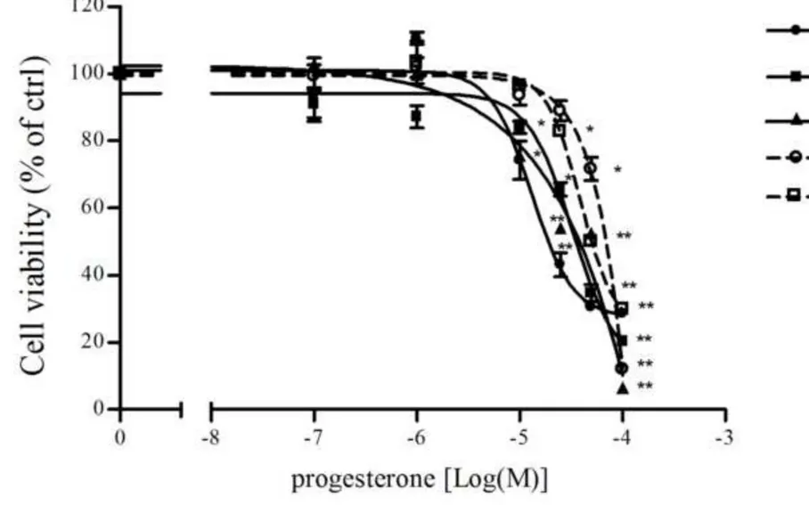
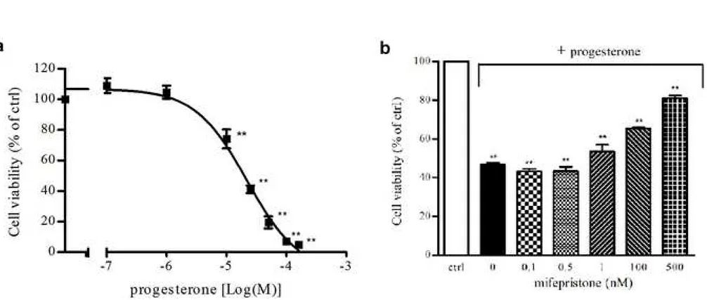
Progesterone effects on NCI-H295R cell viability
197
The predominant expression of the intracellular full length PgR B isoforms was previously described 198
in NCI-H295R cells [10]. The exposure of NCI-H295R cells to increasing concentrations of 199
progesterone (0.1 - 160 µM) for 4 days led to a reduction in NCI-H295R cell viability in a 200
concentration-dependent manner (Fig.1a). Sigmoidal concentration-response function was applied to 201
calculate the IC50 value of progesterone in NCI-H295R cells, which was 25.5 μM (95% confidence 202
interval [CI], 19.9 to 32.9). Time course experiments in NCI-H295R cells treated with progesterone 203
at the IC50 value demonstrated that the reduction of cell viability reached its maximum at 4 days, with
204
no significant change up to 6 days (IC50 value of 29.6 µM; 95% CI 23.6 to 37.0) (data not shown).
205
Pretreatment of NCI-H295R cells with increasing concentration of the PgR antagonist mifepristone 206
(0.1 -500 nM) antagonized the cytotoxic effect elicited by progesterone at its IC50 for 4 days (Fig.1b).
207
This provides evidence that the antineoplastic activity of progesterone requires the stimulation of the 208
PgRs. Mifepristone alone (0.1 – 500 nM; 4 days) did not affect NCI-H295R cells viability (data not 209
shown). 210
Progesterone induces NCI-H295R cells apoptosis, without inducing changes in the cell cycle
211
distribution
212
To provide explanation on the mechanism underlying the progesterone-induced NCI-H295R cell 213
toxicity, cells were treated with progesterone (25 μM; 4 days) and stained with AO/EtBr. 214
Progesterone deeply increased the number of apoptotic cells 39 ± 2%; while necrotic and living cells 215
were 2 ± 2% and 59 ± 3% respectively (apoptotic cells: untreated vs treated cells: P < 0.001 Fig.2a). 216 1 2 3 4 5 6 7 8 9 10 11 12 13 14 15 16 17 18 19 20 21 22 23 24 25 26 27 28 29 30 31 32 33 34 35 36 37 38 39 40 41 42 43 44 45 46 47 48 49 50 51 52 53 54 55 56 57 58 59 60 61 62
Time-course experiments were conducted and our results demonstrated that after 2 days of treatment, 217
the effect is as follow: 93 ± 1% living cells, 7 ± 1% apoptotic cells and no necrotic cells (apoptotic 218
cells: untreated vs treated cells: P < 0.05; Supplemental Fig.1a). Progesterone-induced apoptotic 219
cytotoxicity reached its maximum after 4 days of treatment, as above indicated, and it was not 220
modified if cells were exposed to progesterone up to 6 days: 47 ± 1% living cells, 43 ± 3% apoptotic 221
cells, 10 ± 1% necrotic cells (apoptotic cells: untreated vs treated cells: P < 0.001; Supplemental 222
Fig.1b). We next examined the expression of total Caspase-3 and the cleaved-Caspase-3, that play a 223
central role in the execution phase of cell apoptosis [30], in progesterone-treated NCI-H295R cells in 224
comparison to untreated cells (Fig.2b). Progesterone exposure for 48 hrs significantly increased the 225
expression of cleaved-Caspase-3 (% of increase: 23.9 ± 1.6) while total Caspase-3 levels were not 226
affected. The analyses of the cell cycle progression by flow cytometry in untreated and progesterone-227
treated NCI-H295R cells did not show significant differences in cell distribution up to 4 days of 228
treatment (Fig.2c). However, we observed that treatment with progesterone for 4 days increased the 229
proportion of cells in the sub-G0 phase: 29.7 ± 4.6% untreated cells, 50.3 ± 5.1% progesterone-treated 230
cells (P < 0.05), suggestive of DNA fragmentation. Taken together these observations suggest that 231
apoptosis is the main mechanism mediating the progesterone cytotoxicity. 232
Effect of progesterone on -catenin nuclear translocation in NCI-H295R cells
233
NCI-H295R cells were treated with progesterone and analyzed for -catenin localization using 234
immunofluorescence analyses. At baseline, β-catenin was highly expressed in the nucleus (Fig.3a), 235
whereas the cell exposure to progesterone at its IC50 reduced -catenin nuclear localization and
236
increased its retention into cytoplasm (Fig.3b). The effect of progesterone in sequestering -catenin 237
in cytoplasm was counteracted by 100 nM mifepristone (Fig.3c). Immunofluorescence quantification 238
using ImageJ software, reported in Table 2, demonstrated that progesterone significantly reduced -239
catenin nuclear localization. The progesterone-induced reduction of nuclear β-catenin induced the 240
decrease of mRNA expression of some of its target genes, namely MYC and survivin, while the 241 1 2 3 4 5 6 7 8 9 10 11 12 13 14 15 16 17 18 19 20 21 22 23 24 25 26 27 28 29 30 31 32 33 34 35 36 37 38 39 40 41 42 43 44 45 46 47 48 49 50 51 52 53 54 55 56 57 58 59 60 61 62
mRNA expression level of another gene, CCND1, resulted unchanged by progesterone treatment 242 (Supplemental Fig.2). 243 mPR and PGRMC1 244
As underlined in the Introduction, accumulating evidence suggests that rapid progesterone responses 245
are mediated by activation of mPRs [31]. In order to evaluate whether or these receptors could 246
contribute to the observed progesterone cytotoxic effect on NCI-H295R, we firstly evaluated their 247
expression. As shown in Supplemental Fig.3a, NCI-H295R expressed mPRs. These receptors were 248
functionally active, as, when cells were treated with the IC50 value of progesterone, we observed a
249
reduction of phospho-Erk protein level (% of decrease: 30.39 ± 1.14) at very early time, namely 15’ 250
after progesterone exposure (Supplemental Fig.3b). Finally, we demonstrated that NCI-H295R 251
expressed as well the PGRMC1 (Supplemental Fig.3a). 252
Progesterone enhanced NCI-H295R cytotoxicity induced by mitotane in drug-combination
253
treatments
254
To evaluate whether progesterone treatment of NCI-H295R cells could enhance the cytotoxicity of 255
mitotane, the combination index (CI) was calculated according to the Chou-Talalay method [21]. 256
NCI-H295R cells were firstly exposed to increasing concentrations of mitotane (25 nM - 40 M) for 257
4 days and analyzed for cell viability by MTT assay. Sigmoidal concentration-response function was 258
used to calculate the IC50 value, which was 3 μM (95% CI, 2.08 to 4.34) (Fig.4a). The cytotoxic effect
259
of progesterone in combination with mitotane was evaluated at the 1:4 fixed molar ratio for 4 days 260
(Fig.4b). We found that in NCI-H295R cells, the combination had a synergistic cytotoxic effect as 261
compared to each single compound at a Fa = 0.09 - 0.86 with range of CI: 0.08 to 0.88 (Fig.4c). 262
Progesterone exerted cytotoxic effect in primary human ACC cells
263
Primary cultures derived from ACC patients were treated with increasing concentrations of 264
progesterone for 4 days and analyzed for cell viability by MTT assay. In cortisol-secreting ACC cells 265
(ACC01, ACC02 and ACC16), progesterone exerted a concentration-dependent inhibition of cell 266
viability with IC50 values of 18 μM (95% CI 11.4 to 31.7), 32.9 μM (95% CI 26.5 to 40.9) and 39.2
267 1 2 3 4 5 6 7 8 9 10 11 12 13 14 15 16 17 18 19 20 21 22 23 24 25 26 27 28 29 30 31 32 33 34 35 36 37 38 39 40 41 42 43 44 45 46 47 48 49 50 51 52 53 54 55 56 57 58 59 60 61 62
μM (95% CI 31.8 to 48.4) respectively. Immunohistochemical analyses of PgR expression in ACC01, 268
ACC02 and ACC16 tumors showed that at least 40% of neoplastic cells were positive for PgR (Table 269
1). By contrast, a lesser cytotoxic effect of progesterone was observed in the non-secreting human 270
ACC cells, ACC03 and ACC08, with IC50 values of 73.4 μM (95% CI 46.1 to 116.8) and 80.8 μM
271
(95% CI 50.5 to 129.5) respectively (Fig.5). PgR expression in these cells was detected in less than 272
5% of ACC cells (Table 1). 273
Progesterone effect on SW13 cell line.
274
Finally, as an internal control, we tested the effect of progesterone in the non steroidogenic SW13 275
cell line, that belongs from a small cell carcinoma of adrenal and which exact histopathological 276
features are still under investigation [32]. We firstly analyzed the mRNA expression of PgRs and 277
results indicated that SW13 cells were devoid of PgR: indeed, q-RT-PCR analysis revealed a not 278
detectable PgR mRNA expression in this cell line compared to the ΔCt of 9.01 ± 0.25 in NCI-H295R 279
cells, used as positive control. The western blot analysis of mPR and PGRMC1 expression in SW13 280
cell line indicated that these receptor proteins were expressed in this cell line (Supplemental Figure 281
4a). When exposed to increasing concentrations of progesterone within the same range of 282
concentrations used for NCI-H295R cells, a cytotoxic effect could be observed, although it was non 283
concentration-dependent (Supplemental Fig.4b). 284
285
Discussion
286
In the present study, we demonstrated that progesterone, through its receptors, exerted a 287
concentration-dependent and time-dependent inhibition of ACC cell viability, and this effect was, at 288
least in part, counteracted by the PgR antagonist mifepristone. The role of PgR is further supported 289
by data published by our group, where the PgR silencing induces the almost complete disappearance 290
of the effect of abiraterone on cell viability [10]. The cytotoxic effect of progesterone was observed 291
in the NCI-H295R cells, an ACC cell line that mainly express the full length PgR B isoform [10, 33] 292
and confirmed in primary cell cultures derived from cortisol-secreting ACC that are characterized by 293 1 2 3 4 5 6 7 8 9 10 11 12 13 14 15 16 17 18 19 20 21 22 23 24 25 26 27 28 29 30 31 32 33 34 35 36 37 38 39 40 41 42 43 44 45 46 47 48 49 50 51 52 53 54 55 56 57 58 59 60 61 62
a marked expression of the PgR. Indeed, the cytotoxic effect of progesterone was less evident in non-294
secreting ACC tumors in which PgR expression was low. Intriguingly, we demonstrated that NCI-295
H295R cells expressed also the mPR and PGRMC1 component, suggesting that the progesterone 296
effects that we observed in ACC cells, both in cell line and primary cultures, could be a result of a 297
multifactorial process involving both genomic and non-genomic actions, dependent on both 298
membrane and intracellular progesterone receptor arrangement expressed by each ACC tumor. The 299
scenario is even more complex than expected, in light of the evidence showing that the different effect
300
of progesterone depends on many variables, such as the type of cells, the genomic and/or non genomic
301
effects linked to the progesterone receptor expression and the concentration of hormone present [34]. 302
As a matter of fact , we observed that the non steroidogenic SW13 cell line, established from a small 303
cell carcinoma of the adrenal [35], was responsive to the cytotoxic effect of progesterone (although 304
without displaying a concentration-dependent curve ) despite they expressed only mPR and 305
PGRMC1. On the basis of these preliminary results the characterization of expression and function 306
of all receptor components of progesterone pathway in our experimental models is now undergoing 307
in our lab. 308
The present results confirm and extend our previous study [10] showing that the in vitro 309
antineoplastic activity of abiraterone is mediated by the drug-induced increase in progesterone levels. 310
PgRs,therefore, could represent a novel promising target in the management of ACC. 311
In PgR-positive breast cancer cell lines, progestins can induce a growth arrest due to decreased 312
expression and activity of cyclin-dependent kinase (cdk) complexes [36]. In the present study, we 313
showed that the progesterone-induced cytotoxicity of NCI-H295R ACC cells was not cell cycle 314
mediated, but apoptosis represented the main molecular events, with a significant increase of the 315
proapoptotic cleaved-Caspase-3 levels in the initial phase of the treatment. The ability of progesterone 316
in modulating apoptotic events, both in vitro and in vivo, was previously demonstrated in several 317
tumor cells [37-41]. On the light of these results, we therefore explored the possible molecular 318
mechanism regulating the progesterone-induced apoptosis in ACC. 319 1 2 3 4 5 6 7 8 9 10 11 12 13 14 15 16 17 18 19 20 21 22 23 24 25 26 27 28 29 30 31 32 33 34 35 36 37 38 39 40 41 42 43 44 45 46 47 48 49 50 51 52 53 54 55 56 57 58 59 60 61 62
The Wnt/β-catenin pathway is frequently altered in ACC, which is characterized by CTNNB1 320
mutations leading to β-catenin accumulation in the nucleus, where it binds with the T cell factor (Tcf) 321
and enhances its transcriptional activity. In the NCI-H295R cell line, harboring the activating 322
CTNNB1 p.S45P mutation, we found that progesterone treatment partially inhibited the β-catenin 323
translocation into the nucleus, thus suggesting the involvement of this pathway in the progesterone 324
antineoplastic activity. These data are in line with our previous in vitro experiments showing that the 325
increased levels of progesterone in NCI-H295R cell culture microenvironment, induced by the block 326
of the CYP17A1 by abiraterone, significantly inhibited the β-catenin migration into the nucleus. 327
Further evidence comes from studies showing that progesterone is able to inhibit the Wnt/β-catenin 328
pathway in endometrial carcinoma [42]. The functional effect of the β-catenin modification of 329
traslocation is the down-regulation of the expression of some β-catenin target genes, namely MYC 330
and survivin, while CCND1 was not modified. 331
Taken together these data are suggestive for an involvement of β-catenin inhibition in the 332
progesterone induced apoptosis of ACC cells. These data, however, are not exhaustive and the full 333
evidence of the inhibitory effect would require the demonstration of a modulation of the expression 334
of other specific β-catenin target genes in NCI-H295R cells by progesterone treatment. These further 335
experiments are outside the scope of the present paper and will be a matter of a future study. 336
Finally, the in vitro demonstration of the synergistic cytotoxic effect of the combination mitotane + 337
progesterone could be of considerable interest for its possible clinical application, as progesterone 338
and its derivatives are already part of the supportive approach in cancer patients . These preclinical 339
data provides the rationale for a new trial testing the efficacy of progesterone n association with 340
current systemic therapies in the management of ACC patients. 341
In conclusion, the present study shows that progesterone exerts a cytotoxic activity in ACC cells, by 342
inducing apoptosis via activation of the progesterone receptors. Both the genomic and non-genomic 343
effects of progesterone seemed to mediate the cytotoxicity, although this point is still under 344 1 2 3 4 5 6 7 8 9 10 11 12 13 14 15 16 17 18 19 20 21 22 23 24 25 26 27 28 29 30 31 32 33 34 35 36 37 38 39 40 41 42 43 44 45 46 47 48 49 50 51 52 53 54 55 56 57 58 59 60 61 62
investigation. The synergistic cytotoxic activity of progesterone with mitotane provides the rationale 345
for testing this combination in a prospective clinical study. 346
347
References
348
1. C.L. Ronchi, M. Kroiss, S. Sbiera, T. Deutschbein, M. Fassnacht:. EJE prize 2014: current and 349
evolving treatment options in adrenocortical carcinoma: where do we stand and where do we want to 350
go? Eur. J. Endocrinol. 171, R1-R11 (2014) 351
2. M. Terzolo, F. Daffara, A. Ardito, B. Zaggia, V. Basile, L. Ferrari, A. Berruti: Management of 352
adrenal cancer: a 2013 update. J. Endocrinol. Invest. 37, 207-17 (2014) 353
3. A. Berruti, E. Baudin, H. Gelderblom, H.R. Haak, F. Porpiglia, M. Fassnacht, G. Pentheroudakis: 354
ESMO Guidelines Working Group. Adrenal cancer: ESMO Clinical Practice Guidelines for 355
diagnosis, treatment and follow-up. Ann. Oncol. 23 Suppl 7, 131-138 (2012) 356
4. M. Fassnacht, M. Terzolo, B. Allolio, E. Baudin, H. Haak, A. Berruti, S. Welin, C. Schade-357
Brittinger, A. Lacroix, B. Jarzab, H. Sorbye, D.J Torpy, V. Stepan et al: Combination chemotherapy 358
in advanced adrenocortical carcinoma. N. Engl. J Med. 366, 2189-2197 (2012) 359
5. A. Berruti, M. Terzolo, P. Sperone, A. Pia, S. Della Casa, D.J. Gross, C. Carnaghi, P. Casali, F. 360
Porpiglia, F. Mantero, G. Reimondo, A. Angeli, L. Dogliotti: Etoposide, doxorubicin and cisplatin 361
plus mitotane in the treatment of advanced adrenocortical carcinoma: a large prospective phase II 362
trial. Endocr. Relat. Cancer. 12, 657-666 (2005) 363
6. A. Berruti, M. Fassnacht, H. Haak, T. Else, E. Baudin, P. Sperone, M. Kroiss T. Kerkhofs, A.R. 364
Williams, A. Ardito, S. Leboulleux, M. Volante, T. Deutschbein, R. Feelders, C. Ronchi, S. Grisanti, 365
H. Gelderblom, F. Porpiglia, M. Papotti, G.D. Hammer, B. Allolio, M. Terzolo: Prognostic role of 366
overt hypercortisolism in completely operated patients with adrenocortical cancer. Eur. Urol. 65, 367
832-838 (2014) 368
7. R. Libé, I. Borget, C.L. Ronchi, B. Zaggia, M. Kroiss, T. Kerkhofs, J. Bertherat, M. Volante, M. 369
Quinkler, O. Chabre, M. Bala , A. Tabarin, F. Beuschlein et al.: Prognostic factors in stage III-IV 370 1 2 3 4 5 6 7 8 9 10 11 12 13 14 15 16 17 18 19 20 21 22 23 24 25 26 27 28 29 30 31 32 33 34 35 36 37 38 39 40 41 42 43 44 45 46 47 48 49 50 51 52 53 54 55 56 57 58 59 60 61 62
adrenocortical carcinomas (ACC): an European Network for the Study of Adrenal Tumor (ENSAT) 371
study. Ann. Oncol. 26, 2119-2125 (2015) 372
8. M. Volante, C. Buttigliero, E. Greco, A. Berruti, M. Papotti: Pathological and molecular features 373
of adrenocortical carcinoma: an update. J Clin Pathol. 61, 787-793 (2008) 374
9. Puglisi S, Perotti P, Cosentini D, Roca E, Basile V, Berruti A, Terzolo M. Decision-making for 375
adrenocortical carcinoma: surgical, systemic, and endocrine management options. Expert Rev 376
Anticancer Ther. 2018 Aug 21:1-9. 377
10. C. Fiorentini, M. Fragni, P. Perego, S. Vezzoli, S.A. Bonini, M. Tortoreto, D. Galli, M. Claps, 378
G.A. Tiberio, M. Terzolo, C. Missale, M. Memo, G. Procopio, N, Zaffaroni, A. Berruti, S. Sigala: 379
Antisecretive and Antitumor Activity of Abiraterone Acetate in Human Adrenocortical Cancer: A 380
Preclinical Study. J. Clin. Endocrinol. Metab. 101, 4594-4602 (2016) 381
11. G. Attard, A.H. Reid, R.J Auchus, B.A. Hughes, A.M. Cassidy E. Thompson, N.B. Oommen, E. 382
Folkerd, M. Dowsett W. Arlt, J.S. de Bono: Clinical and biochemical consequences of CYP17A1 383
inhibition with abiraterone given with and without exogenous glucocorticoids in castrate men with 384
advanced prostate cancer. J. Clin. Endocrinol. Metab. 97, 507-516 (2012) 385
12. A. Pia, F. Vignani, G. Attard, M. Tucci, P. Bironzo, G. Scagliotti, W. Arlt, M. Terzolo, A. 386
Berruti:Strategies for managing ACTH dependent mineralocorticoid excess induced by abiraterone. 387
Cancer. Treat. Rev. 39, 966-973 (2013) 388
13. M. Claps, B. Lazzari, S. Grisanti, V. Ferrari M. Terzolo, S. Sigala, S. Vezzoli, M. Memo, M. 389
Castellano, A. Berruti: Management of severe Cushing’s syndrome induced by adrenocortical 390
carcinoma with abiraterone acetate: a case report. AACE Clinical Reports. 2, 337-341 (2016) 391
14. A. Salomon, M. Keramidas, C. Maisin, M. Thomas: Loss of β-catenin in adrenocortical cancer 392
cells causes growth inhibition and reversal of epithelial-to-mesenchymal transition. Oncotarget 6, 393
11421-11433 (2015) 394
15. D. Garg, S.S.M Ng K.M. Baig, P. Driggers, J. Segars: Progesterone-Mediated Non-Classical 395
Signaling. Trends Endocrinol. Metab. 28, 656-668 (2017)
396 1 2 3 4 5 6 7 8 9 10 11 12 13 14 15 16 17 18 19 20 21 22 23 24 25 26 27 28 29 30 31 32 33 34 35 36 37 38 39 40 41 42 43 44 45 46 47 48 49 50 51 52 53 54 55 56 57 58 59 60 61 62
16. R.L. Ashley, C.M. Clay, T.A. Farmerie, G.D. Niswender, T.M. Nett: Cloning and characterization 397
of an ovine intracellular seven transmembrane receptor for progesterone that mediates calcium 398
mobilization. Endocrinology. 147, 4151-4159 (2006) 399
17. V. Boonyaratanakornkit, N. Hamilton, D.C. Márquez-Garbán, P. Pateetin, E.M. McGowan, R.J. 400
Pietras: Extranuclear signaling by sex steroid receptors and clinical implications in breast cancer. 401
Mol. Cell. Endocrinol. 466, 51-72 (2018) 402
18. W.E. Rainey, K. Saner, B.P. Schimmer: Adrenocortical cell lines. Mol. Cell. Endocrinol. 228, 23-403
38 (2004) 404
19. E.N. Kornaga A.C. Klimowicz, N. Guggisberg, T. Ogilvie, D.G. Morris, M. Webster, A.M. 405
Magliocco: A systematic comparison of three commercial estrogen receptor assays in a single clinical 406
outcome breast cancer cohort. Mod. Pathol. 29, 799-809 (2016) 407
20. T.C. Chou, P. Talalay: Quantitative analysis of dose-effect relationships: the combined effects of 408
multiple drugs or enzyme inhibitors. Adv. Enzyme. Regul. 22, 27–55 (1984) 409
21. T.C. Chou: Theoretical basis, experimental design, and computerized simulation of synergism 410
and antagonism in drug combination studies. Pharmacol. Rev. 58, 621-681 (2006) 411
22. J. Hofman, D. Ahmadimoghaddam, L. Hahnova, P. Pavek, M. Ceckova, F. Staud: Olomoucine II 412
and purvalanol A inhibit ABCG2 transporter in vitro and in situ and synergistically potentiate 413
cytostatic effect of mitoxantrone. Pharmacol. Res. 65, 312-319 (2012) 414
23. S. Sigala, S. Bodei, C. Missale, D. Zani, C. Simeone, S.C. Cunico, P.F. Spano: Gene expression 415
profile of prostate cancer cell lines: effect of nerve growth factor treatment. Mol. Cell. Endocrinol. 416
284, 11-20 (2008)
417
24. C. Fiorentini, S. Bodei, F. Bedussi, M. Fragni, S.A. Bonini, C. Simeone, D. Zani, A. Berruti, C. 418
Missale, M. Memo, P.F. Spano, S. Sigala: GPNMB/OA protein increases the invasiveness of human 419
metastatic prostate cancer cell lines DU145 and PC3 through MMP-2 and MMP-9 activity. Exp. Cell. 420 Res. 323, 100-111 (2014) 421 1 2 3 4 5 6 7 8 9 10 11 12 13 14 15 16 17 18 19 20 21 22 23 24 25 26 27 28 29 30 31 32 33 34 35 36 37 38 39 40 41 42 43 44 45 46 47 48 49 50 51 52 53 54 55 56 57 58 59 60 61 62
25. V. Porrini I. Sarnico, M. Benarese, C. Branca, M. Mota, A. Lanzillotta, A. Bellucci, E. Parrella, 422
L. Faggi, P.F. Spano, B.P. Imbimbo, M. Pizzi: Neuroprotective and Anti-Apoptotic Effects of CSP-423
1103 in Primary Cortical Neurons Exposed to Oxygen and Glucose Deprivation. Int. J. Mol. Sci. 18 424
(2017) 425
26. M. Babagana, S. Johnson, H. Slabodkin, W. Bshara, C. Morrison, E.S. Kandel: P21-activated 426
kinase 1 regulates resistance to BRAF inhibition in human cancer cells. Mol. Carcinog. 56, 1515-427
1525 (2017) 428
27. N.M. Bashour S. Wray:Progesterone Directly and Rapidly Inhibits GnRH Neuronal Activity via 429
Progesterone Receptor Membrane Component 1 Endocrinology. Endocrinology. 153, 4457–4469 430
(2012) 431
28. V. Lodde, J.J. Peluso: A novel role for progesterone and progesterone receptor membrane 432
component 1 in regulating spindle microtubule stability during rat and human ovarian cell mitosis. 433
Biol. Reprod. 84, 715-722 (2011) 434
29. A. Chimento A, Sirianni R, Casaburi I, Zolea F, Rizza P, Avena P, Malivindi R, De Luca A, 435
Campana C, Martire E, Domanico F, Fallo F, Carpinelli G, Cerquetti L, Amendola D, Stigliano A, 436
Pezzi V. GPER agonist G-1 decreases adrenocortical carcinoma (ACC) cell growth in vitro and in 437
vivo. Oncotarget. 6, 19190-19203 (2015) 438
30. R.S.Y. Wong: Apoptosis in cancer: from pathogenesis to treatment. J. Exp. Clin. Cancer. Res. 30-439
87 (2011) 440
31. A.O. Mueck, X. Ruan, H. Seeger, T. Fehm, H. Neubauer: Genomic and non-genomic actions of 441
progestogens in the breast. J. Steroid. Biochem. Mol. Biol. 142, 62-67 (2014) 442
32. T. Wang, W.E. Rainey: Human adrenocortical carcinoma cell lines. Mol Cell Endocrinol. 351, 443
58-65 (2012) 444
33. K.M. Scarpin, J.D. Graham, P.A. Mote, C.L. Clarke: Progesterone action in human tissues:
445
regulation by progesterone receptor (PR) isoform expression, nuclear positioning and coregulator
446
expression. Nucl. Recept. Signal. 7: e009 (2009)
447 1 2 3 4 5 6 7 8 9 10 11 12 13 14 15 16 17 18 19 20 21 22 23 24 25 26 27 28 29 30 31 32 33 34 35 36 37 38 39 40 41 42 43 44 45 46 47 48 49 50 51 52 53 54 55 56 57 58 59 60 61 62
34. J.H. Check: The role of progesterone and the progesterone receptor in cancer. Expert Rev
448
Endocrinol Metab. 12, 187-197 (2017) 449
35. A. Leibovitz, W.M. 3rd McCombs, D. Johnston, C.E. McCoy, J.C. Stinson: New human cancer 450
cell culture lines. I. SW-13, small-cell carcinoma of the adrenal cortex. J. Natl. Cancer Inst. 51, 691-451
697 (1973) 452
36. V. Boonyaratanakornkit, E. McGowan L. Sherman, M.A. Mancini, B.J. Cheskis, D.P. Edwards: 453
The role of extranuclear signaling actions of progesterone receptor in 454
mediating progesterone regulation of gene expression and the cell cycle. Mol. Endocrinol. 21, 359-455
375 (2007) 456
37. S.Z. Bu, D.L. Yin, X.H. Ren, L.Z. Jiang, Z.J. Wu, Q.R. Gao, G. Pei: Progesterone induces 457
apoptosis and up-regulation of p53 expression in human ovarian carcinoma cell lines. Cancer. 79, 458
1944-1950 (1997) 459
38. V. Syed, S.M. Ho: Progesterone-induced apoptosis in immortalized normal and malignant human 460
ovarian surface epithelial cells involves enhanced expression of FasL. Oncogene. 22, 6883-6890 461
(2003) 462
39. B. Formby, T.S. Wiley: Bcl-2, survivin and variant CD44 v7-v10 are downregulated and p53 is 463
upregulated in breast cancer cells by progesterone: inhibition of cell growth and induction of 464
apoptosis. Mol. Cell. Biochem. 202, 53-61 (1999) 465
40. K. Horita, N. Inase, S. Miyake, B. Formby, H. Toyoda, Y. Yoshizawa: Progesterone induces 466
apoptosis in malignant mesothelioma cells. Anticancer Res. 21, 3871-3874 (2001) 467
41. F. Atif, S. Yousuf, D.G. Stein: Anti-tumor effects of progesterone in human glioblastoma 468
multiforme: role of PI3K/Akt/mTOR signaling. J. Steroid. Biochem. Mol. Biol. 146, 62-73 (2015) 469
42. Y. Wang, P. Hanifi-Moghaddam, E.E. Hanekamp, H.J. Kloosterboer, P. Franken, J. Veldscholte, 470
H.C. van Doorn, P.C. Ewing, J.J. Kim, J.A. Grootegoed, C.W. Burger, R. Fodde, L.J. Blok: 471
Progesterone inhibition of Wnt/beta-catenin signaling in normal endometrium and endometrial 472
cancer. Clin. Cancer. Res. 15, 5784-5793 (2009) 473 1 2 3 4 5 6 7 8 9 10 11 12 13 14 15 16 17 18 19 20 21 22 23 24 25 26 27 28 29 30 31 32 33 34 35 36 37 38 39 40 41 42 43 44 45 46 47 48 49 50 51 52 53 54 55 56 57 58 59 60 61 62
43. P. De Cremoux, D. Rosenberg, J. Goussard, C. Brémont-Weil, F. Tissier, C. Tran-Perennou L. 474
Groussin, X. Bertagna, J. Bertherat, M.L. Raffin-Sanson : Expression of progesterone and estradiol 475
receptors in normal adrenal cortex, adrenocortical tumors, and primary pigmented nodular 476
adrenocortical disease. Endocr. Relat. Cancer. 15, 465–674 (2008) 477
478
Figure Legends
479 480
Fig. 1: Cytotoxic effect of progesterone in NCI-H295R cells. a. NCI-H295R cells were treated with
481
increasing concentration of progesterone (0.1-160 μM) for 4 days. Cell viability was analyzed by 482
MTT assay. Results are expressed as percent of viable cells vs control (ctrl) cells. Data are the mean 483
± S.E.M. of three experiments performed in triplicate. **P < 0.001 vs ctrl. b. Cells were treated with 484
progesterone (25 μM) for 4 day in the presence of the PgR antagonist mifepristone (0.1 nM – 500 485
nM). Cell viability was analyzed by MTT assay. Results are expressed as percent of viable cells vs 486
control (ctrl) cells. Data are the mean ± S.E.M. of three experiments performed in triplicate. **P < 487
0.001 vs ctrl. 488
Fig. 2: Progesterone promotes apoptotic events in NCI-H295R cells, with no influences on cell
489
cycle phases. a. NCI-H295R cells were treated with progesterone (25 µM) for 4 days. Untreated (C)
490
and progesterone-treated (T) cells were then stained with AO/EtBr. Viable (green), apoptotic (yellow) 491
and necrotic (red) cells were scored under a confocal laser-scanning microscope. Magnification, 10x. 492
The images were representative of at least three independents experiments, with superimposable 493
results. b. Cells were treated with progesterone (25 µM) for 1-3 days and analysed for Caspase-3 and 494
clevead-Caspase-3 expression using Western Blot (WB). The human α-Tubulin was used as internal 495
control. A representative WB is shown. Densitometric analysis of blots (n = 3) with specific levels of 496
cleaved-Caspase-3 normalized to the corresponding tubulin levels. Bars represent the mean ± S.E.M. 497
**P < 0.01 vs untreated cells. c. NCI-H295R cells were treated with progesterone (25 µM), stained
498
with propidium iodide and analyzed for DNA content by flow cytometry. Histograms representative 499
of one out of three experiments were shown in the figure. 500 1 2 3 4 5 6 7 8 9 10 11 12 13 14 15 16 17 18 19 20 21 22 23 24 25 26 27 28 29 30 31 32 33 34 35 36 37 38 39 40 41 42 43 44 45 46 47 48 49 50 51 52 53 54 55 56 57 58 59 60 61 62
Fig. 3: Progesterone treatment affects the subcellular localization of -catenin in NCI-H295R
501
cells. Cells were treated with progesterone (25 µM) alone or in combination with mifepristone (100
502
nM) for 3 days. Untreated (a), progesterone-treated (b) progesterone-mifepristone-treated (c) cells 503
were analyzed for β-catenin localization following by incubation with Hoechst for nuclear staining. 504
Panels a, d, g: Hoechst; panel b, e, h: -catenin; panel c, f, i: merge. The scale bar of 50 µm is 505
automatically inserted by the software ZEN Black. 506
Fig. 4: Effect of the combination of progesterone with mitotane in H295R cells. a.
NCI-507
H295R cells were treated with increasing concentration of mitotane (25 nM - 40 μM) for 4 days. Cell 508
viability was analyzed by MTT assay. Results are expressed as percent of viable cells vs control (ctrl) 509
cells. Data are the mean ± S.E.M. of three experiments performed in triplicate. **P < 0.001 vs ctrl. 510
b. NCI-H295R cells exposed to increasing concentrations of progesterone and mitotane alone or in
511
combination at fixed concentration at 1:4 molar ratio (progesterone: mitotane) for 4 days. Data are 512
expressed as percent of viable cells vs control (ctrl) cells. Data are the mean ± S.E.M. of three 513
experiments performed in triplicate. c. Cell viability from B was converted to Fraction affected (Fa) 514
values and resulting data were analyzed with CompuSyn software to obtain combination index (CI) 515
plot. Fa = 0, 100% cell viability; Fa = 1, 0% cell viability; CI value < 0.9, synergism, CI = 0.9-1.1 516
additive effect and CI > 1.1 antagonism. 517
Fig. 5: Cytotoxic effect of progesterone in human ACC primary cultures. Cortisol secreting
518
ACC01 (), ACC02 (■), ACC16 (▲) and non secreting ACC03 (○), ACC08 () primary cultures of 519
human ACC cells were treated with increasing concentrations of progesterone (0.1 – 100 µM) for 4 520
days. Cell viability was evaluated by MTT assay. Results are expressed as percent of viable cells vs 521
control (ctrl) cells. Data are the mean ± S.E.M. of three independent experiments performed in 522 triplicate. *P < 0.01 vs ctrl; **P < 0.001 vs ctrl. 523 524 525 526 1 2 3 4 5 6 7 8 9 10 11 12 13 14 15 16 17 18 19 20 21 22 23 24 25 26 27 28 29 30 31 32 33 34 35 36 37 38 39 40 41 42 43 44 45 46 47 48 49 50 51 52 53 54 55 56 57 58 59 60 61 62
Table 1: Clinical characteristics of ACC patients.
Primary culture identification
Tumor specimen
Histology Disease stage Hormone hypersecretion PgR expression ACC01 Female 66 yr old Primary ACC Weiss score 8 Mitotic index: >50/50 HPF Ki67 70% Stage IV (hepatic metastases) Cortisol (severe Cushing’s syndrome) 40% ACC02 Female 63 yr old Peritoneal metastases
Weiss score not available Mitotic index: >50/50 HPF Ki67 50% Stage IV (peritoneal dissemination)
Cortisol (mild clinical signs of hypercortisolism) 70% ACC16 Male 55 yr old Primary ACC
Weiss score not available Mitotic index: 10/50 HPF Ki67 50%
Stage IV
(bone and multiple abdominal lymphonodal metastases) Cortisol (severe Cushing’s syndrome) 40% ACC03 Male 59 yr old Local relapse of ACC Weiss score 8 Mitotic index: 25/50 HPF Ki67 20% Stage IV (left hypochondrium soft tissue relapse and peritoneal dissemination) No secretion 3-5% ACC08 Female 50 yr old Lung metastases Weiss score 8 Oncocytic features Mitotic index: 10/50 HPF Ki67 20%
Stage IV (lung and bone metastases)
No secretion 1-2%
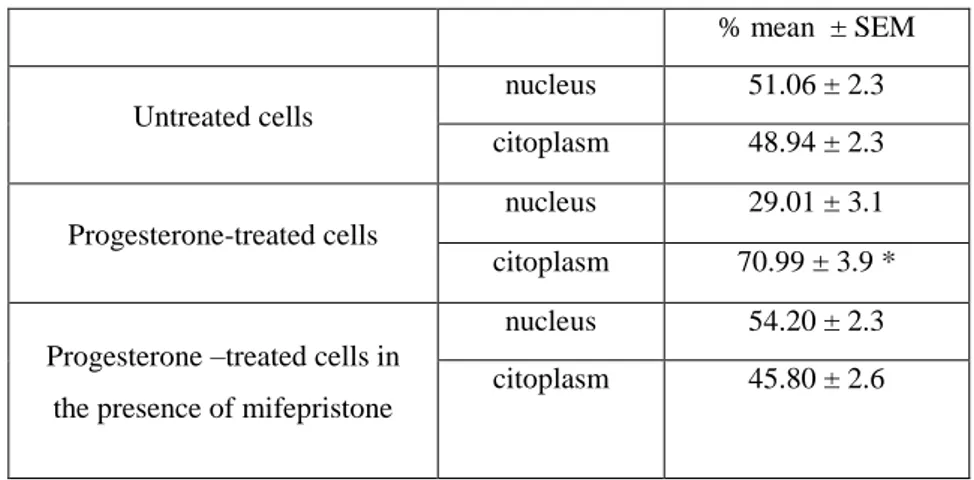
Table 2. Immunofluorescence quantification of β-catenin expression. % mean ± SEM Untreated cells nucleus 51.06 ± 2.3 citoplasm 48.94 ± 2.3 Progesterone-treated cells nucleus 29.01 ± 3.1 citoplasm 70.99 ± 3.9 *
Progesterone –treated cells in the presence of mifepristone
nucleus 54.20 ± 2.3 citoplasm 45.80 ± 2.6
Cells were treated with progesterone (25 µM) alone or in combination with mifepristone (100 nM) for 3 days. Quantification was performed using the ImageJ software. Several cells in different fields, randomly chosen, were quantified. *P < 0.001 vs nuclear localization
Supplementary Material