Prevention 0:
The best way
to prevent
peri-implant disease
?
Prof Magda Mensi, Timothy Ives & Dr Gianluca Garzetti, Italy
The philosophy of prevention in all medical professions is increasing from a global perspective. In fact, prevention of chronic non-communicable diseases, the major bur-den of illness and disability in almost all countries in the world, has been strengthened in recent years.1 The
mo-tivation is to ensure a better quality of life for people and to reduce public health expenditures.
In dentistry, periodontitis is one of the major chronic non-communicable diseases. World experts in periodon-tics and science have published several principles re-garding the prevention of periodontal diseases.2
Peri-implantitis is a twenty-first-century version of peri-odontitis and increasing in occurrence as implant place-ment is increasing (Figs. 1–3). Like periodontitis, it is a biofilm-associated pathological condition, but instead of affecting periodontal ligaments and bone, it is charac-terised by inflammation in the peri-implant mucosa and subsequent progressive loss of supporting bone.3 The
main reasons for concerns in this area are an aetiology in which several risk factors can play a determining role4
and a lack of a gold standard therapy. Primary and sec-ondary preventative measures are really important to prevent mucositis and peri-implantitis and to avoid re-currences, but there are many details to consider before placing implants to mitigate iatrogenic problems. There are many different prosthetic solutions besides implants
that dental professionals could propose to patients if consideration is given from the beginning to the entire situation. Implants may not always be in the best inter-est of the patient.
For these reasons, every clinician, before placing an implant, should consider not only patient- and site-spe-cific aspects, but also surgeon, prosthodontist, dental hygienist and dental technician skills in order to minimise the possibility of peri-implantitis in the future.
The following should be considered before primary and secondary prevention, and it is the proposal of the au-thors that this approach be called “Prevention 0”.
Patient-specific considerations
When deciding to rehabilitate a patient with dental im-plants, before surgical planning, we have to carefully in-form the patient about the characteristics of this pro-cedure. It is important to underline that personal daily maintenance at home and appropriate compliance re-garding follow-up controls and dental hygiene therapies are effective preventative measures.5 Procedure
aware-ness and compliance are the foundation for success, but the clinician must also inform the patient about the impact of systemic disorders (osteogenesis imperfecta, ectodermal dysplasia, diabetes), medications
(bisphos-Fig. 1a
Figs. 1a & b: Implant in position #14 affected by peri-implantitis: peri-implant probing a) with the prosthetic crown in situ and b) after prosthetic crown
removal. Fig. 2: Radiographic examination of the implant. Fig. 3: Excess resin cement around the implant.
Fig. 1b Fig. 2 Fig. 3
|
research
phonates), therapies (radiotherapy in the jawbone), hab-its (smoking, poor biofilm control) and a history of ag-gressive periodontitis6 as being relevant risk factors for
peri-implant disease.7
Site-specific considerations
The healing process after tooth loss leads to a variable reduction of the alveolar process, inducing hard- and soft-tissue deficiencies. The clinician must evaluate care-fully all sites exposed to the following factors, because they have the potential for major healing deficiencies: loss of periodontal support, endodontic infections, longitudi-nal root fractures, thin buccal bone plates, buccal/lingual tooth position in relation to the arch, extraction with addi-tional trauma to the tissue, injury, pneumatisation of the maxillary sinus, medications and systemic diseases re-ducing the amount of naturally formed bone, agenesis of teeth and pressure from soft tissue-supported remov-able prostheses.
Other site considerations relate to anatomical knowl-edge and in respect to the suitable anatomical structure of the area (maxillary sinus, inferior alveolar nerve), endo-dontic and periodontal health of adjacent teeth, and patient phenotype. According to Linkevicius et al. there is significant evidence that thin soft tissue leads to in-creased marginal bone loss compared with thick soft tis-sue around implants.3, 8 Lack of bone has led to the
de-velopment of various alternative surgical techniques to
avoid large bone regenerations or grafts, such as short implants, tilted implants, pterygoid implants and palatal implant mesh, with questionable results, but definitely decreasing the cleanability and maintainability of im-plants and prostheses.
Dental hygienist skills and devices
This professional figure plays a key role in disease pre-vention and oral health promotion.9 Dental hygienists
should not limit their activities to being an oral cleaner, but act as the patient’s dental coach or personal oral trainer, motivating patients not only in dental habits but also in lifestyle, for example regarding smoking cessation and diet. This is a friendly expert who strengthens patient fidelity to the dental office, even in fearful patients, and maintains restorative work and rehabilitations undertaken by the dentist.10
To perform professional care in a minimally invasive way, wearing loupes and using plaque disclosing agents and appropriate devices are mandatory, especially if prosthetic rehabilitation is difficult for the patient to main-tain. Correct and periodic biofilm removal should be con-sidered the standard of care for prevention and manage-ment of peri-implant disease.11 For this reason, patients
should be motivated and instructed in daily implant main-tenance, which should begin before implant placement and be continued after treatment within a regular, per-sonalised recall regime (Figs. 4 & 5).
Figs. 5a–c: Professional peri-implant biofilm removal by a) AIRFLOW with erythritol powder (PLUS powder, EMS), b) PERIOFLOW with PLUS powder and c) with PEEK tip (PI, EMS).
Fig. 4a
Fig. 5a
Figs. 4a–c: Peri-implant home care with a) AirFloss (Philips), b) X-Floss (ROEN) and c) interdental brush (TePe). Fig. 4b Fig. 5b Fig. 4c Fig. 5c
15
4 2018Surgeon skills
Nowadays, especially in Italy, a new professional fig-ure has appeared: the implantologist, who is a gradu-ate dentist, generally a co-worker, and goes to different dental offices or clinics and mainly places implants, often without sufficient expertise in periodontal and prosthetic fields. That means, in some cases, implant misposition-ing, resulting in reconstructive and maintenance prob-lems. In order to avoid fabrication of specific prosthetic parts, unrestored implants and surgical interventions to remove or reposition them in favourable prosthetic posi-tions, this surgical intervention should only be performed by an elite clinician.7 This is an expert dentist with the
necessary surgical skills to manage both soft and hard tissue (before and after implant placement) perfectly and with adequate expertise in the prosthetic field to allow a prothesis-guided implant surgery and, subsequently, a functioning, not overloaded, patient-tailored, cleanable and aesthetically pleasant rehabilitation.
Prosthodontist skills
Skilled clinicians know that there is no such thing as a gold standard prosthesis, but every patient needs a tailored rehabilitation, which takes into consideration his or her resources and requirements and which has to be planned before surgical intervention. After data collection and decision planning regarding the numbers of implants requested, Toronto versus overdenture, cemented ver-sus screwed work, with a motivated and aware patient, the surgical and prosthetic work with careful load man-agement can start.12 Only careful and considerate
plan-ning can prevent poor outcomes (Fig. 6).
Prosthesis fabrication and cementation
Dental technicians should work in direct contact with prosthodontists in order to create aesthetically pleasant, patient-tailored and comfortable cleaning spaces. After dental hygienist instruction and training, patients should be
able to clean their prostheses daily with minimal effort to maintain healthy mouths.13 Another important factor
asso-ciated with clinical signs of peri-implant disease is excess cement.14–17 To avoid excess cement, restoration margins
should be located at or above the peri-implant mucosal margin; otherwise, excess cement must to be removed.18
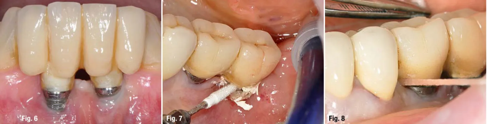
Despite world literature demonstrating an increased interest in excess cement as one of the key factors in aetio-pathogenesis of peri-implant disease, a standard protocol guiding clinicians in this delicate removal proce-dure is still needed. From the authors’ point of view, the cementation procedure requires time, attention, loupes and meticulousness. For these reasons, an accurate protocol, dependent on cement composition, should be published (Figs. 7 & 8).
Conclusion
Implant rehabilitation provides a therapeutic alternative that is more similar to natural teeth than other alterna-tives. Nevertheless, while an implant-supported prosthe-sis can be a permanent successful solution, it lasts only if carefully planned with the patient, properly surgically per-formed, correctly loaded, and
con-stantly maintained by the patient and the dental professionals. Successful results can be achieved only by an expert, patient-centred dental team.
Fig. 7 Fig. 8
Fig. 6: Improper planning led to poor performance. Fig. 7: Careful removal of excess cement after prosthesis cementation using a PEEK tip (PI). Fig. 8: Careful
removal of excess cement with dental floss after prosthesis cementation.
Fig. 6