Immunocytochemical detection
of dentin matrix proteins
in primary teeth from patients
with dentinogenesis imperfecta
associated with osteogenesis
imperfecta
G. Orsini,1A. Majorana,2A. Mazzoni,3 A. Putignano,1M. Falconi,4A. Polimeni,5 L. Breschi4
1Department of Clinical Sciences and Stomatology, Polytechnic University of Marche, Ancona
2Department of Medical-Surgical Specialties, Radiologic Sciences and Public Health, University of Brescia 3Department of Medical Sciences, University of Trieste
4Department of Biomedical and Neuromotor Sciences, University of Bologna
5Department of Oral Sciences, Prosthodontics Unit, La Sapienza University of Rome, Italy
Abstract
Dentinogenesis imperfecta determines structural alterations of the collagen structure still not completely elucidated. Immunohisto -chemical analysis was used to assay type I and VI collagen, various non-collagenous proteins distribution in human primary teeth from healthy patients or from patients affected by type I dentinogenesis imperfecta (DGI-I) asso-ciated with osteogenesis imperfecta (OI). In sound primary teeth, an organized well-known ordered pattern of the type I collagen fibrils was found, whereas atypical and disorganized fibrillar structures were observed in dentin of DGI-I affected patients. Expression of type I collagen was observed in both normal and affected primary teeth, although normal dentin stained more uniformly than DGI-I affected dentin. Reactivity of type VI collagen was sig-nificantly lower in normal teeth than in dentin from DGI-I affected patients (P<0.05). Expressions of dentin matrix protein-1 (DMP1) and osteopontin (OPN) were observed in both normal dentin and dentin from DGI-I affected patients, without significant differ-ences, being DMP1 generally more abundantly expressed. Immuno labeling for chondroitin sulfate (CS) and biglycan (BGN) was weaker in dentin from DGI-I-affected patients com-pared to normal dentin, this decrease being significant only for CS. This study shows ultra-structural alterations in dentin obtained from
patients affected by DGI-I, supported by immunocytochemical assays of different col-lagenous and non-colcol-lagenous proteins.
Introduction
Type I collagen is the major extracellular matrix protein in dentin comprising 85-90% of the organic matrix. Type I collagen is a triple helix containing two alpha 1 and one alpha 2 polypeptides, which are expressed from COL1A1 and COL1A2. Mutations in these genes cause osteogenesis imperfecta (OI), an autosomal dominant form of brittle bone
dis-ease.1OI is usually classified on the basis of
clinical and radiologic criteria in 4 types.1Each
of the 4 types of OI is further subdivided on the presence or absence of the developmental den-tal defect known as dentinogenesis imperfecta (DGI), in which three types can be recognized: DGI-I, DGI-II, and DGI-III.1-3
DGI-I is the dental phenotype recorded in patients affected by OI, which has been noted to be OI types III and IV in approximately 80%.1
Clinically, teeth show marked discoloration, tendency of normal enamel to crack off, bul-bous crowns, short roots and attrition in both the deciduous and permanent dentitions.3
Pulpal obliteration occurs soon after eruption or prior to tooth eruption,3varying, even within
a single individual, from total pulpal oblitera-tion to normal pulp dimensions.1Although
pre-vious studies largely discussed on the genetic changes occurring in OI, little is known on phenotype changes of dentin structure and ultrastructure in patients also showing DGI-I.4,6 Dentin of DGI-I affected patients has been
reported to show irregular texture of dentinal areas, abnormal number and structure of dentin tubules, areas of atubular dentin,5and
ultrastructural abnormalities in the appear-ance and organization pattern of collagen fibers.4 Immuno-electron microscopy studies
have assayed reactivity for different types of collagen, showing presence of type VI collagen and decreasing of type I collagen expression in DGI-I affected dentin.2,6Moreover, DGI-I
affect-ed patients displayaffect-ed reactive zones for the non-collagenous protein fibronectin, alternat-ing layerwise or concentrically with non-reac-tive ones.2
Non-collagenous proteins (NCPs) have been shown to play fundamental roles in actively promoting, controlling, and regulating fibrillo-genesis, crystal growth, and mineralization during dentinogenesis.7 However, no recent
immunocytochemical studies have defined whether there is a correlation among the dif-ferent alterations described in collagenous and non-collagenous dentin components in DGI-I affected patients. Among the NCPs, special
interests have been given to some small leucine-rich proteoglycans (including chon-droitin 4/6 sulphate (CS)-rich decorin and biglycan), and to certain glycoproteins (includ-ing the prominent members of the SIBLINGs family: dentin matrix protein-1 and osteopon-tin), during predentin fibrillogenesis and dentin mineralization, respectively.8,9
Therefore, the aim of the present study was to elucidate morphological alterations of defec-tive dentin in patients affected by DGI-I and OI, by ultrastructural and immunocytochemical analyses. Type I and VI collagen, dentin matrix protein-1 (DMP-1), osteopontin (OPN), bigly-can (BGN), and chondroitin sulfate (CS) were investigated. The tested hypothesis was that no correlation exists between the ultrastruc-tural abnormalities of dentin affected by type I DGI (associated with OI) and defective produc-tion of both collagenous and NCPs.
Materials and Methods
All reagents were purchased from Sigma Aldrich (St. Louis, MO, USA), unless otherwise specified.
Correspondence: Prof. Lorenzo Breschi, Biomedical and Neuromotor Sciences, DIBINEM, University of Bologna, via San Vitale 59, 40125 Bologna, Italy.
Tel. +39.051.2088139 - Fax: +39.051.225208. E-mail: [email protected]
Key words: Osteogenesis imperfecta, dentinogen-esis imperfecta, immuno-electron microscopy, collagen, non-collagenous proteins.
Acknowledgments: the authors report no conflict of interest and wish to thank Mr. Aurelio Valmori (University of Bologna) for photographical assis-tance and Dr. Rosa Curci (IOR, Bologna) for lab-oratory processing. Dr. Elena Bardellini (University of Brescia) and Dr. PierCarlo Brunelli (Spedali Civili, Brescia) are also kindly acknowl-edged for helping during Ethical Committee approval and management of young patients affected by osteogenesis imperfecta, respectively. The study was founded with grants from MIUR (Italy): FIRB RBAP1095CR and PRIN 2009SAN9K5 to L. Breschi.
Received for publication: 2 April 2014. Accepted for publication: 10 November 2014. This work is licensed under a Creative Commons Attribution NonCommercial 3.0 License (CC BY-NC 3.0).
©Copyright G. Orsini et al., 2014 Licensee PAGEPress, Italy
European Journal of Histochemistry 2014; 58:2405 doi:10.4081/ejh.2014.2405
Non
commercial
use
Ten primary teeth exfoliated from five patients affected by DGI associated with OI were used in this study. Five human primary teeth exfoliated were used as controls. The subjects had a mean age of 6.1 years. The con-sent form and experimental protocol was approved by the Ethics Committee of the "AO Spedali Civili" of Brescia (Italy). Following extraction, teeth were cut with a low speed dia-mond saw and sectioned to obtain 1 mm-thick dentin disks. The disks were immediately fixed in 4% paraformaldehyde and 0.1% glutaralde-hyde buffered with 0.1 M sodium cacodylate, at pH 7.2 overnight at 4°C.
Immunocytochemical procedure
After fixation, disks were rinsed for 1 h in 0.1 M cacodylate buffer pH 7.4, and decalcified using 4.13% EDTA for three months at 4°C. Specimens were then extensively rinsed with 0.1M sodium cacodylate buffer, dehydrated in graded concentrations (50, 70, 90, 95 and 100%) of ethanol and embedded in LR White resin (London Resin, Berkshire, UK). Semi-thin sections (1 m) were cut with glass knives on a Reichert Jung Ultracut E ultrami-crotome and stained with toluidine blue. Selected areas of the specimens were trimmed for ultra thin sectioning (80 nm) using a dia-mond knife. Sections were mounted on Formvar carbon-coated nickel grids.
Grid-mounted tissue sections were processed for immunocytochemical labeling using the following primary antibodies: rabbit anti-type VI collagen (Fitzgerald Ind. Int., Acton, MA, USA) and mouse anti-type I colla-gen (C2456, Sigma Aldrich), rabbit anti-DMP-1 (LF-anti-DMP-160, generously donated by Dr. Larry Fisher, National Institute of Dental and Craniofacial Research, Bethesda, MD, USA), rabbit anti-OPN (LF-166, generously donated by Dr. Larry Fisher), mouse anti-CS (CS-56, Sigma Aldrich), rabbit anti-BGN (LF-51, gener-ously donated by Dr. Larry Fisher) as previous-ly reported.8,9In brief, sections were immersed
in 0.05 M Tris HCl buffered solutions (TBS) at pH 7.6, pre-incubated with normal goat serum for 30 min, and incubated overnight with each on of the above mentioned primary antibodies at 4°C. Gold labeling was performed using anti-mouse IgG secondary antibodies conjugat-ed with 15-nm nanoparticles and goat anti-rab-bit IgG secondary antibodies conjugated to 15 nm gold nanoparticles (British BioCell International, Cardiff, UK) diluted 1:100 in 0.02 M Tris HCl buffered solutions (TBS) at pH 8.2. After labeling, grids were rinsed and stained with 4% uranyl acetate and lead citrate for examination with a Zeiss EM 109 electron microscope.
Controls consisted of sections incubated a with a secondary antibody only; and substitut-ing the primary antisera with non-immune
serum. For each antigen, gold particle density was calculated in 20 different areas correspon-ding to 20,000X magnification (area of 3898 µm2) using the Philips CM10 microscope and
Megaview software system (FEI Company, Eindhoven, The Netherlands). Statistical dif-ferences were assessed by Student t-test (P<0.05) and it was performed with GraphPad Prism 5.0 software (San Diego, CA, USA).
Results
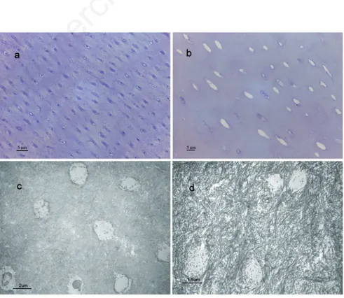
Structural and ultrastructural features and expression patterns of various collagenous and non-collagenous proteins in sound primary teeth and teeth from patients affected by DGI-I, associated with OI were assayed. Light microscopy showed paucity and irregularity of tubules in the dentin of DGI-I affected patients (Figure 1 a,b).
Ultrastructural examination showed the dentin of DGI-I affected patients with thick, sparse and a few lax curvy bundles of cross-striated collagen fibers, whereas uniform dense collagen pattern was recognizable in sound primary dentin (Figure 1 c,d). Table 1 summarizes the quantification of the labeling for the collagenous and non-collagenous
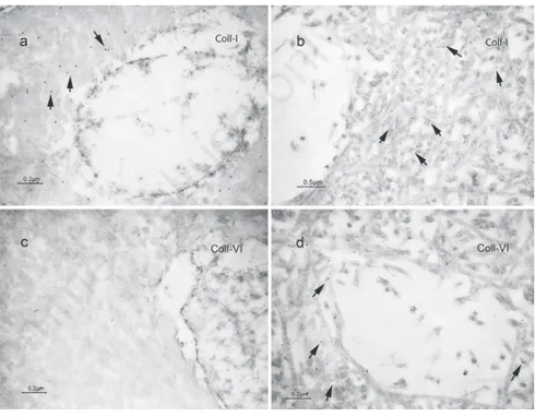
examined proteins. BGN, and CS showed a weaker reactivity in dentin from DGI-I affected patients compared to controls, being signifi-cant only for CS (Figure 2 a,b; Figure 3). Type I collagen was expressed both in sound and DGI-I affected dentin without showing signifi-cant differences. Conversely, type VI collagen labeling was significantly more detected in specimens from DGI-affected patients than in controls (Figure 2 c,d).
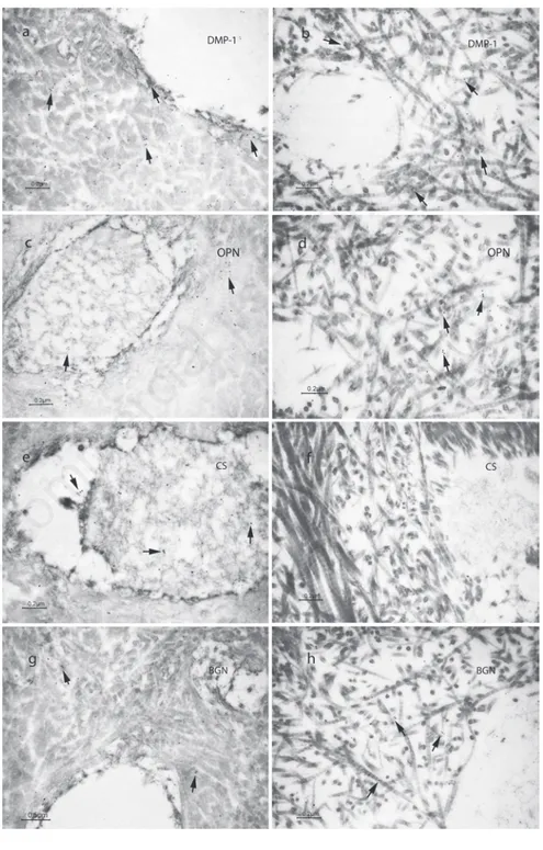
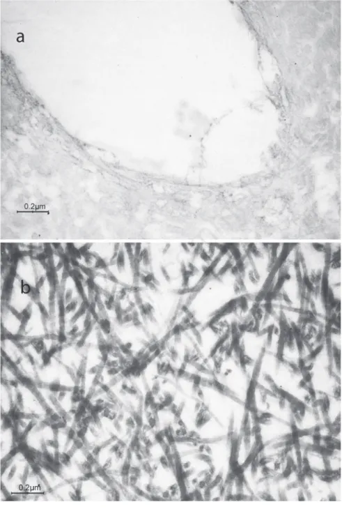
Regarding DMP1 and OPN expression, dentin from DGI-I affected patients showed pattern of loose collagen fibrils, labeled for both DMP1 and OPN, whereas control speci-mens exhibited a more uniform staining, although there was no differences in the count of gold particles for both DMP1 and OPN, being DMP1 generally more abundantly expressed in both sound primary teeth and DGI-I affected ones (Figure 3 a-d). CS was detected in normal dentin, mainly localized inside the dentin tubules (Figure 3e), where-as it wwhere-as slightly detected in the DGI-I affect-ed specimens (Figure 3f), as well as BGN, which was present in controls (Figure 3g) and less present in dentin specimens from DGI-I affected patients (Figure 3h). In all neg-ative controls either no labeling or a very few gold particles were observed over the tissue sections (Figure 4).
Original Paper
Figure 1. Light micrographs showing dentin of a) exfoliated sound primary teeth, pre-senting an uniform tubular structure and b) type I dentinogenesis imperfecta (DGI-I) affected teeth, presenting paucity and irregularity of tubules, toluidine blue staining. c,d) Transmission electron micrographs showing: c) normal dentin with uniform dense colla-gen pattern and tubular structure; d) DGI-I affected dentin characterized by a collacolla-gen fibrillar immature pattern not well arranged in lax curvy bundles.
Non
commercial
use
Discussion
The tested hypothesis that there is no corre-lation between the ultrastructural findings in dentin from DGI-I affected patients and defec-tive expression patterns of collagenous and NCPs is partially rejected, since ultrastructural abnormalities of the dentin matrix from DGI-I affected patients corresponded to a different expression of some of the examined proteins: in particular CS was less expressed and type VI collagen was more expressed in specimens from DGI-I affected patients than in sound pri-mary teeth, respectively. No significant differ-ence was detected in the labeling of type I col-lagen and the NCPs DMP 1, OPN and BGN between normal and DGI-I affected dentin. Our analysis focused on the primary teeth exfoliat-ed by DGI-I affectexfoliat-ed patients since the primary dentition is usually more affected by DGI than the permanent teeth.1 Interestingly, primary
dentin obtained by DGI-I affected patients was poor of dentinal tubules and showed dysplastic dentin, characterized by sparse and coarse col-lagen fibrils, which resemble more the pre-dentin than the pre-dentin typical features.10
As a dentin precursor, predentin is mineral-ized when apatite crystals are deposited within and around collagen fibrils.11 This process
requires mechanisms that control the site and rate of apatite formation. Although the mecha-nisms that control mineralization are largely unknown, NCPs in dentin have been proposed to be critical for this process during dentino-genesis.7,11-13We may speculate that the
abnor-mal distribution of NCPs within the dentin matrix we assayed in DGI-I affected patients may also affect the mineralization process, leading to the suboptimally mineralized areas that characterize DGI-I.14This lack of
mineral-ization has been previously characterized as bone fragility for patients affected by osteoge-nesis imperfecta and was also extensively reported as defective dentin calcification in teeth affected by DGI-I,15-18that wear more
eas-ily and excessively compared to normal teeth.14,19,20
It is well known that identifying and immunolabeling type I collagen fibrils within mineralized tissues is challenging due to the low accessibility and low antigenicity. The labeling protocol employed in this study can be considered highly selective in identifying alter-ations in the protein epitopes as antigenicity of collagen revealed by its specific binding to a monoclonal antibody provides evidence of con-servation of the epitope structure.4,21Epitopes
for type I collagen antibodies can be divided in relation to their ability to interact with the col-lagen structure in helical, central and
termi-nal. Anti-helical type I collagen antibody (such as the one used in this study) recognizes the epitope based on three-dimensional structure, that is strictly related to the intact triple helix. Indeed this type of antibody binds only the native form of collagen type I, while no labeling occurs with denatured collagen molecules.
In previous studies on sound and carious human dentin, anti-type I collagen monoclonal antibody was used to assay the preservation of the collagen structure.9,22-24Interestingly, in the
present study specimens harvested from DGI-I affected patients do not showed a reduced labeling for type I collagen compared to sound teeth. This labeling of the monoclonal
anti-Figure 2. Transmission electron micrographs showing immunolabeling for type I colla-gen (Coll-I) in dentin of: a) sound exfoliated primary teeth; b) type I dentinocolla-genesis imperfecta (DGI-I) affected teeth; c) immunolabeling for type VI collagen in sound dentin used as control; d) DGI-I affected dentin.
Table 1. Comparison of the number of gold particle-antibody complexes/ m2 after labeling for collagenous and non-collagenous proteins in dentin areas of sound primary teeth and teeth affected by type I dentinogenesis imperfecta.
Sound primary teeth DGI-I affected teeth P value (n=20) (n=20)
Collagenous proteins Type I collagen 8.53±2.47 10.44±4.92 0.13 Type VI collagen 4.68±0.15 11.83±3.56 <0.001* Non collagenous proteins
Chondroitin sulphate 8.98±1.81 1.58±0.61 <0.001* Dentin matrix protein 1 21.93±5.65 20.32±6.17 0.22 Osteopontin 6.48±1.77 7.73±2.59 0.083 Biglycan 6.34±0.94 2.30±0.31 0.071
DGI-I, type I dentinogenesis imperfecta. All values are expressed as means and standard deviations (SD). *Statistical difference at P<0.05.
Non
commercial
use
type I collagen (clone COL 1) seems not to be affected by the well-known mutations observed in DGI-I, involving either of the two genes encoding type I collagen.25
Whereas weak in normal teeth, staining for type VI collagen was moderately present in dentin from DGI-affected patients. These find-ings confirm a previous study in which posi-tive reactivity for collagen VI in the dentin of primary teeth affected by both type I DGI (asso-ciated with OI) and type II DGI.6Labeling of
type VI collagen in our specimens was similar to the one observed by Waltimo et al., which reported that the gold particles were often sin-gle, attached to non-striated material with a low electron density and also seen intratubu-larly.6Interestingly, the reported lack of
miner-alization of the dentin matrix obtained by patients affected by DGI-I14,19,20 is not clearly
supported by the labeling detected for the major NCPs proteins implicated in the miner-alization process such as DMP-1 and OPN (that were not significantly different between sound and affected primary teeth), whereas there were significant decrease only of CS expression in the DGI-I affected specimens compared to the normal dentin. Therefore, the main evidences are based on morphological changes and specific features that DGI-I affect-ed specimens present: collagen fibrils in sound dentin showed opaque cross-striations, while collagen fibrils in DGI-dentin showed clear cross-striations. Noteworthy is that the ent report for the first time quantify the pres-ence of some of the collagenous and NCPs present indentin. Indeed, there are no reports examining the role of NCPs in type I DGI, whereas, for instance, dentin sialophosphopro-tein (DSPP) has been previously investigat-ed.26A correlation between the defective
cleav-age of DSPP into DPP, in types II and III DGI, has been assayed whereas no clear correlation have been found between type I DGI and other enzymes cleaving the major NCPs proteins of dentin ECM.26A recent paper has pointed out
that many similarities exist between gene and protein structures of DSPP and DMP-1.27Both
DSPP and DMP-1 contribute to hard tissue mineralization and DMP1 plays a regulatory role in dentin mineralization and can also function as a signaling molecule.28In fact, a
study characterizing DMP-1 null mice, clearly demonstrated that DMP-1 is required for dentin mineralization and that DSPP can be directly or indirectly controlled by DMP-1.12,29
Moreover, Chaussain et al., investigated the potential cleavage of DMP-1 by MMP-2 to release biologically active peptides.30MMP-2 is
a predominant protease in the dentin matrix that plays a prominent role in tooth formation and extracellular matrix degradation.31DMP1,
both in the recombinant form and in its native state within the dentin matrix, was shown to
be a substrate for MMP-2.30Similarly, DMP-1
has been showed to be a possible substrate also for MMP-9.32Future studies may focus on
the expression of MMP-2 nor MMP-9 to clarify their role in the DGI-I disease and to further analyze the expression of the major NCPs and
correlated MMPs in carious tissues of DGI-I affected teeth.33,34
Small leucine-rich PGs, including chon-droitin 4/6 sulphate (CS)-rich decorin and BGN, have some important functional implica-tions in collagen assembly of predentin.9In
Original Paper
Figure 3. Transmission electron micrographs showing immunolabelig for: a) Dentin matrix protein 1 (DMP-1) in dentin of sound primary teeth used as controls; b) DMP-1 in dentin of type I dentinogenesis imperfecta (DGI-I) affected teeth; c) osteopontin (OPN) in sound dentin; d) OPN in DGI-I affected dentin; e) chondroitin sulphate (CS) in sound dentin; f ) CS in DGI-I affected dentin; g) biglycan (BGN) in sound dentin; h) BGN in DGI-I affected dentin.
Non
commercial
use
particular, the suggested role of BGN is to reg-ulate the growth and proliferation of mineral crystals and has been usually found more abundantly in predentin than in mineralized dentin, in which was generally localized in the mineralization front and within the dentinal tubules.8The weak labeling revealed in DGI-I
affected specimens for both CS and BGN (which was significantly weaker only for CS) may suggest that the defective unmineralized dentin does not present the same features of
normal predentin, in which increasing of BGN was expected. These data are in partial agree-ment with the study by Ye et al., in which DMP-1 null mice, characterized by dentin hypomin-eralization and expanded pulp cavities, showed no increasing of BGN production.12
In conclusion, although regulatory mecha-nisms for the expression of NCPs have not been fully elucidated, the present study correlates morphological alteration of dentin from patients affected by type I DGI with immunocytochemical
findings, helping to elucidate the ultrastructural changes observed in dentin defects (currently classified as distinct entities). This may be a con-tribution to understand the pathogenesis of differ-ent types of heritable ddiffer-entin defects as well as aid in early diagnosis of these diseases. Further stud-ies related to the complicated interactions between the extracellular matrix macromolecules of dentin obtained by DGI-I affected patients are needed to fully clarify biochemical alterations that lead to deficient dentin formation.
References
1. O’Connell AC, Marini JC. Evaluation of oral problems in an osteogenesis imper-fecta population. Oral Surg Oral Med Oral Pathol Oral Radiol Endod 1999;87:189-96. 2. Lukinmaa PL, Vaheri A. ED-A
region-con-taining isoform of cellular fibronectin is present in dentin matrix in dentinogenesis imperfecta associated with osteogenesis imperfecta. J Dent Res 1994;73:1187-96. 3. Shields ED, Bixler D, el-Kafrawy AM. A
pro-posed classification for heritable human dentine defects with a description of a new entity. Arch Oral Biol 1973;18:543-53. 4. Waltimo J, Ranta H, Lukinmaa PL.
Ultrastructure of dentin matrix in herita-ble dentin defects. Scanning Microsc 1995;9:185-97; discussion 197-8.
5. Ranta H, Lukinmaa PL, Waltimo J. Heritable dentin defects: nosology, pathol-ogy, and treatment. Am J Med Genet 1993;45:193-200.
6. Waltimo J, Risteli L, Risteli J, Lukinmaa PL. Altered collagen expression in human dentin: increased reactivity of type III and presence of type VI in dentinogenesis imperfecta, as revealed by immunoelec-tron microscopy. J Histochem Cytochem 1994;42:1593-601.
7. Orsini G, Ruggeri A, Mazzoni A, Nato F, Manzoli L, Putignano A, et al. A review of the nature, role and function of dentin non-collagenous proteins: Part 1: proteo-glycans and glycoproteins. Endodontic Topics 2012;21:1-18.
8. Orsini G, Ruggeri A, Jr., Mazzoni A, Papa V, Mazzotti G, Di Lenarda R, et al. Immunohistochemical identification of decorin and biglycan in human dentin: a correlative field emission scanning elec-tron microscopy/transmission elecelec-tron microscopy study. Calcif Tissue Int 2007; 81:39-45.
9. Orsini G, Ruggeri A Jr., Mazzoni A, Papa V, Piccirilli M, Falconi M, et al. Immunohistochemical identification of type I and type III collagen and chondroitin sulphate in human pre-dentine: a
correla-Figure 4. Representative images of the negative controls in dentin specimens of a) sound primary teeth; b) type I dentinogenesis imperfecta affected teeth.
Non
commercial
use
tive FEI-SEM/TEM study. Int Endod J 2007;40:669-78.
10. Kim JW, Simmer JP. Hereditary dentin defects. J Dent Res 2007;86:392-9. 11. Butler WT, Ritchie H. The nature and
func-tional significance of dentin extracellular matrix proteins. Int J Dev Biol 1995;39: 169-79.
12. Ye L, MacDougall M, Zhang S, Xie Y, Zhang J, Li Z, et al. Deletion of dentin matrix pro-tein-1 leads to a partial failure of matura-tion of predentin into dentin, hypomineral-ization, and expanded cavities of pulp and root canal during postnatal tooth develop-ment. J Biol Chem 2004;279:19141-8. 13. Ruggeri A, Orsini G, Mazzoni A, Nato F, Papa
V, Piccirilli M, et al. Immuno histochemical and biochemical assay of versican in human sound predentine/dentine matrix. Eur J Histochem 2009;53:125-33.
14. Tsai CL, Lin YT, Lin YT. Dentinogenesis imperfecta associated with osteogenesis imperfecta: report of two cases. Chang Gung Med J 2003;26:138-43.
15. Salvolini E, Di Giorgio R, Caselli E, De Florio L. [Dentinogenesis imperfecta. Scanning electron microscopic study and microanalysis].[Article in Italian]. Minerva Stomatol 1999;48:87-92. 16. Majorana A, Bardellini E, Brunelli PC,
Lacaita M, Cazzolla AP, Favia G. Dentinogenesis imperfecta in children with osteogenesis imperfecta: a clinical and ultrastructural study. Int J Paediatr Dent 2010;20:112-8.
17. Lukinmaa PL, Ranta H, Ranta K, Kaitila I. Dental findings in osteogenesis imperfec-ta: I. Occurrence and expression of type I dentinogenesis imperfecta. J Craniofac Genet Dev Biol 1987;7:115-25.
18. Kerebel B, Daculsi G, Menanteau J,
Kerebel LM. The inorganic phase in dentinogenesis imperfecta. J Dent Res 1981;60:1655-60.
19. Stephen LX, Beighton P. Dental manage-ment of severe dentinogenesis imperfecta in a mild form of osteogenesis imperfecta. J Clin Pediatr Dent 2002;26:131-6. 20. Rios D, Vieira AL, Tenuta LM, Machado
MA. Osteogenesis imperfecta and dentino-genesis imperfecta: associated disorders. Quintessence Int 2005;36:695-701. 21. Willingham MC. Conditional epitopes. is
your antibody always specific? J Histochem Cytochem 1999;47:1233-6. 22. Breschi L, Gobbi P, Mazzotti G, Falconi M,
Ellis TH, Stangel I. High resolution SEM evaluation of dentin etched with maleic and citric acid. Dent Mater 2002;18:26-35. 23. Breschi L, Perdigao J, Gobbi P, Mazzotti G, Falconi M, Lopes M. Immunocytochemical identification of type I collagen in acid-etched dentin. J Biomed Mater Res A 2003;66:764-9.
24. Suppa P, Ruggeri A, Tay FR, Prati C, Biasotto M, Falconi M et al. Reduced anti-genicity of type I collagen and proteogly-cans in sclerotic dentin. J Dent Res 2006;85:133-7.
25. Barron MJ, McDonnell ST, Mackie I, Dixon MJ. Hereditary dentine disorders: dentino-genesis imperfecta and dentine dysplasia. Orphanet J Rare Dis 2008;3:31.
26. Tsuchiya S, Simmer JP, Hu JC, Richardson AS, Yamakoshi F, Yamakoshi Y. Astacin proteases cleave dentin sialophosphopro-tein (Dspp) to generate dentin phospho-protein (Dpp). J Bone Miner Res 2011;26:220-8.
27. Suzuki S, Haruyama N, Nishimura F, Kulkarni AB. Dentin sialophosphoprotein and dentin matrix protein-1: Two highly
phosphorylated proteins in mineralized tissues. Arch Oral Biol 2012;57:1165-75. 28. Orsini G, Ruggeri A, Mazzoni A, Nato F,
Falconi M, Putignano A, et al. Immuno -histochemical localization of dentin matrix protein 1 in human dentin. Eur J Histochem 2008;52:215-20.
29. Teti G, Salvatore V, Ruggeri A, Manzoli L, Gesi M, Orsini G, et al. In vitro reparative dentin: a biochemical and morphological study. Eur J Histochem 2013;57:e23. 30. Chaussain C, Eapen AS, Huet E, Floris C,
Ravindran S, Hao J, et al. MMP2-cleavage of DMP1 generates a bioactive peptide promoting differentiation of dental pulp stem/progenitor cell. Eur Cell Mater 2009; 18:84-95.
31. Orsini G, Mazzoni A, Orciani M, Putignano A, Procaccini M, Falconi M, et al. Matrix metalloproteinase-2 expression induced by two different adhesive systems on human pulp fibroblasts. J Endod 2011; 37:1663-7.
32. Karadag A, Fedarko NS, Fisher LW. Dentin matrix protein 1 enhances invasion poten-tial of colon cancer cells by bridging matrix metalloproteinase-9 to integrins and CD44. Cancer Res 2005;65:11545-52. 33. Martini D, Trirè A, Breschi L, Mazzoni A,
Orsini G, Teti G, et al. Dentin matrix pro-tein 1 and dentin sialophosphopropro-tein in human sound and carious teeth: an immunohistochemical and colorimetric assay. Eur J Histochem 2013;57:e32. 34. Loreto C, Galanti C, Musumeci G, Rusu
MC, Leonardi R. Immunohistochemical analysis of matrix metalloproteinase-13 in human caries dentin. Eur J Histochem 2014;58:2318.