UNIVERSITY OF SIENA
DEPARTMENT OF MEDICAL BIOTECHNOLOGIES
PhD PROGRAM IN MEDICAL BIOTECHNOLOGY
SECTION OF MICROBIOLOGY AND VACCINE
XXXIII CYCLE
IDENTIFICATION OF NEUTRALIZING EPITOPES
OF TOSV: A MOUSE MODEL
Supervisor: Prof.ssa Maria Grazia Cusi Student: Shibily Prathyumnan
2021
ACKNOWLEDGEMENT
Undertaking this PhD has been a truly life-changing experience for me and it would not have been possible to do without the support and guidance that I received from many people.
Firstly, I would like to express my sincerest gratitude to my supervisor Prof.ssa Maria Grazia Cusi for the continuous support of my PhD study and related research, for her patience, motivation, and immense knowledge. Her guidance helped me in all the time of research and writing of this thesis. I appreciate her contributions of time, ideas, and support to make this thesis productive and stimulating. I could not have imagined having a better advisor and mentor for my PhD study.
I thank my fellow lab mates of Molecular Virology Lab, University of Siena, especially Dr. Gianni Gori Savellini, Dr.ssa Claudia Gandolfo, Dr.ssa Chiara Terrosi and Dr.Gabriele Anichini for the stimulating discussions, for the most cherished time we were working together before deadlines, and for all the fun we have had.
A special word of gratitude to my dearest friend Dr.ssa Valeria Fox, LAMMB, University of Siena, for her relentless encouragement and the quality time she spent with me.
I owe a big hug to my queen, Dr.ssa Karthika Sundaran Shibily for being my side throughout this course and living every minute of it, without whom I would not have the courage to embark on this journey in first place. A kiss to my little prince, Aadi for being such a good little boy and making it possible for me to complete what I started.
My greatest thanks and love to my parents, Dr. Prathyumnan and Mrs. Girija Kumari and also my in-laws, Mr. Sundaresan and Mrs. Remani for their endless support. I could not have reached this far without their prayers and sacrifice. I am forever grateful and indebted for their kindness and generosity.
As everything begins and ends in GOD, I conclude this acknowledgement by thanking HIM for eternal love, care and blessings.
CONTENTS
Abstract 1 Chapter 1 1.1 Arbovirus 5 1.2 Bunyavirales 8 1.3 Phenuiviridae 13 1.4 Toscana Virus 20 1.4.1 Epidemiology 211.4.2 Clinical manifestations of TOSV infection 24
1.4.3 TOSV infection and role of glycoproteins 27
Chapter 2
Aim of the thesis 30
Chapter 3
Materials and Methods
3.1 Cells and Viruses 33
3.2 Human PBMC Isolation 33
3.3 Plasmid Construction 33
3.4 Plasmid Transformation 35
3.5 Protein Purification from Inclusion Bodies 35
3.6 Western Blot 36
3.7 Animal Studies 36
3.8 Enzyme Linked Immunosorbent Assay (ELISA) 38
3.9 Neutralization Test 38
3.10 Indirect Immunofluorescence Assay 39
3.12 Immortalization of PBMC 39
3.13 Statistical Analysis 40
Chapter 4 Results
4.1 Expression and Purification of GC683 and GC727 42
4.2 Humoral Immune response elicited to the peptides in vivo 44
4.3 Serological cross reactivity of immunized mice with other Phleboviruses 46
Chapter 5
Addendum
Modified protocol for the generation of monoclonal antibodies
against TOSV NSs 50 Chapter 6 Conclusion 56 Chapter 7 Bibliography 60 Chapter 8 Annexures 82
1
Emerging and re-emerging viral infections have been an important public health problem in recent years. The Bunyavirales order is one of the largest groups of segmented negative-sense single-stranded RNA viruses, which includes many pathogenic strains that cause severe human diseases. Toscana virus (TOSV), a Phlebovirus belonging to the Phenuiviridae family, is considered an emergent pathogen associated with acute neurological disease, such as meningitis, meningoencephalitis and encephalitis, occurring in the Mediterranean countries (principally Italy, Spain, France) during the summer months (Valassina et al., 2003 a; Valassina et al., 2003 b; Charrel et al., 2005; Sanbonmatsu –Gàmez et al., 2009; Cusi et al., 2010).
The mechanisms of protection against natural infection of Phlebovirus are not known, however it is supposed that a virus neutralizing antibody response against the viral glycoproteins could be needed to block the first stages of infection. Neutralizing antibody responses are critical components of the host defense against viral infections and are recognized as a key element in the protective immune response against infection elicited by many prophylactic vaccines (Burton et al., 2012; Corti et al., 2013). In this setting, we focused our attention on TOSV glycoproteins (Gn and Gc). In a previous work, Prof. Cusi's group was able to localize three neutralizing epitopes on Gn glycoprotein using human mAbs obtained by immortalization of B cells from a subject infected with TOSV. It was postulated that these 3 aminoacid sequences, separated in the primary structure of the protein, had probably a neutralizing activity in the tridimensional structure, in which peptides 1- 2 could be part of a conformational epitope without peptide 3; which instead, is necessary to
2
strengthen the neutralizing activity and hinder the virus replication, by blocking the binding of Gn to the receptor.
The present study was aimed to confirm in vivo the immunogenic efficacy of these three epitopes in various combinations in mouse model and to test if the mice serum obtained show any cross reactivity with other members of Phlebovirus (such as SFNV, PUNV, SNVF, CYPR) and eventually, to evaluate if these cross-reactive sera are also neutralizing against other Phlebovirus. Moreover, these results could be used to design epitopes that can serve as potential targets for the production of epitope-based diagnostics and vaccines against TOSV and other related Phlebovirus.
The study also developed monoclonal antibodies against TOSV NSs protein by immortalization of human B cells by a modified methodology. These monoclonals could be used for better understanding the role of NSs in viral infection of the host and eventually in passive immunization.
4
1.1 Arbovirus
Newly emerging or re-emerging infections continue to pose significant global public health threats. Arthropod-borne pathogens account more than 17% of infectious diseases, affect millions of people around the world each year, causing over 700,000 deaths annually and comprise a significant proportion of emerging human pathogens (Jones et al., 2008; Woolhouse et al., 2008; Rosenberg et al., 2013; WHO Fact Sheet 2020).
A high proportion of arboviruses associated with human and animal disease circulate in tropical and subtropical regions, where arthropods tend to be abundant. However, many arboviruses also circulate among wildlife species in temperate regions of the world. Despite the global distribution of viruses such as West Nile virus (WNV), Dengue Virus (DENV) and Chikungunya Virus (CHIKV), most of the arboviruses are generally endemic but limited to specific regions of the world. Nevertheless, even within this relatively localized distribution, diffusion to distant locations can occur via animal or vector migration (Liang et al., 2015).
Arboviruses already have a well-known history of emergence and will undoubtedly continue to emerge in the future. There are many unidentified arboviruses that, due to their high mutation rates, may emerge as pathogens although they are not present as epidemic strains in the wild environment (Marchi et al.,2018) yet. Considering the history of emergence of some arboviruses, these epidemics have occurred globally as a result of climate and socio-economic changes that have allowed the spread to new geographical areas of viruses previously confined to specific ecological niches. Usually, their dissemination is linked to the natural geographic distribution of the vector, nevertheless climate change and the increasing globalization and habitat modifications, are already resulting in changing
5
epidemiology of a variety of arboviruses and facilitate their spreading to new geographic locations (Whitehorn et al.,2019). Through spillover transmission from enzootic amplification cycles, humans can be infected as incidental and dead-end hosts. By contrast, some arboviruses undergo urban cycle involving humans as amplifying hosts and causing several epidemics in urban areas (Weaver et al., 2010; Coffey et al., 2013; Agarwal et al., 2017).
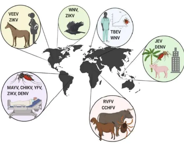
Figure 1: Arbovirus Outbreak. Venezuelan equine encephalitis virus (VEEV), Zika virus (ZIKV), West Nile virus (WNV), tick-borne encephalitis virus (TBEV), Japanese encephalitis virus (JEV), dengue virus (DENV), Rift Valley fever virus (RVFV), Crimean-Congo hemorrhagic fever virus (CCHV), Mayaro virus (MAYV), Chikungunya virus (CHIKV), and Yellow fever virus (YFV).
6
The majority of arboviruses circulate in nature between an amplifying vertebrate host and a vector arthropod without causing harm to either of them. More than 100 arboviruses are known to infect humans and over 40 to infect domestic animals. In general, these represent ‘dead-end infections’ in unnatural vertebrate hosts. Examples of important human pathogens include the four dengue viruses (dengue virus 1–4), West Nile virus, Rift Valley fever, Yellow fever virus, and Japanese encephalitis virus. Nairobi sheep disease, Venezuelan equine encephalitis, and Bluetongue virus are examples of veterinary diseases caused by infection with arboviruses. With the single exception of African swine fever virus, arboviruses have RNA genomes consisting of linear or segmented, positive-sense or negative-sense, and single- or double-stranded molecules. (Miller, 2008).
Although some arboviral infections are asymptomatic or cause mild influenza-like illness, many arboviruses are important human and veterinary pathogens causing serious illness ranging from rash and arthritis to encephalitis and hemorrhagic fever (Hollidge et al., 2010). Arthropod-borne viruses (arboviruses) are transmitted biologically among vertebrate hosts by hematophagous (blood feeding) arthropod vectors such as mosquitoes and other biting flies, and ticks. Biological transmission can be vertical, involving the passage of the virus from an infected female vector to both male and female offspring. Horizontal transmission can be from a vector to a vertebrate host via the saliva during blood feeding. This transmission is the most common for the majority of arboviruses and involves infection of the vector alimentary tract following a viremic blood meal, dissemination of the virus in the vector, and eventual virus replication in the salivary glands (Serena et al., 2018). Most of the arboviruses that cause human/animal diseases belong to four virus families: Togaviridae (genus Alphavirus), Flaviviridae (genus Flavivirus), Bunyaviridae (genera Orthobunyavirus, Phlebovirus and Nairovirus) and Reoviridae (genera Coltivirus and Orbivirus) (Agarwal et al., 2017; Powers 2009). In 2016, the family of Bunyaviridae was changed to Bunyavirales order (Virus Taxonomy: 2016 Release, EC 48, Budapest, Hungary, August 2016; Virus Taxonomy) to accommodate related viruses with segmented, linear, single-stranded,
7
negative-sense or ambisense RNA genomes and classified into twelve families (ICTV 2019 release, https://talk.ictvonline.org/taxonomy/).
1.2 Bunyavirales
The Bunyavirales order is one of the largest groups of segmented negative-sense single-stranded RNA viruses, which includes many pathogenic strains that cause severe human diseases. It is subdivided into 12 families: Arenaviridae, Cruliviridae,
Fimoviridae, Hantaviridae, Leisbuviridae, Mypoviridae, Nairoviridae,
Peribunyaviridae, Phasmaviridae, Phenuiviridae, Tospoviridae and Wupedeviridae comprising of 46 genera. Four families contain members that cause life-threatening diseases in humans: Hantaviridae, Nairoviridae, Peribunyaviridae and Phenuiviridae (Wichgers et al., 2018; Abudurexiti et al., 2019). These families include the species Bunyamwera virus (BUNV), Crimean-Congo haemorrhagic fever virus (CCHFV), Hantaan virus (HTNV), La Crosse virus (LACV), Rift Valley fever virus (RVFV), Severe fever with thrombocytopenia syndrome virus (SFTSV), and Sin Nombre virus (SNV). Three families within Bunyavirales contain members that infect plants as their primary host: Fimoviridae, Phenuiviridae, and Tospoviridae. Within these families, there is one genus of plant infecting viruses: Emaravirus, Tenuivirus, and Orthotospovirus respectively (Herath et al., 2020). Most bunyaviruses are transmitted by insects or ticks, with the exception of members from the family Hantaviridae that are entirely rodent borne (Zhang, 2014). Viruses in the order Bunyavirales infect arthropods, plants, protozoans, and vertebrates. Their RNA genomes are segmented and exhibit negative or ambisense polarity. Depending on the family and genus, bunyaviruses (now referring to all members of the Bunyavirales) has a fixed number of genome segments which range from two to eight, with plant viruses having the largest numbers of segments. (Herath et al., 2020).
The viral particles have a diameter of 80 to 120 nm and are composed of helicoidal nucleocapsids containing three RNA segments that code for a minimum of four structural proteins in a negative-sense orientation named small (S), medium (M) and
8
large (L), reflecting their relative nucleotide length. Viruses within each genus share similar overall segment length and a generally common expression strategy for their encoded protein products (Schmaljohn & Nichol, 2006). The genetic organization of the segments is similar across all genera; each template strand further contains conserved complementary oligonucleotide sequences at their 5’- and 3’-ends (NTRs), allowing the formation of “panhandle” structures and noncovalently closed circular RNAs (Schmaljohn & Elliott 2014). These terminal nucleotide sequences, which surround a single transcriptional unit, are highly conserved among viruses within a genus, but differ from those of viruses in other genera. These structures seem to be important for transcription mechanisms and genome replication or for genome packaging and for virion assembly (Valassina et al.,2003). There are differences in the patterns of sizes of the viral RNAs and structural proteins between the different genera, and the expression strategy of non-structural proteins also differs between different genera. (Elliot et al., 2014).
The coding regions of each genome segment lie between terminal non-translated sequences that vary in length. The 3’ and 5’ genomic RNA termini are essential for RNA synthesis and are typically invariant.
A major difference between bunyaviruses from other enveloped viruses is that the virions are devoid of any classical matrix or capsid protein. The matrix function is performed by the cytoplasmic tail of Gn, one of the two glycoproteins, that not only contains a Golgi localization signal but is also involved in the initiation of the budding process and the packaging of ribonucleoproteins (RNPs) into virus particles (Spiegel et al., 2016).
9
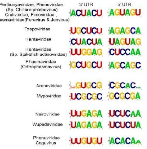
Figure 2: Consensus nucleotide sequence of the 3’ and 5’ termini for each genomic segment of Bunyaviriales.
The S segment (1869 nucleotides) of all bunyaviruses encodes the nucleocapsid (N) protein, whose primary role is to encapsidate the viral RNA-replication products and forms the ribonucleoprotein (RNP) complex. The N protein is the most abundant viral product in virions and infected cells. The S segments of most members of the genera Orthobunyavirus, Tospovirus and Phlebovirus also encode a non-structural protein called NSs, that shows a low conservation across the Phlebovirus genus compared to other viral proteins, with sequence similarities ranging only from approximately 10% to 30% (Bichaud et al., 2016; Yu et al., 2011). The NSs protein is an important virulence determinant, acting as an inhibitor of the antiviral type I IFN system of the mammalian host (Boshra et al., 2011; Weber et al., 2014 a; Weber et al.,2014 b). Hantaviruses and Nairoviruses use the negative-sense coding RNA to
10
express a single protein N from their virion complementary-sense RNA (cRNA) (Schmaljohn & Hooper 2001).
The N and NSs proteins of Orthobunyaviruses are translated from the same mRNA encoded by the S segment genome, whereas the Phlebovirus and Tospovirus N and NSs proteins are translated from separate mRNAs transcribed from the genomic and antigenomic strands, respectively (ambisense gene expression) (Walter & Barr 2011). In particular, a viral complementary, subgenomic mRNA corresponding to the 3’ half of the viral S RNA codes for the N protein, and the viral subgenomic mRNA corresponding to the 5’ half of the viral S RNA codes for the NSs proteins. Among phleboviruses, there is a short intergenic region (IR), a sequence stretch that is proposed to form an irregular double-stranded RNA (dsRNA) structure in the region between the two ORFs (Boshra et al., 2011). Formation of the RNP (ribonucleoprotein) depends on the association of viral RNA with multiple copies of the N protein so that the RNA becomes encapsidated along its entire length (Walter & Barr 2011). Whilst the primary role of the N protein is to provide structural uniformity to the RNA genome, the additional roles include interaction with membrane glycoproteins (Hepojoki et al., 2010; Overby et al., 2007; Ribeiro et al.,2009; Shi et al., 2007; Snippe et al., 2007) and interaction with the viral RNA dependent RNA polymerase (RdRp) to allow access to the RNP during RNA synthesis (Walter & Barr 2011).
The M segment (ranging from 3,600 to 4,900 nucleotides) of all members of the Phenuiviridae family encodes, in a single open reading frame (ORF) of cRNA, a polyprotein precursor, that is processed into the glycoproteins Gn and Gc (referring to the amino-terminal or carboxy-terminal coding of the proteins) (Walter & Barr 2011).The Gn/Gc precursor is translocated, due to a signal sequence preceding Gn, from the cytoplasm into the membrane of the endoplasmic reticulum (ER), where it is cleaved into Gn and Gc components by host-cell proteases (Schmaljohn & Elliott 2014). In the ER, Gn and Gc are glycosylated with N-linked glycans (Kuismanen et al., 1984;Matsuoka et al., 1988) of the high-mannose type, which can be processed
11
into hybrid and complex forms upon import of the two glycoproteins into the Golgi apparatus (Kuismanen et al., 1984; Matsuoka et al., 1988; Madoff et al., 1982; Shi et al., 2005). By virtue of a retention signal, the heavily glycosylated Gn– Gc heterodimer is retained in the Golgi, where it associates with RNPs to mediate assembly and budding of mature virus particles, that are released from infected cells by exocytosis. The Gn–Gc heterodimer performs critical roles in mediating virus assembly, formation of the virus particle and attachment to new target cells (Schmaljohn & Nichol 2006).
Most members of the genera Orthobunyavirus, Phlebovirus and Tospovirus also encode an NSm protein. The Tospovirus NSm protein is translated from a separate mRNA encoded by the antigenome, whereas the Orthobunyavirus and Phlebovirus NSm is cleaved by cellular proteases from the same polyprotein precursor that yields the Gn and Gc proteins (Walter & Barr 2011). Bunyavirus NSm protein is thought to play a role in virus assembly (Shi et al., 2007). In contrast, the Phlebovirus NSm has been shown to be non-essential (Gerrard et al., 2007), but may play accessory roles in the regulation of apoptosis (Won et al., 2007). The M segment of Tospoviruses uses an ambisense coding strategy. The cRNA has a gene order of 5'-Gn-Gc-3', and the vRNA encodes NSm. There are separate, subgenomic messages for the Gc–Gn precursor and for NSm (Law et al., 1992; Kormelink et al., 1992). Hantaviruses have an M segment cRNA gene order of 5'-Gc-Gn-3' (Schmaljohn et al., 1987), while a gene order of 5'-Gn-Gc-3' was determined for the Nairovirus (Marriott et al., 1992).
The M segment gene order and the coding strategy varies among viruses in the Phlebovirus genus (Schmaljohn & Elliott 2014). For Rift Valley fever virus (RVFV) and Toscana virus (TOSV), the cRNA gene order of M fragment is 5'-NSm-Gn-Gc-3' (Collett et al., 1985; Valassina et al., 2003). In general, M-segment gene products have a cysteine content of approximately 4% to 7%. Positions of these residues are highly conserved in the M-segment products of related viruses, suggesting that the positions may be crucial for determining correct polypeptide folding. In the case of TOSV, there are nine sites of glycosylation (Valassina et al., 2003). All M-segment
12
polyproteins display variable numbers of predicted transmembrane regions, and a hydrophobic sequence at the carboxy-terminus, indicative of a membrane anchor region. Therefore, the M segment translation products of viruses in the Phenuiviridae family are typical class I membrane proteins, with the amino terminus exposed on the surface of the virion and the carboxy-terminus anchored in the membrane (Schmaljohn & Elliott, 2014).
The L segment (range in size from about 237kD for Phleboviruses, to 459kD for Nairoviruses) contains a single open reading frame (ORF) that encodes the viral polymerase. The L ORF is expressed via a viral-complementary mRNA (Valassina M. et al., 2003). All L segments of viruses of the family use conventional negative sense coding strategies (Schmaljohn & Elliott 2014). Bunyavirus L proteins perform several complex functions that together result in the generation of RNA-replication and mRNA-transcription products from their respective viral templates (Walter & Barr 2011). Primary transcription of negative-sense viral RNA to mRNA is initiated by interaction of the virion-associated polymerase (L) and the three viral RNA templates (Bouloy & Hannoun 1976; Ranki & Pettersson 1975). Members of the Phenuiviridae family use primers cleaved from cytoplasmic host-cell mRNAs for extension. Cleavage of the capped primers is accomplished by endonucleolytic activity associated with virions (Patterson JL. et al., 1984). Alignments with homologues of other members of the Phenuiviridae family indicate that L protein- associated endonuclease domain is well-conserved, containing metal-binding and catalytic lysine residues, and shows a high degree of sequence similarity to putative endonuclease motifs of L proteins from other segmented, negative-stranded RNA viruses such as Arenaviruses, Emaraviruses and Tenuiviruses (Walter & Barr 2011). 1.3 Phenuiviridae
Phenuiviridae is a virus family belonging to the order Bunyavirales established in 2016. Members of Phenuiviridae are enveloped viruses with helical capsid morphology. Envelope glycoproteins of these viruses are distributed with
icosahedral symmetry (T=12)
13
negative single stranded RNA (-ssRNA) virus family
[https://www.uniprot.org/taxonomy/1980418]. Phenuiviridae family members include human and animal pathogenic viruses transmitted by arthropod vectors, including phlebotomine sandflies, mosquitos and ticks (Matsuno et al., 2015; Palacios et al., 2013). They can cause a variety of clinical syndromes ranging from a brief, self-limiting febrile illness, to retinitis, encephalitis, meningoencephalitis and fatal haemorrhagic fever (Liu et al., 2003).
Replication begins with the attachment of viral proteins to host receptors and entry of virus by endocytosis. Acidification of endocytic vesicles causes uncoating of the virus which is followed by fusion of viral membranes with that of the host endosomes. Primary transcription of viral complementary mRNA occurs and the L, M and S mRNAs are translated. Transcription starts by viral RNA dependent RNA polymerase (L) binding to a promoter on each encapsidated segment, and is terminated by a strong hairpin sequence at the end of each gene. These are capped by L protein during synthesis using cap snatching, but are not polyadenylated. S- segment uses ambisense strategy to encode for several proteins: both genomic and antigenomic RNA are transcribed. The hairpin sequence is a stop polymerase signal which prevents ambisense transcription from producing dsRNA. M segment encodes for several polyproteins by leaky scanning, which are cleaved by host protease into Nsm, Gn and Gc proteins. This is followed by RNA replication and morphogenesis. Mature virions are released by fusion of virus-containing cytoplasmic vesicles with the plasma membrane and budding. (David et al.,2007;
14
Figure:3 Structure of a Phenuiviridae virion (https://viralzone.expasy.org)
Figure:4 Replicative cycle of a Phenuiviridae virion (https://viralzone.expasy.org)
15
Table:1 Taxonomical Classification of Phenuiviridae family Family Phenuiviridae
Genus Species Notable Viruses
Banyangvirus
Guertubanyangvirus Guertu virus (GTV) Heartland banyangvirus Heartland virus (HRTV)
Huaiyangshanbanyangvirus Severe Fever with Thrombocytopenia Syndrome Virus (SFTSV)
Beidivirus Dipteran beidivirus Húběidiptera virus 3 (HbDV-3)
Goukovirus
Cumutogoukovirus Cumuto virus (CUMV)
Gouleakogoukovirus Gouléako virus (GOLV)
Yichang insect goukovirus Yíchāng insect virus (YcIV)
Horwuvirus Horsefly horwuvirus Wǔhàn horsefly virus (WhHV)
Hudivirus Dipteran hudivirus Húběidiptera virus 4 (HbDV-4)
Hudovirus Lepidopteran hudovirus Húběi lepidoptera virus 1 (HbLV-1)
Kabutovirus Huangpikabutovirus Huángpí tick virus 2 (HpTV-2) Kabuto mountain kabutovirus Kabuto mountain virus (KAMV) Laulavirus Laurel Lake laulavirus Laurel Lake virus (LLV)
Mobuvirus Mothramobuvirus Mothra virus (MTHV)
Phasivirus
Badu phasivirus Badu virus (BADUV)
Phasi Charoen-like phasivirus PhasiChaeron-like virus (PCLV) Wutai mosquito phasivirus Wǔtái mosquito virus (WtMV)
Phlebovirus
Bujaruphlebovirus Bujaru virus (BUJV) Munguba virus (MUNV) Candiru phlebovirus
Alenquer virus (ALEV) Ariquemes virus (ARQV) Candirú virus (CDUV)
16
Itaituba virus (ITAV) Jacundá virus (JCNV) Maldonado virus (MLOV) Morumbi virus (MR(M)BV) Mucura virus (MCRV/MRAV) Nique virus (NIQV)
Oriximiná virus (ORXV) Serra Norte virus (SRNV) Turuna virus (TUAV) Chilibrephlebovirus Cacao virus (CACV)
Chilibre virus (CHIV) Frijoles phlebovirus Frijoles virus (FRIV)
Joá virus (JOAV)
Mukawaphlebovirus Mukawa virus (MKWV)
Punta Toro phlebovirus
Buenaventura virus (BUEV) Campana virus (CMAV) Capira virus (CAPIV) Coclé virus (CCLV) Leticia virus (LTCV) Punta Toro virus (PTV)
Rift Valley fever phlebovirus Rift Valley fever virus (RVFV)
Salehabadphlebovirus
Adana virus (ADAV) Adria virus (ADRV) Alcube virus
Arbia virus (ARBV) Arumowot virus (AMTV)
17
Bregalaka virus (BREV) Medjerda Valley virus (MVV) Odrénisrou virus (ODRV) Olbia virus (OLBV) Salehabad virus (SALV) Zaba virus (ZABAV)
Sandfly fever Naples phlebovirus
Arrábida virus (ARRV) Balkan virus (BALKV) Fermo virus (FERV) Gordil virus (GORV)
Granada virus (GRV = GRAV) Massilia virus (MASV)
Punique virus (PUNV) Saddaguia virus (SADV) Saint-Floris virus (SAFV)
Sandfly fever Naples virus (SFNV) Tehran virus (THEV)
Toscana virus (TOSV) Zerdali virus (ZERV)
Uukuniemi phlebovirus
Chizé virus (CHZV)
EgAN 1825–61 virus (EGAV) Fin V 707 virus (FINV)
Oceanside virus (OCV = OCEV) Pontevès virus (PTVV)
St. Abbs Head virus (SAHV) Uukuniemi virus (UUKV)
18
ZalivTerpenyia virus (ZTV)
Pidchovirus Pidgeypidchovirus Pidgey virus (PGYV)
Tenuivirus
Echinochloa hoja blancatenuivirus Echinochloa hoja blanca virus (EHBV) Iranian wheat stripe tenuivirus Iranian wheat stripe virus (IWSV)
Maize stripe tenuivirus Maize stripe virus (MStV = MSpV) Rice grassy stunt tenuivirus Rice grassy stunt virus (RGSV)
Rice hoja blancatenuivirus Rice hoja blanca virus (RHBV)
Rice stripe tenuivirus Rice stripe virus (RSV = RStV)
Urochloa hoja blancatenuivirus Urochloa hoja blanca virus (UHBV)
Wenrivirus Shrimp wenrivirus Wēnzhōu shrimp virus 1 WzSV-1
19
1.4 Toscana Virus
Within the Phenuiviridae family of Bunyavirales order, Phlebovirus genus contains more than 80 viruses, with about half classified into nine antigenic complexes that are regarded as species, whereas 33 are considered as tentative species in the genus. The viruses of the genus Phlebovirus are present throughout the world and their name is associated with their principal vector, phlebotomine sandflies, however there are prominent exceptions, such as RVFV which is primarily associated with Aedes species mosquitoes and Uukuniemi virus (UUKV), which is associated with the tick Ixodesricinus (Schmaljohn& Elliott 2014).
Among sandfly-transmitted viruses, three serotypes have been identified: Naples, Sicilian and Toscana serocomplex. Naples and Sicilian serotypes have the widest geographical distribution which is related to the distribution of the vector (P. pappatasi) and they have been isolated from sandflies in Africa, central Asia, the Americas and Europe.
Toscana Virus (TOSV) was first isolated in 1971 in central Italy (in Monte Argentario, Grosseto Province) from two different species of sandflies, Phlebotomus perniciosus (P. perniciosus) and Phlebotomus perfiliewi (P. perfiliewi) (Verani et al., 1982; Verani et al., 1984; Verani et al.,1988); and it was registered in the International Catalogue of Arbovirus, belonging to the Phlebovirus genus, in 1980. Other strains of Toscana virus were isolated from P. perfiliewi, in other parts of Italy where the insect vectors were present (Verani et al., 1982, 1984a, 1988; Ciufolini et al., 1985). The virus can be transmitted transovarially in the insect vectors, but its animal reservoir has not been identified yet. TOSV isolation from the brain of a bat (Pipistrellus kuhli) has been the only evidence of the possible involvement of this species in the ecology of the virus (Verani et al., 1982, Verani et al., 1984, Ciufolini et al., 1985). However, serological investigations have shown that anti-TOSV specific IgG has been detected in some
20
animal species, indicating that this virus can also infects animals (Ciluna et al., 2007).
TOSV belongs to the Naples serocomplex and is the only virus associated with neuroinvasive infections.
1.4.1 Epidemiology
TOSV is widespread in the Mediterranean basin, and evidence of human infection has been found in Italy, France, Spain, Portugal, Cyprus, and Turkey. Phylogenetic analysis has distinguished two genotypes of TOSV, A and B; the first is circulating mainly in Italy, France, Algeria, Tunisia, Turkey, and the second in Spain, France, Portugal, Morocco, Turkey indicating a different geographic distribution possibly related to the vector. A third lineage, C, has been recently added and identified in a strain circulating in Croatia and Greece (Punda-Polić et al., 2012). As already seen for other arbo-bunyavirus, the diffusion of TOSV is expanding with the geographic distribution of its vector, leading to a higher pathogenicity in non-exposed populations of areas outside the current prevalence (Mediterranean basin) (Collao et al.,2009). This distribution, evolving with the climate, globalization and habitat modification, has implications for the epidemiology of TOSV.
TOSV represents an important emerging pathogen in Europe and Northern Africa (Charrel et al., 2012).The first case of TOSV infection reported from Spain occurred in a Swedish tourist after a visit to Catalonia and was documented by plaque reduction neutralization test (PRNT) (Eitrem et al., 1991), whereas the presence of the virus in Portugal was suspected on the basis of a strain isolated from the cerebrospinal fluid (CSF) of a Swedish patient who was returning to his home country from Portugal (Ehrnst et al., 1985). A large epidemiological study conducted in the province of Granada by a Spanish group (Sanbonmastu et al., 2005) established TOSV as one of the most important etiological agents of CNS diseases. The first case of TOSV infection acquired in France was reported in a German traveler who was returning from southern France (Dobler et al., 1997). An
21
interesting study on seroprevalence was conducted in volunteer blood donors in France, showing that 12% of sera from healthy donors and 18.9% of sera from patients hospitalized for CNS infection were IgG positive for TOSV, confirming that TOSV circulates in southeastern France and that a significant proportion of healthy blood donors has a history of TOSV infection (De Lamballerie et al., 2007). Seroconversion to TOSV was observed in Swedish United Nations soldiers based in Cyprus without any clinical manifestations (Eitrem et al., 1990). Similarly, populations living on the Ionian Islands and western mainland of Greece also showed a seroprevalence of 60% and 35% respectively (Charrel et al., 2005). Although the seroprevalence of antibodies to TOSV was found to be low in Germany, TOSV infection could be considered in patients returning from virus-endemic areas who had fever and headaches or symptoms of meningitis (Schwarz et al., 1995). Some infections imported into Germany by people returning from a vacation in Tuscany, France, Spain or Portugal have been reported (Dobler et al., 1997; Ehrnst et al., 1985; Schwarz et al., 1995; Imirzalioglu et al., 2006; Kuhn et al., 2005; Defuentes et al., 2005; Schwarz et al., 1996). Another case of imported TOSV infection has been recorded in the Netherlands, where a patient with meningoencephalitis was hospitalized after a vacation in central Italy during the summer (Beersma et al., 2004). Other imported TOSV infections acquired by travelers have been reported during the past few years. In Switzerland, a case of meningitis due to TOSV infection was reported in a tourist who had stayed on the coast of Tuscany (Elba Island) during August (Sonderegger et al., 2008). The presence of sandfly fever virus was also investigated in Bosnia and Herzegovina in a serological study of the local population. The presence of specific anti-TOSV antibodies was revealed in a group of patients during 2006-2008 (Hukic et al., 2009).
In Italy, clinical and epidemiological studies have demonstrated human infection by TOSV in Tuscany (Braito et al., 1998; Nicoletti et al., 1991; Terrosi et al., 2009; Valassina et al., 2003; Cusi et al., 2010; Valassina et al.,1996), Marches (Nicoletti
22
et al., 1991), Sicily (Amodio et al., 2012; Calamusa et al., 2012; Colomba et al., 2012; Valassina et al., 1996), Emilia Romagna (Portolani et al.,2002; Vocale et al., 2012), Piedmont (Valassina et al., 2003), Umbria (Baldelli et al., 2004; Francisci et al., 2003), Campania (Di Nicuolo et al., 2005), Sardinia (Venturi et al., 2007), Elba island (Gabriel et al., 2010; Sonderegger et al., 2009), and Calabria (Greco et al., 2012).The rate of TOSV associated meningitis during summer in Italy was demonstrated by an epidemiological study conducted on hospitalized patients residing in Tuscany during the 1995-1998 period. About eighty percent of cases of aseptic meningitis were due to TOSV infection during summer period (Valassina et al., 2000).
All studies agree regarding the monthly distribution of human cases of TOSV infections: the highest risk of acquiring TOSV is in August, then July and September, and finally June and October. Populations living in rural areas and with high levels of outdoor activity are at the greatest risk of TOSV infection. Seroprevalence studies performed in southern Europe indicate that a significant proportion of the exposed population (5 to 50%, depending on the studies and on the regions) possess antibodies that react with Toscana virus. According to these data, TOSV is the most prevalent arthropod-borne virus in Europe far ahead of tick-borne encephalitis virus, West Nile virus, or dengue virus. Hence there is a great need to draw attention to TOSV (Charrel et al., 2005).
23
Figure:5 Toscana Virus in the Mediterranean Basin (Charrel, 2014) 1.4.2 Clinical manifestations of TOSV infection
TOSV infections have an incubation period of three to six days (Sonderegger et al., 2009). The clinical picture is variable. In addition to meningitis and meningoencephalitis, few cases of encephalitis without meningitis have been described (Dioniso et al., 2001). The high seroprevalence suggests that asymptomatic infection is rather common and TOSV therefore is probably under-recognized. In a study performed in the area around Florence, Siena and Arezzo, forestry workers with high occupational risk of TOSV infection showed a seropositivity rate of over 75% with negative history of neurological symptoms (Valassina et al., 2003). This confirms that TOSV infection can be very mild or even completely free of symptoms. TOSV is the major cause of aseptic meningitis (95%) and meningoencephalitis (4.5%) and influenza-like illness during the summer season, especially in July, August and September when the maximum
24
activity of the sandfly vector occurs (Hemmersbach-Miller et al., 2004; Sanbonmatsu –Gàmez et al., 2009).
TOSV was recognized as a causative agent of neurological disease in humans only in 1983, when it was isolated for the first time from a young woman with lymphocytic meningitis (Nicoletti et al., 1991). The most documented clinical form of TOSV infection consists of neuro-invasive cases which are generally hospitalized (Charrel et al., 2012). A wider clinical spectrum of TOSV-associated diseases is now documented, including asymptomatic or mild disease without CNS involvement, such as febrile erythema or influenza-like illness, as well as unusual clinical manifestations or severe sequelae of the neurological infection (Bartels et al., 2011; Brisbarre et al.,2011; Serata et al., 2011). The clinical manifestations of meningitis from TOSV is quite similar to that caused by other viral agents; however, it is found that levels of anti-inflammatory and antiviral mediators were significantly higher in cerebrospinal fluid (CSF) of TOSV-infected patients as compared to patients with other infectious or noninfectious neurological diseases (Varani et al., 2015).
In any case, it is not possible to define a characteristic symptomatology for neurological infections caused by TOSV (Dionisio et al., 2003). Generally, incubation period is influenced by the virus load, ranging from few days to 2 weeks. Disease onset is intense with headache (100%, 18h–5 days), fever (76%–97%), nausea and vomiting (67%–88%) and myalgias (18%). Physical examination may show neck rigidity (53%–95%), Kernig signs (87%), poor levels of consciousness (12%), tremors (2.6%), paresis (1.7%), nystagmus (5.2%). In most cases CSF contained more than 5–10 cells with normoglycorrachia and normoproteinorrachia (Charrel et al., 2012). It is also possible to have abnormal CT or MRI (Rinaldi et al., 2011). Blood samples may show leukocytosis (29%) or leukopenia (6%) (Charrel et al., 2005). The mean duration of the disease is 7 days, and the outcome is usually favorable (Charrel et al., 2012).
25
Although TOSV infection consists of a mild disease with a favorable outcome in most cases, an outcome with severe complications is possible, but only a small number of severe cases have been reported in the literature. Two young brothers and a sister living in Umbria experienced TOSV infection in the form of severe meningoencephalitis with stiff neck, deep coma, maculopapular rash, diffuse lymphadenopathy, hepatosplenomegaly, renal involvement, skin rash with lamellar desquamation, a tendency to bleed, and diffuse intravascular coagulopathy. CNS manifestations occurred after 3 weeks of fever (Baldelli et al., 2004). One case of meningitis, complicated by abducens nerve palsy, was reported (Schwarz et al., 1995). Two cases of ischemic complication followed diagnosis of TOSV infection were reported (Klugman et al., 2009). A case of deafness as a sequela of TOSV infection was also described (Martínez-García et al., 2008). A case in which lymphadenopathy was the main clinical finding at the presentation of symptoms and preceded the onset of meningitis was documented (Rinaldi et al., 2011). Serata et al., 2011 reported a case of psychiatric sequelae following encephalitis due to TOSV infection in a patient without personal or family psychiatric history. Severe and lethal encephalitis was also reported (Bartels & Boni, 2011).
A retrospective study on the antibody prevalence rates of Toscana virus (TOSV) among children and adults was performed in a population living in Tuscany during 1999-2006. The seroprevalence rate was 19.8% in adults and 5.8% in children, showing an age-dependent increase in TOSV specific immunity. Moreover, correlating seroprevalence to the clinical profile, the study indicated that asymptomatic TOSV infection is more frequent in young people (91%) than in adults (31.4%), in whom a higher incidence of severe signs of neurological disorders correlated to TOSV infection are present (Terrosi et al., 2009).
Although the vast majority of cases have a favorable outcome, some severe cases of TOSV infections were described (Depaquit et al., 2010; Charrel et al., 2005; Baldelli et al., 2004; Cusi et al., 2010; Vocale et al., 2012). Various studies have reported a larger extent for neurological symptoms than initially identified. Other
26
neurological manifestations such as deafness, (Martinez-Garcia et al., 2008; Paul et al., 1995) persistent personality alterations (Serata et al., 2011), fasciitis and myositis (Doudier et al., 2011) and speech disorders and paresis (Sanbonmastu-Gamez et al., 2009) were also reported.
1.4.3 TOSV infection and role of glycoproteins
TOSV virus genome is composed of three single-strand RNA segments named S, M and L. The S segment is 1869 nucleotides long and codes for a 253 aminoacid protein, the N protein, in viral complementary sense and for a 316 amino acid protein, a non-structural protein, NSs, in analogy to other members of the same genus (Giorgi et al., 1991). The M segment is 4215 nucleotides long, it has only one open reading frame (ORF) in the viral complementary sense, coding for a putative protein of 1339 amino acid (Gro et al., 1997) co-translationally processed in three proteins: the 30 kDa non structural protein, NSm, and the envelope glycoproteins, Gn and Gc (Di Bonito et al., 1997). Sequence analysis of the L segment showed that it is 6404 nucleotides long and it contains a single ORF, in the viral complementary sense, coding for a protein of 2095 amino acids with a deduced molecular mass of 239 kDa (Accardi et al., 1993) supposed to be the viral polymerase.
TOSV like Phleboviruses and other bunyaviruses use their envelope proteins, Gn and Gc, for entry into target cells and for assembly of progeny particles in infected cells. The glycoproteins are involved in the first step of the bunyavirus replication cycle mediating the viral entry into host cells and are the targets for neutralizing antibodies. Once inside the target cell, exposure to endosomal low pH induces Gc-driven fusion of the viral envelope with the vesicle membranes, leading the release of viral genome in the cytoplasm where the replication cycle starts. Moreover, Gn and Gc also facilitate virion incorporation of the viral genome via their intracellular domains and Gn and Gc interactions allow the formation of a highly ordered glycoprotein lattice on the virion surface. (Spiegel et al., 2016).
27
Gn and Gc are synthesized as a precursor protein, in the secretory pathway of infected cells. The cleavage step is executed by a cellular enzyme, signal peptidase (Lober et al., 2001; Gerrard et al., 2007; Andersson et al., 1997), during import of the Gn/Gc precursor into the endoplasmic reticulum (ER). In the ER, both Gn and Gc are decorated with N-linked glycans (Kuismanen et al., 1984; Matsuoka et al., 1988) of the high-mannose type, which can be processed into hybrid and complex forms upon import into the Golgi apparatus (Kuismanen et al., 1984; Shi et al., 2005; Madoff et al., 1982) which is the site of bunyavirus budding (Kuismanen et al., 1982; Fontana et al., 2008; Smith et al., 1982; Salanueva et al., 2003; Murphy et al., 1973). This process is facilitated by Gn and Gc, which play a key role in particle morphogenesis and genome incorporation (Piper et al., 2011; Overby et al., 2006; Novoa et al., 2005; Spiropoulou et al., 2001; Cifuentes et al., 2014). Finally, infectious particles with Gn and Gc are released from the infected cell by exocytosis.
Considerable progress has been made over the last three decades in understanding the role of the glycoproteins in phlebovirus entry. Although the processing of phlebovirus glycoproteins by signal peptidase is a pivotal step of glycoprotein maturation, only limited experimental data concerning this process is currently available. The subsequent steps in phlebovirus glycoprotein maturation, i.e., disulfide bond formation and N-glycosylation are even less well characterized. Furthermore, the mechanism of how glycoproteins and RNPs interact during virus assembly is poorly understood.
In some phleboviruses, antigenic sites involved in virus neutralization in vitro have been mapped on the envelope glycoproteins and are thought to be conformational (Pifat et al., 1988; Besselaar and Blackburn,1991). The human antibody response against linear epitopes of the envelope glycoproteins has not been studied extensively. In the infection of humans by Sin Nombre hantavirus, an IgG and IgM response is directed almost exclusively against a linear epitope of 31 amino acids
28
of G1, the glycoprotein located at the 3’ end of the M genomic segment (Hjelle et al.,1994).
Usually, the neutralizing activity is a property of the anti Gn-Gc antibodies, as shown for other viruses of this family, like RVFV, La Crosse virus (LACV) and Hantaan virus (HTNV) (Saluzzo et al., 1989a, b; Gonzalez-Scarano et al., 1982; Grady & Kinch 1985; Arikawa et al., 1989). Various studies have been attempted to identify the immunodominant antigen involved in the human immune response against TOSV. The important role of N protein in human infections has been demonstrated by the presence of a strong cytotoxic T cell response against N in patients affected by viral associated meningitis (Cusi et al.,2002). The N protein is considered an important immunogenic antigen, with a partial neutralizing activity of anti-N antibodies (Cusi et al., 2001) but also the anti-glycoproteins response plays a fundamental role to develop a protective immune response, although it apparently seems short lived (Cusi et al., 2002; Di Bonito P. et al., 2002). Despite this, a study (Gori Savellini et al., 2008) demonstrated that a combination of TOSV protein N-Gc, used as vaccine, was able to protect 100% of animals from a lethal intracranial challenge with a neurovirulent strain of the virus.
The mechanisms of protection against TOSV natural infection are not known, however it could be supposed that a virus-neutralizing antibody response against viral glycoproteins would be useful to block the first stages of infection. For these reasons, we decided to focus our attention not only on the N protein, but also on the Gn viral glycoprotein and to identify possible conserved epitopes that can induce a neutralizing activity.
30
Emerging and re-emerging viral infections have been an important public health problem in recent years. The pathogenesis of the Toscana virus (TOSV), circulating in the Mediterranean area, which is responsible for aseptic meningitis, meningoencephalitis and encephalitis is still largely unclear and the mechanisms of protection against this natural infection are still not known, but despite this, the humoral response is thought to help with the cell mediated immune response in order to fight the infection. In particular, a virus-neutralizing antibody response against viral glycoproteins, which are involved in the initial stages of Phlebovirus entry, replication cycle and release, would be necessary to inhibit the first steps of infection.
In a previous work carried out in Prof. Cusi’s laboratory, it was possible to localize three neutralizing epitopes on Gn glycoprotein using human mAbs obtained by immortalization of B cells from a subject infected with TOSV. It was hypothesized that these 3 aminoacid sequences, which are separated in the primary structure of the protein, had probably a neutralizing activity once exposed near one another as a conformational epitope in the natural structure of the protein.
Based on this idea, the aim of this thesis was to in vivo evaluate the immunogenic efficacy of the three peptides in various combinations in a mouse model. The study also aimed at determining the possible cross reactivity of the mice sera immunized with different combinations of the peptides, against viruses such as SFNV, PUNV, SNSV, CYPR which are antigenically related to TOSV. These results could be used to design epitopes that can serve as potential targets for the production of epitope-based diagnostic tools. In addition, the analysis of the possible cross-reactivity with other viruses could be exploited for the design of a vaccine against TOSV and antigenically related other Phleboviruses.
31
Finally, the study also aimed at producing monoclonal antibodies against TOSV NSs protein by a modified EBV immortalization protocol. These monoclonal antibodies could be useful for a better characterization of the NSs protein.
33
3.1 Cells and Viruses
Vero cells (ATCC CCL-81) were grown as a monolayer in Dulbecco’s modified Eagle’s medium (DMEM) (Lonza, Milan, Italy) supplemented with 5% heat inactivated fetal calf serum (FCS) (Lonza) and 100 U/ml penicillin-streptomycin (HyCloneEurope, Milan, Italy) at 37°C. Toscana virus (TOSV) strain 1812 cultured on Vero cells (isolated from a clinical specimen S.Maria alle Scotte Hospital, Siena, Italy) was plaque purified and propagated for viral stocks preparation. B95.8 cells were grown in RPMI 1640 (Hyclone, Cramligton, UK) supplemented with 10% FBS (Lonza) and 100 U/ml penicillin-streptomycin at 37°C with 5% CO2. The
supernatant containing Epstein Barr Virus (EBV) was collected, centrifuged at 1800 rpm for 10 min to remove the cells and filtered through a 0.22 filter. The filter sterile EBV was then stored at -80°C until further use.
3.2 Human PBMC Isolation
PBMCs were isolated from human blood, diluted in an equal volume of phosphate buffered saline (PBS) and further separated on Ficoll-Hypaque gradient by centrifugation at 1800 rpm for 20 minutes at RT. Cells were collected from the plasma/Ficoll interface and resuspended in PBS and centrifuged at 1300 rpm for 10min. at RT. Cells were finally resuspended in RPMI 1640 medium and counted. The cells were either cultured or freezed in liquid nitrogen in 10% DMSO.
3.3 Plasmid Construction
Viral RNA was extracted from TOSV-infected Vero cells using a QIAamp viral RNA minikit (Qiagen). Plasmid vector pGex-2T (Amersham Biosciences) was used for the construction of a plasmid containing the peptide 1 and peptide 2
34
sequences of TOSV Gn glycoprotein. The gene sequence was amplified from the purified viral RNA by reverse transcriptase PCR (RT-PCR) using specific primers. The reaction was carried out using the Super Script III one-step RT-PCR mix (Invitrogen) by one cycle of reverse transcription at 50°C for 30 min and 94°C for 2 min, followed by 40 cycles of PCR (15 min at 94°C, 30 s at 56°Cand 1 min at 68°C) and 5 min at 68°C. The primers used were:
Forward Primer: 5’- AAGGATCCGGAAGTGATATGTCG - 3’ Reverse Primer: 5’- GGGGAATTCTCATCTTCATCTGCTC- 3’
The amplified gene was then cloned into pGex-2T vector linearized with Bam HI and EcoRI. The plasmid was named as GC683.
Another plasmid named GC727 containing peptide 1, 2 and 3 sequences of TOSV Gn glycoprotein was constructed by fusing peptide 3 sequence with GC683. Infusion cloning was performed as per the manufacturer’s instructions (Takara Bio Inc). Briefly, the GC683 was linearized with EcoRI restriction enzyme and the linearized vector was gel eluted using E.Z.N.A.® MicroElute Gel Extraction Kit (Omega Bio-tek), following manufacturer’s instructions. The sequence shown below, used for Infusion cloning, was purchased from Integrated DNA Technologies. The sequence was designed in such a way to introduce a 5 alanine sequence (highlighted) upstream of peptide 3 that was fused with the linearized vector.
5’- GAG CAG ATG AAG ATG AGA ATT CAT GCG GCGGCGGCGGCG TCC TGT GAG GTT AGC AGC TGC CTA TTC TGT GTG CAC GGA CTG CTT AAC TAC CAG TGC CAC ACC TGA CTC GAG GAA TTC ATC GTG ACT GAC TGA CGA - 3’
The fused construct was obtained by PCR using a mix of the digested plasmid, In-Fusion HD Enzyme Premix (Takara bio) and the DNA sequence encoding peptide 3. The reaction mix was incubated for 15 min at 50 °C and then placed in ice for 15 min.
35
3.4 Plasmid Transformation
The GC683 and GC727 plasmids were respectively transformed in XL10 Gold and Top10 competent cells via the heat shock procedure (Froger et al., 2007) and screened for selection. The integrity of the sequence of both constructs was confirmed by sequencing (Sanger et al., 1977).
Protein expression was induced by 1mM IPTG (Thermo Fisher Scientific), until the cultures reached an optical density of 0.5-0.6 at 600 nm measured with the spectrophotometer (Ultrospec 2100 pro, Amersham Biosciences). Cultures were allowed to grow for 3 h, and the cells were harvested by centrifugation at 4500 rpm, for 20 minutes and stored at -80°C.
3.5 Protein Purification from Inclusion Bodies
The protein was isolated from the pellet of IPTG induced cells. The cell pellets were resuspended in STE (50 mM Tris, 150 mM NaCl, 1 mM EDTA) containing 0.1% Triton Buffer and incubated for 30 minutes, with in-between sonications and then centrifuged at 5000 rpm for 15 minutes. Supernatant was discarded while the pellets were resuspended in STE Buffer containing 4 M UREA pH 8.0 (Panreac AppliChem) and incubated for 40 minutes. The supernatant obtained following centrifugation of the incubated mixture was removed, while the pellet was resuspended in STE + 1% Triton Buffer and incubated for 30 minutes. The supernatant obtained after centrifugation was discarded and the pellet was washed with dH2O until the supernatant became clear. The pellet was then resuspended in
10 ml of STE Buffer containing 8M UREA (pH 8.0) and incubated overnight. The overnight sample was then filtered through a 30 kDa cutoff filter (Millipore Sigma) by centrifugation at 10,000 rpm for 15min; the flowthrough was discarded and the filtered protein was washed twice with PBS. The purified protein obtained was then quantified by Bradford reagent (Avantor®) in a spectrophotometer at 595 nm. All the incubations were made in ice and the centrifugations set at 4°C in order to guarantee protein stability.
36
SDS PAGE was then performed to evaluate the purity of the protein by staining the gel with Coomassie Brilliant Blue (CBB) (Bio-rad). The specificity of the protein was confirmed by Western-blot analysis. Protein was then stored at -80°C until further use.
3.6 Western Blot
The proteins separated in the gel were transferred onto a nitrocellulose membrane by electroblotting (Trans-Blot® TurboTM Transfer System, Bio-Rad) with Transfer Buffer 1X (25 mM Tris, 192 mM glycine, 10% methanol) containing 20% ethanol. The nitrocellulose membrane (Santa Cruz Bio- technology, Heidelberg, Germany) was then blocked with 5% milk (in PBS) at room temperature for 1 hour to block the possible non-specific interactions.
Anti- Goat-GST (Sigma, Milan, Italy), diluted 1:1000, was used as primary antibody and incubated with the membrane overnight. Subsequently, the membrane was washed 3 times with PBS-Tween 0.1% and incubated for 1 hour in dark with the secondary antibody, anti-Goat HRP-conjugated, (Santa Cruz Bio- technology, Heidelberg, Germany), diluted 1:2000. After washing off the unbound secondary antibody, the substrate (TMB One Component HRP Membrane Substrate, Tebu-Bio laboratories) was added and the presence of specific band was evaluated. 3.7 Animal Studies
Four-week-old female BALB/c mice (Charles River, Milan, Italy) were used in the immunization experiments. Each experiment was repeated three times to assess the reproducibility of results. Twelve groups of five mice each were immunized every 2 weeks with four intraperitoneal (IP) injections of 100 μg/mice of Gn peptides in different combinations. All animal experiments were approved by the local Ethics Committee and carried out in strict compliance with the Institutional Animal Care and Use Committee (IACUC) guidelines and in accordance with the 2010/63/EUDirective of the European Parliament and the Council for the
37
(http://eurlex.europa.eu/LexUriServ/LexUriServ.do?uri=OJ:L:2010:276:0033:0079:EN:P DF)
Peptide 1, 2 and 3 as single units conjugated with the KLH carrier were purchased from Peptide Facility (CRIBI – Centro di Biotecnologie Università di Padova). Peptides 1 and 2 were expressed and purified in a prokaryotic system by a plasmid named GC683 whereas peptides 1, 2 and 3 by a plasmid named GC727. All the combinations of antigens as described in Table 2 contained an equal volume of Montanide as adjuvant. Two weeks after the last immunization, all mice were sacrificed, blood was drawn and serum o was collected and stored at -20 °C for further analysis.
Table: 2 Mice Immunization Chart Groupof
mice
Antigens injected for immunization
Group 1 Peptide 1-KLHas a single peptide
Group 2 Peptide 2-KLH as a single peptide
Group 3 Peptide 3-KLH as a single peptide
Group 4 Peptide 1-KLH and peptide 2-KLH as a single peptide alternatively
Group 5 Peptide 1-KLH and peptide 3-KLH as a single peptide alternatively
Group 6 Peptide 2-KLH and peptide 3-KLH as a single peptide alternatively
Group 7 Peptide 1-KLH, peptide 2-KLH and peptide 3-KLH as a single peptide
combined
Group 8 Pep GC683purified
Group 9 Pep GC683 purified + Peptide 3-KLH as single peptide
Group 10 GC727purified Group 11 rGn
Group 12 PBS
100g of each antigen was used for immunization at two weeks interval with Montanide as adjuvant.
38
3.8 Enzyme Linked Immunosorbent Assay (ELISA)
Microtiter plates (Labsystem, Helsinki, Finland) were coated with 100μl per well of either purified whole TOSV or purified TOSV Gn glycoprotein protein (1μg/ml conc) in 0.1 M carbonate buffer (pH 9.6) and incubated overnight at 4°C. After three washing steps with phosphate buffer solution (PBS)containing 0.05% Brij, either 100μl of mice sera diluted 1:50 in dilution buffer (PBS + 0.05% Brij + 10% FBS) or 100μl of supernatant from EBV immortalized PBMC diluted 1:2 in dilution buffer (for mAbs screening), were added to each well and the plates were incubated at 37°C for one hour.
After washing, 100μl of either peroxidase conjugated anti-mouse IgG (diluted 1:333) (Sigma-Aldrich) or anti-human IgG + IgM (diluted 1:8000) were added followed by incubation at 37°C for one hour in dark. The plates were then washed and 100μl of 3,3’,5,5’-tetramethylbenzidine (TMB One Component HRP Microwell Substrate, tebu-bio laboratories) was added to each well for 15 min. The reaction was stopped by adding 1NH2SO4 solution and the plates were read
immediately at 450 nm. Dilution buffer was used as negative control and human serum from a positive TOSV patient (diluted 1:20 in dilution buffer) was used as a positive control.
3.9 Neutralization Test
Virus neutralization was carried out on Vero cells in a 96 well microplate. Briefly, two-fold serial dilutions (25μl) of mouse sera were added to an equal volume of a TOSV dilution containing 200 TCID50 and incubated for 90 min at 37°C, then 30 min at 4°C. Fifty microliters of cells (106/ml) suspended in MEM (InVitrogen,
Milan, Italy) with 5% FCS was added to each well. Five days after incubation at 37°C, the cultures were examined microscopically for the presence of cytopathic effect. The 50% end point titer of the serum neutralizing dose was calculated using the Reed and Muench method (Reed & Muench, 1938).
39
3.10 Indirect Immunofluorescence Assay
Cells infected with TOSV strain 1812 were spotted on slides and fixed for 10 min at room temperature with cold methanol/acetone. Mice sera diluted 1:50 were added and the slides were incubated for 30 minutes at 37°C. Subsequently, the slides were washed with PBS + 20% Tween for 5 minutes to remove the unbound primary antibody in the mouse sera. FITC-conjugated anti-mouse secondary antibody (Sigma, Milan, Italy) diluted 1:320 was added to the slides and then incubated for 20 minutes at 37°C in dark. After the final wash, immunofluorescence was visualized using a Diaplan microscope (Leica Microsystems, Milan, Italy). 3.11 Cross reactivity Analysis
The cross reactivity of mouse sera was tested using a commercially available kit “Sandfly fever virus Mosaic 1 types: Sicilian, Naples, Toscana, Cyprus IFA” (EUROPattern) following the manufacturer’s instructions. Briefly, the mouse sera were diluted 1:50 and added to the titer plane slides. The Biochips provided with the test fields coated with the 4 viruses, were positioned above and incubated for 30 minutes at room temperature. After a PBS-Tween wash, the attached antibodies were stained with FITC conjugated anti-mouse secondary antibody for 30 minutes at room temperature in dark. Followed by washing, the results were then evaluated by a Diaplan microscope (Leica Microsystems, Milan, Italy).
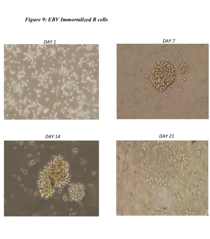
3.12 Immortalization of PBMC
PBMC was isolated upon informed consent, from the blood of a patient with meningitis, infected by TOSV. Using a modified standardized protocol (Steinitz et al., 1977; Corti et al., 2014), Epstein Barr Virus (EBV) was used to immortalize B cells. Isolated PBMC was cultured overnight on a 6 well plate at a cell density of 4x10⁶ cells per well in RPMI supplemented with 10% heat inactivated serum
supplemented with cyclosporin (Sigma) at a concentration of 1g/ml. The supernatant cells were then collected and resuspended in filter purified B95.8 supernatant containing EBV and incubated for one hour at 37oC with 5% CO
40
Following incubation, RPMI medium supplemented with 10% heat inactivated serum was added to the cell suspension containing EBV and plated onto a 96 well plate at a cell density of 5x10⁵ cells per well. The cells were then incubated for
three weeks at 37oC with 5% CO
2 and were routinely monitored for the formation
of clones.
After three weeks, the supernatant was tested for the presence of specific antibodies by ELISA. The cells from the ELISA positive wells were then serially diluted and plated on to a 96 well plate at a cell density of 1 cell per well. Following incubation at 37oC with 5% CO
2 for another three weeks, the supernatant from each well was
again tested for specific monoclonal antibodies by ELISA. 3.13 Statistical Analysis
All experiments were carried out in triplicate. The statistical analysis of differences between means was performed using Stat View statistical software (AbacusConcepts, Berkeley, CA, USA). Neutralization titers are presented as geometric mean±standard deviation. P<0.05 was considered significant.
42
4.1 Expression and Purification of GC683 and GC727
Previous studies (Gandolfo et al., 2019) have identified three regions on TOSV Gn glycoprotein recognized by most of the neutralizing monoclonal antibodies isolated from a patient who was serologically positive to TOSV for both IgM and IgG. The first two regions, Pep 1 and Pep 2, are localized in the amino-terminal of the Gn glycoprotein, while the third region, Pep 3, is positioned close to the transmembrane region, as in Fig.7
The immunogenicity of these three identified peptide sequences were tested in vivo in different combinations either as a single peptide or in combinations. To this aim, Peptide 1 and Peptide 2 were cloned into a prokaryotic expression vector system and named as GC683. Similarly, Peptides 1, 2 and 3 were cloned into another prokaryotic expression vector named GC727. Both the vectors had GST as a carrier sequence. Both the plasmids were expressed in XL10 Gold and Top10 E-coli cells respectively, and proteins were isolated. The concentration of the isolated proteins determined by Bradford method was 5.1g/l for Pep-GC683 and 576ng/l for Pep-GC727. The specificity of the isolated protein was analyzed by western blot using either anti-goat GST or TOSV positive human sera (Fig.8 ).
43 Pep-GC727 40kDa 50kDa 70kDa 100kDa 35kDa 25kDa Pep-GC683 35kDa 25kDa 48kDa 63kDa 75kDa 20kDa
Figure: 7 The three peptide regions of TOSV Gn protein
1NHLLNWPGNG AYTLSDFAES TCTLAYGSEC KSWEHQLDEL SFPFFHSNLD KYSMLEAATE
61 TIPILNKSSA VCTISPSTHS SNACGREASL IKKKCGSDMS AFFYVNLAGQ ITVVKCDTNH 121 VLSNDCGNCI SKTLSGQKIY TPVQDMFCQK GWSESIPSTR YSKDLCSIGL HTVKECKIGT 181 TNFERVGFIV VKGRKMYIEQ MKMRSRQEFS EDQFLCYKSE GSSGSSVKLK KVKVESCKGV 241 TTSSASKCSG DEYFCSRYPC ETANVEAHCI LRRHSAVIEV NVNGVWVVPR CIGYEEVLVR
301 RTSLKVEDTS SRECDTCLWE CGKNKLIVKT HGPKIVYATA CSHGSCKSVM QKPATFVYLP 361 YPGNSEIVGG DIGVHMTEES SPSNIHMVAH CPAKDSCEVS SCLFCVHGLL NYQCHTLFSA 421 LLISTTVMSI LTLLLLLVKG AKDLVKRLFY WLITPLCWLS VFCGWMIRSW KKRVGSAISR
481 TNDTIGWRDN SRRGQDIERA QYTGGAPGAK YSFYGVMILG LLGNVHS
GSDM - Peptide 1
HTVK - Peptide 2
SCEVS - Peptide 3
LFSAL - Transmembrane Region
Figure: 8 Western Blot Analysis of the purified proteins.
(A) (B)
(A) Pep-GC683 (39kDa) (peptide 1-2)
44
4.2 Humoral Immune response elicited to the peptides in vivo
Peptides 1, 2 and 3 either as a single peptide coupled with KLH as carrier or purified proteins as Pep-GC683 and Pep-GC727 or a combination of both were used to immunize 4 weeks-old female BALB/c mice intraperitoneally. Four consecutive immunizations of 5 mice/group were performed at two weeks interval with the peptides and constructs as described in table 2.
Two weeks after the last immunization, mice were sacrificed and the antibody response to the different combinations of peptides were evaluated. Sera obtained from each group were tested for the presence of antibodies against both Gn protein and purified whole TOSV by ELISA. All the mice elicited, at a different extent, an antibody response to Gn after immunization with most of the combinations of the peptides; only pep1-KLH developed a low level of antibodies, while pep2-KLH or other combinations of peptides containing peptide 2 were able to induce a high amount of antibodies reacting with Gn. ELISA results, using the purified whole TOSV as antigen, showed positivity for all the immunized mice groups, although at varying levels. Moreover, the mice group immunized with Pep-GC683 and the combination of Pep-GC683 + Peptide 3-KLH showed a comparatively better response than mice immunized with single combination of peptides or with the recombinant Gn. Immunized mice sera from each group were also tested by immunofluorescence on TOSV infected Vero cells. All the mice immunized with different combinations of peptides elicited antibodies recognizing the Gn glycoprotein in TOSV infected cells, by immunofluorescence.
A neutralization assay was also performed to determine the specific neutralizing activity against TOSV. The mice groups immunized with the single peptides 1, 2 and 3 showed low neutralization titers of 1/10, 1/23 and 1/5 respectively. Similarly, the groups immunized with a combination of all the three single peptides also produced a titer value of 1/8. The mice group immunized with a combination of peptide 2-KLH + peptide 3-KLH showed a titer of 1/24. Likewise, sera from the mice immunized with Pep GC727 containing the sequence of peptides 1, 2 and 3