Università degli Studi di Ferrara
DOTTORATO DI RICERCA IN
FARMACOLOGIA ED ONCOLOGIA MOLECOLARE
CICLO XXII
COORDINATORE Prof. Borea Pier Andrea
Oxidative stress in health and
disease: role of PKCζ and p66
Shc
Settore Scientifico Disciplinare MED/04
Dottorando Tutore
Dott. ZECCHINI ERIKA Dott. RIMESSI ALESSANDRO
SUMMARY
ABSTRACT ... 1
ABSTRACT (ITALIANO) ... 3
INTRODUCTION ... 5
REACTIVE OXYGEN SPECIES ... 5
APOPTOSIS ... 14
THE CROSSTALK BETWEEN APOPTOSIS AND AUTOPHAGY ... 18
PROTEIN KINASE C ... 21
Structure of PKC ... 21
PKC regulation and localization ... 23
Atypical PKC ... 24
Protein kinase C and redox stress ... 26
p66 ... 28
AIMS ... 31
PKCζ RESULTS ... 33
PKCζ-translocation prevents cell death induced by apoptotic stimuli ... 35
Nuclear chimeras of PKCζ prevent apoptotic death by ceramide ... 39
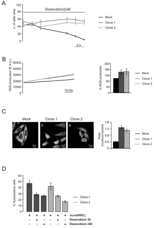
The nuclear PKCζ-inhibitor reverts the apoptotic effect of Doxorubicin in chemotherapeutic-resistant cells ... 43
PKCζ DISCUSSION ... 45
p66 RESULTS ... 49
p66 and the adipogenic differentiation of muscle-derived stem cells ... 50
MATERIAL AND METHODS ... 61
Isolation and Culturing of Myotubes and adipocytes ... 61
Imaging techniques ... 61
Caspase-3 assay ... 61
PKCζ-based constructs ... 62
Intracellular ROS measurements ... 62
Animals and In Vivo Transfection Experiments ... 63
Muscle samples and Immuohistochemical staining ... 63
ABSTRACT
Reactive oxygen species (ROS) are short-living and highly reactive molecules formed by incomplete one-electron reduction of oxygen. Mitochondria produce low levels of ROS as an inevitable consequence of oxidative metabolism. Low levels of ROS are normally reduced by non-enzymatic and non-enzymatic oxidizing agents, such as glutathione, thioredoxin, superoxide dismutase (SOD), catalase and peroxidases. Oxidative stress results from exposure to high levels of ROS, which are not detoxified by cellular antioxidizing agents, and produces cellular damage due to the oxidation of cellular constituents. ROS, however play not only a damaging role: recent studies have demonstrated that they also actively participate in a diverse array of biological processes, including normal cell growth, induction and maintenance of the transformed state, and cellular senescence. Moreover, ROS can also provide a signaling function, by acting as an intracellular messenger involved in the transduction of the signaling of cytokines such as TNF-α and IL-1β. We focused our attention on the effect of ROS in the activities of two proteins: PKCζ and p66Shc. On the one hand, PKCζ is a serine-threonine kinase belonging to the atypical subfamily of PKC proteins. We showed that in an in vitro model redox stress induces PKCζ translocation from the cytosol, where it is located in resting conditions, to the nucleus. Nuclear PKCζ protects cells from apoptotic stimuli such as H2O2 or ceramide and this protective effect is reverted by the selective inhibition of the nuclear pool of the protein. The protective effect of nuclear PKCζ is involved in the chemoresistance of tumor cells to chemotherapic drugs, as demonstrated by the fact that the selective inhibitor of nuclear PKCζ can revert the chemoresistance, suggesting nuclear PKCζ as a suitable target in anticancer therapies. On the other hand p66Shc is a Shc protein involved in stress responses, in particular it is activated by redox stress and it produces ROS itself. Our purpose is to investigate a possible involvement of p66Shc in two different phenomena: autophagy and adipogenic transdifferentiation of skeletal muscle cells by comparing wt mice with p66Shc KO mice. Autophagy is a general term referring to pathways for the degradation of cellular constituents (cytosol and organelles) by the autophagolysosome; it is activated mainly by nutrient starvation and it plays a dual role: it is primarly a surviving mechanism, but it also leads to cell death (called type II cell death) thus possibly acting as an alternative to apoptosis. Starting from this notion, and from
the fact that p66Shc KO cells are protected from apoptosis, we investigated a possible role of p66shc as a key element in the switch from apoptosis to autophagy. We observed that, while in wt cells redox stress induces apoptosis, cells lacking p66Shc in the same condition activate the autophagic pathway. We are now trying to investigate the biological effect of these observation. The second aspect of our work is the investigation of a putative role of p66Shc in the adipogenic transdifferentiation of skeletal muscle precursor cells. To this purpose we used an in vivo model and we observed that mice lacking p66Shc exposed to muscle damage (e.g. freeze injury or redox stress) show lower adipocyte accumulation than wt mice, thus suggesting a role of p66 in the activation of adipogenic differentiation pathway. The key purpose of this work is the evaluation of many different effects of ROS production, looking for possible molecular targets to fight pathological processes such as chemoresistance and adipogenic degeneration of skeletal muscle.
ABSTRACT (ITALIANO)
Le specie reattive dell’ossigeno (ROS) sono piccole molecole altamente reattive e a breve emivita, che si formano in seguito alla riduzione incompleta di molecole di ossigeno. Piccole quantità di ROS vengono fisiologicamente prodotte a livello del mitocondrio come conseguenza inevitabile del metabolismo ossidativo. Bassi livelli di ROS vengono normalmente ridotti da agenti ossidanti, enzimatici e non, quali il glutatione, la tioredoxina, la superossido-dismutasi (SOD), la catalasi e la perossidasi. Lo stress ossidativo è una condizione derivante dall’esposizione ad elevati livelli di ROS, che non vengono completamente eliminati da agenti antiossidanti, e produce danno cellulare dovuto all’ossidazione di componenti cellulari. I ROS, tuttavia, non hanno soltanto un effetto dannoso: studi recenti, infatti, hanno dimostrato che i ROS partecipano attivamente ad un’ampia serie di processi biologici, inclusi la crescita cellulare, l’induzione ed il mantenimento di stati trasformati e la senescenza cellulare. Inoltre, i ROS svolgono anche il ruolo di molecole segnale agendo come messaggeri intracellulari coinvolti nella trasduzione del segnale di citochine, quali TNFa ed IL-1b. Nel presente lavoro, abbiamo focalizzato la nostra attenzione sull’effetto dei ROS sulla funzione di due proteine: PKCζ e p66Shc. PKCζ è una serin-treonin chinasi appartenente alla sottofamiglia delle PKC atipiche. Abbiamo dimostrato che, in vitro, uno stress redox induce la traslocazione di PKCζ dal citosol, dove è localizzata in condizioni di riposo, al nucleo. La porzione di PKCζ nucleare protegge le cellule dagli effetti apoptotici di vari stimoli, quali alte concentrazioni di H2O2 o ceramide, e questo effetto protettivo viene annullato dall’inibizione selettiva della porzione nucleare di PKCζ. L’effetto protettivo di PKCζ, inoltre, risulta essere coinvolto nello sviluppo di chemoresistenza da parte di cellule tumorali, come dimostrato dal fatto che l’inibizione selettiva della porzione nucleare della chinasi può ridurre questo fenomeno, suggerendo che PKCζ potrebbe rappresentare un possibile target nello sviluppo di farmaci coadiuvanti terapie antitumorali. p66, invece, è una proteina adattatrice appartenente alla famiglia delle proteine Shc ed è coinvolta nella risposta cellulare a stress ossidativi; in particolare p66 viene attivata in seguito ad uno stress redox e, quando attivata, può agire da ossido reduttasi mitocondriale producendo ROS. Il nostro principale scopo nel presente lavoro, è stato quello di analizzare un possibile ruolo di p66 in due diversi fenomeni: l’autofagia e il differenziamento adipocitario di precursori miogenici,
attraverso la comparazione di topi wt e di topi KO per p66. Il termine autofagia fa riferimento ad un meccanismo di degradazione di componenti cellulari (citosol ed organelli), da parte di strutture digestive dette autofagolisosomi. L’autofagia viene attivata principalmente in seguito alla deprivazione di nutrienti e svolge principalmente due ruoli: da una parte, infatti, agisce come meccanismo di sopravvivenza in condizioni metaboliche avverse, d’altro canto può culminare nella morte cellulare (è conosciuta come morte cellulare di tipo II), agendo in questo modo come meccanismo alternativo rispetto all’apoptosi. Partendo da questa nozione, nonché dal fatto che cellule KO per p66 sono protette dall’apoptosi, abbiamo analizzato un possibile ruolo di p66 come elemento chiave nello swith fra apoptosi ed autofagia. Abbiamo osservato che, mentre in cellule wt, uno stress redox induce apoptosi, cellule p66KO nelle stesse condizioni attivano la via autofagica; stiamo adesso cercando di comprendere l’effetto biologico di queste osservazioni. Il secondo aspetto del nostro lavoro consiste nell’analisi di un possibile ruolo di p66 nel differenziamento adipocitario di precursori miogenici localizzati a livello del muscolo scheletrico. A tale scopo abbiamo utilizzato un modello in vivo ed abbiamo osservato che topi privi di p66 in seguito ad un danno muscolare (indotto, per esempio, in seguito a freeze-injury o ad uno stress redox), presentano un minor accumulo di adipociti rispetto ai wt, suggerendo un ruolo di p66 nell’attivazione di vie di differenziamento adipogenico. Lo scopo principale di questo lavoro è stata la valutazione di alcuni dei diversi effetti dovuti alla produzione di ROS, attraverso l’analisi di possibili target molecolari utili per intervenire in processi patologici quali la resistenza a farmaci chemioterapici e la degenerazione grassa del tessuto muscolare scheletrico in cui lo stress ossidativo sembra svolgere un ruolo di primo piano.
INTRODUCTION
REACTIVE OXYGEN SPECIES
Reactive oxygen species (ROS) are defined as molecules or ions formed by the incomplete one-electron reduction of oxygen. There are two types of ROS: free radicals, which contains one or more unpaired electron in their external molecular orbital, such as superoxide (O2-) or hydroxyl radicals (HOO-), and non radical species, such as hydrogen peroxide (H2O2). ROS are produced by the cells during their normal metabolism and the major source of ROS are mitochondria, in particular the respiratory chain. The respiratory chain, located in the inner mitochondrial membrane, is composed of four multimeric integral membrane protein complexes (I-IV), coenzyme Q and cytochrome c (cyt c) , a peripheral protein that binds to the outer surface of the inner membrane. The respiratory chain complexes I and III are the primary mitochondrial sources of univalent reduction of O2 into superoxide. Superoxide can be rapidly converted into H2O2 by superoxide dismutase (SOD); two different SOD isoforms exist in the cell: CuZnSOD, that resides in the mitochondrial intermembrane space and in the cytosol and needs Cu or Zn as cofactors, and MnSOD, which is localized in the mitochondrial matrix and needs Mn as cofactor. Once generated, uncharged H2O2 moves across mitochondrial membranes into the cytosol, where it can be converted to water by catalase or glutathione peroxidase. Otherwise, in presence of transition metals (such as Fe2+), H2O2 can be converted to hydroxyl radicals (OH-), which are highly reactive. Other sources of free radicals are the endoplasmic reticulum system, the NADPH oxidase complex and the nitric oxide synthase (NOS), which produces nitric oxide (NO.) from arginine. NO. is reactive a radical having a very short half-life, which can react with superoxide to form peroxynitrite (ONOO-), a non radical species, which is capable of modifying the structure and function of proteins. In
physiological conditions cells employ a number of mechanisms to scavenge and detoxifie ROS to maintain a permissive redox environment. The glutathione redox couple (GSH/GSSG) is the primary cellular redox buffer: GSH is cysteine containing tripeptide that can directly scavenge ROS or act as a cofactor for glutathione peroxidase. GSH is subsequently reduced by glutathione reductase which uses NADPH as a substrate. NADH/NAD+ couple also play a role in redox buffering, as thioredoxin, a disulphide containing protein that can directly scavenge H2O2 as part of thioredoxin reductase and thioredoxin peroxidase system. ROS detoxification also involves enzymes such as SOD, which catalyzes the dismutation of O2- into H2O2 and O2, and catalase, which detoxifies H2O2. ROS are chemically reactive molecules that play both toxic and essential functions in living organisms. A moderate increase in ROS can promote cell proliferation and differentiation, through the regulation of the activity of enzymes such as ribonucleotide reductase, they can mediate inflammation by stimulating cytokine production and they can eliminate pathogens and foreign particles. On the other hand, excessive amount of ROS (condition known as redox stress or oxidative stress) can cause damage to the cells. Many components of cells can be target for ROS-induced oxidation: DNA, in presence of excessive ROS levels, undergoes oxidative damage, which can be assessed by analyzing the levels of 8-hydroxideoxoguanosine which accumulates in DNA; mitochondrial DNA (mtDNA) is particularly susceptible to oxidative damage, probably because it is closer to the site of ROS generation. ROS can also produce oxidative damage to proteins and lipids. Lipid peroxidation consists of the interaction of OH. radical with unsaturated bonds in a membrane lipid and it affects membrane permeability and function. Therefore, maintaining ROS homeostasis is crucial for normal cell growth and survival, for this reason cells finely control the balancing of ROS generation and scavenging. An increase in ROS, indeed, is associated with many pathological states, such as abnormal cancer cell growth and diabetes.
Compared with normal cells, malignant cells seem to function with higher levels of endogenous oxidative stress in culture and in vivo (Szatrowski and Nathan 1991; Kawanishi and Murata 2006). For example, leukemic cells freshly isolated from blood samples show increased ROS production compared with normal lymphocytes (Zhou, McEarchern et al. 2003; Kamiguti, Serrander et al. 2005), while in solid tumors oxidized DNA bases and lipid peroxidation products have been observed ((Patel, Rawal et al. 2007; Kumar, Koul et al. 2008). Moreover, malignant cells show an
alteration in the regulation of redox homeostasis due to a reduction in the activity of ROS-scavenging enzymes. The precise pathway leading to redox stress in cancer cells remains yet unclear, but many intrinsic and extrinsic pathways are thought to be involved in oxidative stress during cancer development. Some examples of intrinsic factors are represented by the activation of oncogenes (such as those associated with tumor transformation like Ras, Bcr-Abl, c-Myc), by the aberrant metabolism, by mitochondrial dysfunctions and by the loss of functional p53. Mitochondrial DNA (mtDNA) mutations have also been shown to be correlated with increased ROS levels in certain types of cancer cells, because several protein components of the electron transport chain are encoded by mtDNA, so mutations of these genes cause impairment in electron transfer, leading to leakage of electrons and generation of superoxide. At an advanced disease stage, redox stress becomes part of a vicious cycle, in which ROS induce gene mutations, thus leading to further metabolic dysfunction and ROS generation. Moreover, in physiological conditions, p53 acts as a transcription factor which regulates, among the others, the expression of genes involved in the regulation of the redox state. One of the most common feature of cancer cells is the loss of function of p53, which produces redox imbalance, high mutagenesis and aggressive tumor growth. Extrinsic mechanism leading redox stress in cancer cells are due to the interaction of cancer cells with the microenvironment: examples of extrinsic factors are represented by the presence of inflammatory cytokines, such as TNFα, by macrophages, by an imbalance of nutrients and by the presence of an hypoxic environment. Increased ROS stress in cancer cells correlates with the aggressiveness of tumor and with poor prognosis and this aspect is in apparent contradiction with the proapoptotic effect of ROS which should, on the contrary, promote the elimination of cancer cells. This contradiction can be explained by the fact that it has been observed that cancer cells can survive oxidative stress thanks to the acquirement of adaptive mechanisms consisting of an increased ROS scavenging activity. During malignant transformation, therefore, oncogenic signals both induce ROS generation to stimulate cell proliferation through redox-sensitive transcriptional factors, and promote antioxidant adaptive mechanisms to minimize oxidative damage. All of these mechanisms also confer to cancer cells an increased capacity to tolerate exogenous stress and insults, so they are involved in the development of drug resistance which is one of the main characteristic of cancer. An increase in ROS production can also influence the viability of cells: dependent on the impact of ROS, the cells can either repair the damage or activate pathways leading cellular suicide through
apoptosis, a form of cell death that occurs during several pathological situations in multicellular organisms, characterized by cell shrinkage, chromatin condensation DNA fragmentation and formation of “apoptotic bodies”. The execution of the apoptotic program consists mainly in the activation of the caspase cascade. Caspases are cysteine-containing, aspartic-acid specific proteases which exist as zymogens in the soluble cytoplasm, mitochondrial intermembrane space and nuclear matrix of all cells. The cascade of caspases ends in the cleavage of many protein targets, for example endonucleases which, once activated, enter the nucleus and are responsible for DNA fragmentation. Many studies investigated the role of redox stress in the regulation of apoptosis, underlying an apparent contradictory effect of ROS. Indeed, incubating cells with exogenous oxidants or adding redox-active compounds triggers apoptosis, but the mechanism is not yet completely clear. One possibility is that ROS produce a damage which, when sensed by the cell, activate the apoptotic machinery; one example is the detection of DNA damage by p53. One other explanation is that ROS, at low concentration, can activate caspases thus inducing apoptosis, but the most important mechanism leading ROS-induced apoptosis is the impairment of mitochondrial function. ROS indeed seem to be involved in the mitochondrial permeability transition (MPT), in particular they increase the efflux of mitochondrial Ca2+ through the MPT and the efflux of Ca2+ disrupts the ion homeostasis and causes drastic changes in mitochondrial ultrastructure and functional activity. MPT results in mitochondrial failure which can lead to necrosis due to ATP depletion, or, if MPT occurs in a subpopulation of mitochondria and the remaining organelle can maintain membrane potential and produce enough ATP, to caspase activation and apoptotic program. Finally, ROS can induce apoptosis by leading the release of cytochrome C from mitochondrial intermembrane space. Cytochrome c, indeed, is normally bound to the inner mitochondrial membrane, associated with the anionic phospholipid cardiolipin: the binding of cytochrome c with cardiolipin is fundamental to maintain cytochrome c into mitochondria. ROS induces cardiolipin oxidation, thus decreasing its binding affinity for cytochrome c, which, after the outer mitochondrial membrane permeabilization, can be released into the cytosol, where it binds to Apaf1 thus forming the so-called apoptosome, which recruits and binds the procaspase 9, which, when activated, starts the apoptotic program.
Oxidative stress moreover, both is affected by and affects autophagy, a process by which eukaryotic cells degrade and recycle macromolecules and organelles. First of all, autophagy is involved in the
clearance of damaged mitochondria, thus decreasing their potential oxidative damage; this process, named mitophagy, plays an important role in protecting cells from ROS-induced cell death. Moreover, oxidized substrate proteins can be eliminated through chaperone-mediated autophagy and they can translocate to lysosomes more efficiently than their unaltered counterparts. ROS, in particular mitochondrial ROS, might also have a signaling role in autophagy, as demonstrated by many evidences collected so far. For example, it has been demonstrated that TNF-α, induces accumulation of H2O2 through the inhibition of mitochondrial electron transfer, thus inducing the expression of Beclin-1 and leading to autophagic cell death. (Djavaheri-Mergny, Amelotti et al. 2006). Moreover, rapamycin in yeast and NGF-deprivation in neurons lead to lipid peroxidation, which in turn activates autophagic cell death (Kirkland, Adibhatla et al. 2002; Kissova, Deffieu et al. 2006). Nutrient starvation, moreover, induces an accumulation of H2O2, probably through phosphoinositide III kinase (PI3K), H2O2 oxidizes Atg4, a protease which activation is necessary for autophagosome formations (Scherz-Shouval, Shvets et al. 2007), thus inducing autophagy.
AUTOPHAGY
The term “autophagy” literally means “self-eating” and it refers to an highly conserved process in eukaryotes by which long-lived cytosol proteins and organelles are delivered to lysosomes for bulk degradation. This process allows the elimination of damaged, aberrant or aggregated proteins, thus protecting cells from their potential damaging effects. At least, three different types of autophagy have been described. The most extensively characterized is macroautophagy, in which portions of cytosol and entire organelles are engulfed by double-membrane structures called autophagosome, which then fuse which lysosomes, thus forming a single-membrane structure called autophagolysosome, in which luminal content is degraded and resulting elements (in particular macromolecules necessary to sustain cell metabolism) return into the cytosol for cell reactions (Klionsky and Emr 2000). The regulation of macroautophagy is complex and it is not yet completely clear. Its major negative regulator is the mammalian Target Of Rapamycin (mTOR), which inhibits the formation of autophagosomes: mTOR acts mainly as an aminoacid sensor and in nutrient starvation conditions, when the availability of aminoacids decreases, it is inhibited thus triggering autophagy . The same effect can be induced by rapamycin itself, a chemical inhibitor of mTOR. In my work I will focus my attention in particular on macroautophagy, so I will refer to it simply as autophagy. The second type of self-eating is microautophagy, in which the engulfment is made directly by the lysosomal membrane; differently from macroautophagy, microautophagy is not activated by nutrient deprivation. One particular type of microautophagy is micropexophagy, the highly selective degradation of peroxisomes described in yeast as crucial in response to oxidative stress. The third type of self-eating is chaperone-mediated autophagy (CMA), in which cytosolic proteins showing a specific pentapeptide lysosomes-targeting motif (the consensus sequence KFERQ) are recognized by a complex of chaperone proteins and targeted to lysosomal membrane, where they bind to the lysosome-associated membrane protein LAMP2a. Substrate proteins are then unfolded and transported to the lysosomal lumen for degradation . One example of protein degraded through this pathway is represented by the amyloid β precursor protein (APP). Autophagy was morphologically first identified in 1960s in mammalian cells, however the molecular machinery regulating this process has not yet been completely elucidated. More than 20 genes, named autophagy-related genes (ATG) involved in the regulation of this process have been
identified both in yeast and in mammals. Autophagy occurs at a basal level in normal growing conditions, however certain types of environmental stress (such as nutrient starvation) result in a dramatic induction. Autophagy mainly consists in a membrane-trafficking process in which a large number of cytoplasmic components are non-selectively enclosed within a double-membrane structure named autophagosome and delivered to the vacuole-lysosome for degradation and recycling. The biogenesis and consumption of autophagosomes can be divided into four steps: 1) induction of vesicle formation mainly by nutrient deprivation; 2) engulfment of the selected cargo by a membrane and fusion of the extremities of the surrounding membrane to generate a double-membrane vesicle; 3) fusion of the autophagosome with the vacuole in yeast or with the lysosome in mammals (this process is mediated by a fusion machinery similar to the one used by Golgi and endosome-derived vescicles to fuse with vacuoles, including SNARE proteins, a RAB-GTPase and a class C vps complex); 4) vesicle breakdown, consisting in the release of the inner vesicle (called autophagic body) into the vacuolar or lysosomal lumen. Each step of this process is finely regulated by specific proteins; the fundamental step allowing the correct execution of the autophagic process is the closure of autophagosome. In this step the protein Atg8 and its mammalian ortologues, LC3, GATE16 and GABARAP, play a key role. Atg8/LC3 is an ubiquitin-like protein and it is produced in an inactive form which serves as a substrate for the cysteine protease Atg4, which cleaves its substrate thus exposing a glycine residue at its C-terminus. This form of Atg8/LC3, named form I, is unconjugated and soluble and it diffuses throughout the cytosol. During autophagy, form-I Atg8/LC3 becomes phosphatidyletanolamine (PE)-conjugated and membrane-bound, thus producing form-II which is bound to the autophagosome membrane. This process is catalyzed by an ubiquitination-like reaction performed by an E1-like enzyme, Atg7, and an E2-like enzyme, Atg3 (Hanada, Satomi et al. 2009). Many studies demonstrated that a defect in LC3 function or activation leads to the failure of autophagosome closure, thus underlying a central role of Atg8/LC3 in the correct formation of autophagosomes (Fujita, Hayashi-Nishino et al. 2008; Sou, Waguri et al. 2008). The translocation of the protein Atg8/LC3 from cytosol to autophagosomal membranes following autophagy induction is largely implied for the study of the apoptotic process: a fusion protein of LC3 with a fluorescent protein (GFP or YFP), indeed, allows to visualize the localization of the protein: in resting condition, LC3 is diffused in the cytosol, while upon starvation it shows a punctuate localization, due to its incorporation in the membrane of the forming vesicles.
Fig. 1: Schematic representation of autophagosome formation and of post-transcriptional LC3 modifications
The protein TOR is one of the most important regulators of the autophagic process. The protein was originally identified by genetic screens in S.cerevisiae and subsequently in other organisms including fungi, mammals, flies and worms (Crespo and Hall 2002; Crespo, Diaz-Troya et al. 2005). TOR is present ad a single gene in most eukaryotes, but some organisms, like yeast and fungi, show two TOR genes. The TOR kinases are large (about 270kDa) proteins that assemble into two structurally and functionally distinct complexes termed TORC1 and TORC2. In yeast TORC1 contains either TOR1 or TOR2, KOG1, TGO89 and LST8, whether TORC2 includes TOR2, LST8, BIT61, AVO1 and AVO3; mammalian TOR (mTOR), similarly, associates with raptor (homologue of KOG1) and mLST8 to form mTORC1, while it associates with mLST8, rictor and SIN1 (homologues respectively of AVO3 and AVO1) to constitute mTORC2. The complex mTORC1 is sensitive to the inhibitory effect of rapamycin: rapamycin, indeed, after forming a complex with FKBP12, can inhibit the proper interaction between mTOR and raptor. This complex is involved in the regulation of cell growth and, under favorable growth conditions it is active and it promotes ribosome biogenesis and initiation of the translation by inducing the phosphorylation of the protein S6 kinase (S6K) (Hara, Maruki et al. 2002). The TORC1 complex is a master controller of protein synthesis integrating signals from growth factors and nutrient conditions of the cell. TORC1, indeed, regulates protein synthesis by directly phosphorylating 4E binding protein 1 (4EBP1) and
p70S6K (S6K) translation initiation factors which are important to mRNA translation, thus increasing the level of proteins involved in proliferation, cycle progression and survival pathway. Moreover, TORC1 acts as an aminoacid sensor and regulates the autophagic process. TORC2, otherwise, regulates cell growth in a rapamycin-insensitive manner; in particular it is involved in the control of cell polarity through the regulation of actin cytoskeleton polarization. I will focus my attention on the complex TORC1, being it involved in the regulation of autophagy. In particular, TORC1 acts as a negative regulator of autophagy by sensing environmental change, in particular, it acts as a sensor for a variety of upstream signals, like growth factors, insulin, aminoacids such as leucine and glutamine, and intracellular levels of ATP, phosphatidic acid, and inorganic polyphosphates . In mammalian cells, mTOR is regulated by pathway PI3K-Akt, in particular it has been shown that Akt indirectly stimulates TORC1 activity (Sekulic, Hudson et al. 2000).
It has been observed that mTOR regulates autophagy by controlling the phosphorylation state of Atg13: in nutrient-rich conditions Atg13 is highly phosphorylated and it shows a low affinity for Atg1, thus repressing autophagy; under starvation conditions or following a rapamycin treatment, mTOR is inhibited and Atg13 is partially and rapidly dephosphorylated and it interacts with Atg1 with an high affinity. Once the complex Atg13-Atg1 is formed, it interacts with multiple proteins, such as Vac8, Atg11 and Atg17 (Hosokawa, Hara et al. 2009). The formation of this multiproteic complex plays an important role in the regulation of the autophagosome biogenesis.
APOPTOSIS
Apoptosis (also known as programmed cell death) is a physiological process used to eliminate superfluous, damaged, infected or aged cells in multicellular organisms. Apoptosis is an highly orchestrated program of cell removal necessary to the organism in many contexts, like immune response, infection or elimination of DNA damage, because it minimizes the damage to surrounding viable tissues. During apoptosis, indeed, the cellular architecture is dismantled in an highly controlled way and apoptotic cells show a series of typical morphological features, like chromatin condensation, phosphatidylserine exposure, mitochondrial fragmentation (due to the action of Drp1, which oligomerizes around mitochondria and induces their fission in a GTP-dependent manner) membrane blebbing and cell shrinkage, which leads to the fragmentation of the cell into small vesicular bodies which can be taken up by macrophages. The apoptotic process is complex and finely regulated, and its dysregulation can lead to many diseases like neurodegeneration, autoimmunity and cancer. In particular, while uncontrolled proliferation and reduced sensitivity to apoptotic signals are classic hallmarks of oncogenic transformation, excessive and inappropriate apoptosis is the basis of neurodegenerative diseases like Alzheimer disease. Molecularly, the execution of the apoptotic program, is due to a family of cysteine proteases called caspases (cysteine aspartic-specific proteases), that cleave substrates at the N-terminal side of a specific aspartic-acid residue. Caspases are synthesized as inactive zymogens (procaspases), that require proteolytical cleavage to form the large and small subunits of the active enzyme; the activation can happen through autoproteolysis or by other activated caspases. Apoptosis in mammals can be initiated through two different pathways: the extrinsic pathway involves extracellular ligands, while the intracellular pathway involves the release of molecules from mitochondria intermembrane space. In both cases, the apoptotic program is a two step proteolytical pathway: the first step consists of the activation of “initiator caspases” (caspase 9 and caspase 8), while the second step consists in the activation of “executioner caspases” (caspase 3 and caspase 7), that cleave a number of cellular proteins to drive forward the biochemical events that culminate in death and dismantling of the cell. Some example of classical downstream target of executioner caspases are: the fibrous protein of nuclear lamin, which cleavage alters the nuclear characteristic of nuclear envelop, ICAD/DFF45, which cleavage determines DNA fragmentation, p21 kinase and
PARP. As just said, apoptosis can be triggered through two different pathways. The extrinsic pathway is activated by extracellular molecules binding to the Fas/APO-1 transmembrane protein, which is a member of the tumor necrosis factor receptor (TNFR): when Fas/APO1 binds to its receptor, it induces the recruitment of procaspase 8 thanks to the adaptor protein FADD (Fas-associated death domain-containing protein). Upon recruitment to the receptor complex, caspase 8 becomes activated through autoproteolysis and subsequently cleaves and activate caspase-3. On the other end, in the intrinsic pathway, a central role is played by mitochondria. The central event in this case, indeed, is the mitochondrial outer membrane permeabilization (MOMP), which allows the escape of proapoptotic molecules from mitochondria, including the second mitochondria-derived activator of caspase (Smac, also known as Diablo) and cytochrome c. In particular, the intrinsic pathway is activated by cellular stresses such as DNA damage, heat shock, oxidative stress and many other forms of cellular damage, which result in caspase activation through the release of cytochrome c from mitochondria following MOMP and the subsequent formation of the apoptosome, a large caspase activating complex, composed of seven Apaf-1 subunits binding cytochrome c, dATP and procaspase 9. Apaf1 is the mammalian homologue of Ced4 and it consists of three functional domains: an N-terminal CARD (caspase-recruitment domain), a central nucleotide-binding domain and a twelve to thirteen WD40 repeats at the C-terminus of the molecule. In the absence of an apoptotic stimulus, Apaf1 exists in a monomeric form and the WD40-repeat region maintains it in an autoinhibited state. When an apoptotic stimulus activates the intrinsic pathway, cytochrome c is released from mitochondria and it binds to the WD40-repeat region of Apaf1. When Apaf1 binds cytochrome c, the autoinhibition is removed and the dATP bound to the nucleotide binding site is hydrolyzed to dADP. In this form Apaf1 can oligomerize, thus forming a seven-member ring acting as a recruitment platform for procaspase9 , which is activated, thus starting the proteolytical apoptotic cascade. Interestingly, inside mitochondria cytochrome c is present in two different location: a minor pool is free in inter-membrane space, and a major pool is enclosed in cristae (Delivani and Martin 2006). The release of cytochrome c after MOMP is performed in two following steps: first the soluble pool and then the pool present in cristae (Scorrano, Ashiya et al. 2002). The opening of cristae junction plays a key role in apoptosis because in some cell types the soluble cytochrome c is not sufficient to induce the formation of the apoptosome. The morphology of cristae junction and their opening during apoptosis are regulated
by Opa1, a large GTPase also involved in the inner mitochondrial membrane opening (Frezza, Cipolat et al. 2006).
The mitochondrial pathway (also known as intrinsic pathway) of apoptosis is regulated by members of the Bcl2 family proteins. The Bcl2 gene was originally identified as a proto-oncogene involved in the translocation of human follicular lymphoma (Tsujimoto, Ikegaki et al. 1987) and, in contrast to many oncogenes it does not trigger cell proliferation but it promotes cell survival under negative conditions. Bcl2 is the prototype of a large family of proteins which share a large degree of homology although they exert many different functions: in particular some of them play an anti-apoptotic role, while some other proteins act as proanti-apoptotic mediators. Anti-anti-apoptotic proteins, like Bcl2, BclxL, BclW and Mcl1, have usually four Bcl2 homology (BH) domains, while proapoptotic proteins display either three BH domains (BH1, BH2 and BH3), like Bax and Bak, or only the BH3 domain, like Bid, Bim and Bad, Noxa and Puma. Anti-apoptotic Bcl2 protein acts by binding and sequestering proapoptotic proteins, including activator BH3-only proteins, but this is not their only mechanism. Our group indeed, showed that Bcl2 is also involved in the regulation of Ca2+ homeostasis, in particular it induces Ca2+ depletion from endoplasmic reticulum, thus avoiding the Ca2+ overload to mitochondria, and the following MOMP (Pinton, Ferrari et al. 2001). On the other hand, Bax and Bak directly participate in the formation of pores in the outer mitochondrial membrane (Chami, Prandini et al. 2004). In healthy cells Bax is located in the cytosol, with a minor pool loosely attached to mitochondria. Upon several apoptotic stimuli, BH3 only proteins like Bim and Bid induce a conformational change in its structure which targets it to the MOM. When it is located to the MOM, Bax can oligomerize, thus forming pores and determining MOMP (Schafer, Quispe et al. 2009). Bak, instead, is constitutively inserted in the MOM, where it is bound to and inhibited by VDAC2, BclxL and Mcl1 (Cheng, Sheiko et al. 2003; Willis, Chen et al. 2005). Bax oligomerization, otherwise, requires BH3 only proteins to induce MOMP (Korsmeyer, Wei et al. 2000). Finally, BH3 only proteins can be divided into two classes: on one hand “activator BH3 only proteins” can directly bind to Bax and Bak and recruit them to the MOM, for example Bid, Bim, Map1 and Puma exert this function; on the other hand, “sensitizers BH3only proteins” act by competitively inhibiting the anti-apoptotic members of the Bcl2 members, for example Bad selectively binds Bcl2 and BclxL, while Noxa specifically binds Mcl-1 (Fletcher and Huang 2006).
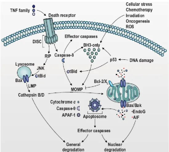
Fig. 2: Schematic representation of the two major apoptotic pathways: the intrinsic pathway and the exstrinsic pathway.
THE CROSSTALK BETWEEN APOPTOSIS AND AUTOPHAGY
Both apoptosis and autophagy are highly regulated processes and increasing evidences suggest a possible relation among these pathways. Apoptosis is a mechanism of controlled cell death which allows the elimination of damaged or unnecessary cells without damaging contiguous cells. Autophagy, on the other end, can be involved in the turnover of long-lived proteins and whole organelles, but it also act as a process by which cells adapt their metabolism to starvation induced by decreased extracellular nutrients or by decreased intracellular metabolite concentrations that result from the loss of growth-factor signaling: through the catabolism of macromolecules, autophagy generates metabolic substrates to sustain the metabolic needs of the cell. In this conditions autophagy de facto suppresses cell death thus avoiding apoptosis, but in several other scenarios it constitutes an alternative pathway to cellular demise, called autophagic cell death (type II cell death). To simplify we can say that two different types of interplay between the two processes can be observed:
9 Both apoptosis and autophagy act as alternative cell death pathways;
9 Autophagy acts as antagonist to block apoptotic cell death by promoting cell survival;
The interconnection between apoptosis and autophagy has been studied in different cell types and in different conditions. A suitable model to study this relation, which is collocated in the first scenario, is represented by the mouse embryonic fibroblasts (MEFs) of Bax-Bak double knockout (Bax
Bak-/-). These cells, lacking two multiprotein members of the Bcl2 family, are unable to
activate the intrinsic apoptotic pathway because these proteins are required for MOMP. It has been observed that, when treated with DNA damaging agents such as etoposide (a topoisomerase-2 inhibitor) Bax
Bak-/- MEFs fail to undergo apoptosis, as expected, but instead they manifest a
massive autophagy followed by massive cell death, thus suggesting that autophagy can act as an alternative pathway to apoptosis (Shimizu, Kanaseki et al. 2004). Another example of this correlation among autophagy and apoptosis was observed both in mouse L929 and in human Jurkat T cell lymphoma, where the chemical inhibition of caspases induces autophagic cell death (Madden, Egger et al. 2007). Otherwise, often autophagy is activated to protect cells from
apoptosis in adverse conditions. For example, in an IL3-dependent immortalized cell line , IL3 withdrawal activates a month long autophagic process causing a severe reduction of cell size and the removal of most cytoplasm: inhibition of autophagy in this case kills these cells, thus demonstrating that in this case autophagy acts as a protective mechanism (Lum, Bauer et al. 2005). This hypothesis is further supported by the observation that both in HeLa and HCT116 cancer cells the inhibition of autophagy, either at early or late stages, results in an accelerated apoptotic cell death (Boya, Gonzalez-Polo et al. 2005; Gonzalez-Polo, Boya et al. 2005). In vivo, moreover, it has been observed that the neuron-specific knockout of Atg5 or Atg7 causes neurodegeneration, accumulation of cytoplasmic inclusion bodies and apoptotic cell death (Hara, Nakamura et al. 2006; Komatsu, Kominami et al. 2006). Autophagy protective effect against apoptosis is not only due to its capacity to defend cells from nutrient deprivation: autophagy, indeed, is also essential to allow the elimination of cell “waste”, represented by protein aggregates and harmful organelles. For example, it has been observed in mutant mice in which Atg5 or Atg7 is depleted an increased development of neurodegeneration also in non stressed cells, due to the formation of inclusion bodies containing protein aggregates (for example of mutant huntingtin, a protein which results mutated in familial forms of Parkinson’s disease). Curiously it has been observed that rapamycin treatment, which induces autophagy, can cause a 50% decrease of cell size thus reducing the susceptibility of cells to apoptotic stimuli; this observation could be explained, for example by the fact that damaged mitochondria are eliminated through mitophagy to protect cells from their potential damaging effects (Fumarola, La Monica et al. 2005).
The crosstalk between apoptosis and autophagy is clearly demonstrated by the evidences collected so far, but it also is suggested by the fact that many common players can be observed among the two processes. For example, Atg5 is one of the basic components of the autophagic machinery and it is essential for the elongation of the autophagosome membrane during vacuole formation. Atg5 overexpression, otherwise, not only induces autophagy, but also can activate apoptotic cell death. It has been observed indeed that Atg5, upon proper stimuli, can undergo proteolysis, thus generating a 24kDa fragment which translocates to mitochondria where it promotes MOMP (Yousefi, Perozzo et al. 2006). Moreover, Atg5 can also directly interact with FADD, thus stimulating the extrinsic apoptotic pathway (Pyo, Jang et al. 2005). Another important player of the autophagic machinery is beclin-1 which, when associated with its binding partners (VPS34 and UVRAG),
regulates the initial steps of autophagy. Interestingly, beclin-1 contains a BH3 domain, similar to that of Bcl2 protein which allows its binding to antiapoptotic Bcl2 homologues. Beclin1, indeed, can be inhibited by multidomain proteins of the Bcl2 family, including Bcl2 itself, BclxL and Mcl1 (Furuya, Yu et al. 2005; Pattingre, Tassa et al. 2005; Maiuri, Criollo et al. 2007; Maiuri, Le Toumelin et al. 2007). The binding of beclin-1 to these proteins avoids its association with its binding partners, thus inhibiting autophagy. The autophagy-inhibitory effects of Bcl2 or BclxL depend on their subcellular localization: only ER-localized but not mitochondrial proteins can bind to and inhibit beclin-1. Signals that promote autophagy induce the disruption of the association among Bcl2/BclxL and beclin1: for example, phosphorylation of beclin1 by the death-associated protein kinase (DAPk) or Bcl2 phosphorylation by JNK trigger the release of beclin-1 and the induction of autophagy; alternatively, other BH3-only proteins, such as BNIP3 competitively displaces beclin-1.
PROTEIN KINASE C
Protein kinase C (PKC) is an ubiquitous family of enzymes that consists of 10 structurally related 70-80 kDa serine/threonine kinases. PKCs have a multitude of cellular substrates and they are involved in a great array of biological processes. The mammalian PKC isotypes have been grouped into three small subfamilies according to their structural properties and their regulation (Mellor and Parker 1998):
9 conventional PKC (cPKCs): α,βI,βII (produced by an alternatively spliced gene) and γ. These isotypes are activated by phosphatidylserine (PS) in a Ca2+-dependent manner and by diacylglycerol (DAG), which increases their specificity for PS and shifts the affinity for Ca2+ into the physiological range. cPKC can also be activated by phorbol ester (PMA), which eliminates the requirement dor DAG and increases the Ca2+ concentration necessary for activation.
9 novel PKC (nPKCs): ε,η , δ, and θ. These proteins are Ca2+-insensitive and they are activated by DAG or PMA in presence of PS.
9 Atypical PKC (aPKCs): λ/ι and ζ. These isoforms are Ca2+-insensitive, nor do they respond to DAG or PMA.
Structure of PKC
The examination of the protein sequence of different PKC isoforms, shows a clear homology between the members of the same subfamily. In all cases highly conserved regions have been shown to define protein domains which confer a specific localization or a specific activating input to the isotype. The single polypeptide chains of each isoform contain conserved (C) and variable (V) regions and are composed of a catalytic and a regulatory domain The catalytic domain is active without cofactors after proteolytical removal of regulatory domain . In the regulatory region we can find different domains playing different roles.
9 C1 domain is defined by two zinc-finger motifs (C1a and C1b) and each motif has a conserved pattern of cysteine and histidine residues, responsible for the coordination of two Zn2+ ions. C1 domain represents the binding site for PMA and DAG, which compete for the
binding to the site. Structural studies demonstrated that in presence of PMA this domain forms an hairpin-like hydrophobic structure that mediates PKC interaction with the membrane. C1 site is present in all PKC isoforms, also in aPKC which are DAG and PMA-insensitive; this can be explained by the fact that aPKC only shows C1a motif which confers lower affinity to PMA and DAG than C1b, as demonstrated by mutational studies.
9 C2 domain is found in the cPKC immediately C-terminal to the C1 domain and it is composed by two four-stranded antiparallel β-sheets forming a compact β-sandwich . This domain binds two Ca2+ ions thanks to five aspartate residues, thus triggering a conformational change which allows the binding o a phospholipid. nPKC and aPKC show C2-like domains in which one or more of the aspartate residues necessary for Ca2+ binding are missing. Probably the role of C2-like domain is the regulation of protein-protein interaction.
9 The pseudosubstrate domain is present in all PKCs subfamilies and it is located at the N-terminus of the C1 domain. The pseudosubstrate is a sequence which retains the hallmarks of a PKC phosphorylation site, but it has an alanine at the predicted serine/threonine phosphorylation site. In resting conditions the pseudosubstrate site interacts with the catalytic domain and it is responsible for the intramolecular suppression of its catalytic activity.
Also in the catalytic region different regions playing different roles can be found:
9 The V3 region is a variable region separating the regulatory domain from the catalytical domain. It is sensitive to the proteolytical activity of Ca2+-dependent proteases like trypsin or calpain and the cleavage of this region allows the removal of regulatory domain and the activation of the protein.
9 The C3 domain is an highly conserved region presenting the binding site for ATP.
9 The C4 domain is the proper kinasic domain where the transfer of a phosphate group to the substrate takes place.
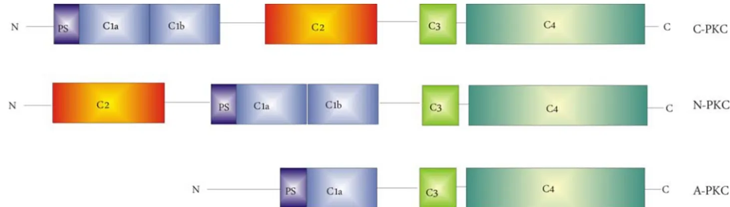
Fig. 3: Schematic representation of different PKC subfamilies structure: classical PKC (C-PKC), novel PKC (N-PKC) and atypical PKC (A-(N-PKC).
PKC regulation and localization
PKCs are produced as inactive precursors which are then phosphorylated in three different sites. Once fully processed and phosphorylated, PKCs can respond to different second messengers, depending on their C1 or C2 domains, and they phosphorylate their downstream targets. Activated PKCs are subjected to phosphatase activity and downregulation by ubiquitination and proteasomal degradation. PKCs display different sensitivities toward activators. The most important activators of PKC are Ca2+, PMA, DAG and PS, which bind to their specific site (see PKC structure), by inducing a conformational variation and the subsequent dissociation of the regulatory domain. Removal of regulatory domain can also be produced by the protelytical cleavage of V3 region. Phorbol esters mimic the action of second-messenger DAG but, while production of second messenger DAG in response to physiological stimuli is transient due to the rapid metabolic conversion of DAG, phorbol esters are more stable, so their action is much more prolonged. DAG and PMA are not the only lipidic mediators involved in PKC activation: nPKC and aPKC, for example, can be activated by products of Phosphoinositide 3-kinase (PI3-K), while aracidonic acid increases DAG effects at low Ca2+ concentrations; ceramide, finally, can activate both members of aPKC family. Another interesting aspect of PKC regulation is the localization of different isoforms which results from the interaction with specific proteins. There are many proteins binding kinases and, while some of them act as a substrate, some others are involved in defining their localization to specific cellular compartments. For example, while vinculine, MARKS (myristoylated alanine-rich C kinase substrate), α and γ adducine,
glycogen syntase and annexin I and II act as PKC substrates, RACKS (receptors for activated kinases), upon binding to specific sequences, drive PKC activation state and define their subcellular distribution, thus leading the association with phospholipide to form stable membrane complexes. Binding of PKC to membrane induces conformational changes that expose the binding site of the kinase domain. The subsequent downstream events include activation of the MEK-ERK and PI3K-AKT pathways. PKCs activation therefore is accompanied by their translocation, so to better understand the specific action of each isoform it is very important to know their localization in resting condition and upon stimulation. Through the construction of chimeric proteins given by the fusion of different PKC isoforms with Green Fluorescent Protein (GFP) it was possible to define the localization of different PKCs isoforms by using digital microscopy techniques, (Rizzuto, Carrington et al. 1998). In resting conditions PKCs localization is mainly cytosolic, but for PKCδ and PKCε it was also described a localization at the Golgi apparatus, while for PKCδ a nuclear localization was described. Upon stimulation, represented for example by oxidative stress, the α, β, γ and δ isoforms translocate to the plasma membrane, while λ and ζ isoforms translocate to the nucleus (Rimessi, Rizzuto et al. 2007). The nuclear translocation of some PKC isoforms is made possible by the fact that in the regulatory domain of these isoform two sequences named Nuclear Localization Sequence (NLS) and Nuclear Export Sequence (NES) can be found and these sequences regulate the trafficking of the protein into and out of the nucleus (Perander, Bjorkoy et al. 2001).
Atypical PKC
Our study focused the attention on the ζ member of the atypical subfamily of PKC. In the last years many studies were performed to enlighten the role of this protein in cell life and the mechanisms in which it is involved.
First of all PKCζ (and its homologue λ) is a critical mediator of mitogenic signal, so it is directly involved in cell proliferation. The mechanism is not yet completely clear, but it seems to be based on the interaction of the enzyme with RAS protein. Moreover PKCζ can activate both MAPK and MEK, thus inducing cycline synthesis and, consequently, cellular proliferation (Kampfer, Windegger et al. 2001; Cohen, Lingen et al. 2006).
Another important aspect is the involvement of PKCζ in the physiological action of insulin. A study performed on 3T3-L1 cells demonstrated that upon insulin stimulation PKCζ is activated through the action of lipidic mediators such as PI3K (Bandyopadhyay, Sajan et al. 2002). Moreover it has been demonstrated that PKCζ overexpressing cells, upon insulin stimulation, show an increased translocation of GLUT-4, demonstrating an involvement of the protein in the regulation of glucose homeostasis insulin-regulated (Liu, Yang et al. 2007). PKCζ, finally, is involved in the synthesis insulin-stimulated of proteins, through the activation of the kinase p70S6K which regulates the proteic transcription (Chou, Hou et al. 1998).
Our work is focused on the involvement of PKCζ in the regulation of apoptosis. Apoptosis is a mechanism of programmed cell death in which, following cell damage, mitochondria release cytochrome C, which allows the activation of APAF-1 and the consequent activation of caspase cascade. In resting conditions, caspase 9 is inactive thanks to the phosphorylation of the serine 144. PKCζ is one the kinases responsible for the phosphorylation of caspase 9, so it is involved in its inactivation. This effect collocates PKCζ among proteins with an antiapoptotic action (Brady, Allan et al. 2005).
PKCζ, therefore, is involved in two basic processes of cell life: cell proliferation and apoptosis. Interestingly the altered regulation of these processes represents the basis for the tumorigenic process, thus suggesting a possible involvement of PKCζ in this event. Many studies have been performed to investigate this possible role. For example Filomenko, in 2002, demonstrated that, in leukemic cells (U937), the treatment with chemoterapic agents such TNFα and etoposide (a topoisomerase II inhibitor) induces a transient increase in PKCζ phosphorylation and subsequent activation, thus reducing cell sensitivity to the antitumoral agent. The same effect was described on cancer cells from colon (HT29) (Filomenko, Poirson-Bichat et al. 2002). In the same year Bezombes performed another study on leukemic cells (HL60 and U937) which demonstrated that PKCζ increases the resistance of cells to chemoterapic drugs such as Daunorubicine (DNR) and 1-b-D arabinifuranosylcitosine (ara-c), two drugs commonly used in antileukaemic therapies, thanks to their ability to induce apoptosis through sphingomyelinase activation and ceramide production. PKCζ, indeed, can reduce apoptosis by inactivation of
sphingomyelinase and subsequent reduction of ceramide production (Bezombes, de Thonel et al. 2002). More recently, the same group showed that the overexpression of PKCζ resulted in the abrogation of UV-C induced sphingomyelinase activation and subsequent lack of ceramide production and apoptosis inhibition; moreover PKCζ overexpression results in a decrease in UV-C induced ROS production (Charruyer, Jean et al. 2007). All these studies underline a central role of PKCζ in the development of resistance to chemoterapic drugs, thus suggesting the kinase as a possible target to increase the effectiveness of anticancer therapies.
Protein kinase C and redox stress
Several unique structural aspects of PKC make it an highly susceptible direct target for oxidants as well as chemopreventive oxidants. Both the regulatory and the catalytic domains of PKC contain cysteine-rich regions that are targets for redox regulation. Currently the accumulating evidences suggest a model in which selective oxidative modifications of the regulatory domain leads to constitutive activation , whereas higher concentrations of oxidants react with catalytically important cysteine residues and inactivate the enzyme. For example the high concentration of cysteine residues in the zinc fingers of the regulatory domain make it an attractive target for redox regulation; moreover the strong binding of redox-inert zinc to thiolates enhances their redox sensitivity: the charged zinc-thiolate are, indeed, susceptible to oxidation by negatively charged oxidants. Oxidative modifications of zinc thiolates releases zinc from these proteins, thus presumably modifying their structure and functions. In the case of PKCs oxidants destroy the zinc-finger conformation, thus producing the loss of phorbol ester binding activity and of autoinhibitory function and permitting a cofactor-independent catalytic activity. The catalytic domain of PKC also contains several cysteines that are required for functional kinase activity. Unlike the zinc-thiolates, catalytic domain cysteine are uncoordinated and they are free to react with alkylating agents and antioxidants in their oxidized form. Certain chemopreventive and growth-inhibiting agents can inactivate PKC by oxidizing the thiols that are needed for catalytical activity. One of the most used substances to mimic redox stress is represented by hydrogen peroxide (H2O2). Both regulatory and catalytic domain of PKC are susceptible to oxidative modification of H2O2. In particular at lower concentrations H2O2 selectively modifies the regulatory domain; as a consequence, the kinase activity becomes
cofactor-independent. On the contrary, sustained peroxide treatment causes PKC downregulation. For example PKCβ can be activated by low H2O2 concentrations (100μM), as demonstrated by two independent studies performed by our group . In 2007, indeed we demonstrated that in presence of low H2O2 concentrations PKCβ is activated resulting in the phosphorylation of p66Shc which can be recognized by the isomerase Pin and can translocate to mitochondria where it can act as an oxidoreductase (Pinton, Rimessi et al. 2007). In 2008 we published a work demonstrating that redox stress induced by hyperglycemia, but also low concentration of H2O2, induce PKCβ activation, and the subsequent activation of the adipogenic differentiation program (Aguiari, Leo et al. 2008).
p66
In mammalians, three different Shc genes have been found: ShcA, ShcB and ShcC. In my work I focused my attention on the ShcA gene, coding for two mRNA species: p66Shc and p46/p52Shc (which generates two proteins, p46Shc and p52Shc, because of an alternative translation start). Each ShcA protein shows three identical functional domains:
9 An N-terminal phospho-tyrosine-binding domain (PTB), which can bind to phospholipids, implying a role of PI3K in the activation of Shc proteins; this domain is slightly truncated in the p46Shc isoform;
9 A central proline-rich domain (CH1);
9 A carboxyl terminal Src homology 2 (SH2) domain, which is important for the interaction with some receptors, such as EGF-receptor and ErbB-2.
The p66Shc isoform differs from p46 and p52 for an additional N-terminal proline-rich domain (CH2).
All three ShcA proteins, but in particular p46 and p52, are involved in the mitogenic signaling and, as a consequence, in oncogenesis, by regulating the signaling mediated by the receptor tyrosine kinase. The activation of the ShcA proteins is due to their phosphorylation which can be induced through different pathways: on one hand insulin induces tyrosine phosphorylation of Shc proteins via a PI3K-dependent mechanism involving the PTB domain (Kavanaugh and Williams 1994), on the other hand, the phosphorylation of tyrosines in the CH1 domain of Shc is due to the interaction among the SH2 domain of the protein and the EGF-receptor, which acts as a tyrosine-kinase. Tyrosine phosphorylation of p46 and p52 enables them to bind to the adaptor protein Grb2, which then recruits the guanine nucleotide exchange factor SOS, causing Ras activation and the subsequent activation of the MAP kinase cascade. The isoform p66 only plays a marginal role in this pathway: it competes, indeed, with p46 and p52 for Grb2 binding, thus acting as a dominant negative regulator of p46 and p52-mediated Ras signaling (Bonfini, Migliaccio et al. 1996; Migliaccio, Mele et al. 1997; Faisal, Kleiner et al. 2004; Yannoni, Gaestel et al. 2004). The main role of p66 is due to its involvement in the regulation of the redox stress response. p66, indeed, is mainly localized into the cytoplasm and it has been demonstrated that, in redox stress conditions, it
phosphorylated, p66 is recognized by the prolyl-isomerase Pin1 and it can translocate to mitochondria, where it acts as an oxidoreductase and increases mitochondrial ROS production (Pinton, Rimessi et al. 2007). When located in the mitochondrial intermembrane space, p66 oxidizes cytochrome c, making it unavailable to reduce oxygen to water (Nemoto, Combs et al. 2006). A fraction of the mitochondrial electron flow is, therefore, deviated to the production of H2O2, which induces the opening of the PTP, the subsequent MOMP and the release of cytochrome c which induces apoptosis . So we can say that p66 sensitizes the cells to the deleterious effects of ROS, thus increasing their sensitivity to apoptosis. Interestingly, it has been observed that the knocking out of the p66 gene in mice determines a lifespan extension of 30%, without alterations in food intake or weight gain (Migliaccio, Giorgio et al. 1999): wild-type mice, indeed, have an average lifespan of 761 days, whereas heterozygous or homozygous mice have, respectively, an average lifespan of 815 or 973 days. Functional studies suggest that this effect on the lifespan should be due to an higher capacity of p66Shc-/- to perform detoxification from ROS and to repair damaged DNA. p66Shc-/- mice, moreover, survive 40% longer than wt mice to an intraperitoneal injection of paraquot, an oxidant-generating compound, thus suggesting that p66Shc is involved in the resistance to an acute redox stress. At the cellular level, p66Shc plays a crucial role in the regulation of oxidative stress response and apoptosis. Mouse embryonic fibroblasts (MEFs) derived from p66Shc-/-, indeed, show a basal level of ROS production comparable to that of wt cells, but p66Shc-/- cells show a lower level of ROS production upon H2O2 treatment and an increased resistance to apoptosis (Migliaccio, Giorgio et al. 1999; Pinton, Rimessi et al. 2007).
ROS play a crucial role in many processes, thus suggesting that p66 should act as a central actor in many phenomena. For example, many pathological conditions are accompanied by an increase of ROS production, so the study of a possible p66Shc involvement in these phenomena should suggest a possible molecular target to attend. First of all, diabetes affects more than 150 million people worldwide and it is estimated it would increase in the next years. Among the full spectrum of biochemical effects of high glucose, generation of ROS is one of the main pathophysiological mechanisms linking hyperglycemia to atherosclerosis, nephropathy and cardiomyopathy, which are the main complications accompanying diabetes. For this reason, a possible involvement of p66Shc in these phenomena was hypothesized. In 2003 Pelicci and his group investigated the role of p66 in atherosclerosis, thus underlying that p66 loss reduces vascular cell apoptosis and early
atherogenesis in mice following high-fat diet (Napoli, Martin-Padura et al. 2003). The same group observed that p66 is involved both in age-related and in diabetes-induced endothelial dysfunction, thus suggesting that p66-produced ROS play a central role in age-associated diseases and also that p66Shc acts as a downstream target of hyperglycemia-activated PKCβ (Francia, delli Gatti et al. 2004; Camici, Schiavoni et al. 2007). Diabetes-associated hyperglycemia also causes an enhanced glomerular cell death by apoptosis, generating the so called diabetic nephropathy. In this regard a study performed in 2006 showed that p66 loss protects from this complication by blocking hyperglycemia-induced ROS production and oxidant-dependent renal tissue injury (Menini, Amadio et al. 2006). Finally, it has been demonstrated that p66 ablation prevents oxidative damage in cardiac progenitor cells and myocytes, thus preserving cardiac function in diabetes and reducing the complications consisting of the reduction of left ventricular compliance and consequent impairment of systolic function and heart failure which are associated with this pathology (Cesselli, Jakoniuk et al. 2001). A recent study performed by Pelicci and co-workers also underlines an involvement of p66 in the development of obesity: in adipocytes, when activated by insulin indeed p66 generates H2O2 thus reducing mitochondrial oxygen consumption and favoring triglyceride accumulation. However, mice lacking p66, show an increased basal metabolism, a reduced fat development, an increased insulin sensitivity of peripheral tissues and a reduction of body weight (Berniakovich, Trinei et al. 2008). Finally, a study performed on the skeletal muscle of mice, demonstrated that p66 deletion results in a faster regeneration of skeletal muscle following ischemia or cardiotoxin treatment due to reduced redox stress; moreover, satellite cells of p66Shc-/- mice proliferate faster and display an higher rate of spontaneous differentiation that wt cells (Zaccagnini, Martelli et al. 2007).
All of this observation suggest an appearently unsolvable contradiction: why does the natural selection conserved a gene which apparently only plays damaging functions? The answer to this question is not yet known and for this reason many efforts are concentrated to clarify all possible p66Shc action, in order to understand why a so damaging gene has been conserved by the nature.
AIMS
ROS are highly reactive molecules or ions formed by the incomplete one-electron reduction of oxygen. In healthy conditions ROS are produced by the cell mainly at the mitochondrial level, as a consequence of the activity of the respiratory chain, and then they are eliminated by a series of scavenging molecules and enzymes in order to minimize their damaging effects. Many pathological conditions are characterized by an imbalance among ROS production and ROS scavenging, which results in an accumulation of ROS. Tumor development is an example of condition characterized by an accumulation of ROS. Malignant cells, indeed, show higher levels of endogenous oxidative stress, mainly due to a decrease in the activity of the scavenging enzymes and to an increase of ROS production as a consequence of an impaired activity of the respiratory chain due to an accumulation of mtDNA mutation. Also diabetes is characterized by an increase in cellular ROS production determined by hyperglycemia, which is a key feature of this disease. Protein kinase C ζ (PKCζ) is a serine-threonine kinase belonging to the atypical subfamily of PKC. PKC is an ubiquitous family of kinases consisting of 10 members performing a multitude of functions in cell life and death. All the members of the PKC family are sensitive to the redox state and they can be activated by an increase of radical oxygen species (ROS) . In this work we focused our attention on one particular isoform of the atypical subfamily: PKCζ. In particular we have investigated the behavior of this protein in condition of redox stress and its effects on cell life: we observed that redox stress induces a translocation of PKCζ from the cytosol, where it is located in resting condition, to the nucleus and that this translocation exerts a protective effect from apoptotic stimuli. Many previous studies suggested that PKCζ is directly involved in the development of chemoresistance of cancer cells (Bezombes, de Thonel et al. 2002; Filomenko, Poirson-Bichat et al. 2002). The purpose of our work was to confirm an hypothesis which puts in the same picture all the previous observations: in cancer cells an increase of ROS production and a reduction of the scavenging systems activity produces an oxidative stress which activates PKCζ thus inducing its translocation to the nucleus; this translocation is a key event to determine an increased resistance to chemotherapic drugs, as observed by other groups.
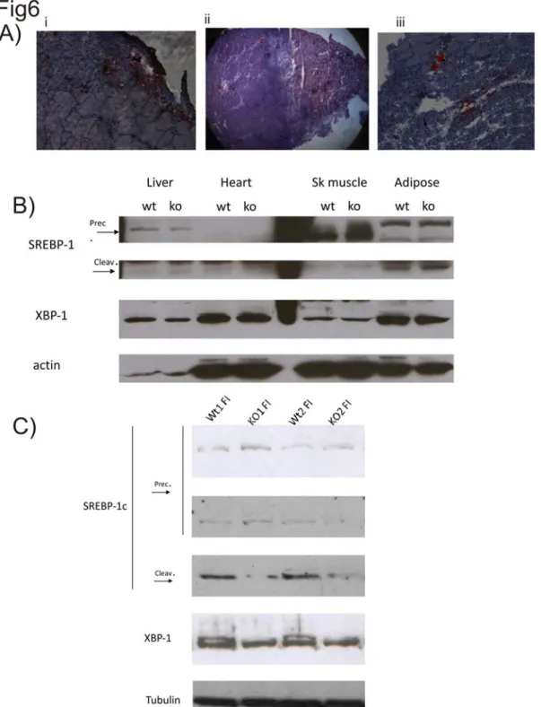
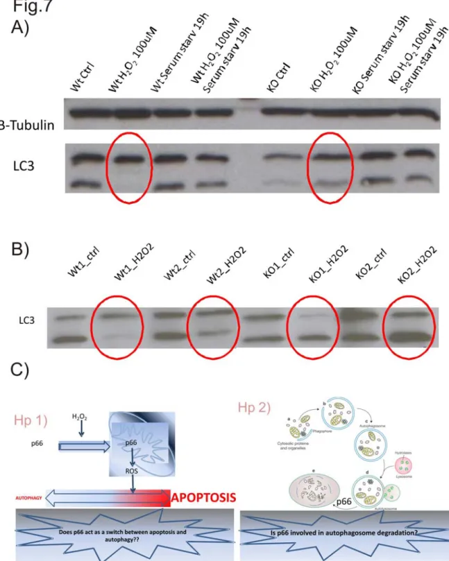
Another protein involved in redox stress regulation and sensitivity is p66, an adaptor protein known to be involved in aging and in many pathological conditions like diabetes and its complications. Many studies in the last years were focused in dissolving the apparent contradiction around p66: why the evolutionary selective pressure conserved this appearently damaging gene? Mice knockout for p66, indeed, show a lifespan extension of 30% compared to the wt mice, p66 Shc-/-mice, moreover, are protected by obesity and by the complications associated with diabetes. Our group investigated the effect of p66 on cell viability showing that, following a redox stress which activates the kinase PKCβ, p66 is phosphorylated on the Ser36 and, in this form, it is recognized by the prolyl isomerase Pin1, which modifies its conformation thus allowing p66 translocation to mitochondria, where it acts as an oxidoreductase and it increases mitochondrial ROS production, thus rendering cells more sensitive to the apoptotic effects of an exogenous redox stress. P66Shc-/-, indeed, are more resistant to the apoptotic effects of H2O2, thus confirming a central role of the protein in the sensitivity to apoptosis (Pinton, Rimessi et al. 2007). The following year our group published an other work showing that hyperglycemia induces an increase in ROS production which activates PKCβ thus inducing the adipogenic differentiation of muscle derived stem cells, which were expected to differentiate into myotubes, thus suggesting a central role for PKCβ in this “transdifferentiation” program (Aguiari, Leo et al. 2008). In both cases ROS-induced PKCβ activation plays a key role and, intuitively, p66 is also involved in the adipogenic transdifferentiation of muscle derived stem cells. The purpose of our work was double: on one hand we wanted to investigate a possible role of p66 in the adipogenic transdifferentiation of muscle-derived stem cells as downstream target of PKCβ and to reproduce our previous observation in an in vivo model; on the other hand we wanted to investigate a possible role of p66 in regulating autophagy and in particular we hypothesized a possible role of p66 as a switch among the apoptotic and the autophagic pathways.