Redistribution of DNA Topoisomerase II
b
After In Vitro
Stabilization of Human Erythroleukemic Nuclei by Heat
or Cu
11
Revealed by Confocal Microscopy
LUCA M. NERI,1,2* ALBERTO M. MARTELLI,3ANDNADIR M. MARALDI2 1Istituto di Anatomia Umana Normale, Universita´ Di Ferrara 44100 Ferrara Italy
2Istituto di Citomorfologia Normale e Patologica del C.N.R., c/o I.O.R., 40137 Bologna, Italy 3Dipartimento di Morfologia Umana Normale, Universita` di Trieste, 34138 Trieste, Italy
KEY WORDS nuclear matrix; topoisomerase; confocal microscopy; 3-D reconstruction
ABSTRACT Using confocal laser scanning microscope and a monoclonal antibody we have examined by means of indirect immunofluorescence techniques the distribution of DNA topoisomer-ase IIb (the 180-kDa nucleolar isoform of topoisomerase II) following stabilization of isolated nuclei by exposure to moderate heat (37° or 42°C) or Cu11. In intact cells the antibody specifically decorated the nucleoli. The same pattern was maintained if nuclei were incubated at 0°C in a buffer containing spermine/spermidine/KCl or stabilized by means of 0.5 mM Cu11for 10 minutes at 0°C
in the same buffer. On the contrary, if stabilization was performed by incubating the nuclei either at 37° or 42°C, the immunoreactivity dispersed all over the nucleus, forming numerous speckles. This phenomenon was not detected if, in addition to spermine/spermidine/KCl, the incubation buffer also contained 5 mM Mg11and the temperature was 37°C. If the stabilization was performed at 42°C,
Mg11failed to maintain the original distribution of DNA topoisomerase IIb, as seen in intact cells. The analysis on 2-D optical section showed the alteration of the nucleolar profile, particularly at 37°C, even when the samples were treated with Mg11. The 3-D reconstruction figured out the irregularity of the surface at 37°C and the variations of the volume occupied by the fluorescent figures. These were in close proximity to each other both in intact cells and in 0°C incubated nuclei; they showed a certain degree of shrinkage in 0°C plus Cu11exposed samples (220% of the volume), and, on the contrary, the labeled structures were scattered in a volume increased two- or threefold when exposed to 37° or 42°C, respectively. The addition of Mg11 restored the original spatial relationship and volume at 37°C, but not at 42°C, where the volumetric analysis showed an increase of about 50%. Our results demonstrate that heat stabilization of isolated nuclei in a buffer without Mg11 (i.e., a technique often employed to prepare the nuclear matrix or scaffold) cannot be
considered an optimal procedure to maintain the original distribution of protein within the nucleus. Microsc. Res. Tech. 36:179–187, 1997. r1997 Wiley-Liss, Inc.
INTRODUCTION
Evidence gained in several laboratories during the last few years indicate that a variety of nuclear func-tions (such as DNA replication and repair, transcrip-tion, and splicing of RNA, etc.) take place within the nucleus not in a diffuse manner but localized at discrete sites (e.g., Antoniou et al., 1993; Jackson et al., 1993, 1994; Nakayasu and Berezney, 1989; Spector et al., 1991; Wansink et al., 1994) even though such a hypoth-esis has been recently challenged (Zhang et al., 1994). For this reason, it is commonly thought that the nucleus should contain an underlying structure respon-sible for maintaining the spatial organization of the aforementioned functions (van Driel et al., 1991). Many investigators feel that such a structure might be repre-sented by the nuclear matrix, a mainly proteinaceous insoluble framework remaining after isolated nuclei are exposed to non-ionic detergents, nucleases, and solutions of high-ionic strength (Berezney, 1984). In-deed, evidence deriving mainly from in vitro experi-ments has shown that this insoluble network could be
involved in genome duplication, RNA synthesis and processing, anchoring of DNA loops, gene expression regulation, protein phosphorylation, and a plethora of other functions (Berezney, 1991). The nuclear matrix is mainly composed from nonhistone proteins, of which several have been characterized over the last years (e.g., Bisotto et al., 1995; Feuerstein et al., 1988; Nickerson et al., 1992; Wan et al., 1994). Ultrastruc-tural studies have shown that the nuclear matrix usually contains three distinct domains: an outer lamina, an inner fibrogranular network, and residual nucleoli (Kaufmann and Shaper, 1984; Maraldi et al., 1986). The nuclear matrix has been prepared from many tissues and cell lines but its existence in vivo is still under debate, because nuclei are exposed to non-physiological environments before the final structure is
*Correspondence to: Dr. Luca M. Neri, Istituto di Anatomia Umana Normale, via Fossato di Mortara 66, 44100 Ferrara, Italy.
L.M. Neri and A.M. Martelli contributed equally to this work. Received 7 January 1996; Accepted in revised form 28 January 1996.
obtained and there is a strong risk of creating in vitro artifacts (e.g., Cook, 1991). Twelve years ago, Evan and Hancock (1985) for the first time showed that when isolated nuclei are briefly exposed to the physiological temperature of 37°C a distinct subset of proteins be-came insoluble in buffers of high-ionic strength (Evan and Hancock, 1985). Such a phenomenon occurs in many cell lines, but despite quite an intensive research, the molecular mechanisms underlying it are far from being elucidated (Berrios and Fisher, 1988; Littlewood et al., 1987; Martelli et al., 1994a).
It is important to emphasize that exposure of isolated nuclei to 37°C in vitro was intentionally used to demon-strate that the nuclear matrix contains specific DNA sequences, called SARs (for Scaffold Associated Re-gions) that would represent stretches of nucleotides, A-T rich (over 70%), conceivably defining the base of DNA loops (Izuarralde et al., 1988; Mirkovitch et al., 1984). Our current interest lies in establishing whether or not the methods used for nuclear matrix isolation can maintain in the final structures the spatial distribu-tion of nuclear polypeptides as seen in whole cells (Martelli et al., 1994b; Neri et al., 1994, 1995). We noticed that while some investigators did not use Mg11 during heat stabilization of isolated nuclei, employing instead a buffer containing KCl and the polycations spermine and spermidine, others included it (e.g., Bel-grader et al., 1991; Izuarralde et al., 1988; Mirkovitch et al., 1984; Neri et al., 1994). The use of Mg11 in solutions used for preparation of nuclei has been criti-cized, because this cation induces chromatin condensa-tion and activacondensa-tion of endogenous nucleases (Cook, 1988; Laval and Bouteille, 1973).
Here, we have examined whether changes in the distribution of a nuclear matrix polypeptide, the 180-kDa isoform of DNA topoisomerase II, or II b, previ-ously found as a component of the nucleolar remnant (Zini et al., 1992, 1994), occur when nuclei from K562 human erythroleukemia cells are stabilized at 37° or 42°C in the absence of Mg11. In addition, we have
verified the influence, on the same antigen, of another well-established stabilizing agent, Cu11(Izuarralde et al., 1988; Mirkovitch et al., 1984).
We demonstrate that when Mg11 is not present
during the heat stabilization, a redistribution of topoisomerase II b takes place. The KCl/spermine/ spermidine buffer thus might not be the optimal choice for studying some proteins belonging to the nuclear matrix. However, the changes can be lowered when Mg11ions are added to the KCl/spermine/spermidine buffer.
These observations were made possible by the optical sectioning ability of confocal microscopy that on 2-D images allowed to identify the profile of fluorescent nucleoli as well as the distance among them and their areas. By 3-D image processing the spatial relationship among the nucleoli was studied including their volume occupancy and surface morphology. By these param-eters (profile, area, distance, surface, spatial relation-ship, and volume) it was possible to describe in detail the effect of thermal stabilization and of Cu11or Mg11
treatment of isolated nuclei.
MATERIALS AND METHODS Cell Culture
K562 human erythroleukemia cells were grown in RPMI-1640 medium supplemented with 10% newborn calf serum at 37°C in a humidified atmosphere contain-ing 5% CO2. They were seeded at a density of 105/ml
and used 4 days later when they reached a density of 106/ml.
Isolation of Nuclei
Cells were washed once in Dulbecco’s phosphate buffered saline (PBS, pH 7.4, free of Mg11and Ca11).
Cells were lysed in a Dounce type tissue homogenizer (Wheaton, Millville, NJ) with 15 strokes using a B pestle in an isolation buffer containing 3.75 mM Tris/ HCl (pH 7.4), 0.05 mM spermine, 0.125 mM spermi-dine, 20 mM KCl, 0.5 mM EDTA/KOH (pH 7.4), 1% thiodiglycol, 1 µg/ml aprotinin and leupeptin plus 0.1% digitonin (water-soluble, Fluka, Buchs, Switzerland) as described by Mirkovitch et al. (1984). Nuclei were immediately checked for cytoplasmic contamination by means of a phase-contrast microscope. Nuclei were pelletted at 400g for 8 minutes and washed twice in nuclear washing buffer (3.75 mM Tris/HCl [pH 7.4], 20 mM KCl, 0.05 mM spermine, 0.125 mM spermidine, 0.1% digitonin, 0.5 mM EDTA/KOH [pH 7.4], 1% thiodi-glycol, 1 mM phenylmethylsulfonyl fluoride plus aproti-nin and leupeptin as above). They were then resus-pended at 200 µg DNA/ml in nuclear washing buffer without EDTA/KOH and stabilized for 20 minutes at either 37° or 42°C. In some cases stabilization was performed with 0.5 mM CuSO4 for 10 minutes at 0°C
(Mirkovitch et al., 1984).
Source of Antibody
The anti-180-kDa nucleolar isoform of DNA topoisom-erase II (topoisomtopoisom-erase II b) was a kind gift by Dr. G.C.B. Astaldi Ricotti, Istituto di Genetica Biochimica ed Evoluzionistica del C.N.R., Pavia, Italy, and its specificity has been demonstrated in previously pub-lished papers (Negri et al., 1992; Zini et al., 1992, 1994).
Immunofluorescent Staining
Cells in PBS and nuclei in nuclear washing buffer were plated onto 0.1% poly-L-lysine-coated glass slides and adhesion was allowed to proceed for 30 minutes at 37°C for cells or at room temperature for nuclei and nuclear matrices. Samples were fixed in methanol for 15 minutes at room temperature. After several washes with PBS, aspecific binding of antibodies was blocked by 30 minutes incubation at 37°C with PBS, 2% bovine serum albumin (BSA), 5% normal goat serum (NGS). Slides were then incubated for 3 hours at 37°C with the primary antibody diluted in PBS, 2% BSA, 5% NGS. After washing three times in PBS, specimens were reacted with a fluorescein (FITC)-conjugated anti-mouse IgG, diluted 1:45 in PBS, 2% BSA, 5% NGS for 1 hour at 37°C. Samples were subsequently washed three times in PBS, stained with 1 µg/ml 48-6-diamidino-2-phenylindole (DAPI) in PBS and mounted in 20 mM
Tris-HCl, pH 8.2, 90% glycerol containing 2.3% of the antifading agent 1,4-diazobicyclo-[2.2.2]-octane.
Confocal Laser Scanning Microscope (CLSM) Analysis
Samples were imaged by a PHOIBOS 1000-SARASTRO (Molecular Dynamics, Sunnyvale, CA) CLSM mounted on a Nikon Optiphot microscope (Ni-kon, Tokyo, Japan). This confocal system was coupled with a 25 mW multiline Argon ion laser as a light source. This laser produces two major lines at 488 and 514 nm. The first one was selected with a band pass filter and was used to reveal FITC signal. The laser power was tuned at 10 mW to obtain the highest light stability and the laser beam was attenuated to 30% of transmission with a neutral density filter to limit bleaching of the FITC fluorescence.
Samples were observed with a3100, 1.4 numerical aperture (NA) planapochromat objective lens. The opti-cal resolution of the confoopti-cal microscope is dependent on the wavelength of the incident light and on the NA of the objective. To obtain the highest resolution we have employed the lowest laser light wavelength suitable and the highest NA objective [(0.46l)/NA]. The refrac-tive index of the immersion oil was 1.518 (Nikon); the oil was dissolved up to a concentration of 20% in the mounting medium to minimize distortion of the confo-cal spot during the laser beam penetration inside the specimen (Carlsson, 1991).
In the detection path the emitted fluorescent light was focused on a back pinhole aperture with a diameter of 50 µm in front of the detector, a photomultiplier tube (PMT). To block any unwanted contribution signal, when observing FITC, a 515 OG long pass filter has been inserted before the PMT, as a barrier filter. The PMT was set at 864 mV. Settings were rigorously maintained for all experiments. Images were acquired, frame by frame, with a scanning mode format of 5123 512 pixels. Pixel values were recorded in the range of 0–255 (8 bits) as previously described. Images were reconstructed as follows: slides were scanned from left to right on the x-y plane moving down with steps of 0.3 µm from top to bottom in the z direction of samples using the motor drive focusing system to a position unaffected by the last horizontal pass. Each frame had a scan time of 0.5 seconds and every optical section was the result of a single scan. The microscope table was set manually so that the first z section was collected just at the top of the structures to be analyzed.
Image Processing Analysis
Digitalized optical sections, i.e., Z series of confocal data (‘‘stacks’’) were transferred from the CLSM to the graphics workstation Indigo Irix XS24 (Silicon Graph-ics, Mountain View, CA) and stored on the graphics workstation with a scanning mode format of 5123 512 pixels and 256 grey levels. The image processing and the volume rendering were performed using the Image-Space software (Molecular Dynamics). To reduce the unwanted background noise generated by the photomul-tiplier signal amplification, all the image stacks were treated with a three-dimensional filter (Gaussian filter-ing) that was carried out on each voxel, with a mask of 3 pixel in the x, y, and z direction (33 3 3 3).
The Z-series sections were then vectorized and pro-jected consecutively to produce a three-dimensional representation of the whole specimen (resulting from 21 sections) (Neri et al., 1992). A projection method, called ‘‘Surface Shading’’ was employed. This method models the sample as if it had an opaque, reflective surface and shows the details of the surface.
A software tool to analyze distances, areas, and volumes of sections or of section series has been used. Distances along a straight line have been measured to obtain the length of the line in pixels and micrometers, the x, y pixel location of the start and end points, and the starting and ending section numbers. By drawing a free-form object, an arbitrary area has been generated that allows for measurements on a single section of a series or on each section of a series, and data about the area measured in µm2were collected. The volume was
measured by a tool that allows the drawing of an arbitrary area (around the profile of the object) and execution the measurement on the whole section series. The data are obtained in µm3.
The FITC signal was elaborated to optimize the contrast, the brightness, and the intensity of the im-ages. Photographs were taken by a digital video re-corder Focus ImageCorder Plus (Focus Graphics, Fos-ter City, CA) using 100 ASA TMax black and white film (Kodak Limited, Rochester, NY).
RESULTS
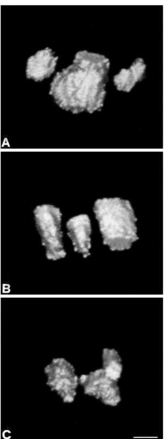
The distribution of topoisomerase IIb was studied by means of a specific monoclonal antibody and CLSM. In agreement with previous reports (Zini et al., 1994; Neri et al., 1994, 1995), in intact cells the antibody stained the nucleoli, while a very faint, sparse and speckled reaction was seen in the nucleoplasm (Fig. 1A). The number of nucleoli was usually two or three and the immunoreaction was characterized by a larger area associated with smaller ones or by two/three areas of the same size with a fairly regular, round shape. The edge of the immunofluorescent areas displayed some small pits, but in general it was quite regular. Such a pattern was well maintained in nuclei kept at 0°C for 20 minutes (Fig. 1B). Also in nuclei kept for 10 minutes in the presence of Cu11(0.5 mM), the fluorescent pattern was essentially unchanged in comparison with intact cells (Fig. 1C).
On the other hand, if the stabilization was performed at 37°C for 20 minutes, in the absence of Mg11, it was possible to see that the immunofluorescent pattern consisted of numerous speckles, of different size and shape, dispersed all over the nuclear area (Fig. 2A). It should be noted that all the speckles had a very irregular contour with finger-like protrusions.
When the incubation temperature was 42°C we no-ticed a similar number of speckles distributed over an even larger nuclear area. The shape of the speckles was less irregular than at 37°C, but numerous tiny dots were also detectable, dispersed within the nucleus (Fig. 2B).
It is important to emphasize that transmission elec-tron microscope analysis revealed in heat-stabilized nuclei that the nucleoli did not appear different in number and in size when compared with nuclei kept at 0°C (data not shown). Such an observation ruled out the possibility that the dispersion of the fluorescent
immu-noreactivity was due to a fragmentation of nucleoli occurring in the absence of Mg11.
If the 37°C incubation was performed in the presence of 5 mM Mg11, in addition to spermine/spermidine/
KCl, the fluorescent pattern due to topoisomerase IIb was reminiscent of that seen when nuclei were incu-bated at 0°C, even though the positivity was restricted to somewhat smaller areas, corresponding to nucleoli; some positivity was found in the nucleoplasm, appear-ing as small dots of different sizes (Fig. 2C). The edges of the nucleoli were very irregular. Nevertheless, the presence of Mg11was not sufficient to avoid the disper-sion of the immunoreactivity, as well as the irregularity of the shapes, when the stabilization was executed at 42°C (Fig. 2D). In the nuclear interior some small, randomly distributed spots were observed.
These observations were corroborated by 3-D recon-struction of cells and nuclei. In the Surface Shading modelling, the imaginary light rays are on the axis of the viewer’s line of sight. To determine which struc-tures should be considered as part of the surface, each voxel is taken into account if it has a value larger than the threshold and if it is on an edge. The computer checks the 26 surrounding voxels in a 3 3 3 3 3 neighbourhood; at least one neighbouring voxel on one side must have a lower value than the voxel in question. If the voxel passes both tests, it is considered part of the surface. If it does not, the next voxel along the ray is tested. The calculation of the surface orientation is based on the intensity gradient. In particular, the angle of a line perpendicular to the surface (the surface normal) is calculated for each surface pixel. It is based on the direction of the intensity gradient at that point. The surface is then shaded so that surfaces with a large tilt are dark and surfaces perpendicular to the viewer’s line of sight are bright. It is important to set correctly the range of pixel intensities included in the calcula-tion. If the threshold is too high, part of the sample could be excluded from the 3-D reconstruction. On the contrary, background noise recorded from areas physi-cally outside the specimen may be considered part of the surface, because the imaginary light ray reaches these voxels before hitting the specimen. To find this threshold level, a histogram of all pixel intensities has been made to analyze in detail the intensity values representing background noise that were not included in the projection calculations.
The 3-D distribution of nucleolar topoisomerase IIb showed a close spatial relationship in intact cells, as well as in 0°C treated nuclei and in 0°C exposed to Cu11 (Fig. 3). In the last case nucleoli were closer one to each other. The surface-shaded reconstruction of the immu-nofluorescence is viewed from different angles (of lati-tude, longilati-tude, and rotation) to better render not only the spatial distribution but also the texture of nucleolar surface. It could be visualized as a regular, round surface sometimes grooved along the nucleolus major axis. Some small protuberances emerged from the nucleoli and were more pronounced at the upper or
Fig. 1. A: Topoisomerase IIb distribution in intact cells. One major fluorescent nucleolar area is accompanied by two smaller areas, one of which is not rounded but almost elliptical. The profile of the fluores-cence is fairly regular and the three nucleolar areas are closely associated. B: 0°C exposed nuclei. The pattern is almost identical to that described in intact cells. C: Nuclei treated at 0°C with Cu11: three
rounded nucleoli appear in very close proximity, almost touching each other. Bar5 1 µm.
lower extremity (Fig. 3A,B), whereas in samples ex-posed to Cu11 they were detected also in the central region (Fig. 3C).
Changes occurring for the 37° and 42°C treated nuclei can be better demonstrated by the 3-D reconstruc-tion. The immunofluorescence appeared scattered throughout a larger nuclear volume, especially in the case of 37°C incubation (Fig. 4A). The single immunoflu-orescence figures were well spaced one from each other and occupied the majority of the nucleoplasm. However, the volume of every single figure is much smaller and more irregular than the 3-D reconstruction of the nucleoli of intact cells. In fact, the surface showed not only finger-like protrusions but also round spots emerg-ing from the edges of the structures and depressions that sometimes appeared to be very deep. An analogous nuclear volume was occupied by the figures in the case
of 42°C treatment and their number was even higher (Fig. 4B). The surface was not as irregular as in the 37°C incubated samples, with some round or finger-like protrusions but no depressions.
The addition of Mg11during the 37°C incubation was capable of preventing the scattering effect of the heat treatment and reduced to three the number of nucleoli, which were characterized by many rounded structures protruding from the surface (Fig. 4C). Various finger-like structures emerged from the extremities of the figures. Some small round spots were also observable in the nucleoplasm. On the contrary, the same treatment did not block the fragmentation of the immunofluores-cence taking place at 42°C (Fig. 4D). The solid figures had a fairly regular round shape, with few rounded and finger-like protrusions. The Mg11 only partially pre-vents the diffusion of the fluorescence to the
nucleo-Fig. 2. A: Topoisomerase IIb distribution in 37°C treated nuclei. Numerous speckles, characterized by very irregular edges, are distrib-uted on a large nuclear area. B: Treatment at 42°C: a similar fluorescent scattering is observable, but single fluorescent areas have a more rounded profile. In addition many tiny dots are present in the nucleoplasm. C: Addition of Mg11at 37°C. In isolated nuclei a close
association among three fluorescent structures is restored, but their
contour is characterized by very irregular protuberances and pits. The nucleoplasm is labeled by spots of various size, randomly distributed.
D: Nuclei exposed to 42°C with the addition of Mg11. Cation addition
is unable to prevent the scattering of topoisomerase IIb. The shape of the fluorescent areas is very irregular as described for the 37°C plus Mg11. Some dots are observable in the nucleoplasm. Bar5 1 µm.
plasm both in terms of speckled, irregularly sized figures and of spotted, rounded structures.
We also performed a 2-D and 3-D image analysis of the samples as shown in Table 1. The distance between
the two most distant immunofluorescent points has been measured in each nucleus, as a representative sample of a larger statistical analysis (data not shown). It was very similar in intact cells, 0°C, 0°C plus Cu11, and 37°C, Mg11 treated nuclei. This distance was almost doubled in nuclei exposed to 37° or 42°C without Mg11and increased about 50% in 42°C, Mg11treated nuclei. The area on which the immunofluorescence was distributed showed a threefold increase in 37° and 42°C treated nuclei and a 30% increase in 42°C plus Mg11 nuclei. The most interesting information was obtained from the volume analysis. In fact, in the intact cells and the 0°C nuclei, the values were similar, but the 0°C plus Cu11nuclei showed a certain degree (around 20%) of shrinkage in the global volume of the solid structures. As far as heat treatment was concerned we recorded a twofold increase in the volume of 37°C incubated samples, while a threefold increment was measured with an incubation temperature of 42°C. At 37°C, the addition of Mg11reduced the volume to that of control
nuclei while, at 42°C, the cations enlarged it around 50% over the control value.
DISCUSSION
By definition, the nuclear matrix, or scaffold, is the final product of harsh extraction techniques, during which cells or nuclei are exposed to highly unphysiologi-cal environments. Therefore, it is of the utmost impor-tance to rule out that during these procedures artifacts are not induced. To this end, a critical control is to assess whether or not the original distribution of nuclear constituents, as seen in intact cells, is maintained during the various steps of the extraction protocols. Surprisingly enough, however, this has been done when the matrix is prepared in situ from adherent-growing cells (Kallajoki et al., 1991; Nakayasu and Berezney, 1991; Staufenbiel and Deppert, 1984), but only rarely in matrices obtained in a more traditional way, i.e., by first isolating nuclei from cells growing as suspension cul-tures, followed by nuclease digestion and by extraction with different agents to remove soluble nuclear pro-teins (Neri et al., 1994, 1995). In the past, major concern had been usually expressed regarding the type of extracting agent (salts, detergents, or polyanions) employed to remove soluble nuclear proteins (e.g., Cook, 1988; Mirkovitch et al., 1984). More recently, we have focused our attention on the nuclear stabilization step that precedes extraction. Our results have shown that changes in the distribution of nuclear proteins can occur during the stabilization step of isolated nuclei performed at 37°C. Here, we have looked at the effect of heat stabilization (37° or 42°C) when nuclei are incu-bated in a buffer that does not contain Mg11,
substi-Fig. 3. A: 3-D reconstruction (Surface Shading) of topoisomerase II
b nucleolar distribution in intact cells. The 2-D rounded aspect is substituted by a more elliptical shape showing a major axis. The surface is fairly homogeneous and is characterized by few small rounded beads and by some grooves. The volume rendering clarifies the spatial relationship among these three structures that occupy distinct nuclear regions but are neighbouring each other with a non-random distribution. B: The Surface Shading reconstruction of topoisomerase IIb in isolated nuclei exposed to 0°C is very similar to that of intact cells. C: Addition of Cu11. Appearance of some more
irregularities on the surface, including some small round spots and pits especially at the extremities. Bar5 1 µm.
tuted by spermine/spermidine/KCl. Such a buffer is routinely employed to obtain matrices suitable for studying interactions between scaffold proteins and specific DNA sequences, SARs, that could represent
stretches of DNA through which the chromatin is bound to the nucleoskeleton.
To our surprise, we have seen that heat stabilization of isolated nuclei kept in a similar buffer could induce a massive redistribution of DNA topoisomerase IIb. On the contrary, stabilization with Cu11does not produce
such a marked rearrangement of the antigen. Thus, it seems that such a stabilization could be preferentially used when studying the properties of this nucleolar matrix component. Also, when nuclei are kept at 0°C in the absence of Cu11, there is no redistribution of topoisomerase IIb, but in this case, when extraction is subsequently performed (for example with a ionic deter-gent), no internal matrix or nucleolar remnants are seen (data not shown), in agreement with the literature (Belgrader et al., 1991; Lude´rus et al., 1992).
Fig. 4. A: Volume rendering reconstruction of the spatial
distribu-tion of topoisomerase IIb after 37°C treatment. Irregular edges with finger-like protuberances, pits, and some very evident and deep grooves (arrow) can be easily observed. Also noticeable is the very enlarged volume occupied by the reconstructed structures. The close association of the figures is completely lost and they are randomly scattered in the nuclear volume. B: 42°C exposure induced in a very large volume a very similar scattering of the fluorescent structures that were characterized by a smoother surface than in A. C: The 3-D
reconstruction of topoisomerase II b immunofluorescence in 37°C exposed nuclei plus Mg11shows a very irregular surface as in 37°C
alone. Three structures only are observable in close association, but in the nucleoplasm some rounded structures are present. D: A fairly irregular surface is observable at 42°C plus Mg11after 3-D image
processing. Many structures of smaller size are distributed in a volume larger than in C but smaller than in B. Also some rounded structures are present in the nucleoplasm. Bar5 1 µm.
TABLE 1. Analysis of volume, area, and distance on intact cells and nuclei exposed to different treatments1
Sample Volume (µm3) Area (µm2) Distance (µm)
Cell 412 52 13.9 Nuclei 0°C 430 65 11.6 Nuclei 0°C Cu11 347 48 11.1 Nuclei 37°C 908 180 20.4 Nuclei 42°C 1,220 199 21.7 Nuclei 37°C Mg11 393 56 11.5 Nuclei 42°C Mg11 654 76 16.2
The use of CLSM analysis coupled to 3-D reconstruc-tion gave new insight into DNA topoisomerase II b distribution before and after the different treatments. The sections obtained through the middle plane of the fluorescent structures demonstrated with sharp detail that isolated nuclei contain two or three fluorescent nucleolar areas and also showed the relationship among them. In fact, they appeared closely related one to each other in intact cells, as well as in 0°C and 0°C Cu11 incubated samples. It has been previously shown that this antibody in intact cells is able to label the regions corresponding to nucleoli (Zini et al., 1994). The contour of the nucleoli, studied by CLSM, became a new param-eter of analysis, both in terms of distances and of areas. In a previous work the Ag-NOR proteins were analyzed by means of confocal microscopy but they displayed a beaded organization and it was impossible to trace back to the nucleolar profile (Robert-Fortel et al., 1993). The heat treatment scattered the DNA topoisomerase IIb distribution and furthermore the outline of every single fluorescent area became more irregular. CLSM allowed us also to observe that Mg11treatment at 37°C main-tained the proximity among the nucleoli but not their original profile.
These observations were strengthened by the 3-D analysis that showed for the first time, as far as we know, the spatial relationship among nucleoli within a single nucleus. The surface shaded reconstruction evi-denced that the distribution of this enzyme is very sensitive to any manipulation of the environment. In fact, 2-D analysis has demonstrated the aforemen-tioned variations (profile, area, distance), but in addi-tion the 3-D reconstrucaddi-tion was useful to confirm the observed increase of irregularity of the nucleolar edges/ profile and to display the spatial increase of nuclear volume occupied by the fluorescent areas corresponding to DNA topoisomerase IIb.
The effects of the buffer containing polyamines are somewhat unexpected because it has long been thought that Mg11could be held responsible for the formation of
artifacts during matrix preparations (Cook, 1988). Thus, it would appear desirable to avoid using this divalent cation if the matrix needs to be isolated, by substituting it with polyamines such as spermine and spermidine which, in addition, have been reported to be compo-nents of the nucleus in vivo (Hougaard et al., 1987; Quemener et al., 1992). Moreover, experiments per-formed with polyamine-depleting agents, have hinted at a possible physiological role played by these multiva-lent cations in regulating interactions between the nuclear matrix and negatively charged DNA (Basu et al., 1986). In this connection it should be recalled that Sen and Crothers (1986) have reported a similar effect for Mg11 and polyamines as far as compacting of chromatin was concerned but our data clearly show the existence of marked differences on the distribution of a nucleolar antigen. Mg11can oppose the effect of
poly-amines during heat stabilization, but only partially and when the incubation temperature is performed at 37°C, while at 42°C they do not produce any sizable effect. As far as Cu11are concerned, we notice that our data are in agreement with the suggestion that this cation might offer specific properties in regulating nuclear structure in vivo (Sen and Crothers, 1986).
It should be emphasized that our previous results have demonstrated that an isoosmotic buffer (0.25 M sucrose) containing 5 mM Mg11can be used for stabiliz-ing nuclei by heat (37°C) or the cross-linkstabiliz-ing agent sodium tetrathionate without affecting at all the spatial distribution of DNA topoisomerase II b (Neri et al., 1994, 1995). Therefore, we believe that such a solution should be considered the optimal choice for analyzing this protein, whereas polyamine containing buffers should not be used.
ACKNOWLEDGMENTS
The authors thank Mr. Aurelio Valmori and Mrs. Giovanna Baldini for the illustrations. This work was supported by Italian CNR grant CT12.00184.94, PF ACRO, Italian MURST 60% grant to Universita` di Ferrara, and Trieste and Fondi AIRC 95.
REFERENCES
Antoniou, M., Carmo-Fonseca, M., Ferreira, J., and Lamond, A.I. (1993) Nuclear organization of splicing snRNPs during differentia-tion of murine erythroleukemia cells in vitro. J. Cell Biol., 123:1055– 1068.
Basu, H.S., Wright, W.D., Deen, D.F., Roti-Roti, J., and Marton, L.J. (1993) Treatment with a polyamine analog alters DNA-matrix association in HeLa cell nuclei: A nucleoid halo assay. Biochemistry, 32:4073–4076.
Belgrader, P., Siegel, A.J., and Berezney, R. (1991) A comprehensive study on the isolation and characterization of the HeLa S3 nuclear matrix. J. Cell Sci., 98:281–291.
Berezney, R. (1984) Organization and functions of the nuclear matrix. In: Chromosomal Nonhistone Proteins, Vol. IV. L.S. Hnilica, ed. CRC Press, Boca Raton, FL, pp. 119–180.
Berezney, R. (1991) The nuclear matrix: A heuristic model for investi-gating genomic organization and function in the cell nucleus. J. Cell. Biochem., 47:109–123.
Berrios, S., and Fisher, P.A. (1988) Thermal stabilization of putative karyoskeletal protein-enriched fractions from Saccharomyces
cerevi-siae. Mol. Cell. Biol., 8:4573–4575.
Bisotto, S., Lauriault, P., Duval, M., and Vincent, M. (1995) Colocaliza-tion of a high molecular mass phosphoprotein of the nuclear matrix (p255) with spliceosomes. J. Cell Sci., 108:1873–1882.
Carlsson, K. (1991) The influence of specimen refractive index, detector signal integration and non-uniform scan speed on the imaging properties in confocal microscopy. J. Microsc., 163:167–178. Cook, P.R. (1988) The nucleoskeleton: artifact, passive framework or
active site? J. Cell Sci., 90:1–6.
Cook, P.R. (1991) The nucleoskeleton and the topology of DNA replication. Cell, 66:627–635.
Evan, G.I., and Hancock, D.C. (1985) Studies on the interaction of the human c-myc protein with cell nuclei: p62c-mycas a member of a
discrete subset of nuclear proteins. Cell, 43:253–261.
Feuerstein, N., Spiegel, S., and Mond, J.J. (1988) The nuclear matrix protein, numatrin (B23), is associated with growth factor-induced mitogenesis in Swiss 3T3 fibroblasts and with T lymphocyte prolif-eration stimulated by lectins and anti-T cell receptor antibody. J. Cell Biol., 107:1629–1642.
Hougaard, D.M., Bolund, L., Fujiwara, K., and Larsson, L.I. (1987) Endogenous polyamines are intimately associated with highly con-densed chromatin in vivo. A fluorescence cytochemical and immuno-cytochemical study of spermine and spermidine during the cell cycle and in reactivated nuclei. Eur. J. Cell Biol., 44:151–155.
Izuarralde, E., Mirkovitch, J., and Laemmli, U.K. (1988) Interaction of DNA with nuclear scaffold in vitro. J. Mol. Biol., 200:111–125. Jackson, D.A., Hassan, B.A., Errington, R.J., and Cook, P.A. (1993)
Visualization of focal sites of transcription within human nuclei. EMBO J., 12:1059–1065.
Jackson, D.A., Baljee, A.S., Mullenders, L., and Cook, P.R. (1994) Sites in human nuclei where DNA damaged by ultraviolet light is repaired: Visualization and localization relative to the nucleoskel-eton. J. Cell Sci., 107:1745–1752.
Kallajoki, M., Weber, K., and Osborn, M. (1991) A 210 kDa nuclear matrix protein is a functional part of the mitotic spindle; a microin-jection study using SPN monoclonal antibodies. EMBO J., 10:3351– 3362.
nuclear proteins reversibly stabilized by the sulfhydryl cross-linking reagent tetrathionate. Polypeptides of the nuclear matrix. Exp. Cell Res., 155:477–495.
Laval, M., and Bouteille, M. (1973) Synthetic activity of isolated rat liver nuclei. I. Ultrastructural activity study at various steps of isolation. Exp. Cell Res., 76:337–348.
Littlewood, T.D., Hancok, D.C., and Evan, G.I. (1987) Characteriza-tion of a heat shock-induced insoluble complex in the nuclei of cells. J. Cell Sci., 88:65–72.
Lude´rus, M.E.E., de Graaf, A., Mattia, E., den Blauween, J.L., Grande, M.A., de Jong, L., and van Driel, R. (1992) Binding of matrix attachment regions to lamin B1. Cell, 70:949–959.
Maraldi, N.M., Marinelli, F., Cocco, L., Papa, S., Santi, P., and Manzoli, F.A. (1986) Morphometric analysis and topological organi-zation of nuclear matrix in freeze-fractured electron microscopy. Exp. Cell Res., 163:349–362.
Martelli, A.M., Neri, L.M., Zamai, L., Bareggi, R., Manzoli, L., and Cocco, L. (1994a) 6-iodoacetamidofluorescein labelling to assess the state of sulfhhydryl groups after thermal stabilization of isolated nuclei. Histochem. J., 26:179–188.
Martelli, A.M., Bareggi, R., Riederer, B.M., Marugg, R.A., and Nar-ducci, P. (1994b) The effect of in vitro heating on the distribution of nuclear matrix polypeptides in HeLa cells. Cell Biol. Int., 18:151– 158.
Mirkovitch, J., Mirault, M.-E., and Laemmli, U.K. (1984) Organiza-tion of the higher-order chromatin loop: Specific DNA attachment sites on nuclear scaffold. Cell, 39:223–232.
Nakasayu, H., and Berezney, R. (1989) Mapping replicational sites in the eucaryotic cell nucleus. J. Cell Biol., 108:1–11.
Nakasayu, H., and Berezney, R. (1991) Identification of the major nuclear matrix proteins. Proc. Natl. Acad. Sci. U.S.A., 88:10312– 10316.
Negri, C., Chiesa, R., Cerino, A., Bestagno, M., Sala, C., Zini, N., Maraldi, N.M., and Astaldi-Ricotti, G.C.B. (1992) Monoclonal anti-bodies to human DNA topoisomerase I and the two isoforms of DNA topoisomerase II: 170- and 180-kDa isozymes. Exp. Cell Res., 200:452–459.
Neri, L.M., Martelli, A.M., Previati, M., Valmori, A., and Capitani, S. (1992) From two dimensional to three dimensional analysis by confocal microscopy. Liver, 12:268–279.
Neri, L.M., Santi, S., Marugg, R.A., Riederer, B.M., Capitani, S., Cataldi, A., and Martelli, A.M. (1994) In vitro heat exposure induces a redistribution of nuclear matrix proteins in human K562 erythro-leukemia cells. Exp. Cell Res., 213:275–285.
Neri, L.M., Riederer, B.M., Marugg, R.A., Capitani, S., and Martelli,
A.M. (1995) The effect of sodium tetrathionate stabilization on the distribution of three nuclear matrix proteins in human K562 erythroleukemia cells. Histochem. Cell Biol., 104:29–36.
Nickerson, J.A., Krockmalnic, G., Wan, K.M., Turner, C.D., and Penman, S. (1992) A normally masked nuclear matrix antigen that appears at mitosis on cytoskeleton filaments adjoining chromo-somes, centrioles and midbodies. J. Cell Biol., 116:977–987. Quemener, V., Blanchard, Y., Lescoat, D., Havouis, R., and Moulinoux,
J.P. (1992) Depletion in nuclear spermine during spermatogenesis, a natural process of cell differentiation. Am. J. Physiol., 263:343–347. Robert-Fortel, I., June´ra, H.R., Ge´raud, G., and Hernandez-Verdun, D. (1993) Three-dimensional organization of the ribosomal genes and Ag-NOR proteins during interphase and mitosis in PtK cells studied by confocal microscopy. Chromosoma, 102:146–157.
Sen, D., and Crothers, D.M. (1986) Condensation of chromatin: Role of multivalent cations. Biochemistry, 25:1495–1503.
Spector, D.L., Fu, X.-D., and Maniatis, T. (1991) Associations between distinct pre-mRNA splicing components and the cell nucleus. EMBO J., 10:3467–3481.
Staufenbiel, M., and Deppert, W. (1984) Preparation of nuclear matrices from cultured cells: Subfractionation of nuclei in situ. J. Cell Biol., 98:1886–1894.
van Driel, R., Humbel, B., and de Jong, L. (1991) The nucleus: A black box being opened. J. Cell. Biochem., 47:311–316.
Wan, K.M., Nickerson, J.A., Krockmalnic, G., and Penman, S. (1994) The B1C8 protein is in the dense assemblies of the nuclear matrix and relocates to the spindle and pericentriolar filaments at mitosis. Proc. Natl. Acad. Sci. U.S.A., 91:594–598.
Wansink, D.G., Manders, E.E.M., van der Kraan, I., Aten, J.A., van Driel, R., and de Jong, L. (1994) RNA polymerase II transcription is concentrated outside replication domains throughout S-phase. J. Cell Sci., 107:1449–1456.
Zhang, G., Taneja, K.L., Siger, R.H., and Green, M.R. (1994) Localiza-tion of pre-mRNA splicing in mammalian nuclei. Nature, 372:809– 812.
Zini, N., Martelli, A.M., Sabatelli, P., Santi, S., Negri, C., Astaldi-Ricotti, G.C.B., and Maraldi, N.M. (1992) The 180-kDa isoform of topoisomerase II is localized in the nucleolus and belongs to the structural elements of the nucleolar remnant. Exp. Cell Res., 200:460–466.
Zini, N., Santi, S., Ognibene, A., Bavelloni, A., Neri, L.M., Valmori, A., Mariani, E., Negri, C., Astaldi-Ricotti, G.C.B., and Maraldi, N.M. (1994) Discrete localization of different DNA topoisomerases in HeLa and K562 cell nuclei and subnuclear fractions. Exp. Cell Res., 210:336–348.