https://doi.org/10.1177/1098612X19868029 Journal of Feline Medicine and Surgery 1 –10
© The Author(s) 2019 Article reuse guidelines:
sagepub.com/journals-permissions DOI: 10.1177/1098612X19868029 journals.sagepub.com/home/jfm This paper was handled and processed by the European Editorial Office (ISFM) for publication in JFMS
Detection of multidrug resistance
and
extended-spectrum/plasmid-mediated AmpC beta-lactamase
genes in Enterobacteriaceae isolates
from diseased cats in Italy
Francesco Lo Piccolo
1*, Adriana Belas
2, Maria Foti
1,
Vittorio Fisichella
1, Cátia Marques
2and Constança Pomba
2Abstract
Objectives The aim of this study was to determine the antimicrobial susceptibility of Enterobacteriaceae isolated from cats affected by diseases commonly encountered in practice, and to characterise the third-generation cephalosporin (3GC)-resistance molecular mechanisms involved.
Methods Clinical samples (n = 100) included 58 rectal swabs from cats with diarrhoea, 31 nasal swabs from cats with clinical signs of upper respiratory tract disease, four ear swabs from cats with otitis, three conjunctival swabs from cats with conjunctivitis, two oral swabs from cats with stomatitis, one swab from a skin abscess and one urine sample from a cat with cystitis. A total of 125 Enterobacteriaceae were isolated from 90 cats. Escherichia
coli was the most frequently isolated species (n = 65), followed by Enterobacter species (n = 20), Proteus species (n = 13), Citrobacter species (n = 12) and others (n = 15). Bacterial susceptibility testing was performed with respect to eight antimicrobial classes. Beta(β)-lactamase genes were identified by PCR and nucleotide sequencing. Results Overall, the higher frequency of resistance was to amoxicillin–clavulanate (61.3%), trimethoprim/ sulfamethoxazole (33.6%) and cefotaxime (32.8%). Thirty-six percent of the isolates (n = 45) were resistant to 3GCs. Of these isolates, 34 were tested by PCR and nucleotide sequencing and 23 were confirmed as encoding β-lactamase genes. Fourteen 3GC-resistant isolates harboured extended-spectrum β-lactamases (ESBLs) belonging to groups CTX-M-1 (n = 12, two of which were CTX-M-79), CTX-M-2 (n = 1) and CTX-M-9 (n = 1), as well as SHV-12 (n = 1) and TEM-92 (n = 1). Nine isolates had CMY-2 plasmid-mediated AmpC β-lactamases (pAmpC). Thirty-one percent (n = 39) of the isolates were multidrug-resistant (MDR) and were isolated from 34% (n = 31/90) of the cats.
Conclusions and relevance A high frequency of MDR and ESBL/pAmpC β-lactamase-producing Enterobacteria-ceae were detected among bacteria isolated from a feline population in southern Italy with a variety of common clinical conditions, which poses limitations on therapeutic options for companion animals. We describe the first detection of CTX-M-79 and TEM-92 ESBL genes in isolates from cats.
Keywords: Multidrug resistance; Enterobacteriaceae; beta-lactamases; gastrointestinal disease; upper respiratory disease
Accepted: 11 July 2019
1Section of Microbiology and Infectious Diseases, Department of Veterinary Sciences, University of Messina, Messina, Italy 2CIISA, Centre of Interdisciplinary Research in Animal Health, Faculty of Veterinary Medicine, University of Lisbon, Lisbon, Portugal
* Current address: Clinic of Small Animal Medicine, Centre for Clinical Veterinary Medicine, Faculty of Veterinary Medicine, Ludwig Maximilian University of Munich, Germany
Corresponding author:
Constança Pomba DVM, MSc, PhD, Laboratory of Antibiotic Resistance, CIISA, Centre of Interdisciplinary Research in Animal Health, Faculty of Veterinary Medicine, University of Lisbon, Avenida da Universidade Técnica, Polo Universitário Alto da Ajuda, Lisbon 1300-477, Portugal
Email: [email protected]
Introduction
Members of the family Enterobacteriaceae can be isolated from healthy and diseased companion animals suffering from several clinical conditions.1 Resistance to antimicro-bials in these microorganisms, namely to third-generation cephalosporins (3GCs), is an increasing global public health threat for both humans and animals.2,3 Companion animals represent an important element in the ecology of antimicrobial resistance through close contact with humans.4 In addition, a selection pressure is determined by mutual exposure of humans and pets to antimicrobial agents for the treatment and prophylaxis of disease.5
In Enterobacteriaceae, beta(β)-lactamases are the most frequent mechanism of resistance to 3GCs. These are con-sidered the highest priority critically important antimicro-bials for humans, and resistance is frequently associated with the production of β-lactamases.6,7 The most impor-tant β-lactamases among human and animal Enterobac-teriaceae are extended-spectrum β-lactamases (ESBLs), plasmid-mediated AmpC β-lactamases (pAmpCs) and carbapenemases.8 Plasmid-encoded β-lactamases often carry genes encoding resistance mechanisms to other anti-microbial classes, with the consequent phenomenon of multidrug resistance, which may lead to failure of antimi-crobial treatment.9 β-Lactamase-producing Enterobact-eriaceae in companion animals have been reported worldwide, both in healthy10–14 and diseased animals.15–22
Cats are the most represented species among house-hold pet animals in Europe.23 Yet, most studies describ-ing the occurrence of ESBL-producdescrib-ing Enterobacteria- ceae in cats also included other domestic animals such as dogs, horses and farm animals.12,13,18,24–32 Moreover, in these and other studies, isolates were obtained from tissues samples at necropsy,28 faeces from healthy cats12,13,33,34 or from urinary tract infections.32,35,36 Susceptibility testing results from canine and feline bac-terial isolates are frequently presented together.13 Until now, only one study has been conducted that included samples obtained exclusively from cats.36
This study aimed to investigate the antimicrobial sus-ceptibility of Enterobacteriaceae isolates from a popula-tion of cats affected by diseases commonly encountered in practice. Furthermore, 3GC resistance was evaluated, and the molecular mechanisms of resistance were characterised.
Materials and methods
Study design and samplingFrom November 2014 to February 2015, samples were collected from 100 European domestic shorthair cats admitted to the Veterinary Teaching Hospital of the Department of Veterinary Sciences of Messina (Italy) and to private veterinary practices in the cities of Palermo and Messina (Italy). Only one sample per animal was collected. Sampling included 58 rectal swabs from cats
with diarrhoea (gastrointestinal disease [GID]), 31 nasal swabs from cats with clinical signs of upper respiratory tract disease (URTD), four ear swabs from cats with oti-tis, three conjunctival swabs from cats with conjunctivi-tis, two oral swabs from cats with stomaticonjunctivi-tis, one swab from a skin abscess and one urine sample from a cat with cystitis.
Enterobacteriaceae isolation and identification
Swabs were enriched in buffered peptone water and then subcultured onto MacConkey agar overnight at 37°C. All bacteria showing distinct morphology in MacConkey agar were isolated. The bacterial isolates were identified by matrix-assisted laser desorption/ ionisation–time of flight (MALDI–TOF) mass spectrom-etry, designed to provide rapidly an accurate and relia-ble bacterial identification. The resulting spectra were analysed with a VITEK MS system (bioMérieux), using Axima (Shimadzu) software and the Spectral ARchive and Microbial Identification System (SARAMIS) data-base (AnagnosTec). The spectra were acquired in posi-tive linear mode in the range of 2000–20,000 m/z. The isolated colonies were seeded in a 48-well metal plate with disposable loops, using as a reference strain
Escherichia coli ATCC 8739 (Biomérieux Italia). Antimicrobial susceptibility testing
Susceptibility testing and interpretation were performed using the disc diffusion method according to Clinical and Laboratory Standards Institute guidelines.37,38
The following antimicrobial discs were used: amikacin 30 µg; amoxicillin–clavulanate 30 µg; aztreonam 30 µg; cefotaxime 30 µg; ceftazidime 30 µg; ceftriaxone 30 µg; ciprofloxacin 5 µg; chloramphenicol 30 µg; meropenem 10 µg; and trimethoprim/sulfamethoxazole 25 µg.
Isolates displaying resistance to at least one anti-microbial in at least three different antianti-microbial classes were considered as multidrug resistant (MDR).39
Detection of β-lactam resistance genes in Enterobacteriaceae and phylogenetic group determination of E coli
3GC-resistant isolates (n = 34) were screened by PCR for the presence of β-lactamase genes of groups CTX-M, and SHV and TEM.40,41 The bla
CTX-M-group1, blaCTX-M-group9,
blaCTX-M-group2, blaCTX-M-group8 and blaCTX-M-group25 genes were identified by PCR with specific primers, and positive amplicons were submitted for nucleotide sequencing.42
Furthermore, a multiplex PCR for the detection of pAmpC-coding genes of CIT, FOX, DHA, MIR, ACT and MOX groups was performed, using specific prim-ers as previously described.43 Isolates that were positive for the group CIT were submitted for nucleotide sequencing after a specific PCR targeting the entire
Escherichia coli harbouring β-lactamase genes were categorised into phylogroups (A, B1, B2 or D) by multi-plex PCR.44
Statistical analysis
Results are expressed as percentages. A Fisher’s exact test using an alpha level of 0.05 was performed to exam-ine the relationship between antimicrobial treatment and isolation of MDR isolates in cats with GID and cats with URTD.
Results
Feline population and Enterobacteriaceae isolates Of the samples collected from 100 cats, only 90 were cul-ture positive (56 cats with GID, 24 cats with URTD, four cats with otitis, two cats with conjunctivitis, two cats with stomatitis, one cat with a skin abscess and one cat with cystitis). Of these 90 cats, 49% (n = 44) were female and 51% (n = 46) were male, with a median age of 3.8 years (range 8 months to 12 years). Thirty-eight percent (n = 34) of cats were from households and 62% (n = 56) were from shelters.
GID (n = 56) and URTD (n = 24) cats were the most numerous groups within the studied population. GID cats had a median age of 3.4 years (range 8 months to 8 years), 46% (n = 26) were female, 54% (n = 30) were male, 48% (n = 27) were household cats, 52% (n = 29) were shelter cats and 9% (n = 5) were under antimicro-bial treatment at the time of sample collection. URTD cats had a median age of 3.8 years (range 1–8 years), 54% (n = 13) were female, 46% (n = 11) were male, 17% (n = 4) were household cats, 83% (n = 20) were shelter Table 1 Enterobacteriaceae isolates recovered from diseased cats
Species URTD GID Other*
Escherichia coli (n = 65) 12 40 13 Enterobacter species (n = 13) 3 10 0 Enterobacter cloacae (n = 7) 3 4 0 Proteus mirabilis (n = 12) 1 11 0 Proteus vulgaris (n = 1) 0 1 0 Citrobacter species (n = 12) 5 7 0 Providencia alcalifaciens (n = 3) 0 3 0 Providencia rustigianii (n = 2) 0 2 0 Providencia rettgeri (n = 1) 1 0 0 Buttiauxella agrestis (n = 2) 1 1 0 Kluyvera species (n = 2) 0 2 0 Serratia liquefaciens (n = 2) 2 0 0 Hafnia alvei (n = 1) 0 1 0 Klebsiella oxytoca (n = 1) 1 0 0 Leclercia adecarboxylata (n = 1) 0 1 0
*Abscess, conjunctivitis, cystitis, otitis, stomatitis
URTD = upper respiratory tract disease; GID = gastrointestinal disease
cats and 42% (n = 10) were under antimicrobial treat-ment at the time of sample collection.
A total of 125 bacteria were isolated (Table 1). A single bacterial species was isolated in 79/90 samples (88%); two or more bacterial species were isolated in 11/90 samples (12%). Fifty-two percent (n = 65) of the isolates were E coli, 16% (n = 20) were Enterobacter species, 10% (n = 13) were Proteus species, 10% (n = 12) were
Citrobacter species and 12% (n = 15) belonged to other bacterial species (Table 1). Isolates from cats with GID and URTD represented 66% (n = 83) and 24% (n = 29) of total isolates, respectively. Ten percent of isolates (n = 13, E coli) were from a cat with an abscess, two cats with conjunctivitis, one cat with cystitis, four cats with otitis and two cats with stomatitis (Table 1).
Antimicrobial susceptibility testing
The highest frequency of resistance among all isolates was observed against amoxicillin–clavulanic acid (61.3% [n = 57/93]; Citrobacter species and Enterobacter species are intrinsically resistant to amoxicillin–clavulanic acid and therefore were excluded), trimethoprim/sulfameth-oxazole (33.6% [n = 42]) and cefotaxime (32.8% [n = 41]) (Table 2). Although lower, resistance to amikacin (31.2% [n = 39]), aztreonam (28.0% [n = 35]), ceftazidime (28.0% [n = 35]), ceftriaxone (24.0% [n = 30]), chloramphenicol (21.6% [n = 27]) and ciprofloxacin (20.0% [n = 25]) was also relevant (Table 2). Thirty-six percent of isolates (n = 45) were resistant to at least one 3GC. All isolates were susceptible to meropenem. Frequency of antimicro-bial resistance of E coli, Citrobacter species, Enterobacter species and Proteus species is shown in Table 2.
Multidrug resistance was displayed by 31.2% of total isolates (n = 39), which included 76.9% of Proteus species (n = 10/13), 27.7% of E coli (n = 18/65), 25.0% of
Enterobacter species (n = 5/20) and 16.7% of Citrobacter species (n = 2/12) (Table 2).
Eighty percent of MDR isolates (n = 31/39) were resistant to 3GC. Among MDR isolates showing resist-ance to 3GC, 64% (n = 25/39), 46% (n = 18/39), 44% (n = 17/39) and 44% (n = 17/39) also showed resistance to trimethoprim/sulfamethoxazole, ciprofloxacin, ami-kacin and chloramphenicol, respectively.
MDR Enterobacteriaceae were detected in 34.4% of cats (n = 31/90). These were affected by GID (n = 20), URTD (n = 6), otitis (n = 2), abscess (n = 1), cystitis (n = 1) and stomatitis (n = 1). All GID cats receiving anti-microbial treatment (n = 5) harboured MDR isolates. Among GID cats not receiving antimicrobial treatment, 29.4% (n = 15/51) harboured MDR isolates. Among URTD cats under antimicrobial treatment, 40% (n = 4/10) harboured MDR isolates, whereas only 14% (n = 2/14) of URTD cats not under antimicrobial treat-ment harboured MDR bacteria. GID cats and URTD cats receiving an antimicrobial treatment were significantly more likely to carry MDR isolates than untreated GID and URTD cats (P = 0.004 and P = 0.01, respectively). ESBL/pAmpC genes and E coli phylogenetic groups
Genes encoding ESBL/pAmpC enzymes, which are the main mechanisms of resistance to 3GC, could be identi-fied in 67.6% (n = 23/34) of 3GC-resistant isolates stud-ied (Table 3). The 23 ESBL/pAmpC-producing Entero- bacteriaceae identified belonged to eight cats with GID, five cats with URTD, one cat with otitis, one cat with an
abscess, one cat with stomatitis and one cat with cystitis (Table 3).
Fourteen 3GC-resistant isolates harboured CTX-M ESBLs belonging to groups CTX-M-1 (n = 12), CTX-M-2 (n = 1) and CTX-M-9 (n = 1). The rest of ESBL-producer isolates harboured blaSHV-12 (n = 1) and blaTEM-92 (n = 1) genes (Table 3). Nine isolates were pAmpC CMY-2-producers, with two isolates also harbouring blaCTX-M-79 or blaCTX-M-9 genes (Table 3). Overall, blaCTX-M-15 genes were the most commonly detected, followed by blaCMY-2.
A high percentage of ESBL/pAmpC-producing isolates were MDR (n = 17/23 [74%]), and 8/23 were fluoroquinolone resistant.
The majority of ESBLs and AmpC-producing E coli isolates belonged to the phylogenetic group B2 (n = 8), followed by group D (n = 5), group B1 (n = 4) and group A (n = 2) (Table 3).
ESBL/pAmpC β-lactamase-producing isolates were recovered from 17 cats. Feline clinical data are presented in Table 4.
Discussion
In Italy, only three studies have been conducted to detect β-lactamases in companion animals including cats.28,36,45 This present study highlights that a large number of cats from Italy affected by a variety of clini-cal conditions commonly encountered in practice carry MDR Enterobacteriaceae (34%). Furthermore, a high percentage of cats carrying 3GC-resistant isolates were confirmed as carriers of Enterobacteriaceae har-bouring MDR ESBL/pAmpC genes. Although the production of ESBL/pAmpC β-lactamases is the main mechanism of resistance to 3GCs, permeability changes in the outer bacterial membrane owing to the Table 2 Antimicrobial susceptibility of Enterobacteriaceae associated with diseased cats
Resistant isolates Antimicrobials E coli (n = 65) Enterobacter species (n = 20) Proteus species (n = 13) Citrobacter species (n = 12) Enterobacteriaceae (n = 125) Amoxicillin–clavulanate 36 (55.4) NA 12 (92.3) NA 57/93 (61.3) Amikacin 25 (38.5) 4 (20.0) 2 (15.4) 5 (41.7) 39 (31.2) Aztreonam 20 (30.8) 7 (35.0) 3 (23.1) 2 (16.7) 35 (28.0) Cefotaxime 20 (30.8) 7 (35.0) 6 (46) 6 (50.0) 41 (32.8) Ceftazidime 17 (26.2) 5 (25.0) 4 (30.8) 5 (41.7) 35 (28.0) Ceftriaxone 20 (30.8) 2 (10.0) 3 (23.1) 4 (33.3) 30 (24.0) Chloramphenicol 9 (13.8) 3 (15.0) 9 (69.2) 2 (16.7) 27 (21.6) Ciprofloxacin 9 (13.8) 4 (20.0) 5 (38.5) 2 (16.7) 25 (20.0) Trimethoprim/ sulfamethoxazole 23 (35.4) 5 (25.0) 9 (69.2) 3 (25.0) 42 (33.6) Multidrug-resistance 18 (27.7) 5 (25.0) 10 (76.9) 2 (16.7) 39 (31.2) Data are n (%) NA = intrinsic resistance
presence of efflux proteins or to alteration or loss of porins are a possible cause for 3GC resistance in iso-lates lacking all the ESBL/pAmpC β-lactamases tested.6
The very high frequency of resistance to antimicro-bials that are categorised as critically important in human medicine (ie, amoxicillin–clavulanic acid, 3GCs and fluoroquinolones), and that are commonly used
compounds in small animal and human practice is an alarming finding.7
Amoxicillin–clavulanic acid is the most prescribed antimicrobial worldwide for companion animal treat-ment.4,46,47 The results of this study are in line with the high frequency of resistance to amoxicillin–clavulanic acid reported for clinical E coli isolates of different ori-gins worldwide.48,49
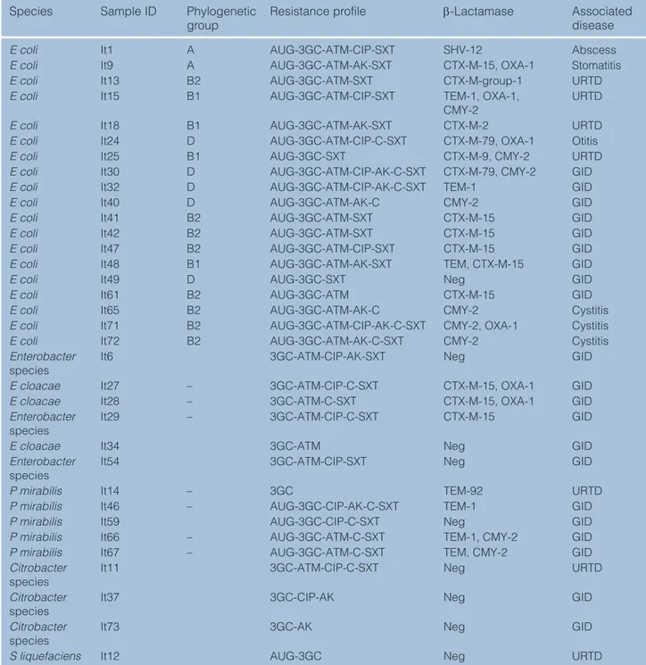
Table 3 β-Lactam resistance mechanisms and antimicrobial resistance profile of 34 third-generation cephalosporin (3GC)-resistant Enterobacteriaceae strains
Species Sample ID Phylogenetic
group Resistance profile β-Lactamase Associated disease
E coli It1 A AUG-3GC-ATM-CIP-SXT SHV-12 Abscess
E coli It9 A AUG-3GC-ATM-AK-SXT CTX-M-15, OXA-1 Stomatitis
E coli It13 B2 AUG-3GC-ATM-SXT CTX-M-group-1 URTD
E coli It15 B1 AUG-3GC-ATM-CIP-SXT TEM-1, OXA-1,
CMY-2 URTD
E coli It18 B1 AUG-3GC-ATM-AK-SXT CTX-M-2 URTD
E coli It24 D AUG-3GC-ATM-CIP-C-SXT CTX-M-79, OXA-1 Otitis
E coli It25 B1 AUG-3GC-SXT CTX-M-9, CMY-2 URTD
E coli It30 D AUG-3GC-ATM-CIP-AK-C-SXT CTX-M-79, CMY-2 GID
E coli It32 D AUG-3GC-ATM-CIP-AK-C-SXT TEM-1 GID
E coli It40 D AUG-3GC-ATM-AK-C CMY-2 GID
E coli It41 B2 AUG-3GC-ATM-SXT CTX-M-15 GID
E coli It42 B2 AUG-3GC-ATM-SXT CTX-M-15 GID
E coli It47 B2 AUG-3GC-ATM-CIP-SXT CTX-M-15 GID
E coli It48 B1 AUG-3GC-ATM-AK-SXT TEM, CTX-M-15 GID
E coli It49 D AUG-3GC-SXT Neg GID
E coli It61 B2 AUG-3GC-ATM CTX-M-15 GID
E coli It65 B2 AUG-3GC-ATM-AK-C CMY-2 Cystitis
E coli It71 B2 AUG-3GC-ATM-CIP-AK-C-SXT CMY-2, OXA-1 Cystitis
E coli It72 B2 AUG-3GC-ATM-AK-C-SXT CMY-2 Cystitis
Enterobacter
species It6 3GC-ATM-CIP-AK-SXT Neg GID
E cloacae It27 – 3GC-ATM-CIP-C-SXT CTX-M-15, OXA-1 GID
E cloacae It28 – 3GC-ATM-C-SXT CTX-M-15, OXA-1 GID
Enterobacter
species It29 – 3GC-ATM-CIP-C-SXT CTX-M-15 GID
E cloacae It34 3GC-ATM Neg GID
Enterobacter
species It54 3GC-ATM-CIP-SXT Neg GID
P mirabilis It14 – 3GC TEM-92 URTD
P mirabilis It46 – AUG-3GC-CIP-AK-C-SXT TEM-1 GID
P mirabilis It59 AUG-3GC-CIP-C-SXT Neg GID
P mirabilis It66 – AUG-3GC-ATM-C-SXT TEM-1, CMY-2 GID
P mirabilis It67 – AUG-3GC-ATM-C-SXT TEM, CMY-2 GID
Citrobacter
species It11 3GC-ATM-CIP-C-SXT Neg URTD
Citrobacter
species It37 3GC-CIP-AK Neg GID
Citrobacter
species It73 3GC-AK Neg GID
S liquefaciens It12 AUG-3GC Neg URTD
E coli = Escherichia coli; E cloacae = Enterobacter cloacae; P mirabilis = Proteus mirabilis; S liquefaciens = Serratia liquefaciens; AUG = amoxicillin–clavulanate; ATM = aztreonam; AK = amikacin; CIP = ciprofloxacin; C = chloramphenicol; SXT = trimethoprim/ sulfamethoxazole; URTD = upper respiratory tract disease; GID = gastrointestinal disease; (–) = not applicable; Neg = negative
An association between ESBL/pAmpC β-lactamase genes and resistance to fluoroquinolones was found in our study and has also been shown in a previous study,50 which identified new mechanisms of resistance and plasmid-mediated fluoroquinolone resistance determi-nants in ESBL-producing E coli of canine and feline origin. In this present study, we did not characterise the mechanisms of resistance to fluoroquinolones. Yet, the high prevalence of ESBL/pAmpC β-lactamase-producing Enterobacteriaceae showing resistance to fluoroquin olones should stimulate a more focused mon-itoring of fluoroquinolone resistance.
MDR was displayed by a high percentage of isolates (31%), and a high percentage of isolates carrying ESBLs/ pAmpC β-lactamase genes were MDR (74%). These results constitute additional evidence for an association between MDR and ESBL/pAmpC production, which allows a co-selection process, because ESBL/pAmpC genes are usually encoded on plasmids that frequently carry aminoglycoside-, tetracycline-, sulfonamide- and/ or fluoroquinolone-resistant genes.51
GID and URTD cats receiving antimicrobial treat-ment were more likely to host MDR isolates than cats not receiving antimicrobials. These results underline the importance of rational use of antimicrobials to pre-vent the selection of resistant bacteria. Although E coli are part of the normal intestinal microflora, they can be
associated with gastroenteritis in the presence of bacterial virulence factors and impaired local or sys-temic immunity. Many E coli strains and other Enterobacte riaceae have been isolated from small ani-mals with and without diarrhoea, and the role of many of these strains in disease causation in dogs and cats is poorly defined.52 In our study, we characterised phylo-genetic groups of some E coli isolates and found that two isolates belonged to phylogenetic group B1, which usually includes commensal strains exhibiting an increased drug resistance pattern but possessing few virulence genes.53 Eight isolates belonged either to groups B2 or D, which usually include pathogenic E coli strains that cause extra-intestinal infections, possess several pathogenicity-associated islands and express multiple virulence factors such as adherence factors including biofilm production and high surface hydro-phobicity, toxins and siderophore production, but are more susceptible to antibiotics.54 Moreover, six E coli isolates (from two cats with URTD, one cat with otitis and one cat with cystitis) belonged either to phyloge-netic groups B2 or D. Another five E coli isolates (from one cat with an abscess, one cat with stomatitis, three cats with URTD) belonged either to phylogenetic groups A or B1. For cats with URTD, otitis, stomatitis, abscess and cystitis, E coli isolates could have been directly associated with the disease condition, but we Table 4 Clinical data of 17 cats harbouring extended-spectrum β-lactamase/plasmid-mediated AmpC β-lactamase-producing Enterobacteriaceae
Cat ID Disease Isolated bacteria
(ID number of the strain[s])
Age (years) Sex Habitat Antimicrobial
therapy
C2588 GID E coli (It30) 4 M H –
C2600 Otitis E coli (It24) 12 F H –
C2608 URTD E coli (It25) 5 F S Amoxicillin–
clavulanate
C2610 Abscess E coli (It1) 8 M S –
C2613 GID E coli (It40) 2 F S –
C2616 GID E coli (It41, It42) 3 F S –
C2618 URTD E coli (It15) 4 F S –
C2621 Stomatitis E coli (It9) 3 M S Cefovecin
C2637 URTD E coli (It18) 5 F S –
C2639 GID E coli (It47) 6 M H Spiramycin and
metronidazole
C2642 URTD E coli (It13) 6 F S –
C2644 GID E coli (It48) 5 F S Cefovecin
C2657 GID P mirabilis (It66, It67) 5 M S –
C2670
C2761 GIDURTD E coli (It61)P mirabilis (It14) 43 FM SH – C2855 GID E cloacae (It27, It28,
It29) 3 F H –
C2977 Cystitis E coli (It65, It71, It72) 4 M H –
URTD = upper respiratory tract disease; GID = gastrointestinal disease; E coli = Escherichia coli; P mirabilis = Proteus mirabilis; E cloacae =
cannot exclude completely their role in a secondary infection complicating a primary non-infectious dis-ease condition or their commensal state. Characterisation of virulence factors of E coli and other Enterobacteriaceae isolates in cats with diarrhoea warrants further studies.
Feline URTD, frequently encountered in clinical prac-tice, represented the second–most common disease of cats included in the study. Detection of Enterobacte-riaceae strains harbouring MDR ESBL/pAmpC genes in cats with URTD is described for the first time in the lit-erature. One of the E coli isolates from cats with URTD harboured a blaCTX-M-15 gene and belonged to netic group B2, while the others belonged to phyloge-netic group B1. These MDR ESBL/pAmpC-producing
E coli may possibly complicate the feline URTD by caus-ing infections that are difficult to treat.
The CTX-M enzymes were the most common ESBL and different types of these enzymes were detected, indicating some diversity of CTX-M-encoding genes in Enterobacteriaceae from diseased cats in Italy. By con-trast, CMY-2 was the only pAmpC found in cats from this study. The high prevalence of CMY-2 is of concern as this β-lactamase confers resistance to a wide range of extended-spectrum cephalosporins used for the treat-ment of serious infections in humans and animals, because they are not inhibited by β-lactamase inhibitors and because they can easily be transferred between dif-ferent bacterial species leading to its rapid dissemination.4,22,55
Of the ESBL genes, blaCTX-M-79 was harboured by an
E coli isolate from one cat with GID and one cat with oti-tis, both of which were from households and had not received any previous antimicrobial treatment. To the best of our knowledge, we describe for the first time the detection of an E coli isolate harbouring the blaCTX-M-79 gene in cats. Previous reports described the blaCTX-M-79 gene from Enterobacteriaceae isolated from wild birds (corvids), hospital waste, human patients and farmed fish in Asia,56–58 and from the faeces of cattle in the USA.59
Furthermore, we also report here for the first time the isolation of Proteus species encoding a blaTEM-92 gene from a nasal swab of a cat with URTD. The TEM-92 ESBL was first described in two bacterial strains, Proteus mirabilis
mirabilis and Providencia stuartii, isolated in 1998 from the urinary tracts of two human patients.60 Since then, TEM-92 has been reported only in isolates from human patients;61 where therapeutic failure and mortality may occur when associated with bloodstream infections.62
In our study, Buttiauxella species, Serratia liquefaciens,
Kluyvera species and Providencia species were MDR. Although the above-mentioned bacteria have been rarely reported to cause clinical infections in companion animals, the detection of MDR isolates highlights that these rarely pathogenic bacteria may pose important
therapeutic limitations when causing opportunistic infections.
All isolates from this study were susceptible to mero-penem. Production of carbapenemases, which repre-sents an additional hazard to public health and veterinary medicine, has so far remained a rare phe-nomenon among Gram-negative bacteria isolated from companion animals.63–66 Nevertheless, based on the advancing spread of this type of resistance in human isolates, this hazard could arise in the future in compan-ion animals.
Conclusions
The prevalence of Enterobacteriaceae carrying ESBL or pAmpC genes identified in this study was higher than that documented in previous reports in Italy, which indicates that ESBL- and pAmpC-producing Enterobac-teriaceae might be widespread. The spread of these enzymes can be attributed to the transfer of plasmids and other mobile genetic elements between bacterial species. The findings indicate that diseased cats may be reservoirs of 3GC-resistant Enterobacteriaceae and/or act as shedders of bacteria resistant to critically impor-tant antimicrobials in humans.
The problem posed by emerging resistance, under-lined in this study by the discovery of β-lactamase genes not previously described in companion animals, the prospect of losing important antimicrobial efficacy and the possibility of an already attested interspecies spread emphasise the urgent need to define the epidemiology of β-lactamase-producing bacteria in companion ani-mals.66–68 The emergence of ESBL-/pAmpC-producing MDR Enterobacteriaceae could pose major limitations in therapeutic options for companion animals. Awareness should be raised among companion animal general practitioners about the threat they may encounter in their daily clinical activities.
Furthermore, these results highlight once more the need for bacteriological examination and susceptibility testing before instituting empirical antimicrobial ther-apy if a bacterial infection is suspected, in order to establish accurate treatment and avoid the antimicro-bial selection pressure on the animal microbiota. This could reduce costs by avoiding resorting to ineffective compounds, and play a role in the control and monitor-ing of antimicrobial resistance in companion animal medicine.
Acknowledgements We thank all the participants for agreeing to enrol and providing the samples and epidemiologi-cal data as requested.
Author note Part of this work was presented as an oral communication at the 26th Annual Congress of the European College of Veterinary Internal Medicine – Companion Animals.
Conflict of interest The authors declared no potential conflicts of interest with respect to the research, authorship, and/or publication of this article.
Funding This work was supported by FEDER funds through the Programa Operacional Factores de Competitividade (COM-PETE) and by national funds through the FCT (Fundação para a Ciência e a Tecnologia CIISA Project [UID/CVT/00276/2019]), and PhD grants SFRH/BD/77886/2011 (CM) and SFRH/ BD/113142/2015 (AB).
Ethical approval This work involved the use of non- experimental animals only (owned or unowned), and followed established internationally recognised high standards (‘best practice’) of individual veterinary clinical patient care. Ethical approval from a committee was not necessarily required.
Informed consent Informed consent (either verbal or writ-ten) was obtained from the owner or legal custodian of all animal(s) described in this work for the procedure(s) under-taken. No animals or humans are identifiable within this publication, and therefore additional informed consent for publication was not required.
ORCID iD Constança Pomba https://orcid.org/0000-0002-0504-6820
References
1 Koenig A. Gram-negative bacterial infections. In: Greene CE (ed). Infectious diseases of the dog and cat. 4th ed. St Louis, MO: Saunders Elsevier, 2012, p 349.
2 Iredell J, Brown J and Tagg K. Antibiotic resistance in
Enterobacteriaceae: mechanisms and clinical implica-tions. BMJ 2016; 352: h6420.
3 Rendle D and Page S. Antimicrobial resistance in
compan-ion animals. Equine Vet J 2018; 50: 147–152.
4 Guardabassi L, Schwarz L and Lloyd DH. Pet animals as
reservoirs of antimicrobial-resistant bacteria: review. J Antimicrob Chemother 2004; 54: 321–332.
5 DeVincent SJ and Reid-Smith R. Stakeholder position
paper: companion animal veterinarian. Prev Vet Med 2006; 73: 181–189.
6 Ruppé E, Woerther PL and Barbier F. Mechanisms of
anti-microbial resistance in Gram-negative bacilli. Ann Inten-sive Care 2015; 5: 21. DOI: 10.1186/s13613-015-0061-0. 7 World Health Organization. Critically important
anti-microbials for human medicine – 5th rev. https://www. who.int/foodsafety/publications/antimicrobials-fifth/ en/ (2017, accessed July 22, 2019).
8 European Centre for Disease Prevention and Control.
Anti-microbial resistance surveillance in Europe 2014. https:// ecdc.europa.eu/en/publications-data/antimicrobial-resis tance-surveillance-europe-2014 (2015, accessed July 22, 2019). 9 Rubin JE and Pitout JD. Extended-spectrum β-lactamase,
carbapenemase and AmpC producing Enterobacteriaceae in companion animals. Vet Microbiol 2014; 170: 10–18. 10 Schmiedel J, Falgenhauer L, Domann E, et al.
Multi-resistant extended-spectrum β-lactamase-producing Enterobacteriaceae from humans, companion animals and horses in central Hesse, Germany. BMC Microbiol 2014; 14: 187. DOI: 10.1186/1471-2180-14-187.
11 Seni J, Falgenhauer L, Simeo N, et al. Multiple ESBL-
producing Escherichia coli sequence types carrying
qui-nolone and aminoglycoside resistance genes circulating in companion and domestic farm animals in Mwanza, Tanzania, harbor commonly occurring plasmids. Front Microbiol 2016; 7: 142. DOI: 10.3389/fmicb.2016.00142. 12 Costa D, Poeta P, Brinas L, et al. Detection of CTX-M-1 and
TEM-52 beta-lactamases in Escherichia coli strains from
healthy pets in Portugal. J Antimicrob Chemother 2004; 54: 960–961.
13 Murphy C, Reid-Smith RJ, Prescott JF, et al. Occurrence
of antimicrobial resistant bacteria in healthy dogs and cats presented to private veterinary hospitals in southern Ontario: a preliminary study. Can Vet J 2009; 50: 1047–1053. 14 Belas A, Salazar AS, Gama LT, et al. Risk factors for faecal
colonisation with Escherichia coli producing
extended-spectrum and plasmid-mediated AmpC β-lactamases in dogs. Vet Rec 2014; 175: 202.
15 Teshager T, Domínguez L, Moreno MA, et al. Isolation
of an SHV-12 beta-lactamase-producing Escherichia coli
strain from a dog with recurrent urinary tract infections.
Antimicrob Agents Chemother 2000; 44: 3483–3484.
16 Sanchez S, McCrackin Stevenson MA, Hudson CR, et al.
Characterization of multidrug-resistant Escherichia coli
isolates associated with nosocomial infections in dogs. J Clin Microbiol 2002; 40: 3586–3595.
17 Timofte D, Dandrieux J, Wattret A, et al. Detection of
extended-spectrum-beta-lactamase-positive Escherichia coli in bile isolates from two dogs with bacterial
cholan-giohepatitis. J Clin Microbiol 2011; 49: 3411–3414.
18 Dierikx CM, van Duijkeren E, Schoormans AH, et al.
Occurrence and characteristics of extended-spectrum-β-lactamase- and AmpC-producing clinical isolates derived from companion animals and horses. J Antimicrob Chemother 2012; 67: 1368–1374.
19 Harada K, Nakai Y and Kataoka Y. Mechanisms of
resis-tance to cephalosporin and emergence of O25b-ST131 clone harboring CTX-M-27 β-lactamase in extraintestinal pathogenic Escherichia coli from dogs and cats in Japan. Microbiol Immunol 2012; 56: 480–485.
20 Nebbia P, Tramuta C, Odore R, et al. Genetic and
phe-notypic characterisation of Escherichia coli producing
cefotaximase-type extended-spectrum β-lactamases: first evidence of the ST131 clone in cats with urinary infec-tions in Italy. J Feline Med Surg 2014; 16: 966–971.
21 Bogaerts P, Huang TD, Bouchahrouf W, et al.
Characteriza-tion of ESBL- and AmpC-producing Enterobacteriaceae from diseased companion animals in Europe. Microb Drug Resist 2015; 21: 643–650.
22 Marques C, Belas A, Franco A, et al. Increase in
antimi-crobial resistance and emergence of major international high-risk clonal lineages in dogs and cats with urinary tract infection: 16 year retrospective study. J Antimicrob Chemother 2018; 73: 377–384.
23 The European Pet Food Industry. Facts & figures 2018. http://www.fediaf.org/images/FEDIAF_Facts__and_Fig ures_2018_ONLINE_final.pdf (2018, accessed August 22, 2019). 24 Hordijk J, Schoormans A, Kwakernaak M, et al. High
prev-alence of fecal carriage of extended spectrum β-lactamase/ AmpC-producing Enterobacteriaceae in cats and dogs.
25 Sallem RB, Gharsa H, Slama KB, et al. First detection of
CTX-M-1, CMY-2, and QnrB19 resistance mechanisms in fecal Escherichia coli isolates from healthy pets in
Tuni-sia. Vector Borne Zoonotic Dis 2013; 13: 98–102.
26 O’Keefe A, Hutton TA, Schifferli DM, et al. First detection
of CTX-M and SHV extended-spectrum beta-lactamases in Escherichia coli urinary tract isolates from dogs and
cats in the United States. Antimicrob Agents Chemother 2010; 54: 3489–3492.
27 Shaheen BW, Nayak R, Foley SL, et al. Molecular
charac-terization of resistance to extended-spectrum cephalospo-rins in clinical Escherichia coli isolates from companion
animals in the United States. Antimicrob Agents Chemother 2011; 55: 5666–5675.
28 Donati V, Feltrin F, Hendriksen RS, et al. Extended-
spectrum-beta-lactamases, AmpC beta-lactamases and plasmid mediated quinolone resistance in Klebsiella
spp. from companion animals in Italy. PLoS One 2014; 9: e90564. DOI: 10.1371/journal.pone.0090564.
29 Harada K, Shimizu T, Mukai Y, et al. Phenotypic and
molecular characterization of antimicrobial resistance in Klebsiella spp. isolates from companion animals
in Japan: clonal dissemination of multidrug-resistant extended-spectrum β-lactamase-producing Klebsiella pneumoniae. Front Microbiol 2016; 7: 1021. DOI: 10.3389/ fmicb.2016.01021.
30 Harada K, Shimizu T, Mukai Y, et al. Phenotypic and
molecular characterization of antimicrobial resistance in
Enterobacter spp. isolates from companion animals in
Japan. PLoS One 2017; 12: e0174178. DOI: 10.1371/journal. pone.0174178.
31 Karkaba A, Grinberg A, Benschop J, et al. Characterisation
of extended-spectrum β-lactamase and AmpC β-lactamase-producing Enterobacteriaceae isolated from companion animals in New Zealand. N Z Vet J 2017; 65: 105–112. 32 Zogg AL, Simmen S, Zurfluh K, et al. High prevalence of
extended-spectrum β-lactamase producing Enterobacteria-ceae among clinical isolates from cats and dogs admitted to a veterinary hospital in Switzerland. Front Vet Sci 2018; 5: 62. DOI: 10.3389/fvets.2018.00062.
33 Costa D, Poeta P, Sáenz Y, et al. Prevalence of
antimicro-bial resistance and resistance genes in faecal Escherichia coli isolates recovered from healthy pets. Vet Microbiol 2008; 127: 97–105.
34 Gandolfi-Decristophoris P, Petrini O, Ruggeri-Bernardi N, et al. Extended-spectrum β-lactamase-producing
Entero-bacteriaceae in healthy companion animals living in nursing homes and in the community. Am J Infect Control 2013; 41: 831–835.
35 Huber H, Zweifel C, Wittenbrink MM, et al.
ESBL-pro-ducing uropathogenic Escherichia coli isolated from
dogs and cats in Switzerland. Vet Microbiol 2013; 162: 992–996.
36 Nebbia P, Tramuta C, Odore R, et al. Genetic and
phe-notypic characterisation of Escherichia coli producing
cefotaximase-type extended-spectrum β-lactamases: first evidence of the ST131 clone in cats with urinary infec-tions in Italy. J Feline Med Surg 2014; 16: 966–971.
37 Clinical and Laboratory Standards Institute (CLSI). Perfor-mance standards for antimicrobial disk and dilution sus-ceptibility tests for bacteria isolated from animals. 4th ed. Approved Standard VET01-A4. Wayne, PA: CLSI, 2013. 38 Clinical and Laboratory Standards Institute (CLSI).
Per-formance standards for antimicrobial susceptibility testing M100S. Wayne, PA: CLSI, 2016.
39 Schwarz S, Silley P, Simjee S, et al. Editorial: assessing the
antimicrobial susceptibility of bacteria obtained from animals. J Antimicrob Chemother 2010; 65: 601–604.
40 Edelstein M, Pimkin M, Palagin I, et al. Prevalence and
molecular epidemiology of CTX-M extended-spectrum β-lactamase-producing Escherichia coli and Klebsiella pneu-moniae in Russian hospitals. Antimicrob Agents Chemother 2003; 47: 3724–3732.
41 Pomba C, Mendonça N, Costa M, et al. Improved multiplex
PCR method for the rapid detection of beta-lactamase genes in Escherichia coli of animal origin. Diagn Microbiol Infect Dis 2006; 56: 103–106.
42 Woodford N, Fagan EJ and Ellington MJ. Multiplex PCR
for rapid detection of genes encoding CTX-M extended-spectrum (beta)-lactamases. J Antimicrob Chemother 2006; 57: 154–155.
43 Pérez F and Hanson N. Detection of plasmid-mediated
AmpC beta-lactamase genes in clinical isolates by using multiplex PCR. J Clin Microbiol 2002; 40: 2153–2162. 44 Doumith M, Day MJ, Hope R, et al. Improved multiplex
PCR strategy for rapid assignment of the four major Esch-erichia coli phylogenetic groups. J Clin Microbiol 2012; 50: 3108–3110.
45 Carattoli A, Lovari S, Franco A, et al. Extended-spectrum
β-lactamases in Escherichia coli isolated from dogs and
cats in Rome, Italy, from 2001 to 2003. Antimicrob Agents Chemother 2005; 49: 833–835.
46 Murphy CP, Reid-Smith RJ, Boerlin P, et al. Outpatient
antimicrobial drug use in dogs and cats for new disease events from community companion animal practices in Ontario. Can Vet J 2012; 53: 291–298.
47 Escher M, Vanni M, Intorre L, et al. Use of antimicrobials
in companion animal practice: a retrospective study in a veterinary teaching hospital in Italy. J Antimicrob Chemother 2011; 66: 920–927.
48 Belmar-Liberato R, Gonzalez-Canga A, Tamame-Martin P, et al. Amoxicillin and amoxicillin-clavulanic acid
resis-tance in veterinary medicine – the situation in Europe: a review. Vet Med Czech 2011; 56: 473–485.
49 Marques C, Gama LT, Belas A, et al. European multicenter
study on antimicrobial resistance in bacteria isolated from companion animal urinary tract infections. BMC Vet Res 2016; 12: 213. DOI: 10.1186/s12917-016-0840-3.
50 Shaheen BW, Nayak R, Foley SL, et al. Chromosomal and
plasmid-mediated fluoroquinolone resistance mecha-nisms among broad-spectrum-cephalosporin-resistant
Escherichia coli isolates recovered from companion
animals in the USA. J Antimicrob Chemother 2013; 68: 1019–1024.
51 Cantón R and Coque TM. The CTX-M beta-lactamase
52 Marks SL, Rankin SC, Byrne BA, et al.
Enteropatho-genic bacteria in dogs and cats: diagnosis, epidemiol-ogy, treatment, and control. J Vet Intern Med 2011; 25: 1195–1208.
53 Picard B, Garcia JS, Gouriou S, et al. The link between
phy-logeny and virulence in Escherichia coli extraintestinal
infection. Infect Immun 1999; 67: 546–553.
54 Smith JL, Fratamico PM and Gunther NW. Extraintestinal
pathogenic Escherichia coli. Foodborne Pathog Dis 2007; 4: 134–163.
55 Shaheen BW, Boothe DM, Oyarzabal OA, et al.
Antimicro-bial resistance profiles and clonal relatedness of canine and feline Escherichia coli pathogens expressing
multi-drug resistance in the United States. J Vet Intern Med 2010; 24: 323–330.
56 Hasan B, Olsen B, Alam A, et al. Dissemination of
the multidrug-resistant extended-spectrum β-lactamase-producing Escherichia coli O25b-ST131 clone and the role
of house crow (Corvus splendens) foraging on hospital
waste in Bangladesh. Clin Microbiol Infect 2015; 21: 1000. DOI: 10.1016/j.cmi.2015.06.016.
57 Guo TS, Cui EB, Bao CM, et al. Distribution of genotypes
in ESBLs producing E. coli strains isolated from
post-hepatitic cirrhosis’ patients with bloodstream infection
[article in Chinese]. Zhonghua Shi Yan He Lin Chuang Bing Du Xue Za Zhi 2013; 27: 348–350.
58 Jiang HX, Tang D, Liu YH, et al. Prevalence and
character-istics of β-lactamase and plasmid-mediated quinolone resistance genes in Escherichia coli isolated from farmed
fish in China. J Antimicrob Chemother 2012; 67: 2350–2353. 59 Wittum TE, Mollenkopf DF, Daniels JB, et al. CTX-M-type
extended-spectrum β-lactamases present in Escherichia coli from the feces of cattle in Ohio, United States. Food-borne Pathog Dis 2010; 7: 1575–1579.
60 De Champs C, Monne C, Bonnet R, et al. New TEM variant
(TEM-92) produced by Proteus mirabilis and Providencia
stuartii isolates. Antimicrob Agents Chemother 2001; 45: 1278– 1280.
61 Biendo M, Thomas D, Laurans G, et al. Molecular diversity
of Proteus mirabilis isolates producing extended-spectrum
beta-lactamases in a French university hospital. Clin Micro-biol Infect 2005; 11: 395–401.
62 Endimiani A, Luzzaro F, Brigante G, et al. Proteus mirabi-lis bloodstream infections: risk factors and treatment
outcome related to the expression of extended-spectrum beta-lactamases. Antimicrob Agents Chemother 2005; 49: 2598–2605.
63 Shaheen BW, Nayak N and Boothe M. Emergence of a
New Delhi metallo-β-lactamase (NDM-1)-encoding gene in clinical Escherichia coli isolates recovered from
com-panion animals in the United States. Antimicrob Agents Chemother 2013; 57: 2902–2903.
64 Stolle I, Prenger-Berninghoff E, Stamm I, et al. Emergence
of OXA-48 carbapenemase-producing Escherichia coli
and Klebsiella pneumoniae in dogs. J Antimicrob Chemother 2013; 68: 2802–2808.
65 Pomba C, Endimiani A, Rossano A, et al. First report of
OXA-23-mediated carbapenem resistance in sequence type 2 multidrug-resistant Acinetobacter baumannii
asso-ciated with urinary tract infection in a cat. Antimicrob Agents Chemother 2014; 58: 1267–1268.
66 Ljungquist O, Ljungquist D, Myrenas M, et al. Evidence of
household transfer of ESBL-/pAmpC producing
Entero-bacteriaceae between humans and dogs – a pilot study. Infect Ecol Epidemiol 2016; 6. DOI: 10.3402/iee.v6.31514. 67 Carvalho AC, Barbosa AV, Arais LR, et al. Resistance
pat-terns, ESBL genes, and genetic relatedness of Escherichia coli from dogs and owners. Braz J Microbiol 2016; 47: 150–158.
68 Pomba C, Rantala M, Greko C, et al. Public health risk of
antimicrobial resistance transfer from companion ani-mals. J Antimicrob Chemother 2017; 72: 957–968.