2015 American Thyroid Association Management
Guidelines for Adult Patients with Thyroid Nodules
and Differentiated Thyroid Cancer
The American Thyroid Association Guidelines Task Force
on Thyroid Nodules and Differentiated Thyroid Cancer
Bryan R. Haugen,1,* Erik K. Alexander,2Keith C. Bible,3Gerard M. Doherty,4Susan J. Mandel,5 Yuri E. Nikiforov,6Furio Pacini,7Gregory W. Randolph,8Anna M. Sawka,9Martin Schlumberger,10
Kathryn G. Schuff,11Steven I. Sherman,12 Julie Ann Sosa,13David L. Steward,14 R. Michael Tuttle,15and Leonard Wartofsky16
Background: Thyroid nodules are a common clinical problem, and differentiated thyroid cancer is becoming
increasingly prevalent. Since the American Thyroid Association’s (ATA’s) guidelines for the management of
these disorders were revised in 2009, significant scientific advances have occurred in the field. The aim of these
guidelines is to inform clinicians, patients, researchers, and health policy makers on published evidence relating
to the diagnosis and management of thyroid nodules and differentiated thyroid cancer.
Methods: The specific clinical questions addressed in these guidelines were based on prior versions of the
guidelines, stakeholder input, and input of task force members. Task force panel members were educated on
knowledge synthesis methods, including electronic database searching, review and selection of relevant
cita-tions, and critical appraisal of selected studies. Published English language articles on adults were eligible for
inclusion. The American College of Physicians Guideline Grading System was used for critical appraisal of
evidence and grading strength of recommendations for therapeutic interventions. We developed a similarly
formatted system to appraise the quality of such studies and resultant recommendations. The guideline panel
had complete editorial independence from the ATA. Competing interests of guideline task force members were
regularly updated, managed, and communicated to the ATA and task force members.
Results: The revised guidelines for the management of thyroid nodules include recommendations regarding
initial evaluation, clinical and ultrasound criteria for fine-needle aspiration biopsy, interpretation of fine-needle
aspiration biopsy results, use of molecular markers, and management of benign thyroid nodules.
Re-commendations regarding the initial management of thyroid cancer include those relating to screening for
thyroid cancer, staging and risk assessment, surgical management, radioiodine remnant ablation and therapy,
and thyrotropin suppression therapy using levothyroxine. Recommendations related to long-term management
1
University of Colorado School of Medicine, Aurora, Colorado.
2
Brigham and Women’s Hospital, Harvard Medical School, Boston, Massachusetts.
3
The Mayo Clinic, Rochester, Minnesota.
4Boston Medical Center, Boston, Massachusetts. 5
Perelman School of Medicine, University of Pennsylvania, Philadelphia, Pennsylvania.
6University of Pittsburgh Medical Center, Pittsburgh, Pennsylvania. 7
The University of Siena, Siena, Italy.
8
Massachusetts Eye and Ear Infirmary, Massachusetts General Hospital, Harvard Medical School, Boston, Massachusetts.
9
University Health Network, University of Toronto, Toronto, Ontario, Canada.
10
Institute Gustave Roussy and University Paris Sud, Villejuif, France.
11
Oregon Health and Science University, Portland, Oregon.
12
University of Texas M.D. Anderson Cancer Center, Houston, Texas.
13Duke University School of Medicine, Durham, North Carolina. 14
University of Cincinnati Medical Center, Cincinnati, Ohio.
15Memorial Sloan Kettering Cancer Center, New York, New York. 16
MedStar Washington Hospital Center, Washington, DC. *Chair.
Authors are listed in alphabetical order and were appointed by ATA to independently formulate the content of this manuscript. None of the scientific or medical content of the manuscript was dictated by the ATA.
ª American Thyroid Association ª Mary Ann Liebert, Inc. DOI: 10.1089/thy.2015.0020
of differentiated thyroid cancer include those related to surveillance for recurrent disease using imaging and
serum thyroglobulin, thyroid hormone therapy, management of recurrent and metastatic disease, consideration
for clinical trials and targeted therapy, as well as directions for future research.
Conclusions: We have developed evidence-based recommendations to inform clinical decision-making in the
management of thyroid nodules and differentiated thyroid cancer. They represent, in our opinion, contemporary
optimal care for patients with these disorders.
INTRODUCTION
T
hyroid nodules are a common clinical problem. Epidemiologic studies have shown the prevalence of palpable thyroid nodules to be approximately 5% in women and 1% in men living in iodine-sufficient parts of the world (1,2). In contrast, high-resolution ultrasound (US) can detect thyroid nodules in 19%–68% of randomly selected individ-uals, with higher frequencies in women and the elderly (3,4). The clinical importance of thyroid nodules rests with the need to exclude thyroid cancer, which occurs in 7%–15% of cases depending on age, sex, radiation exposure history, family history, and other factors (5,6). Differentiated thyroid cancer (DTC), which includes papillary and follicular cancer, com-prises the vast majority (>90%) of all thyroid cancers (7). In the United States, approximately 63,000 new cases of thyroid cancer were predicted to be diagnosed in 2014 (8) compared with 37,200 in 2009 when the last ATA guidelines were published. The yearly incidence has nearly tripled from 4.9 per 100,000 in 1975 to 14.3 per 100,000 in 2009 (9). Almost the entire change has been attributed to an increase in the inci-dence of papillary thyroid cancer (PTC). Moreover, 25% of the new thyroid cancers diagnosed in 1988–1989 were £1 cm compared with 39% of the new thyroid cancer diagnoses in 2008–2009 (9). This tumor shift may be due to the increasing use of neck ultrasonography or other imaging and early diag-nosis and treatment (10), trends that are changing the initial treatment and follow-up for many patients with thyroid cancer. A recent population-based study from Olmsted County re-ported the doubling of thyroid cancer incidence from 2000 to 2012 compared to the prior decade as entirely attributable to clinically occult cancers detected incidentally on imaging or pathology (11). By 2019, one study predicts that PTC will become the third most common cancer in women at a cost of $19–21 billion in the United States (12). Optimization of long-term health outcomes and education about potential prognosis for individuals with thyroid neoplasms is critically important. In 1996, the American Thyroid Association (ATA) pub-lished treatment guidelines for patients with thyroid nodules and DTC (13). Over the last 15–20 years, there have been many advances in the diagnosis and therapy of both thyroid nodules and DTC, but clinical controversy exists in many areas. A long history of insufficient peer-reviewed research funding for high-quality clinical trials in the field of thyroid neoplasia may be an important contributing factor to existing clinical uncertainties (12). Methodologic limitations or con-flicting findings of older studies present a significant chal-lenge to modern-day medical decision-making in many aspects of thyroid neoplasia. Although they are not a specific focus of these guidelines, we recognize that feasibility and cost considerations of various diagnostic and therapeutic optionsalso present important clinical challenges in many clinical practice settings.
AIM AND TARGET AUDIENCE
Our objective in these guidelines is to inform clinicians, patients, researchers, and health policy makers about the best available evidence (and its limitations), relating to the dia-gnosis and treatment of adult patients with thyroid nodules and DTC. These guidelines should not be applied to children (<18–20 years old); recent ATA guidelines for children with thyroid nodules and DTC were published in 2015 (14). This document is intended to inform clinical decision-making. A major goal of these guidelines is to minimize potential harm from overtreatment in a majority of patients at low risk for disease-specific mortality and morbidity, while appropriately treating and monitoring those patients at higher risk. These guidelines should not be interpreted as a replacement for clinical judgement and should be used to complement in-formed, shared patient–health care provider deliberation on complex issues. It is important to note that national clinical practice guidelines may not necessarily constitute a legal standard of care in all jurisdictions (15). If important differ-ences in practice settings present barriers to meaningful im-plementation of the recommendations of these guidelines, interested physicians or groups (in or outside of the United States) may consider adapting the guidelines using estab-lished methods (16,17) (ADAPTE Collaboration, 2009; www.g-i-n.net). The ADAPTE Collaboration is an interna-tional group of researchers, guideline developers, and guide-line implementers who aim to promote the development and use of clinical practice guidelines through the adaption of existing guidelines. Because our primary focus was review-ing the quality of evidence related to health outcomes and diagnostic testing, we decided a priori not to focus on eco-nomic resource implications in these guidelines. As part of our review, we identified some knowledge gaps in the field, with associated future research priorities.
Other groups have previously developed clinical practice guidelines, including the American Association of Clinical Endocrinologists, Associazione Medici Endocrinologi, and the European Thyroid Association (18), the British Thyroid Association and The Royal College of Physicians (19), and the National Comprehensive Cancer Network (www.nccn .org). The European Thyroid Association has published con-sensus guidelines for postoperative US in the management of DTC (20). The Society for Nuclear Medicine and Molecular Imaging (21) and the European Association of Nuclear Med-icine have also published guidelines for radioiodine (RAI) therapy of DTC (22). The Japanese Society of Thyroid Sur-geons and the Japanese Association of Endocrine SurSur-geons
have recently revised guidelines on treatment of patients with thyroid tumors (23). Given the existing controversies in the field, differences in critical appraisal approaches for existing evidence, and differences in clinical practice patterns across geographic regions and physician specialties, it should not be surprising that the organizational guidelines are not in com-plete agreement for all issues. Such differences highlight the importance of clarifying evidence uncertainties with future high quality clinical research.
METHODS
ATA Thyroid Nodules and Differentiated Thyroid Cancer guidelines were published in 2006 (24) and revised in 2009
(25). Because of the rapid growth of the literature on this topic, plans for revising the guidelines within approximately 4 years of publication were made at the inception of the project. A task force chair was appointed by the ATA Pre-sident with approval of the Board. A task force of specialists with complementary expertise (endocrinology, surgery, nu-clear medicine, radiology, pathology, oncology, molecular diagnostics, and epidemiology) was appointed. In order to have broad specialty and geographic representation, as well as fresh perspectives, one-third of the task force is replaced for each iteration of the guidelines, per ATA policy. Upon discussion among the panel members and the Chair with other Chairs of other ATA guideline committees, the American College of Physicians’ (ACP) Grading System was adopted Table1. Interpretation of the American College of Physicians’ Guideline Grading
System (for Therapeutic Interventions)
Recommendation Clarity of risk/benefit Implications
Strong recommendation Benefits clearly outweigh harms and burdens, or vice versa.
Patients:Most would want course of action; a person should request discussion if an intervention is not offered. Clinicians:Most patients should receive the recommended
course of action.
Policymakers: The recommendation can be adopted as policy in most circumstances.
Weak recommendation Benefits closely balanced with harms and burdens.
Patients:Many would want course of action, but some may not; the decision may depend on individual circumstances. Clinicians:Different choices will be appropriate for different
patients; the management decision should be consistent with patients’ preferences and circumstances.
Policymakers: Policymaking will require careful consideration and stakeholder input.
No recommendation Balance of benefits and risks cannot be determined.
Decisions based on evidence cannot be made.
Table 2. Recommendations (for Therapeutic Interventions) Based on Strength of Evidence Recommendation
and evidence quality Description of supporting evidencea Interpretation
Strong recommendation
High-quality evidence RCT without important limitations or overwhelming evidence from observational studies
Can apply to most patients in most circumstances without reservation Moderate-quality evidence RCT with important limitations
or strong evidence from observational studies
Can apply to most patients in most circumstances without reservation Low-quality evidence Observational studies/case studies May change when higher-quality
evidence becomes available Weak recommendation
High-quality evidence RCT without important limitations or overwhelming evidence from observational studies
Best action may differ based on circumstances or patients’ values Moderate-quality evidence RCT with important limitations
or strong evidence from observational studies
Best action may differ based on circumstances or patients’ values Low-quality evidence Observational studies/case studies Other alternatives may be equally
reasonable
Insufficient Evidence is conflicting, of poor
quality, or lacking
Insufficient evidence to recommend for or against
a
This description of supporting evidence refers to therapy, therapeutic strategy, or prevention studies. The description of supporting evidence is different for diagnostic accuracy studies.
for use in these guidelines, relating to critical appraisal and recommendations on therapeutic interventions (26) (Tables 1 and 2). An important component of these guidelines was judged to be critical appraisal of studies of diagnostic tests; however, the ACP Guideline Grading System is not designed for this purpose. We reviewed a number of appraisal systems
for diagnostic tests, but some of the complexity and the time-consuming nature of some systems limited their feasibility for implementation in our group (27–31). We drafted, re-vised, and piloted the use of a newly developed diagnostic test appraisal system that was acceptable to panel members. This system included consideration of the following Table3. Interpretation of the American Thyroid Association Guideline Grading
System for Diagnostic Tests
Recommendation
Accuracy of diagnostic information versus risks
and burden of testinga Implications
Strong
recommendation
Knowledge of the diagnostic test result clearly outweighs risks and burden of testing or vice versa.
Patients:In the case of an accurate test for which benefits outweigh risks/burden, most would want the diagnostic to be offered (with appropriate counseling). A patient should request discussion of the test if it is not offered. In contrast, for a test in which risks and burden outweigh the benefits, most patients should not expect the test to be offered. Clinicians:In the case of an accurate test for which
benefits outweigh risks/burden, most patients should be offered the diagnostic test (and provided relevant counseling). Counseling about the test should include a discussion of the risks, benefits, and uncertainties related to testing (as applicable), as well as the implications of the test result. In contrast, for a test in which risks and burden outweigh the perceived benefits, most patients should not be offered the test, or if the test is discussed, the rationale against the test should, for the particular clinical situation, be explained.
Policymakers: In the case of an accurate test for which benefits outweigh risks/burden, availability of the diagnostic test should be adopted in health policy. In contrast, for a test in which risks and burden outweigh the perceived benefits, some restrictions on circumstances for test use may need to be considered. Weak
recommendation
Knowledge of the diagnostic test result is closely balanced with risks and burden of testing.
Patients:Most would want to be informed about the diagnostic test, but some would not want to seriously consider undergoing the test; a decision may depend on the individual circumstances (e.g., risk of disease, comorbidities, or other), the practice environment, feasibility of optimal execution of the test, and consideration of other available options.
Clinicians:Different choices will be appropriate for different patients, and counseling about the test (if being considered) should include a discussion of the risks, benefits, and uncertainties related to testing (as applicable), as well as the implications of the test result. The decision to perform the test should include consideration of the patients’ values, preferences, feasibility, and the specific circumstances. Counseling the patient on why the test may be helpful or not, in her/his specific circumstance, may be very valuable in the decision-making process.
Policymakers: Policymaking decisions on the avail-ability of the test will require discussion and stake-holder involvement.
No recommendation Balance of knowledge of the diagnostic test result cannot be determined.
Decisions on the use of the test based on evidence from scientific studies cannot be made.
a
Frequently in these guidelines, the accuracy of the diagnosis of thyroid cancer (relative to a histologic gold standard) was the diagnostic outcome unless otherwise specified. However, prognostic, disease staging, or risk stratification studies were also included in the grading scheme of diagnostic studies. For disease staging systems, the implication for use would be on the part of the clinician, in reporting results in the medical record and communicating them to the patient (at the applicable time point in disease or follow-up trajectory), as opposed to offering a specific choice of staging/risk stratification system to the patient.
methodologic elements: consecutive recruitment of patients representative of clinical practice, use of an appropriate ref-erence gold standard, directness of evidence (e.g., target population of interest, testing procedures representative of clinical practice, and relevant outcomes), precision of diag-nostic accuracy measures (e.g., width of confidence intervals for estimates such as sensitivity, specificity), and consistency of results among studies using the same test (Tables 3 and 4). In the majority of circumstances (unless otherwise specified), the outcome of interest for the diagnostic test was the diag-nosis of thyroid cancer (relative to a histologic gold stan-dard). However, prognostic studies were also graded using the diagnostic study critical appraisal framework. In terms of strength of recommendation for use of diagnostic studies, we modeled our approach on the ACP system for therapeutic studies, as previously described, but the target outcome was
the accuracy in establishing a definitive diagnosis, largely relating to the diagnosis of new or recurrent malignancy (unless otherwise specified). Diagnostic tests or risk stratifi-cation systems used for estimation of prognosis were also appraised using the diagnostic test grading system. An im-portant limitation of our diagnostic test appraisal system is that it does not specifically examine the clinical utility of a test in improving long-term health outcomes by execution of the test as part of an intended therapeutic strategy (unless specifically noted). However, as much as possible, we tried to separate recommendations on the diagnostic accuracy of a test from therapeutic management based on the test result, with the latter grading being more rigorous and based on longer term outcomes (whenever possible). It is important to note that according to our diagnostic test grading system, a body of well-executed nonrandomized diagnostic accuracy Table 4. Recommendations (for Diagnostic Interventions) Based on Strength of Evidence
Recommendation and
evidence quality Methodologic quality of supporting evidence Interpretation
Strong recommendation
High-quality evidence Evidence from one or more well-designed nonrandomized diagnostic accuracy studies (i.e., observational—cross-sectional or cohort) or systematic reviews/meta-analyses of such observational studies (with no concern about internal validity or external generalizability of the results)
Implies the test can be offered to most patients in most applicable circumstances without reservation.
Moderate-quality evidence Evidence from nonrandomized diagnostic accuracy studies (cross-sectional or cohort), with one or more possible limitations causing minor concern about internal validity or external generalizability of the results
Implies the test can be offered to most patients in most applicable circumstances without reservation. Low-quality evidence Evidence from nonrandomized diagnostic accuracy
studies with one or more important limitations causing serious concern about internal validity or external generalizability of the results
Implies the test can be offered to most patients in most applicable circumstances, but the utilization of the test may change when higher-quality evidence becomes available. Weak recommendation
High-quality evidence Evidence from one or more well-designed nonrando-mized diagnostic accuracy studies
(i.e., observational—cross-sectional or cohort) or systematic reviews/meta-analyses of such ob-servational studies (with no concern about internal validity or external generalizability of the results)
The degree to which the di-agnostic test is seriously considered may differ de-pending on circumstances or patients’ or societal values.
Moderate-quality evidence Evidence from nonrandomized diagnostic accuracy studies (cross-sectional or cohort), with one or more possible limitations causing minor concern about internal validity or external generalizability of the results
The degree to which the diag-nostic test is seriously con-sidered may differ depending on individual patients’/ practice circumstances or patients’ or societal values. Low-quality evidence Evidence from nonrandomized diagnostic accuracy
studies with one or more important limitations causing serious concern about internal validity or external generalizability of the results.
Alternative options may be equally reasonable.
Insufficient Evidence may be of such poor quality, conflicting, lacking (i.e., studies not done), or not externally generalizable to the target clinical population such that the estimate of the true effect of the test is uncertain and does not permit a reasonable conclusion to be made.
Insufficient evidence exists to recommend for or against routinely offering the diag-nostic test.
studies could be considered high-quality evidence; yet, a therapeutic strategy incorporating the use of the diagnostic test would require one or more well-executed randomized controlled trials (RCTs) to be considered high-quality evi-dence. In developing and applying our diagnostic test critical appraisal system, we considered American societal values, relating to the importance of informing patients about po-tentially helpful tests developed for their clinical situation (with counseling on relevant limitations) and the role of patients in informed, shared decision-making relating to diagnostic and therapeutic strategies. Such input was based on thoughtful consideration of stakeholder input, including input from physician stakeholders who were committee members. Because this was a preliminary pilot utilization of this diagnostic test critical appraisal system by our group, we have labeled recommendations using this system in the manuscript (diagnostic test recommendation). Moreover, we anticipate that the future iterations of these guidelines will likely incorporate further refinements to the system, or even possible adoption of another system, if it is superior and feasible to execute by contributing physicians.
Prior to initiating the reviews, all task force members were provided written and verbal group advice on conducting electronic literature searches, critical appraisal of articles, and rationale for formulating strength of recommendations from a panel member with epidemiology and systematic re-view expertise (via e-mail documents, a teleconference meeting on February 21, 2012). For each question, a primary reviewer performed a literature search, appraised relevant literature, generated recommendations, accompanying text, and a relevant bibliography. This was then reviewed by the secondary reviewer, revised as needed, and presented for review by the entire panel. Feedback and suggestions for revisions from the Chair and panel members were obtained via e-mail, regularly scheduled teleconferences, and face-to-face meetings held in conjunction with scientific meetings. Once the manuscript was drafted, all suggestions for
revi-sions were regularly reviewed by all panel members in the form of a tracked changes draft manuscript and teleconfer-ences. The draft document continued to be revised until no further suggestions for further revisions were requested by any panel members. Thus, general consensus on acceptability of recommendations and manuscript text was achieved, with the fundamental understanding that not all recommendations may be feasible in all practice settings.
Formal stakeholder input in development of these guide-lines was sought from ATA membership in an online survey distributed in October 2011. Thyroid cancer survivor group leadership input was sought from three North American thyroid cancer groups via e-mail correspondence in January to March of 2012. We also reviewed any letters, editorials, or reviews of the 2009 iteration of the guidelines (25) that were collected by the current Chair of the committee. Pre-publication verbal feedback on some of the key guideline recommendations was received at a formal Satellite Sym-posium held in conjunction with the Endocrine Society meeting in Chicago on June 19, 2014. The guideline man-uscript was reviewed and approved by the ATA Board of Directors, then made available to the ATA membership for review and comments in September 2014. Substantive comments were received from 33 members representing endocrinology, surgery, pathology, and nuclear medicine. Feedback and suggestions were formally discussed by the panel, and revisions were made to the manuscript prior to journal submission. The organization of management guide-line recommendations is shown in Table 5.
The medical opinions expressed here are those of the au-thors, and the committee had complete editorial indepen-dence from the ATA in writing the guidelines. No funding was received by individual committee members from the ATA or industry for work on these guidelines. Competing interests of all committee members were reviewed at incep-tion of the group, yearly, and upon compleincep-tion of the guidelines and are included with this document.
Table5. Organization of the 2015 ATA Guidelines for Thyroid Nodules and Differentiated Thyroid Cancer
Page Location key Sections and subsections Itema
10 [A1] THYROID NODULE GUIDELINES
10 [A2] What is the role of thyroid cancer screening in people with familial follicular cell–derived DTC?b
R1b 10 [A3] What is the appropriate laboratory and imaging evaluation for
patients with clinically or incidentally discovered thyroid nodules?
10 [A4] Serum thyrotropin measurement R2
11 [A5] Serum thyroglobulin measurement R3
11 [A6] Serum calcitonin measurement R4
11 [A7] [18F]Fluorodeoxyglucose positron emission tomographyb R5b
12 [A8] Thyroid sonography R6
12 [A9] US for FNA decision-making R7
12 [A10] Recommendations for diagnostic FNA of a thyroid nodule based on sonographic patternc
R8cF1c, F2c, T6c 16 [A11] What is the role of FNA, cytology interpretation, and molecular
testing in patients with thyroid nodules?c
R9c, F1c, T7c
17 [A12] Nondiagnostic cytology R10
17 [A13] Benign cytology R11
18 [A14] Malignant cytology R12
19 [A15] Indeterminate cytology (AUS/FLUS, FN, SUSP)c
Table5. (Continued)
Page Location key Sections and subsections Itema
19 [A16] What are the principles of the molecular testing of FNA samples?b
R13–14
21 [A17] AUS/FLUS cytologyc R15c
22 [A18] Follicular neoplasm/suspicious for follicular neoplasm cytologyc R16c
23 [A19] Suspicious for malignancy cytologyc R17c
23 [A20] What is the utility of18FDG -PET scanning to predict malignant or benign disease when FNA cytology is indeterminate (AUS/ FLUS, FN, SUSP)?b
R18b
23 [A21] What is the appropriate operation for cytologically indeterminate thyroid nodules?c
R19–20c 25 [A22] How should multinodular thyroid glands (i.e., two or more
clinically relevant nodules) be evaluated for malignancy?
R21–22 25 [A23] What are the best methods for long-term follow-up of patients
with thyroid nodules?
25 [A24] Recommendations for initial follow-up of nodules with benign FNA cytologyc
R23A–Cc 25 [A25] Recommendation for follow-up of nodules with two benign FNA
cytology resultsb
R23Db
26 [A26] Follow-up for nodules that do not meet FNA criteriab R24b
27 [A27] What is the role of medical or surgical therapy for benign thyroid nodules?
R25–29
27 [A28] How should thyroid nodules in pregnant women be managed?
27 [A29] FNA for thyroid nodules discovered during pregnancy R30
28 [A30] Approaches to pregnant patients with malignant or indeterminate cytology
R31
28 [B1] DIFFERENTIATED THYROID CANCER: INITIAL
MANAGEMENT GUIDELINES
29 [B2] Goals of initial therapy of DTC
29 [B3] What is the role of preoperative staging with diagnostic imaging and laboratory tests?
29 [B4] Neck imaging—ultrasound R32 F3, T6, T8b
30 [B5] Neck imaging—CT/MRI/PETc R33c
31 [B6] Measurement of serum Tg and anti-Tg antibodies R34
31 [B7] Operative approach for a biopsy diagnostic for follicular cell–derived malignancyc
R35c
33 [B8] Lymph node dissection R36–37, F3
35 [B9] Completion thyroidectomy R38
35 [B10] What is the appropriate perioperative approach to voice and parathyroid issues?b
35 [B11] Preoperative care communicationb R39b
35 [B12] Preoperative voice assessmentb R40–41b, T9b
36 [B13] Intraoperative voice and parathyroid managementb R42–43b
37 [B14] Postoperative careb R44–45b
37 [B15] What are the basic principles of histopathologic evaluation of thyroidectomy samples?b
R46b 40 [B16] What is the role of postoperative staging systems and risk
stratification in the management of DTC?
40 [B17] Postoperative staging R47
40 [B18] AJCC/UICC TNM staging T10
41 [B19] What initial stratification system should be used to estimate the risk of persistent/recurrent disease?c
R48c, T11b, T12c 43 [B20] Potential impact of specific clinico-pathologic features on the risk
estimates in PTCb
44 [B21] Potential impact of BRAFV600Eand other mutations on risk of estimates in PTCb
45 [B22] Potential impact of postoperative serum Tg on risk estimatesb 46 [B23] Proposed modifications to the 2009 ATA initial risk stratification
systemb
T12c
46 [B24] Risk of recurrence as a continuum of riskb F4b
46 [B25] How should initial risk estimates be modified over time?b R49
47 [B26] Proposed terminology to classify response to therapy and clinical
Table 5. (Continued)
Page Location key Sections and subsections Itema
implicationsb
47 [B27] Excellent response: no clinical, biochemical, or structural evidence of disease after initial therapy (remission, NED)b
T13b 50 [B28] Biochemical incomplete response: abnormal Tg values in the absence
of localizable diseaseb
T13b 51 [B29] Structural incomplete response: persistent or newly identified
loco-regional or distant metastasesb
T13b 52 [B30] Indeterminate response: biochemical or structural findings that
cannot be classified as either benign or malignant (acceptable response)b
T13b
52 [B31] Using risk stratification to guide disease surveillance and therapeutic management decisionsb
53 [B32] Should postoperative disease status be considered in decision-making for RAI therapy for patients with DTC?
R50 53 [B33] Utility of postoperative serum Tg in clinical decision-making
54 [B34] Potential role of postoperative US in conjunction with postoperative serum Tg in clinical decision-making
54 [B35] Role of postoperative radioisotope diagnostic scanning in clinical decision-making
55 [B36] What is the role of RAI (including remnant ablation, adjuvant therapy, or therapy persistent disease) after thyroidectomy in the primary management of differentiated thyroid cancer?
R51 T14 58 [B37] What is the role of molecular marker status in therapeutic RAI
decision-making?b
R52b
58 [B38] How long does thyroid hormone need to be withdrawn in
preparation for RAI remnant ablation/treatment or diagnostic scanning?
R53
59 [B39] Can rhTSH (Thyrogen) be used as an alternative to thyroxine withdrawal for remnant ablation or adjuvant therapy in patients who have undergone near-total or total
thyroidectomy?
R54
60 [B40] What activity of131I should be used for remnant ablation or adjuvant therapy?c
R55–56c
63 [B41] Is a low-iodine diet necessary before remnant ablation? R57
63 [B42] Should a posttherapy scan be performed following remnant ablation or adjuvant therapy?
R58 64 [B43] Early management of DTC after initial therapy
64 [B44] What is the appropriate degree of initial TSH suppression? R59
65 [B45] Is there a role for adjunctive external beam radiation or chemotherapy?
65 [B46] External beam radiation R60
65 [B47] Systemic adjuvant therapy R61
65 [C1] DTC: LONG-TERM MANAGEMENT AND ADVANCED
CANCER MANAGEMENT GUIDELINES
65 [C2] What are the appropriate features of long-term management?
66 [C3] What are the criteria for absence of persistent tumor (excellent response)?
66 [C4] What are the appropriate methods for following patients after initial therapy?
66 [C5] What is the role of serum Tg measurement in the follow-up of DTC?c R62–63c
66 [C6] Serum Tg measurement and clinical utility
68 [C7] Anti-Tg antibodies
68 [C8] What is the role of serum Tg measurement in patients who have not undergone RAI remnant ablation?
R64 69 [C9] What is the role of US and other imaging techniques (RAI
SPECT/CT, CT, MRI, PET-CT) during follow-up?
69 [C10] Cervical ultrasonography R65
69 [C11] Diagnostic whole-body RAI scans R66–67
70 [C12] 18FDG-PET scanning R68
71 [C13] CT and MRIb R69b
72 [C14] Using ongoing risk stratification (response to therapy) to guide disease long-term surveillance and therapeutic management decisionsb
Table5. (Continued)
Page Location key Sections and subsections Itema
72 [C15] What is the role of TSH suppression during thyroid hormone therapy in the long-term follow-up of DTC?c
R70c T15b 74 [C16] What is the most appropriate management of DTC patients with
metastatic disease?
74 [C17] What is the optimal directed approach to patients with suspected structural neck recurrence?
R71
74 [C18] Nodal size threshold
75 [C19] Extent of nodal surgery
75 [C20] Ethanol injectionb
75 [C21] Radiofrequency or laser ablationb
75 [C22] Other therapeutic optionsb
76 [C23] What is the surgical management of aerodigestive invasion? R72
76 [C24] How should RAI therapy be considered for loco-regional or distant metastatic disease?
76 [C25] Administered activity of131I for loco-regional or metastatic diseasec R73c 77 [C26] Use of rhTSH (Thyrogen) to prepare patients for131I therapy for
loco-regional or metastatic disease
R74–75
77 [C27] Use of lithium in131I therapy R76
77 [C28] How should distant metastatic disease to various organs be treated?
78 [C29] Treatment of pulmonary metastases R77–78
78 [C30] RAI treatment of bone metastases R79
79 [C31] When should empiric RAI therapy be considered for Tg-positive, RAI diagnostic scan–negative patients?
R80–82
79 [C32] What is the management of complications of RAI therapy? R83–85
80 [C33] How should patients who have received RAI therapy be
monitored for risk of secondary malignancies?
R86 80 [C34] What other testing should patients receiving RAI therapy undergo? R87
80 [C35] How should patients be counseled about RAI therapy and
pregnancy, breastfeeding, and gonadal function?
R88–90
81 [C36] How is RAI-refractory DTC classified?b R91b
82 [C37] Which patients with metastatic thyroid cancer can be followed without additional therapy?b
R92b 82 [C38] What is the role for directed therapy in advanced thyroid cancer?c R93c
84 [C39] Treatment of brain metastases R94
84 [C40] Who should be considered for clinical trials?b R95b
84 [C41] What is the role of systemic therapy (kinase inhibitors, other selective therapies, conventional chemotherapy,
bisphosphonates) in treating metastatic DTC?c
85 [C42] Kinase inhibitorsb R96b, T16b
87 [C43] Patients for whom first-line kinase inhibitor therapy failsb R97b
87 [C44] Management of toxicities from kinase inhibitor therapyb R98b, T17b
87 [C45] Other novel agentsb R99
87 [C46] Cytotoxic chemotherapy R100
88 [C47] Bone-directed agentsc R101c
89 [D1] DIRECTIONS FOR FUTURE RESEARCH
89 [D2] Optimizing molecular markers for diagnosis, prognosis, and therapeutic targets
89 [D3] Active surveillance of DTC primary tumors
90 [D4] Improved risk stratification
90 [D5] Improving our understanding of the risks and benefits of DTC treatments and optimal implementation/utilization
90 [D6] Issues with measurement of Tg and anti-Tg antibodies
90 [D7] Management of metastatic cervical adenopathy detected on US 91 [D8] Novel therapies for systemic RAI-refractory disease
91 [D9] Survivorship care
a
F, figure; R, recommendation; T, table.
b
New section/recommendation.
c
Substantially changed recommendation compared with 2009.
ATA, American Thyroid Association; AUS/FLUS, atypia of undetermined significance/follicular lesion of undetermined significance; CT, computed tomography; DTC, differentiated thyroid cancer; FN, follicular neoplasm; FNA, fine-needle aspiration; 18FDG-PET, [18F]fluorodeoxyglucose positron emission tomography; MRI, magnetic resonance imaging; NED, no evidence of disease; PET, positron
emission tomography; RAI, radioactive iodine (radioiodine); rhTSH, recombinant human thyrotropin; SPECT/CT, single photon emission computed tomography–computed tomography; SUSP, suspicious for malignancy; Tg, thyroglobulin; TSH, thyrotropin; US, ultrasound.
[A1] THYROID NODULE GUIDELINES
A thyroid nodule is a discrete lesion within the thyroid gland that is radiologically distinct from the surrounding thyroid pa-renchyma. Some palpable lesions may not correspond to distinct radiologic abnormalities (32). Such abnormalities do not meet the strict definition for thyroid nodules. Nonpalpable nodules detected on US or other anatomic imaging studies are termed incidentally discovered nodules or ‘‘incidentalomas.’’ Non-palpable nodules have the same risk of malignancy as do so-nographically confirmed palpable nodules of the same size (33). Generally, only nodules>1 cm should be evaluated, since they have a greater potential to be clinically significant cancers. Occasionally, there may be nodules<1 cm that require further evaluation because of clinical symptoms or associated lymph-adenopathy. In very rare cases, some nodules<1 cm lack these sonographic and clinical warning signs yet may nonetheless cause future morbidity and mortality. This remains highly un-likely, and given the unfavorable cost/benefit considerations, attempts to diagnose and treat all such small thyroid cancers in an effort to prevent exceedingly rare outcomes is deemed to cause more harm than good. In general, the guiding clinical strategy acknowledges that most thyroid nodules are low risk, and many thyroid cancers pose minimal risk to human health and can be effectively treated.
[A2] What is the role of thyroid cancer screening in people with familial follicular cell–derived DTC?
& RECOMMENDATION 1
Screening people with familial follicular cell–derived DTC may lead to an earlier diagnosis of thyroid cancer, but the panel cannot recommend for or against US screening since there is no evidence that this would lead to reduced morbidity or mortality.
(No recommendation, Insufficient evidence)
Screening programs for patients at risk of oncological disease are usually advocated based on the following evi-dence: (a) a clear demonstration that the patient is indeed at risk; (b) demonstration that screening allows the detection of the disease at an earlier stage; (c) early diagnosis has an impact on subsequent outcome, both recurrence and survival.
Family members of patients with nonmedullary DTC may be considered at risk based on epidemiological evidence showing that 5%–10% of DTCs have a familial occurrence. However, in most of the pedigrees only two members are affected. There is controversy on whether two family members are sufficient to define a real familial disease rather than a fortuitous association. The probability estimates reported by Charkes (34) suggests that when only two first-degree family members are affected, the probability that the disease is sporadic is 62%. This probability decreases when the number of affected family members is three or more. In contrast, the study by Capezzone et al. (35), which was statistically adjusted to minimize risk of ‘‘insufficient follow-up bias,’’ demonstrates that even when only two family members are affected, the disease displays the features of ‘‘ge-netic anticipation’’ (occurrence of the disease at an earlier age and with more aggressive presentation in the subsequent gener-ation compared with the first genergener-ation), which is considered good evidence for a distinct clinical entity possibly representing
true familial disease. Appearance of the disease at an earlier age has also been found by Moses et al. (36). More advanced disease at presentation and slightly worse outcomes have been reported in familial cases by Capezzone et al. (35). More frequent mul-ticentricity has been reported by Ito et al. (37), but disease-free and overall survival were similar to sporadic cases. In the study by Park et al. (38), familial follicular cell–derived DTC patients with parent–offspring relationship were found to have a higher recurrence rate compared with sporadic cases and the second generation had even higher rates compared with the first gen-eration. Mazeh et al. (39) found that familial DTC patients had more aggressive disease compared with sporadic cases re-gardless of the number of family members affected. In contrast, Robenshtok et al. (40) found that staging at diagnosis and out-comes were not different in familial DTC patients compared with sporadic DTC patients. Syndromes associated with DTC (e.g., PTEN [phosphatase and tensin homolog] hamartoma tu-mor syndrome [Cowden’s disease], familial adenomatous polyposis [FAP], Carney complex, multiple endocrine neopla-sia [MEN] 2, Werner syndrome/progeria) in a first-degree rel-ative, warrant screening based on various components of that syndrome (41).
It is not possible to speculate on the impact of screening in preventing or reducing recurrence and deaths, since no in-terventional screening programs have ever been reported in at-risk family members. Patients with familial DTC should have a careful history and directed neck examination as a part of routine health maintenance. One should also consider thyroid cancer syndromes as noted above (41).
[A3] What is the appropriate laboratory and imaging evaluation for patients with clinically or incidentally discovered thyroid nodules?
[A4] Serum thyrotropin measurement
& RECOMMENDATION 2
(A) Serum thyrotropin (TSH) should be measured during the initial evaluation of a patient with a thyroid nodule. (Strong recommendation, Moderate-quality evidence) (B) If the serum TSH is subnormal, a radionuclide (pref-erably 123I) thyroid scan should be performed. (Strong recommendation, Moderate-quality evidence)
(C) If the serum TSH is normal or elevated, a radionuclide scan should not be performed as the initial imaging evaluation. (Strong recommendation, Moderate-quality evidence) With the discovery of a thyroid nodule, a complete history and physical examination focusing on the thyroid gland and adjacent cervical lymph nodes should be performed. Perti-nent historical factors predicting malignancy include a his-tory of childhood head and neck radiation therapy, total body radiation for bone marrow transplantation (42), exposure to ionizing radiation from fallout in childhood or adolescence (43), familial thyroid carcinoma, or thyroid cancer syndrome (e.g., PTEN hamartoma tumor syndrome [Cowden’s dis-ease], FAP, Carney complex, Werner syndrome/progeria, or MEN 2, a risk for medullary thyroid cancer [MTC]) in a first-degree relative, rapid nodule growth, and/or hoarseness.
Pertinent physical findings suggesting possible malignancy include vocal cord paralysis, cervical lymphadenopathy, and fixation of the nodule to surrounding tissue.
With the discovery of a thyroid nodule>1 cm in any di-ameter, a serum TSH level should be obtained. If the serum TSH is subnormal, a radionuclide thyroid scan should be obtained to document whether the nodule is hyperfunctioning (‘‘hot,’’ i.e., tracer uptake is greater than the surrounding normal thyroid), isofunctioning (‘‘warm,’’ i.e., tracer uptake is equal to the surrounding thyroid), or nonfunctioning (‘‘cold,’’ i.e., has uptake less than the surrounding thyroid tissue) (44). Since hyperfunctioning nodules rarely harbor malignancy, if one is found that corresponds to the nodule in question, no cytologic evaluation is necessary. If overt or subclinical hyperthyroidism is present, additional evaluation is required. A higher serum TSH level, even within the upper part of the reference range, is associated with increased risk of malignancy in a thyroid nodule, as well as more advanced stage thyroid cancer (45,46).
[A5] Serum thyroglobulin measurement
& RECOMMENDATION 3
Routine measurement of serum thyroglobulin (Tg) for initial evaluation of thyroid nodules is not recommended. (Strong recommendation, Moderate-quality evidence) Serum Tg levels can be elevated in most thyroid diseases and are an insensitive and nonspecific test for thyroid cancer (47–49).
[A6] Serum calcitonin measurement
& RECOMMENDATION 4
The panel cannot recommend either for or against routine measurement of serum calcitonin in patients with thyroid nodules.
(No recommendation, Insufficient evidence)
The utility of serum calcitonin has been evaluated in a series of prospective, nonrandomized studies (50–54). These data suggest that the use of routine serum calcitonin for screening may detect C-cell hyperplasia and MTC at an earlier stage, and overall survival consequently may be improved. However, most studies relied on pentagastrin stimulation testing to increase specificity. This drug is not available in the United States, Canada, and some other countries, and there remain unresolved issues of sensitivity, specificity, assay performance, cut-offs using calcium stim-ulation (55), and cost effectiveness. Two retrospective stud-ies have shown improved survival in patients diagnosed with MTC after routine calcitonin testing compared with historical controls (53,56), but they were unable to show a decreased number of MTC-related deaths. A cost-effectiveness analysis suggested that calcitonin screening would be cost effective in the United States (57). However, prevalence estimates of MTC in this analysis included patients with C-cell hyper-plasia and microMTC, which have uncertain clinical signif-icance. Based on the retrospective nature of the survival data, unresolved issues of assay performance, lack of availability of pentagastrin in North America, and potential biases in the
cost-effective analysis, the task force cannot recommend for or against the routine measurement of serum calcitonin as a screening test in patients with thyroid nodules, although there was not uniform agreement on this recommendation. There was, however, agreement that serum calcitonin may be considered in the subgroup of patients in whom an elevated calcitonin may change the diagnostic or surgical approach (i.e., patients considered for less than total thyroidectomy, patients with suspicious cytology not consistent with PTC). If the unstimulated serum calcitonin determination has been obtained and the level is greater than 50–100 pg/mL, a diag-nosis of MTC is common (58).
There is emerging evidence that a calcitonin measure-ment from a thyroid nodule fine-needle aspiration (FNA) washout may be helpful in the preoperative evaluation of patients with a modestly elevated basal serum calcitonin (20–100 pg/mL) (59).
[A7] [18F]Fluorodeoxyglucose positron emission tomography scan
& RECOMMENDATION 5
(A) Focal [18F]fluorodeoxyglucose positron emission to-mography (18FDG-PET) uptake within a sonographically confirmed thyroid nodule conveys an increased risk of thyroid cancer, and FNA is recommended for those nod-ules‡1 cm.
(Strong recommendation, Moderate-quality evidence) B) Diffuse 18FDG-PET uptake, in conjunction with so-nographic and clinical evidence of chronic lymphocytic thyroiditis, does not require further imaging or FNA. (Strong recommendation, Moderate-quality evidence) 18
FDG-PET is increasingly performed during the evalua-tion of patients with both malignant and nonmalignant ill-ness. While18FDG-PET imaging is not recommended for the evaluation of patients with newly detected thyroid nodules or thyroidal illness, the incidental detection of abnormal thyroid uptake may nonetheless be encountered. Importantly, incidental 18FDG-PET uptake in the thyroid gland can be either focal or diffuse. Focal18FDG-PET uptake in the thy-roid is incidentally detected in 1%–2% of patients, while an additional 2% of patients demonstrate diffuse thyroid uptake (60–62).
Focal thyroid uptake most often corresponds to a clinically relevant thyroid nodule, and US examination is thus rec-ommended to define thyroid anatomy. Importantly, focal 18
FDG-PET uptake increases malignancy risk in an affected nodule, and therefore clinical evaluation and FNA of nodules ‡1 cm is recommended.18FDG-PET positive thyroid nodules <1 cm that do not meet FNA criteria (see Recommendation 8) can be monitored similarly to thyroid nodules with high-risk sonographic patterns that do not meet FNA criteria (see Rec-ommendation 24A). A recent meta-analysis confirmed that approximately one in three (*35%) 18FDG-PET positive thyroid nodules proved to be cancerous (60), with higher mean maximum standardized uptake value in malignant compared to benign nodules (6.9 vs. 4.8, p< 0.001). In con-trast, diffuse thyroid uptake most often represents benign disease corresponding to inflammatory uptake in the setting
of Hashimoto’s disease or other diffuse thyroidal illness. However, if detected, diffuse 18FDG-PET uptake in the thyroid should also prompt sonographic examination to en-sure there is no evidence of clinically relevant nodularity. Most patients with diffuse 18FDG-PET uptake demonstrate diffuse heterogeneity on sonographic examination, and no further intervention or FNA is required. It is appropriate to evaluate thyroid function in these patients.
[A8] Thyroid sonography
& RECOMMENDATION 6
Thyroid sonography with survey of the cervical lymph nodes should be performed in all patients with known or suspected thyroid nodules.
(Strong recommendation, High-quality evidence) Diagnostic thyroid/neck US should be performed in all patients with a suspected thyroid nodule, nodular goiter, or radiographic abnormality suggesting a thyroid nodule inci-dentally detected on another imaging study (e.g., computed tomography [CT] or magnetic resonance imaging [MRI] or thyroidal uptake on 18FDG-PET scan) (www.aium.org/ resources/guidelines/thyroid.pdf). Thyroid US can answer the following questions: Is there truly a nodule that corre-sponds to an identified abnormality? How large is the nodule? What is the nodule’s pattern of US imaging characteristics? Is suspicious cervical lymphadenopathy present? Is the nodule greater than 50% cystic? Is the nodule located posteriorly in the thyroid gland? These last two features might decrease the accuracy of FNA biopsy performed with palpation (63,64).
Ultrasound should evaluate the following: thyroid paren-chyma (homogeneous or heterogeneous) and gland size; size, location, zand sonographic characteristics of any nodule(s); the presence or absence of any suspicious cervical lymph nodes in the central or lateral compartments. The US report should convey nodule size (in three dimensions) and location (e.g., right upper lobe) and a description of the nodule’s so-nographic features including composition (solid, cystic pro-portion, or spongiform), echogenicity, margins, presence and type of calcifications, and shape if taller than wide, and vascularity. The pattern of sonographic features associated with a nodule confers a risk of malignancy, and combined with nodule size, guides FNA decision-making (65,66) (see Recommendation 8).
In the subset of patients with low serum TSH levels who have undergone radionuclide thyroid scintigraphy suggesting nodu-larity, US should also be performed to evaluate both the pres-ence of nodules concordant with the hyperfunctioning areas on the scan, which do not require FNA, as well as other nonfunc-tioning nodules that meet sonographic criteria for FNA (67).
[A9] US for FNA decision-making
& RECOMMENDATION 7
FNA is the procedure of choice in the evaluation of thyroid nodules, when clinically indicated.
(Strong recommendation, High-quality evidence) FNA is the most accurate and cost-effective method for evaluating thyroid nodules. Retrospective studies have reported
lower rates of both nondiagnostic and false-negative cytology from FNA procedures performed using US guidance compared to palpation (68,69). Therefore, for nodules with a higher likelihood of either a nondiagnostic cytology (>25%–50% cystic component) (64) or sampling error (difficult to palpate or posteriorly located nodules), US-guided FNA is preferred. If the diagnostic US confirms the presence of a predominantly solid nodule corresponding to what is palpated, the FNA may be performed using palpation or US guidance.
[A10] Recommendations for diagnostic FNA of a thyroid nodule based on sonographic pattern
Figure 1 provides an algorithm for evaluation and man-agement of patients with thyroid nodules based on sono-graphic pattern and FNA cytology, which is discussed in subsequent sections.
& RECOMMENDATION 8
I. Thyroid nodule diagnostic FNA is recommended for (Fig. 2, Table 6):
(A) Nodules‡1 cm in greatest dimension with high sus-picion sonographic pattern.
(Strong recommendation, Moderate-quality evidence) (B) Nodules‡1 cm in greatest dimension with intermedi-ate suspicion sonographic pattern.
(Strong recommendation, Low-quality evidence) (C) Nodules‡1.5 cm in greatest dimension with low sus-picion sonographic pattern.
(Weak recommendation, Low-quality evidence) II. Thyroid nodule diagnostic FNA may be considered for
(Fig. 2, Table 6):
(D) Nodules‡2 cm in greatest dimension with very low suspicion sonographic pattern (e.g., spongiform). Ob-servation without FNA is also a reasonable option. (Weak recommendation, Moderate-quality evidence) III. Thyroid nodule diagnostic FNA is not required for
(Fig. 2, Table 6):
(E) Nodules that do not meet the above criteria.
(Strong recommendation, Moderate-quality evidence) (F) Nodules that are purely cystic.
(Strong recommendation, Moderate-quality evidence) Thyroid US has been widely used to stratify the risk of malignancy in thyroid nodules, and aid decision-making about whether FNA is indicated. Studies consistently report that several US gray scale features in multivariate analyses are associated with thyroid cancer, the majority of which are PTC. These include the presence of microcalcifications, nodule hypoechogenicity compared with the surrounding thyroid or strap muscles, irregular margins (defined as either infiltrative, microlobulated, or spiculated), and a shape taller than wide measured on a transverse view. Features with the highest specificities (median>90%) for thyroid cancer are microcalcifications, irregular margins, and tall shape,
although the sensitivities are significantly lower for any single feature (70–77). It is important to note that poorly defined margins, meaning the sonographic interface between the nod-ule and the surrounding thyroid parenchyma is difficult to de-lineate, are not equivalent to irregular margins. An irregular margin indicates the demarcation between nodule and paren-chyma is clearly visible but demonstrates an irregular, infil-trative or spiculated course. Up to 55% of benign nodules are hypoechoic compared to thyroid parenchyma, making nodule hypoechogenicity less specific. In addition, subcentimeter be-nign nodules are more likely to be hypoechoic than larger nodules (71). Multivariable analyses confirm that the proba-bility of cancer is higher for nodules with either microlobulated margins or microcalcifications than for hypoechoic solid nod-ules lacking these features (70). Macrocalcifications within a nodule, if combined with microcalcifications, confer the same malignancy risk as microcalcifications alone (70,74). However, the presence of this type of intranodular macrocalcification alone is not consistently associated with thyroid cancer (78). On the other hand, a nodule that has interrupted peripheral calci-fications, in association with a soft tissue rim outside the cal-cification, is highly likely to be malignant, and the associated pathology may demonstrate tumor invasion in the area of dis-rupted calcification (79,80).
In a recent study where 98% of the cancers were PTC, intranodular vascularity did not have independent predictive
value for malignancy in multivariate logistic regression model including gray-scale features (72). Two other studies and a meta-analysis with higher proportions of follicular thyroid cancer (FTC) (10%–22%) have shown that intranodular vas-cularity was correlated with malignancy (66,74,81). FTC ex-hibits other differences in sonographic features compared to PTC. These tumors are more likely to be iso- to hyperechoic, noncalcified, round (width greater than anterioposterior di-mension) nodules with regular smooth margins (82). Similarly, the follicular variant of papillary cancer (FVPTC) is also more likely than conventional PTC to have this same appearance as FTC (79). Distant metastases are rarely observed arising from follicular cancers<2 cm in diameter, which therefore justifies a higher size cutoff for hyperechoic nodules (83).
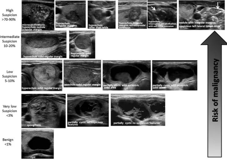
The vast majority (82%–91%) of thyroid cancers are solid (70,73,75,77,84). Of 360 consecutively surgically removed thyroid cancers at the Mayo clinic, 88% were solid or mini-mally cystic (<5%), 9% were <50% cystic, and only 3% were more than 50% cystic (85). Therefore, FNA decision-making for partially cystic thyroid nodules must be tempered by their lower malignant risk. In addition, evidence linking sonographic features with malignancy in this subgroup of nodules is less robust, originating from univariate rather than multivariate analyses. However, an eccentric rather than concentric position of the solid component along the cyst wall, an acute rather than obtuse angle interface of the FIG. 1. Algorithm for evaluation and management of patients with thyroid nodules based on US pattern and FNA cytology. R, recommendation in text.
Table6. Sonographic Patterns, Estimated Risk of Malignancy, and Fine-Needle Aspiration Guidance for Thyroid Nodules
Sonographic pattern US features
Estimated risk of malignancy, %
FNA size cutoff (largest dimension) High suspicion Solid hypoechoic nodule or solid hypoechoic
component of a partially cystic nodule with one or more of the following features: irregular margins (infiltrative, microlobu-lated), microcalcifications, taller than wide shape, rim calcifications with small extru-sive soft tissue component, evidence of ETE
>70–90a Recommend FNA at‡1 cm
Intermediate suspicion Hypoechoic solid nodule with smooth mar-gins without microcalcifications, ETE, or taller than wide shape
10–20 Recommend FNA at‡1 cm
Low suspicion Isoechoic or hyperechoic solid nodule, or partially cystic nodule with eccentric solid areas, without microcalcification, irregular margin or ETE, or taller than wide shape.
5–10 Recommend FNA at‡1.5 cm
Very low suspicion Spongiform or partially cystic nodules with-out any of the sonographic features de-scribed in low, intermediate, or high suspicion patterns
<3 Consider FNA at ‡2 cm Observation without FNA is also a reasonable option
Benign Purely cystic nodules (no solid component) <1 No biopsyb
US-guided FNA is recommended for cervical lymph nodes that are sonographically suspicious for thyroid cancer (see Table 7).
a
The estimate is derived from high volume centers, the overall risk of malignancy may be lower given the interobserver variability in sonography.
b
Aspiration of the cyst may be considered for symptomatic or cosmetic drainage. ETE, extrathyroidal extension.
solid component and cyst, and the presence of micro-calcifications consistently confer a higher risk of malignancy (85–87). Other findings such as lobulated margins or increased vascularity of the solid portion are risk factors that are not as robust (86,87). However, a spongiform appearance of mixed cystic solid nodules is strongly correlated with benignity (66,70,71,88). A spongiform appearance is defined as the ag-gregation of multiple microcystic components in more than 50% of the volume of the nodule (71). Spongiform and other mixed cystic solid nodules may exhibit bright reflectors on US imaging, caused by colloid crystals or posterior acoustic en-hancement of the back wall of a microcystic area. These may be confused with microcalcifications by less proficient sono-graphers, and a recent meta-analysis confirmed that operator experience is correlated with accurate evaluation of internal calcifications (66). Therefore, because of potential for mis-classification, FNA may still be considered for nodules inter-preted as spongiform, but with a higher size cutoff. Lastly, pure cysts, although rare (<2% of thyroid lesions), are highly likely to be benign (66,89,90).
Given the nuances in sonographic appearances of different thyroid cancer histologies, as well as the challenges posed by partially cystic nodules, some authors have suggested risk stratification based upon a constellation of sonographic fea-tures (89–91). In the absence of sonographically suspicious cervical lymph nodes, features associated with the highest risk for thyroid cancer can be used to triage smaller nodules for fine-needle biopsy, whereas nodules with sonographic appearance suggesting lower risk might be considered for fine-needle biopsy at a larger size as determined by maximal diameter (Figs. 1 and 2, Table 6). The sonographic appearance for the vast majority of thyroid nodules can be generally classified in the following categories of US patterns, which combine several individual sonographic characteristics. Since the interobserver variability in reporting individual charac-teristics is moderate even within controlled studies (72), the use of patterns exhibiting correlated sonographic features is more robust. Two recent studies have reported substantial interobserver correlation for identification for nodule sono-graphic patterns (multirater kappa statistics>0.6) (92,93).
High suspicion [malignancy risk>70%–90% (89,90,94)]. High suspicion of malignancy is warranted with a solid hy-poechoic nodule or a solid hyhy-poechoic component in a par-tially cystic nodule with one or more of the following features: irregular margins (specifically defined as infiltra-tive, microlobulated, or spiculated), microcalcifications, tal-ler than wide shape, disrupted rim calcifications with small extrusive hypoechoic soft tissue component, or evidence of extrathyroidal extension (Fig. 2, Table 6). A nodule demon-strating this US pattern is highly likely to be a PTC. Nodules with the high suspicion pattern and measuring‡1 cm should undergo diagnostic fine-needle biopsy to refute or confirm malignancy. However, in the absence of evidence of extra-thyroidal extension, metastatic cervical lymph nodes, or distant metastases, micropapillary thyroid cancers (<1 cm) often have an indolent course, but this may depend upon patient age (95). Although no distant metastases or deaths occurred in a recent observational series of 1235 Japanese patients with biopsy-proven PTC, tumor growth and new appearance of lymph node metastases occurred more fre-quently in patients younger than 40 years of age compared
with those over age 60 (5.9% vs. 2.2% for size increase; 5.3% vs. 0.4% for new nodal metastases, p< 0.05). Thus although a sonographically suspicious subcentimeter thyroid nodule without evidence of extrathyroidal extension or sono-graphically suspicious lymph nodes may be observed with close sonographic follow-up of the nodule and cervical lymph nodes, rather than pursuing immediate FNA, patient age and preference may modify decision-making (95).
Intermediate suspicion [malignancy risk 10%–20% (89,90,94)]. Intermediate suspicion of malignancy is at-tached to a hypoechoic solid nodule with a smooth regular margin, but without microcalcifications, extrathyroidal ex-tension, or taller than wide shape (Fig. 2, Table 6). This appearance has the highest sensitivity (60%–80%) for PTC, but a lower specificity than the preceding high suspicion pattern, and fine-needle biopsy should be considered for these nodules‡1 cm to refute malignancy.
Low suspicion [malignancy risk 5%–10% (89,90,94)]. Isoechoic or hyperechoic solid nodule, or partially cystic nodule with eccentric uniformly solid areas without micro-calcifications, irregular margin or extrathyroidal extension, or taller than wide shape prompts low suspicion for malig-nancy (Fig. 2, Table 6). Only about 15%–20% of thyroid cancers are iso- or hyperechoic on US, and these are generally the follicular variant of PTC or FTCs (71). Fewer than 20% of these nodules are partially cystic. Therefore, these appear-ances are associated with a lower probability of malignancy and observation may be warranted until the size is‡1.5 cm.
Very low suspicion [£3% (66,89,90,94)]. Spongiform or partially cystic nodules without any of the sonographic features described in the low, intermediate, or high suspicion patterns have a low risk of malignancy (<3%). If FNA is performed, the nodule should be at least 2 cm. Observation without FNA may also be considered for nodules‡2 cm (Fig. 2, Table 6).
Benign [£1% (89,90,94)]. Purely cystic nodules are very unlikely to be malignant, and fine-needle biopsy is not indi-cated for diagnostic purposes (Fig. 2, Table 6). Aspiration with or without ethanol ablation may be considered as a therapeutic intervention if a cyst is large and symptomatic; cytology should be performed if aspiration is done.
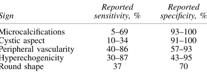
Sonographic evaluation of the anterior cervical lymph node compartments (central and lateral) should be performed whenever thyroid nodules are detected. If US detects cervical lymph nodes that are sonographically suspicious for thyroid cancer (Table 7), FNA of the suspicious lymph node should be performed for cytology and washout for Tg measurement if indicated. In addition, this scenario also warrants US-guided FNA of a subcentimeter nodule that is likely to rep-resent the primary tumor based upon sonographic features.
Although there are several known clinical risk factors for thyroid cancer in patients with thyroid nodules including immobility with swallowing, pain, cough, voice change, growth, lymphadenopathy, and a history of childhood radi-ation therapy (either therapeutic, such as cranial radiradi-ation in childhood leukemia, or for benign conditions, such as en-larged thymus or tonsils) or familial thyroid cancer (96), these have not been incrementally included in multivariate analyses of gray-scale sonographic features and thyroid
cancer risk. However, given the higher pretest likelihood of thyroid cancer associated with these clinical risk factors, FNA can be considered at lower size cutoffs for all of the sonographic appearances described above.
Ultrasound elastography (USE) has similarly been investi-gated for its ability to modify thyroid cancer risk assessment among clinically relevant thyroid nodules. Elastography is a measurement of tissue stiffness. Performance requires an US machine, as well as an elastography computational module that most often must be purchased separately. An initial pro-spective study of 92 selected, nonrandomized patients sug-gested positive and negative predictive values (NPVs) near 100% (97). However, more recently, larger trials have reported substantially different results. Moon and colleagues retro-spectively studied 703 thyroid nodules in comparison to gray-scale US (78). Performance of USE was inferior to that of gray-scale US assessment. The largest prospective study of 706 patients with 912 thyroid nodules was recently published by Azizi et al. (98). In this study, the positive predictive value (PPV) of USE was only 36%, comparable to that of micro-calcifications. The NPV of USE was 97% in a population with cancer prevalence of 9%. Thus, while USE holds promise as a means by which to noninvasively assess cancer risk, its per-formance is highly variable and operator dependent. Perhaps most importantly, USE can only be effectively applied to solid nodules, thus excluding its utility for cystic or partially cystic nodules. Furthermore, to be amenable to direct pressure and determination of tissue strain, the index nodule must not
overlap with other nodules in the anterioposterior plane. Obese patients, those with multinodular goiters and coalescent nod-ules, or patients in whom the nodule is posterior or inferior are not candidates for USE. Thus, at present, USE cannot be widely applied to all thyroid nodules in a similar fashion to gray-scale or Doppler US examination. The committee therefore believes USE (when available) may prove to be a helpful tool for preoperative risk assessment in those patients in whom accurate assessment can be performed. However, the committee cannot presently recommend its universal use or widespread adoption. Importantly, the ability to perform (or not perform) USE should not modify the recommendation for traditional gray-scale sonographic evaluation.
Finally, while most thyroid nodules meeting the preceding sonographic patterns and sizes should undergo FNA, we acknowledge that a conservative approach of active surveil-lance management may be appropriate as an alternative to FNA in selected patients. These may include patients with very low-risk tumors (e.g., no clinical or radiographic evi-dence of invasion or metastases), patients at high surgical risk, or those with a relatively short life span expectancy in whom the benefits of intervention may be unrealized.
[A11] What is the role of FNA, cytology
interpretation, and molecular testing in patients with thyroid nodules?
& RECOMMENDATION 9
Thyroid nodule FNA cytology should be reported using diagnostic groups outlined in the Bethesda System for Reporting Thyroid Cytopathology.
(Strong recommendation, Moderate-quality evidence) To address a significant variability in the reporting of cy-tological findings in thyroid FNA samples, the 2007 National Cancer Institute Thyroid Fine-Needle Aspiration State of the Science Conference provided consensus recommenda-tions known as the Bethesda System for Reporting Thyroid Cytopathology (99,100). The Bethesda system recognizes six diagnostic categories and provides an estimation of cancer risk within each category based upon literature review and expert Table7. Ultrasound Features of Lymph Nodes
Predictive of Malignant Involvementa
Sign Reported sensitivity, % Reported specificity, % Microcalcifications 5–69 93–100 Cystic aspect 10–34 91–100 Peripheral vascularity 40–86 57–93 Hyperechogenicity 30–87 43–95 Round shape 37 70
aAdapted with permission from the European Thyroid
Associa-tion guidelines for cervical ultrasound (20).
Table8. The Bethesda System for Reporting Thyroid Cytopathology: Diagnostic Categories
and Risk of Malignancya
Diagnostic category
Estimated/predicted risk of malignancy by the Bethesda system, %a
Actual risk of malignancy in nodules surgically excised,
% median (range)b
Nondiagnostic or unsatisfactory 1–4 20 (9–32)
Benign 0–3 2.5 (1–10)
Atypia of undetermined significance or follicular lesion of undetermined significance
5–15 14 (6–48)
Follicular neoplasm or suspicious for a follicular neoplasm
15–30 25 (14–34)
Suspicious for malignancy 60–75 70 (53–97)
Malignant 97–99 99 (94–100)
a
As reported in The Bethesda System by Cibas and Ali (1076).
b
Based on the meta-analysis of eight studies reported by Bongiovanni et al. (103). The risk was calculated based on the portion of nodules in each diagnostic category that underwent surgical excision and likely is not representative of the entire population, particularly of nondiagnostic and benign diagnostic categories.