UNIVERSITÀ DEGLI STUDI ROMA TRE
Facoltà di Scienze Matematiche, Fisiche e Naturali Dipartimento di Biologia
Scuola Dottorale in Biologia
Sezione di Biologia Applicata alla Salute
dell’Uomo (BASU) – XXV ciclo
REGULATION OF CELL PHYSIOLOGY THROUGH
THE MODULATION OF ESTROGEN RECEPTORS
ACTIVITIES BY NATURAL AND SYNTHETIC
COMPOUNDS.
REGOLAZIONE DELLA FISIOLOGIA CELLULARE
ATTRAVERSO LA MODULAZIONE DELLE
ATTIVITÀ DEI RECETTORI DEGLI ESTROGENI
INDOTTA DA COMPOSTI NATURALI E SINTETICI.
PhD Student: Dr Piergiorgio La Rosa A.A. 2011/2012
Supervisors: Dr. Filippo Acconcia Pr. Maria Marino
INDEX
SUMMARY……….…..3
RIASSUNTO……….7
1.BACKGROUND………..………..11
1.1 Estrogens………....……....…...11
1.2 Estrogen receptors (ERs)………11
1.3 Genomic signaling…...………..12
1.4 Rapid extra-nuclear signaling………...….13
1.5 ERs post-translational modification……….…..15
1.6 ERs intracellular content……….…...15
1.7 Endocrine Disruptors……….…17
1.8 EDs influence on ERs signaling…...………..…19
2.AIM………...…...21
3. RESULTS………..23
4. DISCUSSION AND CONCLUSIONS………40
Summary
17-β-estradiol (E2) is the most active estrogen in humans and exerts profound effects on the growth, differentiation, and functioning of many reproductive and non-reproductive tissues.
A number of synthetic substances known as xenoestrogens show estrogenic effects; among them, bisphenol A (BPA) is one of the best characterized because human exposure is a risk factor for many disease. Also naturally plant-produced molecules [e.g., naringenin (Nar)] are known to display a mild estrogenic activity, which is exerted in mammals as a consequence of dietary intake. Although “dietary estrogens” are related to a lower predisposition to breast cancer development, reduced incidence in osteoporosis and cardiovascular disease, adverse effects are known. Thus, because of their ability to interfere with many aspects of natural hormones-dependent control of body homeostasis, reproduction, and developmental processes, xenoestrogens and phytoestrogens are also known as endocrine disruptors (EDs).
Two estrogens receptors (ER) are known in humans to interact with endogenous E2 and EDs: these two isoforms, the ERα and the ERβ, modify the expression of specific genes acting as ligand-activated transcription factors. Gene regulation occurs trough ERs recruitment at target gene promoting sequences and follows a “direct genomic pathway”, which expects the ERs to recognize and to bind DNA at the estrogen responsive elements (ERE) sequences, or to interact with transcription factors that in turn bind to DNA in a mechanism known as “genomic indirect pathway”.
In addition, rapid effects of E2 occur within seconds, are insensitive to treatment with inhibitors of transcription and depend on the presence of the ERs to the plasma membrane. Palmitoylation of the ERs determines localization to the plasma membrane, where ERs form multimolecular complexes to trigger rapid E2-activated signal transduction pathways that control proliferative and anti-apoptotic effects (e.g., ERK/MAPK and PI3K/AKT pathways) or pro-apoptotic and differentiating effects (e.g., p38/MAPK pathway), as a function of the ER isoform present.
Also EDs can bind and modulate genomic and rapid ERs activities: in many cancer cell lines Nar impairs ERα-mediated rapid activation of signaling kinases (i.e., ERK/MAPK and PI3K/AKT); in the mean time, the ERα rapid pathways-dependent cyclin D1 transcription is
avoided by blocking AP-1 binding to its promoter. In parallel Nar enhances the persistent phosphorylation of p38/MAPK and, consequently, the induction of a pro-apoptotic cascade. However, Nar does not impair the ERα-mediated transcriptional activity of an ERE-containing promoter and, in addition, Nar acts as E2-mimetic in the presence of ERβ. BPA stimulation mediates the transduction of signaling pathways that culminates with ERK/MAPK and PI3K/AKT phosphorylation in ERα expressing cells, while, in an endogenous ERβ expressing colon cancer cell line (DLD1), BPA behaves as an antagonist by blocking the activation a pro-apoptotic cascade.
Different layers of regulation finely modulate the complexity and the vastness of this signaling network, correctly addressing ERs to the right place in the right moment, through the numerous ER post-translational modifications. Indeed, beside palmitoylation, in response to E2 binding ERs are also phosphorylated on serine (S) residues. Another layer of complexity is introduced by the control of ERs intracellular levels, which represents a crucial step to regulate ER-dependent transcription and hormone-dependent effects. The binding of E2 produces ERα ubiquitination, an event that leads to the 26S-proteasome-mediated receptor degradation, drastically lowering the protein half life. This mechanism is, however, not fully understood and many steps of the process are still to be cleared; there are many evidences that phosphorylation plays a major role in ERα degradation. Less is known about ERβ degradation; there are some proofs that this receptor is degraded by the proteasome, but stronger evidence shows that ERβ does not undergo the ubiquitination processes and that E2 induces the increase in ERβ intracellular content.
Because all the effects of E2 occurs through the above-mentioned ligand-dependent modulation of ERs intracellular content, the goal of the present project was to understand the mechanisms underlying the ligand-dependent modulation of ER intracellular levels to better clarify their modulation abilities in ERα- and ERβ-driven physiological processes.
Results obtained with a wide spectrum of approaches demonstrate that Nar and BPA affect ERα and ERβ protein intracellular content in this way influencing the resulting ERs-dependent effects. In particular, while BPA mimics E2 effects in inducing the 26S proteasome-dependent ERα degradation, Nar induces the receptor accumulation; importantly, this modulation seems to be connected with degradation, as
both E2 as well as EDs induce the ERα mRNA levels down-regulation. Studying ERα palmitoylation, we found that this modification is the upstream structural determinant that guarantees the physiological balance of the ERα protein levels; in the presence of E2, lack of ERα palmitoylation causes faster receptor degradation, thus demonstrating that this receptor posttranslational modification is involved in the regulation of ERα stability and ERα S118 phosphorylation. Our finding demonstrates that E2 maintains both a constant level of S118 phosphorylation, whereas it triggers a significant reduction in total ERα content and a parallel increase in ERα gene transcription. We also show that the rapid E2-dependent activation of the PI3K/AKT pathway but not of the ERK/MAPK pathway regulates ERα phosphorylation and that the effect of the lack of ERα palmitoylation on E2-evoked ERα degradation is mimicked by PI3K/AKT pathway inhibition and unaffected by ERK1/2 inhibitor. Thus, the PI3K/AKT pathway is involved in the regulation of the ERα cellular levels. Reduction in S118 phosphorylation also correlates with a faster E2-induced receptor elimination and is paralleled with an ERα transcription impairment. Indeed both palmitoylation and phosphorylation control ERα activity and stability and are linked each other in a consequent process: E2 induces ERα depalmitoylation and S118 phosphorylation and this process leads to ERα-induced transcription and then to receptor degradation; nevertheless, these two post-translational modifications stabilize the receptor, as the lack of ERα palmitoylation or phosphorylation, leads to a faster ERα degradation.
Our experiments also show that the EDs produce as E2 an increase in ERE-mediated transcription and that the ERα partial antagonist Nar determines ERα phosphorylation as well as the E2 mimetic BPA, that partially stabilizes the receptor. Analysis of the modality by which Nar and BPA affect ERα protein intracellular content reveals that BPA mimics the E2 effects in inducing the 26S proteasome-dependent ERα degradation while Nar induces the receptor accumulation by blocking ERα proteolytic degradation. This mechanism requires Nar to induce the persistent activation of the p38/MAPK, as in the presence of the pharmacological inhibition of the p38/MAPK, Nar acquired the ability to trigger ERα degradation.
More importantly, we also found that the Nar-dependent accumulation of ERα results in an increased receptor transcriptional activity and that, upon Nar stimulation, E2 looses its capacity to regulate
ERα turnover and to physiologically control ERα gene transcription and that both EDs raise ERβ cellular content alone and in co-administration with E2.
These discoveries indicate that in a cellular context exposed to Nar the absolute physiological receptor response or the one in response to E2 is changed because of dysregulated receptor expression. Thus, Nar modulation of ERα cellular content could further affect the E2-dependent regulation of specific cellular processes leading to scenarios that strongly diverge from the physiological ones.
In conclusion, the studies conducted during this PhD project demonstrate that the fine hormone-dependent modulation of ERs intracellular levels is intrinsically connected with all the aspects of the molecular mechanisms (i.e., genomic and extra-nuclear) that ERs uses to transduce the physiological E2 intracellular message and as a consequence that EDs hijack the ERs signaling pathway by deregulating ERs expression. In turn, cells exposed to EDs undergo an altered response if compared to the E2-dependent physiological one.
Riassunto
Il 17-β-estradiolo (E2), l'estrogeno più efficace negli esseri umani, esercita effetti profondi sulla crescita, il differenziamento e il funzionamento di molti tessuti riproduttivi e non.
Sostanze sintetiche, note come xenoestrogeni, presentano effetti estrogenici, tra queste, il bisfenolo A (BPA) è uno dei più caratterizzati in funzione del rischio di esposizione per gli esseri umani. Anche molecole naturali, prodotte da organismi vegetali (come la Naringenina [Nar]) esprimono una lieve attività estrogenica, esercitata, attraverso la dieta. L’assunzione di questi “dietary estrogens” comporta una minor predisposizione allo sviluppo del cancro al seno, una ridotta incidenza di osteoporosi e malattie cardiovascolari, anche se sono noti alcuni effetti collaterali. Per la loro capacità di interferire con molti aspetti del controllo sull’omeostasi ormone-dipendente, la riproduzione e i processi di sviluppo, gli xenoestrogeni ed i fitoestrogeni sono noti anche come interferenti endocrini (EDs).
Negli esseri umani, due recettori interagiscono con estrogeni endogeni e EDs: queste due isoforme, α e β, modificano l'espressione di geni specifici agendo come fattori di trascrizione attivati dal legante. La regolazione genica avviene attraverso il reclutamento degli ER su specifiche regioni promotrici, gli Elementi Responsivi agli Estrogeni (ERE) in una modalità definita “diretta”, o attraverso l’interazione degli ERs con altri fattori trascrizionali in un meccanismo “genomico indiretto”.
É noto, ad ogni modo, che gli ERs scatenano anche effetti rapidi (nell’arco di pochi secondi) e insensibili al trattamento con inibitori della trascrizione. La palmitoilazione dei ERs è il requisito essenziale che determina la localizzazione alla membrana plasmatica degli ERs, dove i recettori formano complessi multimolecolari che innescano eventi di trasduzione del segnale, il cui risultato comporta effetti proliferativi e anti-apoptotici, attivando le vie di trasduzione del segnale ERK/MAPK e PI3K/AKT, o effetti pro-apoptotici e differenziativi attraverso la via p38/MAPK, a seconda della isoforma attivata.
Anche gli EDs legano e modulano l'attività dei ERs: in molte linee cellulari tumorali la Nar compromette l’attivazione ERα-mediata delle chinasi (ad esempio, ERK/MAPK e PI3K/AKT), inoltre, la trascrizione della ciclina D1 ERα-dipendente è impedita, a seguito del blocco di AP-1 sul suo promotore. Negli stessi sistemi cellulari, i flavonoidi compromettono l'attivazione della via ERK/MAPK e della
via PI3K/AKT mediate dal E2, mentre comportano la fosforilazione persistente (60 min) della via p38/MAPK e, di conseguenza, l'induzione di una cascata pro-apoptotica. Tuttavia la Nar non compromettere l’attività trascrizionale mediata dal ERα sui promotori ERE e si comporta come E2-mimetica in presenza del ERβ. Il BPA media la trasduzione di segnali intracellulari che culminano con la fosforilazione delle ERK e della AKT in cellule esprimenti ERα, mentre, in linee cellulari di cancro del colon (DLD1) esprimenti ERβ, BPA si comporta come un antagonista bloccando l'attivazione della cascata pro-apoptotica.
Diversi livelli di regolazione modulano finemente la complessità e la vastità della rete di segnalazione che caratterizza i ERs e indirizzano correttamente i recettori nei distretti subcellulari adeguati, attraverso le numerose modificazioni post-traduzionali che i ER sono in grado di subire. Accanto alla palmitoilazione, in risposta al E2 i ERs sono fosforilati su residui di serina (Ser). Un altro livello di complessità è introdotto dalla modulazione del turnover dei recettori, che rappresenta un passo fondamentale per regolare la trascrizione ER-dipendente e coinvolge sia la degradazione proteica che la trascrizione dei geni dei ERs. Il legame del E2 produce, infatti, l’ ubiquitinazione del ERα, un evento che porta alla degradazione mediata dal proteasoma 26S, riducendo drasticamente l’emivita della proteina. Questo meccanismo non è, tuttavia, pienamente compreso e molti passaggi del processo risultano tuttora oscuri; ci sono molte prove che la fosforilazione del recettore α giochi un ruolo importante nel processo di degradazione. Sul ERβ le informazioni sono ancora più scarse; sebbene alcune prove indichino la degradazione proteasomale di questo recettore, altre dimostrano che il ERβ non va incontro al processo di ubiquitinazione.
Questo progetto dimostra che Nar e il BPA regolano il contenuto intracellulare di ERα rivelando che, mentre il BPA mima gli effetti del E2 nell'indurre la degradazione del recettore, la Nar induce l'accumulo dei livelli proteici del ERα; è importante notare che questa modulazione sembra essere collegata con l’attività del proteasoma, visto che sia il E2 sia gli EDs riducono i livelli di mRNA di ERα. Studiando la palmitoilazione del ERα, è statu ulteriormente scoperto che questa modificazione è il determinante strutturale che garantisce l'equilibrio fisiologico dei livelli proteici del ERα; in presenza del E2, la mancanza di palmitoilazione provoca una rapida degradazione del recettore, dimostrando che questa modificazione post-traslazionale è coinvolta
nella regolazione della stabilità del ERα e della fosforilazione in Ser (S) 118 del recettore. I nostri dati dimostrano che il E2 mantiene un livello costante di fosforilazione in S118, che innesca una riduzione significativa del contenuto totale del ERα ed un parallelo aumento della trascrizione ERα-mediata. Abbiamo inoltre dimostrato che è la rapida attivazione E2-dipendente della via di trasduzione del segnale PI3K/AKT piuttosto che quella delle ERK/MAPK a regolare la fosforilazione di ERα e che l'effetto di aumento della degradazione in seguito alla mancata palmitoilazione del ERα è riprodotto dall’inibizione della via PI3K/AKT e non influenzata da quella delle ERK/MAPK. Anche la riduzione della fosforilazione in S118 è correlata con una più rapida eliminazione E2-indotta del recettore e, di pari passo, con una minore trascrizione ERα-mediata. Dunque palmitolazione e fosforilazione sono reciprocamente correlate nel regolare il processo di controllo della stabilità e dell’attività del recettore α; il E2 induce la depalmitolazione del ERα e la sua fosforilazione, portando alla trascrizione genica e alla successive degradazione. Queste due modificazioni post-traduzionali, tuttavia, stabilizzano il recettore, visto che sia la mancanza di palmitolazione che di fosforilazione producono una più rapida degradazione del recettoere. I nostri esperimenti dimostrano anche che, al pari del E2, i EDs producono un aumento della trascrizione ERE-mediata e che sia la Nar (antagonista parziale del ERα) sia il BPA (mimetico del recettore), determinano la fosforilazione del ERα, stabilizzando così parzialmente il recettore. L’analisi della modalità con cui la Nar ed il BPA regolano i contenuti proteici del ERα rivelano che il BPA imita gli effetti del E2 nell'indurre la degradazione proteasoma-dipendente mentre la Nar induce l'accumulo del recettore bloccandone la degradazione proteolitica. In questo meccanismo sembra fondamentale l’attivazione persistente della via p38/MAPK, dato che, inibendo farmacologicamente la sua attivazione, la Nar acquisita la capacità di attivare la degradazione del ERα. Inoltre, abbiamo ulteriormente dimostrato che l’accumulo Nar-dipendente dei livelli del ERα si esprime in un' aumentata attività trascrizionale del recettore e che, in seguito a stimolazione con la Nar, il E2 perde la sua capacità di regolare il turnover del ERα e la sua attività fisiologica
Il nostro lavoro indica, pertanto, che in un contesto cellulare esposto alla Nar, la normale risposta fisiologica E2-indotta cambia in seguito alla de-regolazione dei livelli di recettore; questa differente
modulazione porta ad una specifica risposta, che diverge da quella fisiologica.
1 Introduction. 1.1 Estrogens.
Estrogens are a class of steroidal hormones synthesized in all vertebrates [1] primarily produced in follicles, corpus luteum and placenta and in smaller amount in the liver, breasts, adrenal gland and fat tissue. The three major endogenous compounds that belong to this class, estrone (E1), 17-β-estradiol (E2) and estriol (E3), share the typical steroid structure (three ciclohexane rings and one ciclopentane ring) and are synthesized from cholesterol, in an enzymatic cascade of reaction in which the last step is driven by the enzyme aromatase [2]; nevertheless, E2 is the most active estrogen in humans and it exerts profound effects on the growth, differentiation, and functioning of many reproductive and non-reproductive tissues, including bone, liver, muscle, cardiovascular system, and brain
[3, 4]. E2 plays a pivotal role not only in female, but also in male reproductive development and physiology, influences metabolism, maintenance of lipids, bone tissues and cardiovascular and neuronal systems[1, 5].
1.2 Estrogen receptors (ERs).
Two estrogens receptors are known in humans to interact with endogenous and esogenous ER ligands: these two isoforms, α and β, are encoded by different genes located on different chromosomes (chromosome 6 locus 6q25.1 and chromosome 14 locus 14q23-24.1 respectively) [6]. In both cases, a mRNA with 8 esonic sequences and 7 introns is spliced and translated in a conserved structure: the A/B region, encoded by exon 1, establishes a protein-protein interaction domain [7] and modulates gene expression of target genes [8]. The AF-1 domain belongs to this region [9] and is directly, or via co-activators/co-repressors, responsible for the binding to the primary transcription machinery. It reacts to the conformational changes that occur in the ligand binding domain (LBD) and the DNA binding domain (DBD) influencing its functional state. Moreover, along with the DBD or beyond its influence, it induces the constitutive modulation of the receptor’s target genes in a ligand-independent fashion, targeting genes that contain, in the promoter region, the right receptor’s consensus sequences [10]; nevertheless, complete receptor transcription activation needs the AF-2 domain cooperation. One of the major difference between the ERα and ERβ results in the divergences in this domain: in
particular, the distinctive response to estrogen-like ligands, such as 4-hydroxytamoxifen, raloxifene and ICI 164,384 (which are partial ERα agonist and ERβ antagonist) could depend on missing parts of the ERβ AF-1 domain which are, instead, present in the ERα receptor sequence [9].
The region that corresponds to the DNA binding domain (DBD) (i.e., the C region) is encoded by exon 2, 3 and a part of exon 4. It is functionally divided into 2 sub-domains [2] and plays a pivotal role regulating the receptor dimerization processes and recognition of specific DNA sequences, known as estrogen responsive elements (ERE) [10]. The minimal consensus ERE sequence, recognized by both receptors on the DNA strand, is a palindromic inverted repetition: 50-GGTCAnnnTGACC-30 (where n corresponds to any nucleotide); nevertheless, immediately flanking sequences are crucial in determining the affinity which the receptor binds to the ERE sequences with [11]. The DBD domain importance is emphasized by the fact that its protein sequence is the most conserved in all ERs from different animal species; for this reason both receptors bind DNA strand with almost the same specificity and affinity [12].
The hinge, or D region, encoded by exon 4, is one of the most variable sequence within ERs. Even if little is known about its function, this domain is the target of many post-translational modifications and is the region that contains the nuclear localization signal (NLS) [13].
The LBD, the AF-2 domain and a part of the nuclear localization region are encompassed in the C-terminal region of the receptor; the last portion of exon 4 and the exons 5-8 encode for it. The E/F region is also responsible for the receptor binding to chaperone proteins (i.e., heat shock proteins), which complex the receptor in the absence of ligands and unmask the steroid binding cleft upon ligand binding. This machinery also facilitates activated-ERs translocation to the nucleus [14]. The LBD folding shows two layers of α-helices (H1-4, H7, H8 and H11) encasing the central core of the structure (α-helices H5-6, H9 and H10); a two-stranded β-sheet (S1 and S2) and H12 complete the ligand-binding portion [15].
1.3 Genomic signaling.
The ERs modify the expression of specific genes acting as ligand-activated transcription factors [16]. Gene regulation occurs trough ERs recruitment at target gene promoters and follows two
different pathways. The direct genomic pathway expects the ERs to recognize and to bind the ERE sequences, that show typical enhancer properties [17]. The sequence itself determines the receptor affinity for the DNA strand and the tightness of the binding, as many E2-regulated genes miss a perfectly corresponding ERE sequence [18]. Another class of genes is modulated by ERs even if ERE sequences are completely missing. The lack of the classic responsive elements imposes the necessity of a second transcription factor to mediate the binding to the DNA strand. Proteins like the stimulating protein 1 (Sp1) or the fos/jun transcription factor complex on the activator protein 1 (AP-1) are involved to stimulate gene expression [19] in a mechanism known as “genomic indirect pathway”.
Nevertheless, ERs gene modulation requires, in both cases, the receptor to interact with a complex of protein with a activator or co-repressor behaviour, exercised trough AF-1 and AF-2 domains; these motifs work as scaffold for other proteins, which have a direct interaction with the DNA strand [20]. Around 50 different proteins form the co-activators group. Among these, the most important family comprises 3 polypeptides: Steroid Receptor Coactivator (SRC) 1, 2 and 3, that, by interacting with ERs via the AF-2 domain, lead to the recruitment of chromatin modification proteins [21]. Co-activators, gathered to gene promoters, enhance the transcription activity through mechanisms that include the recruitment of transcription factors or other proteins with crucial enzymatic activity needed for an efficient transcription, like the ATP-coupled chromatin remodelling the SNF complex, HATs, metiltransferases e ligases [22]. Such interactions lead to chromatin remodelling and to nucleosomal complexes dissociation. It is also possible that co-repressors interact with AF-2 domain of ERs; even if the number of these proteins is relatively low, they exert an important role by negatively regulating the expression of the ERs-regulated genes [23].
ERs interaction with co-activators seems to deeply rely on H12 position to generate a competent AF-2 region [15, 24]. Upon the binding with a natural or a synthetic ligand, the occupancy of the binding cavity produces a H12 displacement that may vary depending on the compound that binds the receptor. In particular, even if partial-agonists (e.g., genistein, 4-hydroxytamoxifen, and raloxifene) and antagonists (e.g., ICI 164,384) can efficiently fill the binding cavity, they produce a H12 non-allosteric displacement [25, 26] due to their chemical structure, that
does not allow a proper accommodation in the confines of the binding site; this is the contrary of what happen when natural and synthetic ERs agonists (i.e., E2 and diethylstilbestrol, respectively) bind ERs. It is also possible to observe a different H12 positioning between the two ERs: some ERs ligands (e.g. the5,11,cis-diethyl-5,6,11,12-tetra-hydrochrysene-2,8-dyol [THC]) act as ERα agonist, producing the same E2-induced H12 positioning, while others do not allow the ERβ LBD to assume the right conformation, thus acting as antagonist [27]. A proper H12 positioning is needed for a transcriptionally competent AF-2 conformation; thus E2-like compounds that bind the ERs lead to co-activators recruitment or impairment depending on whether they act as agonist or as antagonist, giving rise to a different expression patterns depending on the ligand.
1.4 Rapid extra-nuclear signaling.
In addition to genomic effects of E2, that needs at least a couple of hours to be observed at cellular level, rapid effects also take place. These effects occur within seconds or minutes, are insensitive to treatment with inhibitors of transcription (actinomycin D) and translation (cycloheximide) [28]; they are also activated following stimulation with the complex E2-BSA (17β-estradiol conjugated to bovine serum albumin), unable to cross the plasma membrane [30]. These effects require ERs to be localized at the plasma membrane, where receptors are concentrated in caveolae micro-domains, making contact with the scaffolding protein caveolin-1 [31]. At this level, ERs can interact with several proteins involved in signal transduction of hormones and growth factors (e.g., G-proteins, receptor tyrosine kinases, serine/threonine kinases and adapter proteins) and form multimolecular complexes that are required to trigger the rapid signal transduction events [30, 32]. It is well established that E2 is able to induce proliferative and anti-apoptotic effects in different cell lines activating ERK/MAPK and PI3K/AKT pathways, or pro-apoptotic and differentiating effects via p38/MAPK pathway depending on the ER isoform present [33]. Indeed, the E2:ERα complex rapidly (15 min) activates the proliferative ERK/MAPK and PI3K/AKT and the pro-apoptotic p38/MAPK pathways; after 30 min of E2 stimulation the expression of the anti-apoptotic protein Bcl-2 is enhanced and the activation of the p38/MAPK is blocked in an ERK/MAPK-dependent manner, thus enabling the cell cycle progression [34, 35]. Conversely,
the E2:ERβ complex induces the rapid (15 min) and persistent (24 h) phosphorylation of p38/MAPK pathway, driving cells to the apoptotic route [36].
1.5 ERs post-translational modification.
Different layers of regulation finely modulate the complexity and the vastness of the ERs signaling network, as correctly addressing ERs to the right place in the right moment is a critical issue for cells. This problem is elegantly solved through the numerous post-translational modifications that ERs are able to undergo to: palmitoylation is required to promote membrane localization and ERs:caveolin-1 association. Both ERα and ERβ are palmitoylated on cysteine (C) residues with ERα being palmitoylated on C447 and ERβ on C399. The sequences encompassing these two residues are highly homologues with each other [37]. Palmitoylation occurs through the action of two palmitoyl-acyl-transferases (PAT) [38] and the mutation of the C447 to A or the chemical inhibition of PAT activity with 2-bromo-hexadecanoic acid (2-Br) prevent plasma membrane localization and the E2-mediated extra-nuclear signaling [33, 37, 39].
In response to E2 binding, ERs are also phosphorylated. ERα is phosphorylated on many serine (S) residues with S118 and S167 being the main ones; these two residues, belonging to the A/B region (i.e. the AF-1 domain) regulate co-factors recruitment and enhance ERα transcriptional activity [40]. The homologue residue on ERβ, despite the poor homology between ERs in the A/B region, is S87, part of a motif shared with other steroid receptors. ERβ phosphorylation on S87 enhances ERβ interaction with SRC-1 [41].
1.6 ERs intracellular content.
Regulation of ERs intracellular levels is a fundamental cell property to tightly regulate all the effects of E2 and all the effects triggered by the hormone occur in parallel with the concomitant modulation of receptors intracellular levels. ERs level modulation represents a crucial step in order to regulate ER-dependent gene transcription and involves both receptors degradation and ERs gene transcription; however, ligand modulation of receptor intracellular levels is different for ERα or ERβ.
The E2 binding produces ERα ubiquitination an event that leads to the 26S-proteasome-mediated receptor degradation [42]. This event
drastically lowers the protein half life from at least 24 hours, in the unliganded state, to less than 2 hours [43]. ERα is marked for degradation by the proteasome trough the action of protein ligases, which covalently attach ubiquitin to a target lysine [44]. The mechanism that leads to this modification contemplates ubiquitin-activating enzymes (UBA) to activate ubiquitin, before transferring the protein to ubiquitin-conjugating enzymes. Finally the receptor ubiquitination is achieved trough the action of the ligases [45]. In order to proceed with the degradation process, additional ubiquitin moieties are attached to the receptor, thus forming the ubiquitin chain [46]. The polyubiquitinated receptor is finally recognized and degraded by the 26S-proteasome complex. Proofs that upon E2 binding ERα undergoes this kind of regulation are given by treating cells with proteasome inhibitors (e.g., MG-132): thus the major route of clearance of liganded ERα follows this mechanism [44]. Even if in the absence of the hormone ERα undergoes a basal amount of degradation, upon E2 binding the degradation process fastens the protein turnover, which produces a switch in the steady state of the receptor. ERα gene transcription is also regulated by many factors, which include several different transcription factors as well as ERα, which regulates its own expression. ERα mRNA synthesis can be also regulated by its promoter methylation and, more recently, a number of subtype-specific microRNAs (miRNAs) have been found to affect ERα expression [Thomas et al., 2011]. The final goal of this mechanism is the reduction of the total ERα content and in turn the reduction in the cell response to the hormone-induced stimuli, in a typical negative feedback regulation [47]. This mechanism is however not fully understood and many steps of the process are still to be cleared; there are many evidences that receptor phosphorylation plays a major role in its degradation [48]; although this receptor posttranslational modification is required for receptor activation, it is not known if it could be a signal for receptor ubiquitination, following the ERα DNA binding and modulation of target genes [49].
Less is known about ERβ regulation of intracellular content; even if there are some proofs that this receptor is degraded by the proteasome [50], stronger evidence show that ERβ does not undergo the ubiquitination processes [51]. Moreover, following E2 stimulation, ERβ induces its transcription [52] instead of down-regulate it. Thus, the intracellular concentration of the ERs results from a dynamic balance between ER synthesis and ER breakdown [53]; moreover the
hormone-induced stimuli is the result of the balance between ERα and ERβ expression, especially in cells that express both ERs isoform, like muscle myoblasts and myotubes. Nonetheless, even if the fine regulation of the ERs expression comes along with the hormone-induced rapid effects, the knowledge on the contribution of the E2-induced extra-nuclear signaling is still poor.
1.7 Endocrine disruptors (EDs).
A number of synthetic substances known as xenoestrogens show estrogenic effects. This highly heterogeneous group consists of synthetic chemicals used as drugs (e.g., tamoxifen, raloxifene), in industry and agriculture (e.g., pesticides) and acting as food contaminants (e.g., bisphenols). Probably, the best-characterized exponent of this class is bisphenol A (BPA) a molecule widely used in plastic manufacturing and found as environmental contaminant. BPA is one of the most worldwide-produced chemicals, with more than 6 billion pounds/year produced and over 100 tons released into the atmospheare [54]. High temperature and high or low pH make BPA to be released from tin cans and polycarbonate plastic containers allowing the chemical to leach into food and water. Thus, it is a risk factor for human exposed to BPA [55]. Its simple structure, consisting in two benzene rings and two (4, 4’)-OH substituents allows the molecule to bind both ERs binding pocket, with a 10-fold higher affinity to ERβ [56]; nevertheless, E2 affinity to ERs is 10.000-fold higher with respect to BPA [57]. Several reports proved that BPA is capable to induce severe adverse effects (e.g., normal reproductive tract development disruption in rodents [58], inhibition of testosterone synthesis in adult rats BPA-exposed during perinatal periods [59]). Although risk of BPA exposure is not completely assessed, it is clear that human exposure easily occurs: BPA has been detected in pregnant women and also in their foetuses (in maternal and foetal plasma and amniotic fluid, BPA concentration ranges from 1 to 9 ng/ml) [60] and in the 95% of urine samples tested in U.S. at concentrations ≥0.1 µg/L [61].
Not only human-derived compounds are able to mimic estrogenic responses, a large number of naturally plant-produced molecules are known to display a mild estrogenic activity, which is exerted, in mammals, through dietary intake [62]. “Dietary estrogens” or phytoestrogens belong to a large class of compounds that include about 5000 molecules, known as flavonoids and further divided into six
subclasses according to their chemical structure: flavonols, flavones, flavanols, flavanonols, flavanones, and isoflavones [63]. Such vastness and heterogeneity make the characterization of the estrogenic effects of these compounds quite hard: due to their structure divergences, flavonoids can act as estrogen-mimetics, display an anti-estrogenic effect, or even do not affect E2 signaling at all [64]. Moreover, impact on human physiology strongly relies on the nature and the distribution of these compounds in the diet. In particular, citrus fruit and soy are the sole sources of flavonones and isoflavonones, respectively [65]; polyphenols like quercetin are found in almost all vegetables, fruit, wine and tea [66]; others, like naringenin (Nar), are more exclusively distributed and found in orange, grapefruits and tomatoes skin. In most cases, however, food contains complex and poorly characterized mixtures of polyphenols, which level can change depending on food storage, processing and environmental factors [65]. Obviously, habits, and availability is crucial to determine the differences in polyphenol human intake. In particular, flavanones are relevant only in countries where citrus fruit is available, particularly in Southern Europe [65], while isoflavon intake in Asian countries is definitely higher than Europe or America’s (25-40 mg/day versus less than 1 mg/day) [67]. These differences allowed the understanding of important properties of these compounds, such as the lower predisposition of Asian women to breast cancer developing [68]. Moreover, a reduced incidence in osteoporosis [69] and cardiovascular disease [70] is related to a diet rich in flavonoids. These evidence and flavonoids antioxidant properties [66] led to a neat increase in flavonoid usage as dietary components although adverse effects are known [71] and the E2-like and E2 antagonistic effects are not yet fully clarified. Flavonoids are considered potentially able to exert a protective role against the development of E2-dependent pathologies (e.g., endocrine tumors) through the binding to ERs [72]; among others, nutritionally relevant concentrations of Nar are known to induce apoptosis in different cancer cell lines ERs-containing (e.g., colon, breast, and uterus cancer cell lines) [73, 36]. Although flavonoids, as in the case of BPA, show a higher affinity towards ERβ than ERα, these phytochemicals are able to activate both receptors [74]. However, for their ability to interfere with many aspects of natural hormones-dependent control of body homeostasis, reproduction, and developmental processes, xenoestrogens and phytoestrogens are also known as endocrine disruptors (EDs) [74]. EDs act directly via steroid
hormone receptors or indirectly through non-steroid receptors (e.g., neurotransmitter receptors such as the serotonin receptor, dopamine receptor, norepinephrine receptor), orphan receptors [e.g., aryl hydrocarbon receptor (AhR)], and on enzymatic pathways involved in steroid biosynthesis and/or metabolism [75].
1.8 EDs influence on ERs signaling.
Several research groups have demonstrated that Nar triggers the activation of ERE containing genes via both ERα and ERβ [72], in the mean time, it impairs ERα interaction with the transcriptional factors Sp1 and AP-1 [36]. Interestingly, since Sp1 and AP-1-dependent gene expression is strictly dependent on the extra-nuclear pathway activation, xenoestrogen could affect non-ERE containing genes transcription (e.g., cyclin D1) through the modulation of extra-nuclear ERs activities [72]. These evidence, observed in human hepatoma cells (HepG2), which express only endogenous ERα, and in cervix carcinoma cells (HeLa), devoid of any ER isoforms but transiently transfected with the human ERα expression vector, indicate that flavonoids hinder the ERα-dependent proliferative effects.
Data from our laboratory also show that in the same cell systems, flavonoids impair the E2-mediated activation of ERK/MAPK and PI3K/AKT pathway, while they enhance the persistent (60 min) phosphorylation of p38/MAPK and, consequently, the induction of a pro-apoptotic cascade (i.e., caspase-3 activation and PARP cleavage). Thus, Nar decouples the ERα action mechanisms by preventing the activation of proliferative pathways and instead by driving cells to apoptosis [36]. The mechanisms through which flavonoids impair the rapid signals activation involve the decrease in ERα localization at the plasma membrane and the consequent reduction of receptor association to caveolae. In particular, Nar induces ERα de-palmitoylation faster than E2, which results in a rapid ERα dissociation from membrane caveolin-1, thus impairing the receptor association with adaptors and/or signaling proteins (i.e., MNAR and c-Src). This event precludes the activation of mitogenic signaling cascades, while the activation of p38/MAPK is independent from ERα palmitoylation, as demonstrated in HeLa cells transfected with the ERα unpalmitoylable mutant. All these events lead to the activation of the apoptotic cascade [36]. However, Nar does not impair the ERα-mediated transcriptional activity of an ERE-containing promoter [72]. Thus, flavonoids modulate specific ERα mechanisms and
they can be considered as mechanism-specific ligands for ERs [76]. In addition, Nar acts as E2-mimetic in the presence of ERβ by rapidly activating p38/MAPK and the apoptotic cascade in HeLa cells transiently transfected with ERβ expression vector or in endogenous ERβ expressing colon cancer cell line (DLD1) colon adenocarcinoma cells [36]
BPA is also known to display estrogenic activity: it is a weak ligand for both ERα and ERβ [77] even if it principally acts through the nuclear activation of the ERα [78, 79] and does not affect ERβ-based ERE containing gene transcription; this ED is able to prevent the E2:ERβ-mediated activation of ERE-based transcriptional activity [80]. Through ERα binding, it stimulates cell proliferation in ERα-containing breast cancer cells and in ERα-overexpressing HeLa cells, and mimics E2 in enhancing ERα-mediated transcriptional activity of ERE-containing promoters [72]. It has been demonstrated by our group that BPA-dependent proliferative response requires the ERα-mediated extra-nuclear signaling activation. In fact, upon BPA stimulation ERα mediates the transduction of signaling pathways that culminates with ERK/MAPK and PI3K/AKT activation. These effects are not present in empty vector-transfected cells and are completely prevented by the anti-estrogen ICI 182,780. As a whole, these data indicate that BPA acts as an E2-mimetic by binding to ERα leading to the activation of rapid extra-nuclear pathways that drive cells to proliferation [78].
Conversely, in DLD1 cells, BPA behaves as an antagonist by blocking the activation a pro-apoptotic cascade [80]. Our group has reported that the physical association between ERβ, caveolin-1, and the unphosphorylated form of p38/MAPK are present in DLD-1 cell line even in the absence of E2 [33]; after E2 stimulation, ERβ rapidly interacts with p38, leading to the phosphorylation of the kinase and the activation of apoptotic cascades [33]. Intriguingly, both in the absence and in the presence of E2, BPA stimulation specifically prevents the ERβ:p38 association without affecting the ERβ:caveolin-1 complex formation. Thus, BPA decouples ERβ from the downstream signals important for the E2-induced pro-apoptotic cascade. The BPA-induced inhibition of the ERβ:p38/MAPK interaction suggests that different ligands could modulate ER extra-nuclear signals changing in this way the final cellular outcomes by inducing distinct receptor conformational changes. Moreover, E2 induces the ERβ-dependent increase of the pC3 ERE-containing promoter transcription, while BPA does not exert any
transcriptional effect. When added together with E2, BPA prevents the E2-induced ERβ transcriptional activity. Furthermore, BPA prevents the E2 ability to increase ERβ levels, which requires both the genomic and the extra-nuclear ERβ activities. All these data suggests a complete antagonistic role of BPA on ERβ signal transduction pathways [80].
Beside their modulation of ERs rapid and nuclear activities, it is also possible that EDs could also influence ERα and ERβ cellular content like the endogenous ligands regulate receptors intracellular levels, transcription and subsequent ERs-dependent cellular effects. Nonetheless, nothing is known about the regulation that occurs following EDs binding to ERα or ERβ produced by EDs on the ERs intracellular content and expression.
2. Aim.
On this basis, the main goal of the present project was to understand the mechanisms underlying the ligand-dependent modulation of ER intracellular levels to better clarify ligand effects in ERα- and ERβ-driven physiological processes.
The highly regulated cellular mechanisms, which control the ERs intracellular levels, point to a critical role of ERs stability for E2 signaling. Indeed, numerous clinical and in vitro studies suggest that the alteration of ERs expression is an important step in the development and progression of E2-related diseases including different type of cancers [81]. Thus, deregulation of the balance between ERα and ERβ expression could be a critical step in several E2-dependent diseases. Signaling modulation of the ERα and ERβ intracellular levels also occur. Indeed, receptor phosphorylation appears to be required for ERα degradation [82] and the extra-nuclear E2-activated signal transduction cascades have been implicated in the modulation of ERα and ERβ cellular content. In particular, ERK/MAPK and PI3K/AKT pathways are involved in the E2-dependent regulation of the ERα degradation [83] and the activation of the p38/MAPK controls the E2-mediated increase of ERβ mRNA and protein levels [52]. This evidence together with the fact that EDs (e.g., Nar and BPA) affect ERα and ERβ activities, by modulating their extra-nuclear signaling, led to the hypothesis that they could also modulate ERα and ERβ cellular content by modulating ERs mRNA and protein levels. Thus, EDs modulation of ERs cellular content could further affect the E2-dependent regulation of specific
cellular processes (e.g., proliferation, differentiation and apoptosis) suggesting scenarios that strongly diverge from the physiological ones.
To this purpose, in this PhD project we used ductal carcinoma cells (MCF-7) and the colon cancer cell line DLD-1 expressing only one isoform of the receptor (ERα and ERβ, respectively) to avoid the influence that the second isoform could produce on the modulation of ERs signaling network. We also used the Human Embrionic Kidney (HEK293) cell line and the cervix adenocarcinoma HeLa cells that we endowed with wild type ERα or with ERα mutated in the C447 residue to Alanine (A) (i.e., un-palmitoylable mutant) or in the S118 residue to A (i.e., un-phosphorylable mutant). Where necessary, we also used a set of specific inhibitors to block cellular mechanism and to verify their involvement in the specific measured parameter.
3. Results.
3.1 Impact of EDs on cellular physiology.
In order to evaluate differences in the modulation of ERα activities induced by EDs, we have undertaken a high-throughput approach, which allowed us to assess the “signature” that endogenous, natural and synthetic compounds leave on the cell by activating the ERα receptor. Lysates from ductal carcinoma cells (MCF-7 cells), treated with E2, Nar and BPA for 24 hrs were processed in a two-dimensional gel electrophoresis and differences in the resulting cellular proteomes were analysed. More than 575 spots on the gels show differences in their quantification values. More importantly, at least 24 proteins show a significant diversity in their pattern of expression, depending on the substance that binds ERα (data not shown). These data implicate that EDs influence the physiology of cells expressing the ERα and further suggest that the observed differences in Nar- and BPA-induced protein expression pattern could be a consequence of a Nar and BPA direct influence on ERα intracellular levels.
Figure 1: EDs effect on cellular proteome.
Two-dimensional electrophoresys of MCF-7 cells were treated for 24 hrs with vehicle (A), E2 10-8M (B), Nar 10-6M (C) or BPA 10-5M (D). Results were then analyzed to point out
qualitative and quantitative differences in protein expression. Figure shows representative blots.
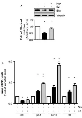
3.2 Effect of EDs on ERα expression.
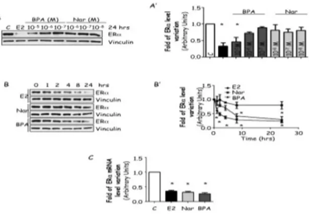
Because EDs could control ERα cellular levels and this control could result in a different protein expression, we evaluated the effect of BPA and Nar on the regulation of ERα protein expression. MCF-7 cells were treated for 24 hrs with BPA or Nar in a concentration range known to engage ERα [78, 84]. As expected, 24 hrs E2 administration reduced ERα protein levels (Fig. 2A and A’). A dose-dependent reduction in ERα intracellular levels were observed in cells exposed to BPA while none of the Nar concentrations significantly affected ERα content (Fig. 2A and A’).
It is widely known that E2 decreases ERα protein half-life from 24 to 2 hrs [53] but ligand-dependent ERα degradation could occur faster than 24 hrs; for this reason, a more detailed time course analysis of the effect of BPA and Nar in MCF-7 cells showed that E2 rapidly (2 hrs) induces ERα degradation while BPA-induced ERα breakdown requires 4 hrs treatment to be apparent. Conversely, no significant changes in ERα protein content were detected when MCF-7 cells were treated with Nar at all the tested time points (Fig. 2B and B’). Because ERα regulates also the mRNA transcription of its own gene, we also evaluated the impact of all ERα ligands on receptor mRNA levels. RT-qPCR analysis showed that in MCF-7 cells 24 hrs treatment E2, Nar and BPA reduces ERα mRNA content (Fig. 2C).
This evidence indicates that BPA mimics E2 in determining ERα down-regulation while Nar reduces ERα mRNA content without affecting receptor protein level.
Figure 2: EDs effect on ERα.
Western blot analysis of ERα levels (A and B) and relative densitometric analysis (A’ and B’) of MCF-7 cells treated for 24 hrs or at indicated time points with E2 10-8M, Nar 10-6M
or BPA 10-5M. (A). RTq-PCR analysis of ERα mRNA levels treated with E2 10-8M, Nar
10-6M or BPA 10-5M for 24 hrs. Loading control was done by evaluating vinculin
expression in the same filter.*indicates significant differences with respect to the relative control sample (P<0.001).Figure shows representative blots.
3.3 Effect of ERα palmitoylation on receptor degradation.
Because Nar and BPA differentially affects ERα expression (Fig. 2) and mainly influence ERα activities by targeting ERα-triggered extra-nuclear effects [36, 72, 77], it is possible that rapid signaling could play a role in controlling ERα intracellular content.
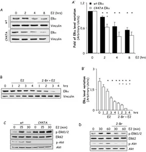
To test this hypothesis, we evaluated the role of ERα membrane localization on receptor intracellular levels by taking advantage of the fact that palmitoylation is required to trigger rapid ERα signaling and that genetic (i.e., mutation of the ERα palmitoylation site C447 to A) or pharmacological (i.e., inhibition of PAT activity) interference of ERα palmitoylation both disrupt E2:ERα extra-nuclear signaling activation. Thus, we stably transfected HEK293 cells with wt ERα and the un-palmitoylable C447A ERα mutant and used the PAT inhibitor 2-Br in MCF-7 cells. In ERα wt HEK293 cells, E2 is capable of inducing a significant reduction in ERα cellular content within the first 4 hours (Fig. 3A and A’). Longer E2 treatment (8 hours) did not further enhance receptor degradation (Fig. 3A). Notably, the difference in the time-dependent E2-mediated receptor degradation between MCF-7 cells and
HEK293 stable cell lines could be ascribed to ERα overexpression. On the contrary, in HEK293 cells stably expressing the C447A mutant ERα, 2 hours of E2 administration were sufficient to determine a significant reduction in ERα levels (Fig. 3A and A’). In order to further demonstrate the impact of ERα palmitoylation on receptor degradation, we analyzed the time-course of E2-dependent ERα breakdown in HEK293 stably expressing the wt ERα both in the presence and in the absence of the PAT inhibitor 2-Br. Under 2-Br pre-treatment, E2 induced an higher reduction of ERα cellular levels than the one observed in the absence of the PAT inhibitor (Fig. 3B and B’) while 2-Br alone did not modify the basal ERα cellular content of stable HEK293 cells (data not shown). These data demonstrate that inhibition of PAT activity as well as mutation of the ERα palmitoylation site determine a receptor pool that undergoes to a faster elimination in response to E2 in stable expressing ERα cells, thus indicating that ERα palmitoylation protects the receptor from E2-dependent degradation.
Accumulating evidence identifies the ERK/MAPK and PI3K/AKT pathways as the principal transduction cascades activated by E2 in many different cell contexts [85]. In line with these notions, time-course analysis revealed that E2 induces a rapid increase in ERK1/2 and AKT phosphorylation in the wt ERα expressing HEK293 cells while the hormone fails to trigger it in the C447A mutant receptor expressing cells (Fig. 3C). Notably, the basal ERK1/2 activation was increased and the basal AKT phosphorylation was reduced when the cells were transfected with the C447A mutant receptor with respect to the wt ERα (Fig. 3C), possibly because of compensatory mechanisms due to the introduction of the exogenous mutated receptor. 2-Br treatment also dampened E2-induced ERK1/2 and AKT phosphorylation in MCF-7 cells (Fig. 3D). These data confirm that ERα palmitoylation is required for the activation of the rapid E2 extra-nuclear signaling [33, 37] and further suggest the notion that a direct link between E2-induced extra-nuclear signaling and ERα degradation could occur.
Figure 3: Effect of palmitoylation on ERα stability.
Western blot analysis of ERα cellular levels, ERK1/2 and AKT phosphorylation in HEK293 cells stably expressing the pcDNA flag-ERα (wt) and the pcDNA flag-ERα C447A (C447A) (A and C) and MCF-7 cells (B and D) treated with E2 10-8M at indicated
time points. Where indicated, cells were treated for 30 min with the palmitoyl-acyl-transferase inhibitor (2-bromopalmitate, 2-Br, 10 µM) before E2 administration. Loading control was done by evaluating vinculin expression in the same filter. * indicates significant differences with respect to the relative control sample; ° indicates significant differences with respect to the corresponding E2 sample (P < 0.05). Figure shows representative blots.
3.4 Effect of phosphorylation on ERα receptor degradation.
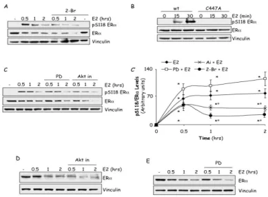
Beside palmitoylation, ERα S118 phosphorylation also plays a role in E2-activated ERα intracellular signaling [49]. Because ERα palmitoylation is involved in the process of E2-evoked ERα elimination (Fig. 3), we sought to determine the impact of ERα palmitoylation on the E2-evoked S118 phosphorylation. MCF-7 cells were pre-treated with the PAT inhibitor 2-Br and than time-course analysis of S118 phosphorylation was performed under E2 stimulation. However, as E2 determines a reduction in ERα cellular content both in the presence and in the absence of 2-Br (Fig. 3B), the receptor S118 phosphorylation was analyzed by quantifying the fraction of the modified ERα with respect to the total observed receptor quantity. Figure 4A and 4C’ shows that E2
treatment induced a rapid increase in the amount of the S118 phosphorylated pool of the ERα within the first 30 min of hormone administration. Although total receptor cellular levels were reduced by E2, the amount of the S118 phosphorylated ERα remained constant for the next 2 hours of E2 administration. 2-Br incubation reduced the amount of the S118 phosphorylated ERα in response to E2 without changing either the overall S118 phosphorylation kinetic or the basal ERα S118 phosphorylation levels (Fig. 4A and 4C’). Accordingly, E2 increased in a time-dependent manner the phosphorylation of the receptor in the S118 residue also in stable wt ERα expressing HEK293 cells but not in HEK293 cells stably expressing the C447A mutated receptor (Fig. 4B). These data indicate that ERα palmitoylation is also required for the E2-dependent phosphorylation of the ERα on the S118 residue.
Next, we evaluated the impact of the E2 extra-nuclear signaling cascades on the ERα S118 phosphorylation status and on E2-induced receptor degradation. In MCF-7 cells, the AKT pathway inhibitor Ai but not the ERK pathway inhibitor PD pre-treatment resulted in a reduction in the amount of the S118 phosphorylated ERα in response to E2 with respect to cells that were treated with the hormone alone (Fig. 4C and C’), without affecting the basal ERα S118 phosphorylation levels (data not shown). Also in this case, the overall E2-dependent ERα S118 phosphorylation kinetic was not changed under either inhibitor treatments (Fig. 4C and C’). Remarkably, as shown in figure 4D, incubation of MCF-7 cells with Ai, induced an increase in the time-dependent E2-evoked reduction of ERα cellular amount with a statistically significant maximum effect (70%) occurring after 30 min of E2 stimulation. On the contrary, PD administration did not change the ability of E2 to induce the reduction of ERα cellular levels (Fig. 4E).
These data indicate that ERα palmitoylation and E2 extra-nuclear-activated PI3K/AKT pathway control S118 phosphorylation and further indicate that inhibition of the PI3K/AKT pathway sensitizes ERα to E2-dependent removal, thus demonstrating that the E2-dependent membrane-ERα-activated PI3K axis activation defends the receptor from E2-mediated degradation.
Figure 4: Effect of phosphorylation on ERα
Western blot analysis of ERα S118 phosphorylation (A,B and C), relative densitometric analysis (C’) and ERα cellular levels (D and E) in MCF-7 cells (A, C, D and E) and in HEK293 cells stably expressing the pcDNA flag-ERα (wt) and the pcDNA flag-ERα C447A (C447A) (B) treated with E2 10-8M at different time points. Where indicated, cells
were treated for 30 min with the palmitoyl-acyl-transferase inhibitor (bromopalmitate, 2-Br, (10 µM), or for 1 h, AKT inhibitor (Akt in, 5µM) or with the ERK1/2 inhibitor PD 98059 (PD, 10µM) before E2 administration. Loading control was done by evaluating vinculin expression in the same filter. * indicates significant differences with respect to the relative control sample; ° indicates significant differences with respect to the corresponding E2 sample (P < 0.05). Figure shows representative blots.
3.5 Effect of palmitoylation and phosphorylation on ERα mediated transcription.
It is well known that ERα S118 phosphorylation is required for full ERα transcription of the ERE-containing genes [40, 86]. Because the lack of ERα palmitoylation prevents ERα S118 phosphorylation, we next studied its impact on E2-dependent ERα transcriptional activity. Real-time qPCR analysis revealed that in MCF-7 cells pre-treatment with the PAT inhibitor 2-Br prevents the increase in the amount of the E2-responsive ERE-containing gene presenelin 2 (pS2/TIFF) mRNA levels observed after 2 hours of E2 administration (Fig. 5A). The cell pre-treatment with either the AKT inhibitor Ai or the ERK1/2 inhibitor
PD also dampened the E2-induced increase in the pS2 mRNA cellular content (Fig. 5A), thus sustaining the notion that rapid E2 extra-nuclear signaling is required for ERα transcriptional activity [87]. Notably, incubation of MCF-7 cells with the inhibitors alone did not affect the total content of pS2/TIFF.
As a transcription factor, ERα cycles on and off its ERE-containing promoters with a frequency of about 30 minutes. E2 rapidly enhances the amount of the ERα associated to its responsive promoters and prolongs the frequency of the ERα:promoter association to about 60 minutes [88]. The data presented above suggest that ERα palmitoylation could be a pre-requisite for E2-activated ERα ERE-containing gene expression. Therefore, it is possible that lack of ERα palmitoylation may impair E2-activated ERα:promoter association. To test this hypothesis, we coupled chromatin immunoprecipitation (ChIP) assays with real-time qPCR analysis in MCF-7 cells to analyze the E2-dependent recruitment of ERα to the pS2/TIFF promoter region both in the presence and in the absence of the PAT inhibitor 2-Br. 2-Br administration completely prevented the E2-induced ERα recruitment to pS2/TIFF promoter without affecting the basal ERα:promoter association (Fig. 5B). Notably, the specificity of the binding of ERα to the pS2/TIFF promoter was determined by using a primer set 1 kb upstream of the ERE in pS2/TIFF, which served as a negative control (data not shown).
These data suggest that ERα palmitoylation rather than S118 phosphorylation is required for ERα-regulated ERE-containing gene expression. Therefore, in order to dissect the relative contribution of ERα palmitoylation and S118 phosphorylation on the E2-dependent ERα-mediated transcriptional activity, mutation of the S118 residue to A was first introduced both in the wild type and in the un-palmitoylable C447A mutant ERα and than the ability of the wt and mutant receptors to modulate E2-dependent ERE-based transcriptional activation was assayed in transiently transfected HeLa cells. As shown in figure 5C, 24 hours of E2 treatment was able to trigger the activation of the artificial promoter containing three repetitions of the ERE sequence (i.e., 3×ERE-TATA, pERE) in the presence of both wt ERα and all the mutant receptors. Although the E2:ERα-mediated activation of the pERE promoter was significantly reduced (40%) in the presence of the S118A ERα mutant than in the presence of the wt receptor, when HeLa cells were transfected with either the C447A mutant ERα or with the S118A,
C447A double mutant receptor, the E2-induced pERE promoter activity was 70% and 50% less stimulated that the one in wt or S118A ERα containing HeLa cells, respectively (Fig. 5C).
Therefore, these data demonstrate a prevalent role of ERα palmitoylation with respect to ERα S118 phosphorylation for receptor transcriptional activity.
Figure 5: Effect of membrane localization on ERα transcription.
RT-qPCR analysis of pS2 mRNA expression normalized on the GAPDH mRNA expressionin MCF-7 cells treated with E2 10-8M for 2 hours (A). Where indicated, cells
were treated for 30 min with the palmitoyl-acyl-transferase inhibitor (bromopalmitate, 2-Br, 10 µM), or for 1 h either with the AKT inhibitor (Akt in, 5µM) or with the ERK1/2 inhibitor PD 98059 (PD, 10µM) before E2 administration. (B) Chromatin Immunoprecipitation analysis of ERα pS2 promoter occupancy normalized on input DNA in MCF-7 cells treated with E2 (10-8M) for 1 hours. (C) Luciferase assay detection on
HeLa cells transiently co-transfected with the reporter plasmid 3xERE TATA and either with the pcDNA flag-ERα (wt), pcDNA flag-ERα S118A (S118A), pcDNA flag-ERα C447A (C447A) or the pcDNA flag-ERα S118A C447A (S118A C447A) expression vectors and than treated 24 hours with E2 (10-8M). * indicates significant differences with
respect to the relative C sample (p < 0.01). ° indicates significant differences with respect to the E2 or wt E2 sample (p < 0.01). # indicates significant differences with respect to the S118A E2 sample (p < 0.01)
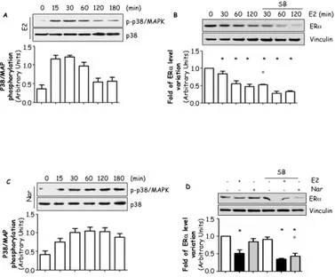
3.6 Effect of EDs on ERα-mediated transcription.
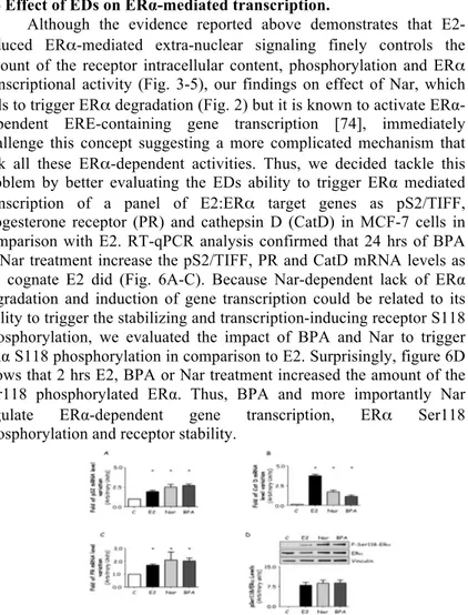
Although the evidence reported above demonstrates that E2-induced ERα-mediated extra-nuclear signaling finely controls the amount of the receptor intracellular content, phosphorylation and ERα transcriptional activity (Fig. 3-5), our findings on effect of Nar, which fails to trigger ERα degradation (Fig. 2) but it is known to activate ERα-dependent ERE-containing gene transcription [74], immediately challenge this concept suggesting a more complicated mechanism that link all these ERα-dependent activities. Thus, we decided tackle this problem by better evaluating the EDs ability to trigger ERα mediated transcription of a panel of E2:ERα target genes as pS2/TIFF, progesterone receptor (PR) and cathepsin D (CatD) in MCF-7 cells in comparison with E2. RT-qPCR analysis confirmed that 24 hrs of BPA or Nar treatment increase the pS2/TIFF, PR and CatD mRNA levels as the cognate E2 did (Fig. 6A-C). Because Nar-dependent lack of ERα degradation and induction of gene transcription could be related to its ability to trigger the stabilizing and transcription-inducing receptor S118 phosphorylation, we evaluated the impact of BPA and Nar to trigger ERα S118 phosphorylation in comparison to E2. Surprisingly, figure 6D shows that 2 hrs E2, BPA or Nar treatment increased the amount of the Ser118 phosphorylated ERα. Thus, BPA and more importantly Nar
regulate ERα-dependent gene transcription, ERα Ser118
phosphorylation and receptor stability.
Figure 6: Effect of EDs on ERα transcription and stability.
RT-qPCR analysis of presenelin 2 (pS2/TIFF) (A), progesterone receptor (PR) (B) and cathepsin D (CatD) (C) mRNA expression, normalized on the GAPDH mRNA expression in MCF-7 cells treated with E2 10-8M, Nar 10-6M or BPA 10-5M for 24 hours. (D) Western
blot analysis of ERα S118 phosphorylation in MCF-7 cells treated with E2, Nar or BPA for 2 hours. The same filter was re-probed with anti-ERα antibody. Loading control was done by evaluating vinculin expression in the same filter. * indicates significant differences with respect to the relative control sample. (P < 0.01)
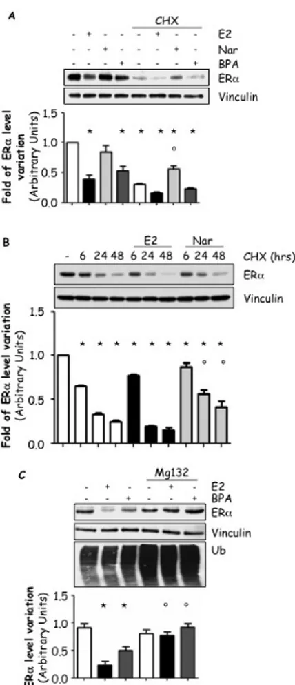
3.7 Effect of BPA and Nar on ERα degradation.
The data presented above indicate that BPA and Nar both induce ERE-containing gene transcription and ERα S118 phosphorylation while BPA induces reduction in ERα protein and mRNA levels and Nar triggers reduction in ERα mRNA levels (Fig. 2 and 6). Thus, it is possible that these EDs could modulate ERα expression through two different post-transcriptional mechanisms. In order to test this hypothesis, we used the protein-biosynthesis inhibitor cycloheximide (CHX) as a tool to study the effect of E2, BPA and Nar on the receptor degradation without the contribution of the neo-synthesized ERα pool (i.e., pre-formend ERα) [89]. As expected, 24 hrs treatment of MCF-7 cells with E2, BPA or CHX induced a significant reduction in total ERα cellular content while Nar did not affect it (Fig. 7A). On the contrary, while E2 and BPA increased the effect of CHX on ERα breakdown, Nar treatment prevented the CHX-induced ERα degradation (Fig. 7A), thus suggesting that BPA, as E2, triggers proteolytic ERα degradation while Nar could induce an ERα intracellular accumulation.
Prompted by these results, we further investigated the Nar effect on the pre-formed ERα cellular pool [89]. Time-course analysis confirmed that prolonged (24-48 hrs) Nar treatment was able to prevent the CHX-dependent reduction in ERα cellular content while E2 further increased it (Fig. 7B). In parallel, we also evaluated if BPA-dependent ERα degradation was ascribable, as in the case of E2, to the action of the 26S proteasome [53]. Pre-treatment of MCF-7 cells with the 26S proteasome inhibitor MG-132 blocked the 24 hrs E2- and BPA-induced ERα reduction in intracellular levels (Fig 7C). As expected, the inhibition of the 26S proteasome induced an increase in the total amount of ubiquitinated proteins (Fig. 7C).
These data demonstrate that BPA mimics the effect of E2 in inducing ERα degradation and that Nar affects ERα intracellular content in a different manner than E2 and BPA.
Figure 7: Effect of EDs on ERα degradation.
Western blot analysis of ERα (A, B and C) or ubiquitin (C) cellular levels in MCF-7 cells treated with E2 10-8M, Nar 10-6M or BPA 10-5M for 24 hours (A and C) or at the indicated
time points (B); Where indicated, cells were treatedfor 60 min with the 26S proteasome inhibitor MG-132 (1 µg/ml) or cycloheximide (CHX) 1 µg/ml before ligand administration (C). Loading control was done by evaluating vinculin expression in the same filter.* indicates significant differences with respect to the relative control sample; ° indicates significant differences with respect to the corresponding CHX (A and B) and E2 or BPA (C) sample.