UNIVERSITÀ DEGLI STUDI DI SASSARI
Dipartimento di Scienze Biomediche
INTERNATIONAL PhD SCHOOL IN BIOMOLECULAR
AND BIOTECHNOLOGICAL SCIENCES
Indirizzo: Microbiologia ed Immunologia
XXVII CICLO
Direttore Prof. Leonardo A. Sechi
Several aspects of pathogenesis of
Trichomonas vaginalis
Tutor: Dottoranda:
Prof. Pier Luigi Fiori Valentina Margarita
Contents
Abstract ... VI List of tables ... VII List of figures ... VII Abbreviations ... VIII
CHAPTER I ...1
General Introduction ...1
I.1 Introduction ...1
I.2 Trichomonas vaginalis ...1
I.3 Trichomonas vaginalis pathogenesis ...4
I.4 Adhesion ...5
I.4.1 Trichomonas vaginalis surface proteins ...5
I.4.1.1 A novel type of surface gluzincin metallopeptidases ...8
I.5 Contact independent mechanisms ... 10
I.6 T.vaginalis interacts with viruses and human-associated bacteria ... 10
I.6 Object of this study ... 12
CHAPTER II ... 13
Characterisation of domains of putative T.vaginalis M60-LIKE protease ... 13
II.1 Introduction ... 13
II.2 Overview about HS-GAG ... 14
II.2.1 GAGs ... 14
II.2.2 Heparin/heparan sulphate glycosaminoglycans ... 16
II.2.3 HSPGs ... 18
II.3 Syndecan families ... 21
II.3.1 Syndecan-1 shedding is implicated in microbial pathogenesis. ... 23
II.4 Overview on matrix metalloproteinases ... 24
II. 4.2 Bacterial strains ... 28
II.4.3 Plasmids ... 28
II.4.3 Growth and selective media ... 29
II.4.4 Sterilization ... 29
II.4.5 Plating bacteria ... 29
II.4.5 Storage of bacteria and DNA samples ... 30
II.4.6 Determination of DNA and protein concentration ... 30
II.4.7 Primers ... 30
II.4.8 Polymerase chain reaction (PCR) ... 31
II.4.9 Agarose gel electrophoresis for analysis or PCR products ... 32
II.4.10 PCR products purification ... 33
II.4.11 Digestion with restriction enzymes... 33
II.4.12 Ligation reactions ... 34
II.4.13 Transformation of competent E.coli cells ... 35
II.4.14 Plasmid DNA purification by mini prep ... 36
II.4.15 DNA sequencing ... 36
II.4.16 Induction of recombinant proteins expression in E.coli and cell lysis37 II.4.17 Purification recombinant N-terminal 6x His-tagged proteins ... 38
II.4.18 Sodium dodecyl sulphate-polyacrylamide gel electrophoresis (SDS-PAGE) 38 II.4.19 Western blotting ... 40
II.4.20 Ion-exchange and concentrating proteins ... 40
II.4.21 Isothermal titration calorimetry (ITC) ... 40
II.4.22 Cleavage assay of Syndecan-1 recombinant protein ... 41
II.4.23 Bioinformatic tools ... 42
II.5 Results ... 43
II.5.2 Carbohydrate binding modules of TVAG339720 M60like proteins. . 44
II.5.3 Cleavage assay of Syndecan-1 ... 46
CHAPTER III ... 48
Trichomonas vaginalis and Mycoplasma hominis: a mutualistic endosymbiotic relationship ... 48
III.1 Introduction ... 48
III.2 Mycoplasma hominis ... 50
III.3 Arginine dihydrolase (ADH) pathway: a common biochemical pathway 51 III.3 Macrophages and nitric oxide: an important host defence against T.vaginalis ... 53
III.4 Objectives ... 54
III.5 Materials and methods ... 55
III.5.1 Materials and reagents ... 55
III.5.2 Parasites and culture condition ... 55
III.5.3 Cell culture and THP-1 differentiation ... 56
III.5.4 Determination in vitro amount of intracellular ATP ... 56
III.5.5 Determination of NO produced by THP-1 co-incubated with infected and not infected T.vaginalis ... 56
III.5.6 Statistical analysis ... 57
III.5.7 Bioinformatic tools ... 57
III.6 Results ... 58
III.6.1 T.vaginalis in symbiosis with M.hominis produces more ATP ... 58
III.6.2 M.hominis-infected T.vaginalis competes with host macrophages for arginine and prevents NO formation ... 60
CHAPTER IV ... 63
Final discussion ... 63
Appendices ... 85
Appendix A: Cloning primers ... 85
Appendix B: Extinction coefficients ... 85
Abstract
The pathobiology of Trichomonas vaginalis involves direct and indirect interactions with host tissue, bacteria and viruses. In this work, we investigated adhesion and interaction with human microbiota, pathogenic mechanisms of protozoan.
We characterized a new protein, TVAG339720, belonging to M60-like/PF13402 domain-containing proteins. M60-like domains are shared by proteins from several mucosal microbes, hypothetically relating with epithelial cells. TVAG339720 is characterized by a signal peptide, a transmembran domain, and putative carbohydrate binding modules, PA14 and GBDL, supposed to bind to heparin and heparan sulphate (HS). HS are sugars forming proteoglycans, a component of epithelial cells glycocalyx. We tested protease activity of TVAG339720 towards proteoglycans and interaction between CBMs, heparin and HS. Although the target of TVAG339720-M60L is still unknown, the bounds between TVAG339720-CBMs and HS suggest that these proteases play a role in adhesion to epithelial layer.
T.vaginalis interactions with microbes of urogenital tract represent another
pathogenic mechanism. We focused on symbiosis established with Mycoplasma
hominis, studying how M.hominis influences T.vaginalis pathobiology in vitro.
Comparing the ATP produced by free-protozoan and T.vaginalis with M.hominis and evaluating mycoplasma ability to influence nitric oxide production by macrophage in T.vaginalis infections, we can assert that this is a mutually beneficial endosymbiotic relationship.
List of tables
Table I.1 - Classification of T.vaginalis
Table II.1 - Classification of HSPGs
Table II.2 - List of MMPs involved in syndecan cleavage in vitro and in vivo.
Table II.3 - Bacterial strain used in this study Table II.4 - Typical PCR reaction set-up Table II.5 - Typical PCR reaction program
Table II.6 - Set-up of a typical restriction enzyme digestion reaction Table II.7 - Set-up of a ligation reaction
List of figures
Figure I.1 - Various cellular forms of T.vaginalis.
Figure I.2 - Hypothetical membrane proteins of T.vaginalis.
Figure I.3 - Structural organization of M60-like containing-proteins from different mucosal microbes.
Figure II.1 - GAGs structure and synthesis. Figure II.2 - HS biosynthesis.
Figure II.3 - Schematic representation of major classes of HSPGs
Figure II.4 - Domain architecture of T.vaginalis M60L entries from PFAM database.
Figure II.5 - PCR products analysed by AGE.
Figure II.6 - Expression of domains from TVAG339720.
Figure II.7 - ITC data of TvPA14-GBDL binding to heparin and heparan sulphate
Figure II.8 - Cleavage assay of human recombinant Syd-1.
Figure III.1 - Intracellular localization of M.hominis within T.vaginalis. Figure III.2 - Arginine dihydrolase pathway in T.vaginalis and
Figure III.3 - Biochemical pathway of NO production in mammalian cells.
Figure III.4 - Intracellular concentration of ATP produced by T.vaginalis
and T.vaginalis infected with M.hominis in different growth phases. Figure III.5 - ATP produced by T.vaginalis G3 and T.vaginalis G3-MPM02 growth in media with different concentration of arginine.
Figure III.6 - NO produced by macrophage after cocolture with pathogens.
Figure III.7 - NO produced by macrophages infected with pathogen in media with different arginine concentrations.
Abbreviations
A280 nm Absorbance at a wavelength of 280 nm
ADH Arginine dihydrolase pathway
ADI Arginine deiminase
AGE Agarose gel electrophoresis
BACON Bacteroidetes-Associated Carbohydrate-binding often N-terminal CBM
BLAST Basic Local Alignment Search Tool BspA Bacteroides surface protein A
CAZy Carbohydrate active enzymes database CBM Carbohydrate-binding module
CFE Cell-free extract
CK Carbamate kinase
cOCT Catabolic ornithine carbamyltransferase
CS Chondroitin sulfate
FT Flow-through sample during IMAC
GBDL Galactose-binding domain-like
GiardiaDB Giardia lamblia genomic databse
GlcA d-Glucuronic acid
GlcN D-glucosamine
GP63 Major membrane glycoprotein Leishmania spp
GR Glucose-restriction
His6-tag Polyhistidine tag
HS Heparan sulphate
HSPG Heparan sulphate proteoglycan
IdoA l-Iduronic acid
IMAC Immobilized metal ion affinity chromatography IPTG Isopropyl-β-D-thiogalactopyranoside
ITC Isothermal titration calorimetry
KEGG Kyoto Encyclopedia of Genes and Genomes
KOD Thermococcus kodakaraensis DNA polymerase enzyme
LPG lipophosphoglycan
LTG Lateral gene transfer
MEROPS On-line database for peptidases
Mh Mycoplasma hominis
MMPs Matrix metalloproteinases
MT1-MMPs membrane type MMPs
NAD+ Nicotinamide adenine dinucleotide (oxidized) NADH Nicotinamide adenine dinucleotide (reduced)
NC Nitrocellulose membrane
NO Nitric oxide
OD600nm Optical Density at 600nm
PA14 CBM named after the anthrax Protective Antigen
PBS Phosphate buffered saline
PCR Polymerase chain reaction
PFAM On-line database of protein families SAGE SDS-agarose gel electrophoresis
SDS-PAGE SDS polyacrylamide gel electrophoresis STD Sexually transmitted diseases
Syd-1 Syndecan-1
TAE Tris-acetate-EDTA
TBE Tris-borate-EDTA
TEMED N,N,N',N'-Tetramethylethylenediamine TrichDB Trichomonas vaginalis database
Tv Trichomonas vaginalis G3
TvBspA BspA-like proteins in T.vaginalis
Tv-MhMPM02 T.vaginalis G3 in symbiosis with M.hominis MPM02
TVV T.vaginalis virus
CHAPTER I
General Introduction
I.1 Introduction
Sexually transmitted diseases (STD) are a major global health problem. Each year, an estimated 500 million people acquire one of four sexually transmitted infections: chlamydia, gonorrhoea, syphilis and trichomoniasis. Moreover, more than 530 million people are living with HSV2 and more than 290 million women have an HPV infection [1].
Trichomoniasis is the most common non-viral, curable, STD worldwide that annually affects millions of people[1]. The causative agent of infection is
Trichomonas vaginalis, obligate extracellular mucosal parasite that induce significant
health sequelae in both men and women. In women, symptoms range from a silent form to important complications including pelvic inflammatory disease [2], invasive cervical cancer [3], sterility, pregnancy and postpartum problems [4, 5]. In men, the infection occurs mainly without symptoms, complicating its diagnosis and control. Recently, trichomoniasis has been associated with aggressive prostate cancers [6-8]. Moreover, T.vaginalis infection is epidemiologically associated with HIV [9-11]. Despite the high prevalence of trichomoniasis and the complication associated with the disease, little is known about parasite or host factors involved in pathogenesis [12, 13].
I.2
Trichomonas vaginalis
Trichomonas vaginalis is an anaerobic flagellated eukaryotic protozoan that infects
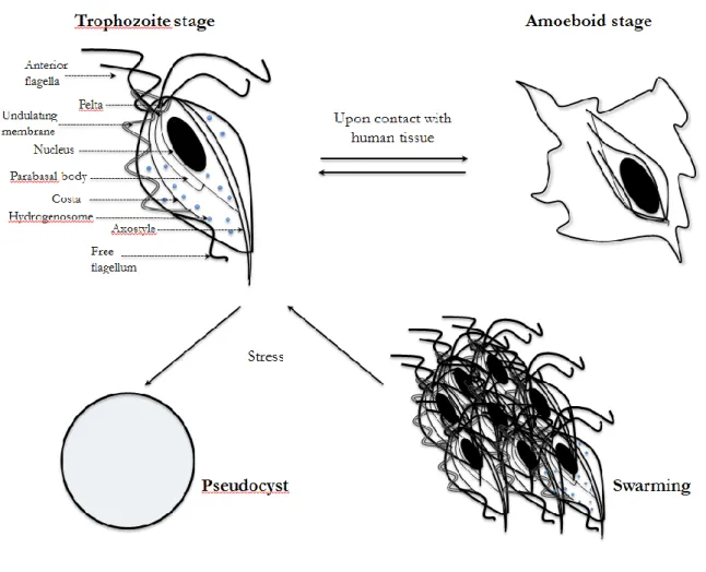
the urogenital tract of human body. This pathogen varies in size and shape; with the average length and width being 10 and 7 m respectively [2]. The two best characterized forms are the trophozoite and the ameboid. The trophozoite is the
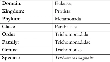
infective form, motile stage of protozoa. In this stage T.vaginalis tends to be uniform, i.e. pyriform or pear-like cell. Upon contact in vivo with epithelial cells from the vagina, cervix, urethra, prostate and extracellular matrix (ECM) proteins, the protozoan can rapidly switch from trophozoite to ameboid form, a pancake shape that allows an increasing of the surface contact [2, 14]. In a process called swarming, trophozoites are able to attach one to another by their pseudopods forming aggregates consisting of numerous cells. The exact role of these aggregates is not yet clear. Pereira-Naves et al have shown the presence of third form of T.vaginalis, which can be induced in vitro upon exposure of trophozoites to cold and drugs [15]. Even for this shape, called pseudocyst, the significance during the infection is unknown [16]. The organism possesses four anterior flagella and a fifth one comes along with its undulating membrane, a large nucleus, and an axostyle, which bisects the protozoan longitudinally. Moreover, T.vaginalis cytosol is glycogen rich and contains other internal organelles such a cytoskeleton, costa, pelta and hydrogenosomes (Figure I.1). The hydrogenosomes are double membrane organelles that share common ancestry with mitochondria, [17, 18], and are involved in production of ATP, acetate, carbon dioxide and hydrogen as end products from pyruvate and malate substrates [19]. T.vaginalis lacks conventional mitochondria and peroxisomes but contains hydrogenosome, which are a typical feature of parabasalid lineage to which T.vaginalis belongs (Table I.1). Hydrogenosomes are also found in several anaerobic eukaryotic microbes [20].
T.vaginalis is an unusual eukaryote showing remarkably similarity to primitive
anaerobic bacteria, in terms of its carbohydrate and energy metabolism. In fact,
T.vaginalis mainly obtains energy from fermentative carbohydrate metabolism
under both anaerobic and aerobic conditions [19]. Glucose is converted to pyruvate in cytosol and subsequently metabolized in the hydrogenosome by fermentative oxidation. Hydrogenosome produces ATP by substrate-level phosphorilation [21]. Trichomonads require unusually high concentrations of iron in in vitro cultures, likely for the dependence of T.vaginalis upon the activities
required from trichomonads to furnish the turnover of FeS proteins, given that this pathogen apparently lacks substantial levels of iron-storage proteins, such as ferritin [23].Several studies have investigated the effect of iron limitation on T.
vaginalis morphology and overall proteome change, showing how cells from
iron-depleted medium displayed altered morphology, including the internalization of flagella and the axostyle and transformation to a larger and rounded shape [24, 25]. Moreover, iron deficiency led to the upregulation of proteins involved in iron-sulfur cluster assembly and the downregulation of enzymes involved in carbohydrate metabolism [26].
Furthermore, the pathogen lacks the ability to synthesize many macromolecules
de novo, particularly purines, pyrimidines, and many lipids, and acquires these
nutrients form vaginal secretions or though phagocytosis of bacteria, vaginal epithelial cells (VECs) and erythrocytes [27-29].
Table I.1 - Classification of T.vaginalis.
Domain: Eukarya Kingdom: Protista Phylum: Metamonada Class: Parabasalia Order Trichomonadida Family: Trichomonadidae Genus: Trichomonas
Figure I.1 - Various cellular forms of T.vaginalis. Trophozoite stage shows the morphological features.
I.3
Trichomonas vaginalis
pathogenesisT.vaginalis is transmitted from person to person through sexually intercourse [14]. The life cycle consists of two stages, the infective and diagnostic stages. The trophozoites attach to mucosal surfaces of urogenital tract and divides by longitudinal binary fission.
Successful colonization of the host mucosa by T.vaginalis is the result of multiple pathogenic mechanisms, including adhesion; secretion of cytotoxic molecules and soluble factors; interaction with member of vaginal microbiome; evasion of
I.4 Adhesion
As mucosal microbial pathogens, T.vaginalis must adhere to epithelial cells as an initial step towards colonizing the host and establishing infections [30]. T.vaginalis adherence to host cell is mediated, in part, by a major lipid-anchored phosphosaccharide, known as lipophosphoglycan (LPG).
LPG is the most abundant component of T.vaginalis surface glycocalyx and the alteration of its sugar content reduces both the ability of adhesion and cytotoxicity of protozoan to host cells [31, 32]. T.vaginalis LPG binds to mammalian protein galectin-1, in a carbohydrate-dependent manner [33]. Galectin-1 is the only identified human receptor for T.vaginalis so far [33].
Moreover, recent compositional and structural analysis of pathogen revealed that LPG has specific domains with proinflammatory properties, modulating inflammatory responses of epithelial cells and macrophages [34].
Although the binding between LPG and galectin-1 may be central in establishing infections, the parasite involves other adhesion factors to establish host-parasite interaction. In fact, surface proteins are expected to be important for initialing and sustaining infections, allowing the parasite to interact with its environment including human epithelial cells, immunocytes and extracellular proteins of the urogenital tract [30].
I.4.1 Trichomonas vaginalis surface proteins
In 2007, the entire genome sequence for the T.vaginalis strain G3 has been sequenced [35], increasing the interest about molecular and cellular mechanisms of T.vaginalis. The genome has ~160 megabases and contains ~60,000 protein coding genes organized into six chromosomes, according to data from the GiardiaDB and TrichDB databases [14, 35]. T.vaginalis genome encodes membrane trafficking machinery, pathogenic proteins for endocytosis of host proteins and phagocytosis of bacteria and host cell [35, 36]. Moreover, a large repertoire of genes consistent with carbohydrate and amino acid metabolism, defense against oxidative stress, transport and pathogenesis are contained into genome [30, 35, 37, 38]. A total of 3000 candidate genes for surface molecules
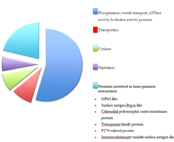
mediating interaction with host tissue, membrane trafficking and signaling have been identified from the sequenced genome and have been divided in ten different protein families [30, 35]. The genome analysis has also indicated that the pathogen is the first eukaryote that does not produce glycosylphosphatidylinositil (GPI) anchor [35]. De Miguel et al, using a proteomic analysis according to BLAST analysis and genome annotation, have identified 411 surface proteins examining six strains of T.vaginalis, showing that 63% of these proteins possess a predicted transmembrane domain and/or signal peptide sequence and 35% of total proteins are membrane proteins predicted to have a possible role in Trichomonas pathogenesis [30, 35] (Figure I.2).
ATPase activity or hydrolase activity; ~8% are transporters. ~8% are cyclases; ~5% are peptidases and ~18% are proteins predicted to be involved in host-parasite interaction. The function of each predicted protein was given through identification of domain that allows the assignment of a predicted function.
BspA-like, GP63-like proteins and adhesins are the major categories of surface proteins and have already been biochemically characterized [14, 30].
Four adhesins, AP65, AP51, AP33 and AP23, are thought to mediate the adhesion of the parasite to the epithelial cells, even if their role as adhesive molecules is highly debatable. These proteins are alternatively expressed on the surface with a highly immunogenic glycoprotein, P270 [2].
TvBspA are the largest gene family encoding potential surface proteins. These proteins share a specific type of leucin-rich repeats (LRRs) named TpLRR (Tp is for Treponema pallidum) and they are also identified into Entamoeba histolytica and
E.dispar genomes [12]. LRR-containing proteins were shown to mediate binding to host epithelial cells and/or ECM proteins and were also implicate in bacterial aggregations [39].
The second largest gene family of T.vaginalis candidate surface proteins encodes GP63-like proteins. GP63 are metalloproteinase belonging to the metzincin class characterized by the motif HExxHxxGxxH, where x represents any amino acid residues. TvGP63 proteases contain the minimal motif HExxH for zincins and their protease activity is inhibited by the cysteine proteinase inhibitor [40]. GP63 proteases likely play a vital role in T. vaginalis infection process, by degrading and binding to various host components.
Another class of surface proteinases is cysteine proteinase, identified by two dimensional (2-D) substrate gel and electrophoresis. CP30 cysteine surface proteinase is a protein of 30kDa involved in binding to HeLa cervical carcinoma cell lines, degrading some ECM proteins and hemoglobin. CP30 is also immunogenic and is secreted into vagina during infection [37].
I.4.1.1 A novel type of surface gluzincin metallopeptidases
Comparative genome studies among bacterial and eukaryotic mucosal microbes, including mutualists and pathogens of invertebrates and vertebrates, have identified several genes and gene families encoding candidate surface proteins and putative enzymes shared through lateral gene transfer (LTG) [30, 41].
One family of T.vaginalis candidate surface proteins was annotated as zinc (Zn)-metallopeptidase sharing a novel protein domain termed „‟M60-like (PF13402) domains‟‟[41]. In profile-profile comparisons, this new domain shows similarity to an existing protein family termed M60-enhancin [MEROPS database][12], characterized by the insect baculovirus (Lymantria dispar nucleopolyhedrovirus) and known to be capable of degrading insect intestinal mucins [41].
In M60-like domain is presented HExxH motif or consensus sequence, typical of zinc metalloproteases belonging to the zincin superfamily or clan [42]. The two histidine residues within consensus sequence are ligands of zinc ion (Zn2+),
while the glutamic acid (E) residue acts as the catalytic active amino acid [43]. An additional conserved glutamate is presented downstream of the HExxH motif, defining pattern HExxHxE. This motif characterizes the gluzincin-like family of Zn-metallopeptidases and the second E functions as a third zinc binding ligand
[41].
InterProScan and Pfam analyses have identified additional domains presents in M60-like containing proteins, the carbohydrate binding modules (CBM). CBMs are involved to binding with specific carbohydrate components within a target substrate and are classified into families on sequence similarities, as defined in the Carbohydrate active enzymes (CAZy)database [41]. CBM families are identified by a number written together with CMB abbreviation, e.g. CBM32 to specify a family with 32 carbohydrate binding module. BACON and PA14 are also carbohydrate binding domains only available from PFAM database and are thought to be involved in glycan binding. Proteins with CBM32 were predominantly associated with vertebrate mucosal surfaces microbes. In many
and one or more transmembrane domains (TMD), suggesting their extracellular or cell surface localization [41].
M60-like domain-containing proteins are shared by a large number of bacterial and eukaryotic microbes [PFAM database] [44] and are present in important mucosal microbes such as T.vaginalis, Bacteroides thetaiotaomicro , Bacteroides fragilis,
Bacteroides caccae, Bacillus anthracis, Clostridium perfringens, Vibrio cholera, Entamoeba histolytica, Cryptosporidium species, suggesting that these proteins might play an
important role in the biology of mucosal microbes as well as in host-microbial interactions (Figure I.3).
Figure I.3 - Structural organization of M60-like containing-proteins from different mucosal microbes.
The length of proteins and their structural features are shown for each pathogen, together with accession number. Species aligned to their N terminus. Several CBMs are associated with M60-like peptidase domains. CMB domains can be found at either the C-terminal or N-terminal side of the M670-like domain compared with the relative position of the protease domain, which is often conserved suggesting this configuration is functionally important. (Image form Nakjang et al, 2012).
I.5 Contact independent mechanisms
T.vaginalis pathogenic mechanisms involve also the contact-independent
cytotoxicity. The pathogen is able to produce a wide range of hydrolase identified as cytoplasmic cysteine proteinase (20-100 kDa). These proteins are released by the parasite and have trypsin-like activity, functioning as cell-detaching factors (CDF) by degrading proteins of the ECM. CDF allow
T.vaginalis to traverse the protective mucus barrier of host epithelium and aid in
the release of host cells from tissue and mucosal desquamation [14, 45].
CDF levels have been shown to correlate with the severity of the clinical symptoms of vaginitis[2].
Another pathogenic mechanism used by T.vaginalis to damage target cell plasma membrane is the secretion of cytotoxic molecules such as perforin like activity, creating pores in erythrocyte membranes. T.vaginalis is also known to excrete different lytic factor having phospholipase A2 activities, to destroy nucleated cells and erythrocytes by degradation of phosphatidylcholine [14].
Recently, a new protein secreted by T.vaginalis was characterized. This protein is homolog of human macrophage migration inhibitory factor (MIF), a versatile proinflammatory cytokine involved in several processes, including immunity, cell proliferation and tumorigenesis. HuMIF is reported to be elevated in prostate cancer. As HuMIF, TvMIF inhibits macrophage migration, is proinflammatory and binds CD74 MIF receptor with high affinity. Moreover, it increases cellular proliferation and invasiveness of BPH-1 and PC3 in vitro. These data indicate that chronic T. vaginalis infections may result in TvMIF-driven inflammation and cell proliferation that contributes to the promotion and progression of prostate cancer [8].
HIV, facilitating HIV entry and transmission in humans by damaging mucosal surface [9-11]. Recently it has been shown that the pathogen can also be infected by four dsRNA viruses (TVV) [46, 47].
T.vaginalis is capable to ingest different mammalian cells [28], including epithelial cells, immunocytes and spermatozoids, obtaining important sources of nutrient and contributes to defense from immune system. [12]
Moreover, the parasite phagocytes several types of bacteria colonizing the lower urogenital tract of human body, inducing an imbalance in the microbial community [48]. During bacterial vaginosis, for example, was observed a correlation between the presence of T.vaginalis and low abundance of protective lactobacilli and higher proportions of Mycoplasma, Prevotella and other bacteria typically involved in bacterial vaginosis [49]. Among several species of human macrobiota, T.vaginalis has shown a clinical association with two different species of Mycoplasma: Mycoplasma hominis [50], and Candidatus Mycoplasma girerdii [51] [52]. Mycoplasmas are the smallest self-replicating organisms lacking cell walls, have small genome and often dependent on their host. In the urogenital tract they are associated with bacterial vaginosis, pelvic inflammatory disease, preterm labor and preterm birth [52]. In spite of these associations with disease,
Mycoplasma hominis and Ureoplasma are also commensal bacteria of the lower
urogenital tract.
Acting as pathogen, M.hominis causes an infection linked with several pregnancy and postpartum complications like spontaneous abortion, endometritis and low birth weight [53-55], as for trichomoniasis.
Many pathogenicity mechanisms of M.hominis are not yet clear. The symbiosis between T.vaginalis and M.hominis is the only endosymbiotic relationship described so far involving two obligated human parasites that produce independent diseases in the same anatomical area. Previous studies have shown that Mycoplasma cells carried by T.vaginalis are able to infect human cells in vitro, suggesting that T.vaginalis could play a role of “Trojan horse” for the bacterium during infection [56]. However, the exact nature and fundamental aspects of this association still have to be cleared.
I.6 Object of this study
The overall aim of this study was to gain new insights into pathogenic mechanisms of T.vaginalis, focusing the attention on adhesion to host cells and interaction with human microbiota.
The specific objectives were as follows;
Characterize of putative M60-like domain-containing protease from Trichomonas
vaginalis using biochemical approaches.
Analyse the functional aspects of CBMs present on putative M60-like domain protein of T.vaginalis.
CHAPTER II
Characterisation of domains of putative
T.vaginalis
M60-LIKE protease
II.1 Introduction
Infections occur when the balance of host-pathogen interaction shifts to favour the pathogen. A multitude of virulence factors is used by pathogens to promote pathogenesis. Many pathogens facilitate their attachment to host tissue components by specific cell surface molecules, called adhesins. Other microbial pathogens secrete enzymes that digest host components to acquire nutrients and to inactivate host defence factors. Some pathogens are able to synthesize toxins that cause both the death of host cells and tissue damage. Several microorganisms express factors that inhibit specific host defence mechanisms or deregulate the host inflammatory response to their advantage. Moreover, many pathogens subvert host components to grow, survive and spread in the host environment. Among many components modulated by microbial pathogens, one common link is represented by heparan sulphate (HS) glycosaminoglycans (GAGs). GAGs are sugars attached to a protein backbone forming proteoglycans, a component of epithelial cell glycocalyx. Proteoglycans along with glycoproteins, glycolipids and transmembrane mucins, constitute glycocalyx of human cells [57]. All component of glycocalyx can act as receptor for microbial adhesion, while the entire structure represents an important second line of defence against invading pathogens, after mucosal surfaces [58].
II.2 Overview about HS-GAG
II.2.1 GAGs
GAGs are terminal carbohydrate structures in the extracellular matrix, comprised of repeating disaccharide units of hexosamine and uronic acid or galactose with various substitutions. GAGs are expressed widely in the human body and a number of bacteria, viral and parasitic pathogens exploit GAGs on key steps of pathogenesis, such as adhesion and invasion of host cells, cell-cell transmission and evasion of host defence mechanisms [59].
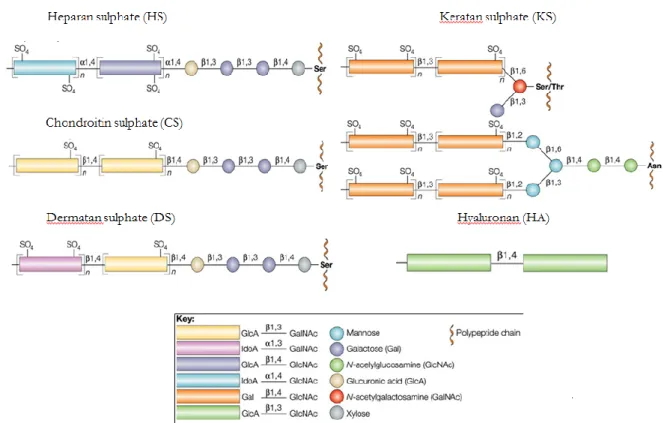
The list of GAGs includes heparan sulphate (HS)/heparin, chondroitin sulphate (CS), dermatan sulphate (DS), keratan sulphate (KS), and hyaluronan (HA). Except for HA, all other polysaccharides are sulphated to various degrees and are covalently complexed to proteoglycans core protein (Figure II.1).
Among GAGs, HS can be considered a crucial common link that many pathogens exploited to infect human hosts and cause diseases, including those originating from STIs. Several studies have shown the multiple roles of HS in microbial invasion of human hosts [60]. A number of etiologic agents of STIs, such as HSV, HIV HPV and Chlamydia trachomatis express surface proteins that interact with HS to mediate their attachment to eukaryotic cells as a primary mechanism during mucosal infections [59, 60].
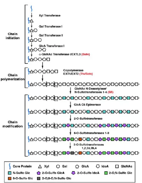
Figure II.1 - GAGs structure and synthesis.
Synthesis of HS, CS and DS start with addition of xylose to serin (Ser), proceeding by addition of two galactose residues and one glucuronic acid. In HS synthesis, there is an alternating addition of N-acetylglucosamine and glucuronic acid (GlcA), while for the assembly of CS and DS there is an alternating addition of N-acetylgalactosamine and GlcA. The polymerization of HS, CS and DS is completed by epimerization of GlcA to iduronic acid (IdoA) and sulphation (SO4) of different residues, resulting in a large micro-heterogeneity within the polymers. KS assembly is the result of alternating attachment of N-acetylglucosamine and galactose to O or N-glycans on the proteoglycan core and of sulphatation of several residues. HA is a large polymer of alternating N-acetylglucosamine and GlcA and is not attached to a core protein. (Image modified from Kleene R et al, 2004).
II.2.2 Heparin/heparan sulphate glycosaminoglycans
Heparin/heparan sulphate glycosaminoglycans, extracellular complex polysaccharides, are attached to a protein core or proteoglycan (HSPG) and are extruded by cells to the cell surface and into the extracellular space [61, 62]. Heparin (highly sulphated polysaccharide) and HS (the less sulphated polysaccharide), are negatively charged linear carbohydrate polymer composed of repeating uronic acid [d-glucuronic acid (GlcA) or l-iduronic acid (IdoA)] and 10-200 disaccharide units of D-glucosamine (GlcN) [61]. Variable patterns of substitution of the disaccharide units with N-sulphate, O-sulphate and N-acetyl groups give rise to a large number of complex sequences.
Heparin is commonly isolated from connective-tissue type mast cells and it is biosynthesized as heparin proteoglycan (Mr 750000–1000000). Multiple polysaccharide chains (Mr 60000–100000) are covalently attached to a unique proteoglycan called serglycin [61, 63]. Serglycin is a secretory vesicle proteoglycans implicated in inflammation, storing mast cell inflammatory mediators [63].
HS is also biosynthesized as a proteoglycan, but it has fewer and shorter polysaccharide chain than heparin proteoglycan [61]. HS proteoglycans are expressed and secreted by most mammalian cells, and are located on cell surfaces and in the extracellular matrix. HS is evolutionarily ancient and its composition has remained relatively constant from Hydra to humans [62]. Moreover, it is involved in important physiological functions, such as lipid metabolism, neurogenesis and cytokine/growth factor interaction [60].
One of the reasons that HS interacts with a diverse group of pathogens relates to the structural and functional diversity of HS originating from extensive modifications during its biosynthesis. The biosynthesis of HS occurs in the Golgi apparatus and is a sequential, multistep process (Figure II.2).
Figure II.2 - HS biosynthesis.
HS chains are synthesized on a core protein by the sequential action of individual glycosyltransferases and modification enzymes, in a three-step process involving chain initiation, polymerization and modification. Chain initiation: assembly of a linkage tetrasaccharide on serine residues in the core polypeptide. The tetrasaccharide is composed by GlcA-galactose (Gal)-Gal-Xylose (Xyl). This process is catalysed by four enzymes (Xyl transferase, Gal transferase I-II and GlcA transferase I), which add individual sugar residues sequentially to the non-reducing end of the growing chain. Chain polymerization: after the assembly of the linkage region, one or more α-GlcNAc transferases add a single α1,4-linked GlcNAc unit to the chain, which initiates the HS polymerization process. HS chain polymerization then takes place by the addition of alternating GlcA and GlcNAc residues, which is catalyzed by the EXT family proteins. Chain modifications: include GlcNAc
N-deacetylation and N-sulfation, C5 epimerization of GlcA to IdoA, and variable O-sulfation at C2 of IdoA and GlcA, at C6 of GlcNAc and GlcNS units, and, occasionally, at C3 of GlcN residues. The HS chains are ∼100 or more sugar units long and have numerous structural heterogeneities Monosaccharaides are synthesized in the cytoplasm and transported into the Golgi, where they are used to synthesize a conserved tetrasaccharide, and then initiate and elongate heparan-sulphate-specific polysaccharide chains (Image from LinX, 2004).
II.2.3 HSPGs
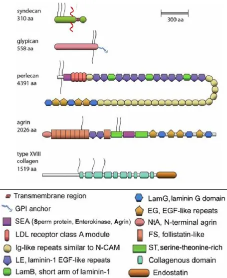
HSPGs are glycoproteins, containing one or more covalently attached HS chains. There are two major subfamilies of cell surface HS proteoglycans (HSPGs), the syndecan (four members) and the glypicans (six members). The syndecan core proteins are transmembrane proteins and virtually all cell types express one or more syndecans [64]. The glypican core proteins are attached to cell membranes by a glycosylphosphatidylinositol (GPI) tail. Perlecan, agrin and collagen XVIII are HS proteoglycans but located in the extracellular matrix. These are also distinguished by their specific core proteins [65] (Figure II.3) (Table II.1 shows HSPGS classification depending on their localization).
Figure II.3 - Schematic representation of major classes of HSPGs.
HSPGs include four syndecans, six glypicans, and one each of perlecan, agrin, and the hybrid HSPG/collagen type XVIII. The size is approximately proportional to the number of amino acid residues (aa). In the bottom panel there are various color-coded modules. HS chains are shown as black lines; CS chains are shown as red lines. (Image modified from Iozzo RV, 2001).
Table II.1 - Classification of HSPGs.
Class Proteoglycan Core mass (kDa) Chain type (number) Membrane-bound Syndecan -1 31-45 3-4 HS 1-2 CS Syndecan -2 31-45 2-3 HS Syndecan -3 31-45 3-4 HS 1-2 CS Syndecan -4 31-45 2-3 HS Glypican-1, Glypican -6 57-69 1-3 HS Betaglycan 110 1-2 HS 1-2 CS Neuropilin-1 130 1 HS or CS CD44v3 37 1 HS
Secretory vesicles Serglycin 10-19 10-15 Heparin 10-15 CS
Extracellular matrix Perlecan 400 1-4 HS
Agrin 212 2-3 HS
Collagen XVIII 150 1-3 HS
HSPGs have multiple activities in cell and tissue, such as to collaborate with other matrix component to define basement membrane structure and to provide matrix for cell migration (perlecan, agrin and collagen XVIII); to maintain proteases in an activate state; and to regulate various biological activities after secretion (serglycin). HPGSs protect cytokines, chemokines, growth factors, and morphogens by binding and act as receptors for proteases and proteases inhibitors, regulating their spatial distribution and activity. Moreover, syndecans and glypicans can cooperate with integrins and other cell adhesion receptors, allowing cell-ECM attachment, cell-cell interactions, and cell motility. They can also act both as endocytic receptors for clearance of bound ligands and as
receptor [64]. The binding takes place both through core protein interaction and through GAG chains modified. One feature shared by many coreceptors is modification of GAG chains, which contribute into interaction between proteoglycans and ligands, extracellular matrix proteins or other cell surface receptors, regulating cell adhesion, migration and invasion also in the human diseases. The most common modifications occur on one or more site of HS and CS chains.
HSPGs signaling coreceptors include CD44, glypicans (Glypicans-1, -6), neurophilins, syndecans (Syndecan -1, -4) and betaglycan.
All transmembrane proteoglycans signalling co-receptor, such as transmembrane syndecans, undergo ectodomain shedding resulting in proteolytic cleavage of core protein near the transmembrane domain that allows releasing of the extracellular domain from the cell surface. The soluble extracellular domains of HSPGs continue to carry their glycosaminoglycan modifications [66]. Syndecan shedding is mediated by matrix metalloproteinases (MMP1, MMP7, MMP9, ADAM17) [67, 68] but even some microorganisms can enhance host cell proteolytic shedding of syndecan-1 (Syd-1), with an increase of bacterial colonization [69, 70]. Moreover, it has been shown that the amount of HS presents on syndecan core proteins regulates both the rate of syndecan shedding and core proteins synthesis [71].
II.3 Syndecan families
Syndecans family is a group of transmembrane HSPGs with a long evolutionary story. The four mammalian members, syndecan-1 to syndecan-4, are composed by core protein with covalently attached GAG chains. The syndecans contain an N-terminal extracellular domain or ectodomain, a hydrophobic transmembrane domain, and a short C-terminal cytoplasmic domain. The ectodomain contains three consecutive consensus Ser-Gly sequences for HS chain attachment close to N terminus, and Ser-Gly sequences for CS chains at site near the plasma membrane [72]. The length of transmembrane and cytoplasmic domain is highly conserved among member of syndecans family, whereas the length of the ectodomains varies considerably. Many syndecan roles are attributed to their HS
chains, which interact with a wide range of ECM and adhesion molecules, chemokines and cytokines [72]. The functions of syndecans include anchorage of cells to ECM with associated HS binding domains; maintenance of epithelial and endothelial morphology; binding to and modulation of activity of HS binding growth factors; modulation of activity of several proteases and their inhibitors; and signaling molecules [69, 72]. Moreover, syndecan family has been proposed to act as adhesion and internalization receptors for pathogenic microorganisms. By shedding, the syndecan ectodomains complete with their GAG chains, are released from the cell surface forming soluble ectodomains that may function either as paracrine or autocrine effectors, or competitive inhibitors of intact proteoglycans [73, 74]. These ectodomains are in fluids accumulating, following injury and inflammation. Matrix metalloproteinases (MMPs) and membrane type MMPs (MT1-MMPs) are involved in the shedding and catabolic process of syndecans, whereas heparanase and endosulfatases modify HS chains within extracellular environment, including the ability of HS chains to bind and sequester growth factors [73] [75]. Moreover, a recent study has shown that the loss of HS chains enhances both the susceptibility of core protein of Syd-1 to proteolitic cleavage by matrix metalloproteinases and a dramatic increase in core protein synthesis [71].
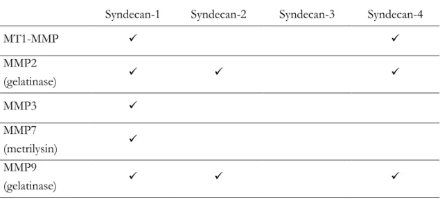
In the table II.2 have listed the MMPs involved in syndecan cleavage both in vitro and in vivo [68, 73]. Moreover, MMP2, MMP7 and MMP9 bind HS chains of syndecan.
Table II.2 - List of MMPs involved in syndecan cleavage in vitro and in vivo.
Syndecan-1 Syndecan-2 Syndecan-3 Syndecan-4
MT1-MMP MMP2 (gelatinase) MMP3 MMP7 (metrilysin) MMP9 (gelatinase)
II.3.1 Syndecan-1 shedding is implicated in microbial pathogenesis.
Syd-1 shedding is one of the general host responses to tissue injury and inflammation [69]. Moreover, the activation of Syd-1 shedding is used by some pathogens as an important virulence mechanism [69, 70, 76]. For example, P.
aeuroginosa actives Syd-1 shedding through LasA, a virulence factor for its lung
infection and S.aureus sheds syndecan-1 through - and toxins, implicated in sthaphylococcal infections. Both of these microorganisms activate a protein-tyrosine kinase (PTK)2 –dependent intracellular signalling mechanism to stimulate the ectodomains cleavage [69, 76]. Streptococcus pneumonia, unlike P.
aeuroginosa and S.aureus, directly sheds Syd-1 ectodomain through zinc
metalloproteinase (ZmpC) [70]. So far, it is not completely clear how Syd-1 shedding promotes microbial pathogenesis.
Moreover, Syd-1 has been identified as a substrate of MT1-MMP that cleaves the Gly82-Leu83 and Gly245-Leu246 bonds of a recombinant syndecan-1 fusion protein [68]. Increased levels of shed Syd-1 are present in sera of patients with some cancer types [71]. Moreover, shed Syd-1 plays an active role in driving tumour progression, stimulating signalling and proliferation in tumour cells, and
enhancing angiogenesis, osteolysis, growth, and spontaneous metastasis of tumour cells [73].
II.4 Overview on matrix metalloproteinases
Matrix metalloproteinases, also called matrixins, are the major enzymes implicated in cleavage of several ECM constituents, as well as non-matrix proteins, regulating cell-matrix composition [77]. In the MEROPS database, MMPs are classified as the metzincin subfamily of zinc metalloproteases family M10. This family is distinguished by a highly conserved motif, HEXXHXXGXXH, where histidines (H), glutamic acid (E) and glycine (G) residues are invariant. The histidines bind zinc at the catalytic site. A conserved methionine also presents in the catalytic domain forms a “Met-turn‟‟, eight residues after the zinc binding motif, forming a base to support the structure around the catalytic zinc. A third zinc binding ligand differentiates the zincin clan further into subclans e.g gluczincins (E), aspzincins (D) or metzincins (H/D) [77].
Metzincin subfamily is further subdivided into four families: serralysins, astacins, ADAMs/adamalysins and MMPs.
The 23 human MMPs typically containing a signal peptide (SP), a propeptide of ~80 amino acid, a catalytic domain of ~170 amino acid, a linker peptide of variable lengths, and a hemopexin (Hpx) domain of ~200 amino acids, with exceptions of MMP-7 (matrilysin 1), MMP-26 (matrilysin 2) and MMP-23, which lack or present changes of domains in the structure [78].
With exception of six membrane -anchored MMPs, the remaining 17 MMPs are destined for secretion into the extracellular milieu [79].
MMP14, a membrane-type MMP (MT1-MMP), exists in a membrane-bound rather than a secreted form [80]. It is expressed on endothelial cells, on fibroblasts, on osteoclasts, and on monocytes. Moreover, MMP14 has been also found to be produced on cancer cell membranes. [81]. The Hpx domain is the
MT1-MMP has a wide substrate specificity, such as aggrecan, elastin, perlecan, and fibronectin; and cleaves and promotes shedding of Syd-1 [68] , of betaglycan
[83] and of MUC1, a transmembrane mucin with a critical role in embryo implantation, protection of mucosal epithelial cells from microbial and enzyme attack, and in several aspects of tumour progression [84].
II.4.1 M60-like domain containing proteins of T.vaginalis
Recent study has shown a novel protein domain termed „M60-like (PF13402) domains‟, shared by several bacterial and eukaryotic mucosal microbes [41]. The presence of the extended consensus HExxHxE in this domain suggested that the M60-like domain containing proteins could be considered as gluzincin metallopeptidases processing extracellular glycoprotein targets. This hypothesis is further supported by evidence that M60-like domain containing proteins represent distant relatives of viral-enhancin proteases known to degrade insect mucins. Moreover, Dr.Didier Ndhe in his PhD thesis has shown that three M60-like domains proteins of Bacteroides thetaiotaomicron, a bacterial gut mutualist of human body, exhibited mucin protease activity. This proteolytic activity was shown to be inhibited in a mutant version of the protein as well as in the presence of Ethylenediaminetetraacetic acid (EDTA), implying BT4244 and its relatives are metal dependent proteases. The CBMs contained in B.
thetaiotaomicron are from family 32 and are capable to bind galacto-configured
sugars that are common to mucin glycans.
A total of 25 TvM60-Like containing proteins were also identified in T.vaginalis and 11 of these possess XExxHxE motif. Among them, six have one TMD with only three of these entries possessing a complete M60-Like domain. A sequence alignment suggested that most entries are likely represented gene fragments or truncated version of longer proteins [41]. Among three complete TvM60-like containing proteins, i.e. TVAG339720, TVAG189150 and TVAG199300, TVAG339720, TVAG189150 were detected on the cell surface by proteomic analysis, and TVAG339720 is the only one identified in all six tested isolates [38]
(figure II.4). Moreover, native TVAG339720 and TVAG189150 proteins had been detected in T. vaginalis cell membrane extracts and the extracellular
localisation of the TVAG339720 protein has indeed been confirmed through immunofluorescence assays by our collaborators in the U.S.A (personal communication, Prof. Robert Hirt).
Using HMM profile-profile searches, PA14-like and CBM32 or galactose- binding domain (GBD) like sequences were identified in M60-like proteins from
T.vaginalis. These domains were also detected in several M60-like
domain-containing proteins of other mucosal microbes, such as C.perfringens and
C.albicans, to target galacto-configured sugars [85] [86]. In Dr. Ndeh thesis is shown that TVAG199300-M60L failed to degrade any of the mucin substrates tested against the protein and the PA14 domain of TVAG339720 binds heparin instead of mucin glycans.
The preference for highly sulphated heparin was an indication that sulphate groups may play a role in heparin recognition by the PA14 domain.
Heparin is a GAG, structurally similar to heparan sulphate. These GAGs form part of well-known epithelial cell surface proteoglycans, such as syndecans and glypicans [64]. These proteoglycans along with mucins, glycoprotein and glycolipids constitute epithelial cell glycocalyx at mucosal surfaces [57].
An important hypothesis is that the actual targets for the TvM60 proteins containing PA14 domains of T.vaginalis are heparan sulphate glycosaminoglycans. Data accumulated so far about M60-like domain-containing proteins of T.
vaginalis suggest that TVAG339720 and its close relatives may represent an
important virulence factors for the organism.
II.5 Objectives
In this chapter the aim of work was tested the hypothesis that TVAG339720, a M60-like domain-containing proteins of T. vaginalis, is a glycoprotein targeted
extracellular zinc-metalloproteases.
Experiments were performed to study proteolytic and carbohydrate binding activity of TvM60L, PA14-GBDL and GBDL domains of T. vaginalis M60-like domain-containing protein.
II.4 Materials and methods
II.4.1 Materials
Heparan sulphate, 10 mg, was obtained from Celsius Laboratories. Plasmin ≥2.0 units/mg protein (human plasma fibrinolysin, EC 3.2.21.7) was obtained from Sigma-Aldrich. Recombinant human Syndecan -1/CD138 was from R&D.
II. 4.2 Bacterial strains
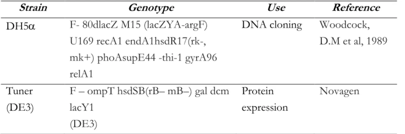
Two different Escherichia coli (E.coli) strains were used to investigate domains from TVAG_339720: DH5 and Tuner strain. The futures are listed in the table II.3 below.
Table II.3 - Bacterial strain used in this study
Strain Genotype Use Reference
DH5 F- 80dlacZ M15 (lacZYA-argF)
U169 recA1 endA1hsdR17(rk-, mk+) phoAsupE44 -thi-1 gyrA96 relA1
DNA cloning Woodcock, D.M et al, 1989 Tuner (DE3) F – ompT hsdSB(rB– mB–) gal dcm lacY1 (DE3) Protein expression Novagen II.4.3 Plasmids
The plasmid pET28a (Novagen) was used for expression of different domains from M60-Like zinc-metallopeptidase of T.vaginalis.
This bacterial expression vector of ~5.4Kb contains a T7lac promoter and a natural promoter and coding sequence for the lac repressor (lacI). Moreover, it carries an N-terminal His tag, thrombin cleavage site, internal T7 epitope tag, C-terminal His tag; kanamycin resistance and restriction enzyme cloning.
II.4.3 Growth and selective media
During this study were used two different growth media: Lurie-Bertani (LB) medium and LB-agar. LB medium is composed by Bacto®tryptone (10g/L), Bacto®yeast extract (5g/L) and NaCl (10g/L). The components were dissolving in 1L of water and the pH of solution was adjusted to 7.4 with NaOH.
LB-agar was prepared added 2g of agar (add information about it) to 100ml of LB medium (2%). All media were sterilized by autoclaving at 121°C, 32 lb / inch -2 for 20 min. After autoclaving, LB medium and LB-agar were left to cool to between 45°and 50°C and then a specific antibiotic was added. For our study we used kanamycin antibiotic in a final concentration of 20g/ml.
LB-agar mixed with antibiotic was poured into 90mm sterile petri dishes and storage at 4°C after solidification.
II.4.4 Sterilization
Different sterilization methods were adopted, depending on materials.
Media, laboratory glassware and some solutions were sterilized by autoclave (Prestige Medical), using steam under high pressure (32 lb / inch -2) at 121°C for 20 min.
Sterile syringe (Plastipak®,Becton Dickinson) and appropriate pore-sized (0.22-1μm) Millipore filter discs (Supor® Acrodisc®) were adopted to sterilize solutions impossible to autoclave.
II.4.5 Plating bacteria
A single inoculum of bacteria suspension (150ul) was spread over the surface of LB-agar plate.
A glass spreader, earlier flame sterilized by dipping in alcohol (100% EtOh) and cooled for about a minute, was placed in contact with the inoculum and was used for distribution and absorption of bacteria suspension into the agar. Plates
were allowed to dry at room temperature and later incubated in an inverted position at 37°C for overnight growth in an incubator. (Name of incubator)
II.4.5 Storage of bacteria and DNA samples
Bacteria colonies growth on LB-agar plates was stored in a fridge at 4°C for a maximum of one week. Plasmids were stored at -20°C in water.
II.4.6 Determination of DNA and protein concentration
The concentration of DNA and proteins was quantitated by measuring the absorption of ultraviolet light at 260nm (for nucleic acid) and 280nm (for protein) using a NanoDrop 2000 UV-Vis spectrophotometer (Thermo Fisher Scientific Inc, USA).
The Beer-Lambert law:
A=εCL
where A is the absorbance at a given wavelength of light (nm), ε is the molar extinction coefficient, C is the molar concentration of sample, and L is the length of the light path (cm), was used to calculate the specific concentration of DNA and protein starting from absorption value at 260nm (A260) or at 280nm (A280).
II.4.7 Primers
Different primers were designed for cloning and sequencing experiments during this study. The lengths of oligonucleotides were between 20 to 40bp and the melting temperatures (Tm) were greater to 50°C, to have the best chance for maintenance of specificity and efficiency during annealing step.
Tm = 64.9°C + 41°C x (number of G‟s and C‟s in the primer – 16.4)/N
where N is the length of the primer.
Primers developed for cloning experiments were designed adding CCGCG, CCGG or CGCG spacer plus restriction sites to the 5‟-ends, to allow the cleavage by restriction enzymes and then the ligation in vectors cut in the same sites.
Primers for cloning were synthesized dry by Sigma (Sigma Aldrich, UK) and resuspended in distilled water to the desired concentration.
II.4.8 Polymerase chain reaction (PCR)
Amplification reactions of the DNA under investigation were performed by PCR technique (Mullis & Faloona, 1987), using the Novagen Hot start PCR kit (Novagen) and PHC-3 thermocylcer (Biorad). A typical PCR reaction set up and the standard program used are schematised as shown in the tables II.4 and II.5 below.
Table II.4 - Typical PCR reaction set-up.
Components and concentrations Volume
Autoclaved distilled water 19μl
10 x KOD buffer minus Mg2+ (10 x ) 5μl
dNTP‟s (2 mM) 5μl
Q-solution (DMSO) 5μl
MgSO4 (25 mM) 4μl
Template DNA (~70 ng/μl) 1μl
Novagen ® KOD DNA Polymerase
(2.5 U/μl) 1μl
Forward oligonucleotide (5 μM) 5μl Reverse oligonucleotide primer (5 μM) 5μl
Table II.5 - Typical PCR reaction program. Program
name Event Temperature Duration
Number of Cycles
Program 1 Denaturation 95 °C 1 min 1
Denaturation 95 °C 1 min
Program 2 Annealing 50 °C 1 min 30
Extension 68 °C 1min/1kbp
fragment size
Program 3 Polishing 68 °C 10 min 1
Program 4 Storage 10°C ≤ 24hr 1
II.4.9 Agarose gel electrophoresis for analysis or PCR products
Agarose gel electrophoresis (AGE) (Meyers et al, 1976) is a technique used for detection and separation of DNA molecules. In this study, AGE was used to analyse the presence or absence of PCR products, of plasmid DNA purified from bacteria, and to check plasmids digested by restriction enzyme (Figure II.5).
The gel was prepared by dissolving of 0.5g of low grade (MELFORD Ltd) or SeaKem® Gold Agarose (Lonza) in 50 ml of TAE (Amersham) in a 200 ml conical flask to obtain a 1 % agarose solution. The solution was mixed, heated in in a microwave oven at 450 watts for about 5min and allowed to cool to about 50°C. After cooling, 5μl (1mg/ml) of an ethidium bromide solution was added to the solution, which was stirred again and poured into a gel casting mould. After solidification, the gel was put in electrophoresis equipment [HU10 Mini Plus Horizontal Gel Unit (SCIE-PLAS Ltd) and BDH horizontal gel mould connected to a Bio-rad Mini-PROTEAN® Tetra Cell power supply (Bio-rad)]
mM Boric acid, 2 mM EDTA pH 8.0 )] and were applied into the gel, alongside a standard (3μl of Bioline HyperLadder™ I markers).
To run an AGE experiment, (70V Biorad machine) Amersham machine was used to constant voltage (100V) for about 1h and the results were visualized in the UV range by Bio-Rad Gel Doc 1000 system (Bio-Rad).
Figure II.5 - PCR products analysed by AGE.
Vector pET28.a (lane 1) and PCR products of TvM60L (lane 2), TvPA14-GBDL (lane 3) and TvGBDL (lane4) were analysed on a 1% of agarose in TAE buffer. Lanes M: Bioline HyperLadder™ I standards (3μl). Lanes 1-a: AGE results from 5 μl of different PCR reactions.
II.4.10 PCR products purification
The kit QIAquick PCR Purification Kit (Qiagen) was used to purify PCR products as described in manufacturer‟s instructions.
II.4.11 Digestion with restriction enzymes
Digestion reactions to PCR products and plasmids containing cleavage sites for restriction enzymes were made in four steps:
1. Digestion with first restriction enzyme
2. Sample purified by QIAquick PCR Purification Kit (Qiagen) and eluted with 30l of distillate water
3. Digestion with second restriction enzyme
4. Sample purified by QIAquick PCR Purification Kit (Qiagen) and eluted with 10l of distillate water
Digestion reactions were incubated in a water bath at 37°C for1.5h. The amount of restriction enzymes was of 15unit (U) and one unit of enzyme is defined as the amount of enzyme required to cleave 1μg of DNA in 1 h at 37 °C. Buffers and restriction enzymes used in this work were ordered from Fermentas (MBI Fermentas, UK).
In the table II.6 is shown a typical set-up of a digestion reaction.
Table II.6 - Set-up of a typical restriction enzyme digestion reaction.
Components Volume
Distilled water 1-2μl
DNA fragment / plasmid(~0.1 – 0.5μg) 50μl Restriction enzyme buffer (10 x) 6μl
Restriction enzyme (10U) 2-3μl
Total volume ~60ul
II.4.12 Ligation reactions
The ligation reactions involve plasmids and PCR products (insert) after digestion with restriction enzyme. Most of these enzymes are able to digest DNA asymmetrically across their recognition sequence, in order to obtain overhangs
to vector is usually used at around 3:1.The ligation reactions were incubated for at least 1h, at 37°C.
In this study, Rapid DNA ligation KIT (Fermentas Life Science, UK) was used to perform ligation reactions. In the table below (table II.7) is schematized an example of ligation reaction set-up.
Table II.7 - Set-up of a ligation reaction.
Components Volume
Vector (10 ng/μl) 2μl
Insert DNA(10 ng/μl) 6μl
5x Ligase buffer 4μl
T4 DNA Ligase(4 U/μl) 1μl
H2O (Nuclease free water) 7μl
Total volume 20μl
II.4.13 Transformation of competent E.coli cells
Chemically competent cells stored in -80°C, were taken from freezer and allowed to thaw on ice for about 5 min. After defrosting, 5μl of plasmid or ligation were mixed with bacteria and allowed on ice for further 1h. Soon after, bacteria were heat-shocked by incubation in a Techne Dri-Block™ DB-2A at 42 °C for 2min and were immediately replaced on ice for ~ 3 min.
Transformed E.coli cells were plated on LB-agar added with appropriate antibiotic (Section I.3 and I.5) and plated cells were growth overnight at 37°C.
II.4.14 Plasmid DNA purification by mini prep
Plasmid purification includes three steps: growth of bacterial culture, harvesting and lysis of the bacteria, and purification of the plasmid DNA.
Single colonies of transformed E.coli cells grown overnight on selective LB-agar, were subcultured overnight, into 5ml or 10ml of LB with appropriate antibiotic at 37°C with shaking at 180 rpm.
Following overnight growth, bacteria were used for plasmid extraction by Qiagen QIAprep ® Spin Miniprep Kit (Qiagen), in accordance with manufacturers‟ instruction.
II.4.15 DNA sequencing
Plasmids DNA were sequenced using Sanger sequencing services (GATC Biotech AG, European Custom Sequencing Centre, Cologne, Germany) to check cloned DNA sequences. The samples, with concentration between 30ng/l and 100ng/l, were put in a 1.5ml Eppendorf tube and labelled with pre-ordered sequencing labels before posting to GATC Biotech.
The primers used for sequencing were either custom-designed or standard sequencing primers available from GATC Biotech website. In this study, standard primers were T7- (TAATACGACTCACTATAGGG) and reverse primers (CTAGTTATTGCTCAGCGGT) complementary to regions within plasmids. DNA and primer custom- designed were sent in labelled separate tubes with total volume of 20ul each, sufficient for up to 8 reactions. Multiple
sequence alignment tools such as Multalign
(http://multalin.toulouse.inra.fr/multalin/) were used to analyse sequencing data, by alignment with the original DNA sequence.
II.4.16 Induction of recombinant proteins expression in E.coli and cell lysis
For recombinant protein expression, E.coli Tuner (DE3) strains were transformed with 1l of sequenced recombinant plasmids and afterwards plated on appropriate selective media for overnight growth at 37°C. The next day a single colonies harbouring plasmids were picked, inoculated into 10ml of LB containing antibiotic and was grown overnight at 37°C with shacking (180rpm). Thereafter, bacteria grown during the night were inoculated in 1L or 2L of LB plus antibiotic in 1L flasks and were grown at 37°C with aeration (180rpm) until an OD600nm between 0.6 and 1. Before induction with 0.1mM IPTG, cells were cooled under running tap water to about 16°C and were grown overnight at the same temperature before protein purification.
For protein purification, cells were harvested in 500 ml centrifuge pots (Nalgene) by centrifugation at 5000 × g for 10 min at 4°C using a JA-10 rotor of a Beckman J2-21 centrifuge (Beckman Coulter, Inc.). After discharged of supernatants, pellet fractions were resuspended in 7ml of Talon buffer (20 mM Tris/HCl pH 8.0 plus 100 mM NaCl) per 400ml of original culture volume. Cells were then lysed by sonication for 45sec on ice for twice; using a B. Braun Labsonic U sonicator (B. Braun, Melsungen, Germany) set at low intensity (~45 watts) and soon after transferred into 50ml centrifuge tubes (Nalgene) for centrifugation. JA25.5 rotor in a Beckman J2-21 centrifuge (Beckman Coulter, Inc.) was used to centrifuge lysed cells at 19000rpm for 10-20min at 4°C. The supernatant or cell free extract (CFE or soluble fraction) was collected for protein purification.
After centrifugation steps, a small amount of pellet fractions were collected, resuspended in 50l of Laemmli sample buffer 1x, boiled at 100°C in a boiling water bath for 5 min and stored at room temperature, for later analyses.
II.4.17 Purification recombinant N-terminal 6x His-tagged proteins
Recombinant N-terminal 6xHis-tagged proteins from CFE were achieved by immobilised metal affinity chromatography (IMAC), using NiNTA Agarose resin (Quigen), containing Nickel Nitrilo-triacetic Acid. Histidine residues in the His-tag bind to the vacant positions in the coordination sphere of the immobilized nickel ions with high specificity and affinity. Briefly, columns were filled with a 2.5 ml bed volume of NiNTA resin and equilibrated with at least 7 ml of Talon buffer (20 mM Tris/HCl pH 8.0 plus 100 mM NaCl).
The CFE solution was then applied onto the resin bed in the column and allowed to drain by gravity. The flow through (FT) was collected and saved for later analyses. The resin was washed with 5 ml of Talon containing 25mM of imidazole. Elution of the bound protein from the resin was achieved by sequential application of 5 ml volumes of Talon buffer containing 50mM, 100mM, 150mM and 250mM of imidazole. All eluted fractions were collected and saved for subsequent analyses by SDS PAGE or for further purification.
II.4.18 Sodium dodecyl sulphate-polyacrylamide gel electrophoresis (SDS-PAGE)
SDS-PAGE, technique described by Laemmli in 1970, was used to analyse protein expression. In the table II.8 are described solutions and buffers prepared for SDS-PAGE experiments.
3μl of pellet fractions, 10μl of CFE, FT, 25mM, 50mM, 100mM and 250mM fractions and a protein standards were applied on 10% of polyacrylamide gels (Acrylogel 3; BDH Electran) using Bio-rad Mini-PROTEAN® Tetra Cell system (Bio-rad) according the manufacturer‟s instructions. The use of protein standards (PageRuler Prestained Protein Ladders, Thermo Scientific) was necessary to estimate the protein molecular weight after staining.
Table II.8 - Solutions and buffer used for preparation of SDS-PAGE.
Component Volume/Amount
~For 4 gels Resolving gel (10%)
0.75 M Tris/HCl buffer, pH 8.8 with 0.2 % SDS 7.5ml 40 % Acrylamide (BDH Electran acrylamide, 3 %
(w/v) bisacrylamide) 4.6ml
d.d. H2O 2.8ml
10 % (w/v) Ammonium persulphate 72l
TEMED 24l
Stacking gel
0.25 M Tris/HCl buffer, pH 8.8 with 0.2 % SDS 3.75ml 40 % Acrylamide (BDH Electran acrylamide, 3 %
(w/v) bisacrylamide) 0.75ml d.d. H2O 3.0ml 10 % (w/v) Ammonium persulphate 60μl TEMED 20μl Sample/Loading buffer SDS 10% (w/v)
0.25 M Tris/HCl buffer, pH 8.8 with 0.2 % SDS 5ml
Glycerol 25% (w/v)
β-mercaptoethanol 2.5ml
Bromophenol blue dye 0.1%
Running buffer
32 mM Tris/190 mM glycine, pH 8.3 350ml