Open Peer Review
Any reports and responses or comments on the article can be found at the end of the article. CASE REPORT
Case Report: “Incognito” proteus syndrome [version 1; peer
review: 2 approved]
Michelangelo Vestita
,
Angela Filoni , Nicola Arpaia , Grazia Ettorre ,
Domenico Bonamonte
2
Section of Plastic and Reconstructive Surgery, Department of Emergency and Organ Transplantation, University of Bari, Bari, 70124, Italy Section of Dermatology, Department of Biomedical Sciences and Clinical Oncology, University of Bari, Bari, 70124, Italy Section of Dermatology, Miulli Regional Hospital, Bari, 70021, Italy Abstract Proteus syndrome (PS) is a postnatal mosaic overgrowth disorder, progressive and disfiguring. It is clinically diagnosed according to the criteria reported by Biesecker et al. We describe the case of a 49-year-old woman who presented with a 10-year history of pauci-symptomatic infiltrating plaque lesions on the sole and lateral margin of the left foot, which had been diagnosed as a keloid. The patient had a positive history for advanced melanoma and a series of subtle clinical signs, such as asymmetric face, scoliosis, multiple lipomas on the trunk, linear verrucous epidermal nevi, and hyperpigmented macules with a mosaic distribution. Even if the clinical presentation was elusive, she had enough criteria to be diagnosed with PS. This case describes the first evidence, to the best of our knowledge, of pauci-symptomatic PS in adulthood, reports its rare association with advanced melanoma, and illustrates the importance of even minor cutaneous clinical signs, especially when atypical, in formulating the diagnosis of a complex cutaneous condition such as this. Keywords proteus syndrome, cerebriform, keloid, diagnosis, elusive, hidden1,2
2
3
2
2
1 2 3 Reviewer Status Invited Reviewers version 1 published 26 Feb 2018 1 2 report report , Sapienza University of Marco Marcasciano Rome, Rome, Italy 1 , University of Catania, Giuseppe Micali Catania, Italy 2 26 Feb 2018, :228 ( First published: 7 ) https://doi.org/10.12688/f1000research.13993.1 26 Feb 2018, :228 ( Latest published: 7 ) https://doi.org/10.12688/f1000research.13993.1v1
Michelangelo Vestita ( )
Corresponding author: [email protected]
: Data Curation, Investigation, Resources, Supervision, Validation, Writing – Original Draft Preparation, Writing – Review &
Author roles: Vestita M
Editing; Filoni A: Investigation, Methodology, Resources, Validation, Writing – Original Draft Preparation, Writing – Review & Editing; Arpaia N: Conceptualization, Formal Analysis, Investigation, Project Administration, Supervision, Validation; Ettorre G: Data Curation, Investigation, Methodology, Validation, Visualization, Writing – Original Draft Preparation, Writing – Review & Editing; Bonamonte D: Conceptualization, Investigation, Methodology, Project Administration, Supervision, Validation, Visualization
No competing interests were disclosed.
Competing interests:
The author(s) declared that no grants were involved in supporting this work.
Grant information:
© 2018 Vestita M . This is an open access article distributed under the terms of the , which
Copyright: et al Creative Commons Attribution License
permits unrestricted use, distribution, and reproduction in any medium, provided the original work is properly cited. Vestita M, Filoni A, Arpaia N
How to cite this article: et al. Case Report: “Incognito” proteus syndrome [version 1; peer review: 2
F1000Research 2018, :228 ( )
approved] 7 https://doi.org/10.12688/f1000research.13993.1
26 Feb 2018, :228 ( )
First published: 7 https://doi.org/10.12688/f1000research.13993.1
Introduction
Proteus syndrome (PS) is a postnatal mosaic overgrowth disorder, which was originally described by Cohen and Hayden in 19791. In 1983, the syndrome was named after a minor Greek
deity who had the power to change his appearance2. The
occurrence of this disorder is sporadic, with a prevalence of less than one per million3. PS is a progressive, disfiguring
disor-der caused by a somatic point mutation in AKT1 leading to gene activation. The product of this gene is involved in cell prolifera-tion and apoptosis suppression, acting through the mammalian target of rapamycin signaling pathway, which may explain the overgrowths in this syndrome4. Clinically, this disorder is
char-acterized by typically asymmetric, disproportionate, postnatal overgrowth of tissues derived from any of the three germ layers. While skin, bone, connective, and adipose tissues are most commonly involved, some patients present with over-growths of the central nervous system, spleen, thymus, or colon. In addition, patients may also present with a range of tumors, pulmonary complications, and a striking predisposition to deep vein thrombosis and pulmonary embolism5. PS is clinically
diagnosed according to the criteria described by Biesecker et al.6
(Table 1). Case
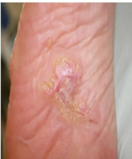
A 49-year-old woman presented with a 10-year history of pauci-symptomatic infiltrating plaque lesions on the sole and lateral margin of the left foot (Figure 1 and Figure 2). The lesions simulated and had been misdiagnosed as keloids, but there was no history of trauma to the area. The patient reported that similar lesions had affected her great-grandmother. The patient had a positive history for stage IV melanoma, and she had finished chemotherapy7 just 3 months before our observation.
Physical examination also revealed multiple lipomas on the trunk, linear verrucous epidermal nevi, and hyperpigmented macules
Table 1. Diagnostic criteria for Proteus syndrome6.
General
criteria ▪ Mosaic lesions ▪ Sporadic disease
▪ Progressive disease Category A ▪ Cerebriform connective tissue nevus Category B ▪ Epidermal nevus (linear verrucous epidermal nevus) ▪ Overgrowth of various body tissues ▪ Tumors (bilateral ovarian cystadenomas or
Figure 1. Proteus syndrome: Modestly developed connective tissue nevus of the left foot, previously misdiagnosed as a keloid.
with a mosaic distribution. Additionally, she presented with an asymmetric face, dysmorphic skull with frontal-parietal hyper-ostosis, dropped shoulders, scoliosis, and a stiff spine. Her legs were asymmetric with disproportionate overgrowth, the left leg being longer than the right one and having ectatic veins. In addition, computed tomography documented uterine fibromas, and abdominal magnetic resonance imaging demonstrated hepatic angiomatosis. A skin biopsy specimen from the left foot stained with hematoxylin and eosin revealed remarkable hyperkeratosis, epidermal hyperplasia, dermoepidermal fibrosis with extensive sclerosis of the reticular dermis, thickened collagen bundles, and fat-cell entrapment (Figure 3). We made the diagnosis of Proteus syndrome. No therapeutic intervention was carried out.
Discussion
To meet the diagnostic criteria for PS, a patient must fulfill all three general criteria, plus a single criterion from category A or two criteria from category B or all three criteria from category C (Table 1). Even though our patient had not previously sought medical care, when she presented to us her condition fulfilled the criteria, and the diagnosis of PS was confirmed.
This case illustrates the importance of even minor cutaneous clinical signs, especially when atypical. They should not be overlooked because, together with other clinical and diagnostic findings, they may lead to the diagnosis of a specific condition. This is especially true in mosaic diseases, such as PS, in which
the wide variety of tissue types and cells that are involved may not be apparent at the first examination. Subtle cutaneous forms of PS have been described in infants4, but to the best of our
knowledge, this is the first case in which the cutaneous signs remained elusive in adulthood. We do not know whether the chemotherapy she had been administered had somehow altered the lesion morphology7, but that seems unlikely, as that occurred
in adulthood and the patient referred no significant changes in shape and volume.
Correct recognition of pauci-symptomatic “incognito” PS is essential, as PS patients are known to be exposed to a higher risk to develop tumors1–4, such as meningiomas, breast and ovarian
cancer, parotid adenoma, and others. We do not know whether melanoma occurrence is facilitated by PS, and the literature provides scarce data on this. The association between PS and melanoma in this case is either a novel finding or an incidental coexistence. At present our patient reached the 24 months follow up with no clinical or instrumental signs of recurrence.
Consent
Written informed consent for publication of their clinical details and clinical images was obtained from the patient.
Competing interests
No competing interests were disclosed. Grant information
The author(s) declared that no grants were involved in supporting this work.
Figure 3. Proteus syndrome: Biopsy from the cerebriform nevus of the foot. Hematoxylin and eosin (magnification x100).
References
1. Cohen MM Jr, Hayden PW: A newly recognized hamartomatous syndrome.
Birth Defects. 1979; 15(5B): 291–296.
PubMed Abstract
2. Nguyen D, Turner JT, Olsen C, et al.: Cutaneous manifestations of Proteus syndrome. Correlations with general clinical severity. Arch Dermatol. 2004; 140(8): 947–953.
PubMed Abstract |Publisher Full Text
3. Doucet ME, Bloomhardt HM, Moroz K, et al.: Lack of mutation-histopathology correlation in a patient with Proteus syndrome. Am J Med Genet A. 2016; 170(6): 1422–1432.
PubMed Abstract |Publisher Full Text |Free Full Text
4. Rodenbeck DL, Greyling LA, Anderson JH, et al.: Early Recognition of Proteus Syndrome. Pediatr Dermatol. 2016; 33(5): e306–310.
PubMed Abstract |Publisher Full Text
5. Biesecker LG, Sapp JC: Proteus Syndrome. 2012.
6. Biesecker LG, Happle R, Mulliken JB, et al.: Proteus Syndrome: diagnostic criteria, differential diagnosis and patient evaluation. Am J Med Genet. 1999; 84(5): 389–395.
PubMed Abstract
7. Guida M, Cramarossa A, Fistola E, et al.: High activity of sequential low dose chemo-modulating Temozolomide in combination with Fotemustine in metastatic melanoma. A feasibility study. J Transl Med. 2010; 8: 115.
PubMed Abstract |Publisher Full Text |Free Full Text
Open Peer Review
Current Peer Review Status:
Version 1
08 March 2018 Reviewer Report
https://doi.org/10.5256/f1000research.15209.r31219
© 2018 Micali G. This is an open access peer review report distributed under the terms of the Creative Commons
, which permits unrestricted use, distribution, and reproduction in any medium, provided the original Attribution License work is properly cited.
Giuseppe Micali
Dermatology Clinic, University of Catania, Catania, Italy
It is an interesting and well-presented case report of pauci-symptomatic, previously overlooked Proteus
syndrome in a 49-year-old woman. Proteus syndrome is a rare, complex disorder with multisystem
involvement and remarkable clinical variability. The authors highlight the relevance of the recognition of
minor cutaneous clinical signs for the diagnosis of minimal forms of the disease, as several complications,
some of which life-threatening, may potentially occur in these patients.
Is the background of the case’s history and progression described in sufficient detail?
Yes
Are enough details provided of any physical examination and diagnostic tests, treatment given
and outcomes?
Yes
Is sufficient discussion included of the importance of the findings and their relevance to future
understanding of disease processes, diagnosis or treatment?
Yes
Is the case presented with sufficient detail to be useful for other practitioners?
Yes
No competing interests were disclosed.
© 2018 Marcasciano M. This is an open access peer review report distributed under the terms of the Creative Commons
, which permits unrestricted use, distribution, and reproduction in any medium, provided the original Attribution License work is properly cited.
Marco Marcasciano
Unit of Plastic, Reconstructive and Aesthetic Surgery, Sapienza University of Rome, Rome, Italy
Proteus syndome is a complex multi-organ mosaic disorder, characterized by progressive overgrowth
and disfiguring. The authors did a nice job in describing the occurrence of such a multi-faceted condition
in its (rare) minimal symptomatic presentation. To suspect and correctly diagnose a Proteus in view of
(apparently) so few clues is indeed challenging. Since the repercussions are relevant (increased
incidence of malignancies) the educational message conveyed here is an important one: never
underestimate the importance and significance of a given set of signs and symptoms, as subtle as they
may be.
Is the background of the case’s history and progression described in sufficient detail?
Yes
Are enough details provided of any physical examination and diagnostic tests, treatment given
and outcomes?
Yes
Is sufficient discussion included of the importance of the findings and their relevance to future
understanding of disease processes, diagnosis or treatment?
Yes
Is the case presented with sufficient detail to be useful for other practitioners?
Yes
No competing interests were disclosed.
Competing Interests:
I confirm that I have read this submission and believe that I have an appropriate level of
expertise to confirm that it is of an acceptable scientific standard.
Reader Comment 13 Jan 2019