Effects of Oregano (Origanum vulgare L.) and Rosemary
(Rosmarinus officinalis L.) Aqueous Extracts On in vitro
Rabbit Immune Responses
Abbreviations:
PB: Peripheral Blood; mAbs: Monoclonal Antibodies; HBSS: Hank’s Balanced Salt Solution; PBMC: Peripheral Blood Mononuclear Cell; PWM: Pokeweed Mitogen; GALT: Gut-Associated Lymphoid TissueIntroduction
Natural antioxidants are receiving increasing attention in human and animal nutrition because of their association with food quality characteristics and immune responses [1,2]. Among a variety of plants bearing anti-oxidative constituents, the Labiatae family (mint plants) has been attracting the greatest interest [3], with particular attention to products from oregano (Origanum vulgare L.) and rosemary (Rosmarinus officinalis L.). Oregano exerts a well documented anti-oxidative activity [4-6], but it also possesses intense in vitro antimicrobial [7] and antifungal [8] properties. Such properties make it an appropriate candidate as a replacement for antibiotic growth promoters and also a promising food additive in order to prevent meat lipid oxidation [9]. Indeed, oregano has been found to improve meat storage stability after slaughter in rabbits [10]. The rosemary extract exerts anti-oxidative activity, but its constituents have also shown a variety of pharmacological activities for cancer chemoprevention and therapy in in vitro and in vivo models [11]. The claim often made of phytogenic feed additives as stimulant of
immune function however faces with a poor specific experimental verification in monogastric animals (Lagomorpha). Therefore there is a lack of data regarding the possible effects of plants extracts on immunity of these species. This study was designed to evaluate: 1) the effect of dietary plant extract (oregano and/or rosemary) and 2) age effect, on some specific immune responses (lymphocytes proliferation test and IgM measurements in vitro) and on peripheral blood lymphocyte subsets development in rabbits
Materials and Methods
Animal dietary treatment and blood sampling
The animals were bred at the experimental facilities of the University of Perugia. A total of 100 New Zealand mixed-sex rabbits were weaned at 30 days of age and immediately split into homogeneous groups submitted to the following dietary treatments: 1) Standard diet (C); 2) C + 150 ppm Vit E (E); 3) C + 0.2% oregano (O); 4) C + 0.2% rosemary (R) and 5) C + 0.1% oregano + 0.1% rosemary (OR). Every diets contained an integration of 50ppm Vit E, CLA 0.5% (from soy oil) + 3% Omega Lin (Mignini & Petrini) + 0.5% mixed vitamins.
The plant derived ingredients were obtained with an enzyme aided extraction of leaves using water as solvent (Phenbiox,
Volume 4 Issue 4 - 2016
1School of Biosciences and Veterinary Medicine, University of
Camerino, Italy
2Rheumatology Unit, Department of Clinical & Experimental
Medicine, University of Perugia, Italy
3Department of Applied Biology, University of Perugia, Italy
*Corresponding author:Daniela Beghelli, School of Biosciences and Veterinary Medicine, University of Camerino, Via Gentile III da Varano, 62032Camerino, Italy, Tel: +39 (0) 373 403438; Fax: + 39 (0) 737 403446; Email:
Received: October 27, 2016 | Published: December 02, 2016
Research Article
Abstract
In order to investigate the effects of some dietary phytoderivates (Origanum vulgare L. and Rosmarinus officinalis L.) on rabbit immune system, 100 New Zealand mixed-sex rabbits weaned at 30 days of age were split into homogeneous groups submitted to the following dietary treatments: 1) Standard diet (C); 2) C +150 ppm Vit E (E); 3) C +0.2% oregano (O); 4) C +0.2% rosemary (R) and 5) C +0.1% oregano + 0.1% rosemary (OR). Blood samples were drawn from rabbits at 30 and 90 days of age (ten rabbits/diet group at Time 0 and 1, respectively) to investigate the blood lymphocyte subset evolution, the in vitro lymphocyte proliferation (in presence or absence of mitogens) responses and the Immunoglobulin M (IgM) concentration in the supernatants of lymphocyte cultures. A diet effect was observed in the O and OR groups, where the lymphocytes proliferation responses to pokeweed mitogen (PWM; ***P<0.001, in the O group) or the interleukin-2 production (IL-2; ***P<0.001, for both groups) at Time 1 were significantly higher. The IgM levels were systematically higher in the O, OR and E cells culture supernatants (**P<0.01). Age did not affect the rabbit lymphocyte subset evolution nor the in vitro lymphocyte proliferation. Data obtained in the present study show that rabbit’s dietary supplementation with oregano elicits positive effects on the adaptive immune response.
Keywords: Dietary phytoderivates; Flow cytometry; IgM; T cells; Lymphocyte proliferation assay; Rabbit; Immune function; Animal nutrition
Calderara di Reno, Bologna). All rabbits were individually housed in flat-deck cages measuring 600 x 250 x 330 mm. Peripheral blood (PB) samples were taken from rabbits at 30 and 90 days of age (ten rabbits/diet group at Time 0 and 1, respectively). These samples were drawn from the marginal ear vein after washing with a 70% ethanol solution, using vacuum heparinized tubes. Diurnal variations in hematological parameters were minimized by collecting blood at approximately the same time (8:00-10:00 am). Blood samples were processed within 1h after sampling.
Lymphocyte labeling and flow cytometry analysis
Commercially available monoclonal antibodies (mAbs) were used for the detection of lymphocyte subsets. The surface staining of blood leukocytes was performed using a PE labeled mouse anti-rabbit CD4+ mAb (KEN-4 clone, AbD Serotec/Bio-Rad LaboratoriesInc., Segrate, Milan) or a FITC labeled mouse anti-rabbit CD8+ mAb
(12.C7 clone, AbD Serotec/Bio-Rad Laboratories Inc., Segrate, Milan), both recognizing T-cells-specific antigen. An APC labeled mouse anti-human CD79α+ mAb (clone HM47, BD, eBioscience
Inc. and BioLegend, San Diego) a cross-reactive antibody against rabbit B+ cells antigen were used for intracellular staining
[12]. Flow cytometry analysis was performed using a standard FACSCalibur flow cytometer (Becton Dickinson, Lincoln Park, NJ) operating the Cell Quest ProTM software. In each sample, 10.000
cells were measured and the data were saved in the list mode. Gating was based on forward angle and right angle scatter signals. The gates of each leucocyte type were adjusted with an isotype negative control. After a 4% paraformaldehyde fixation, the same samples were permeabilized with 0.1% saponin blocking buffer for intracellular staining with APC labeled CD79α+ mAb according
to manufacturer’s instructions and data were again acquired by flow cytometer. Up to three different fluorochromes were analysed in the same vial.
Lymphocyte proliferation test
The lymphocyte proliferation test was performed pooling blood aliquots (2 mL/each) of two individual samples. Mixture of equal volumes of blood samples and NaCl 0.9% were layered on the top of 15 mL of Lympholyte (Cedarlaneâ Burlington,
North Carolina) and centrifuged at 400 x g for 20 min at room temperature. The peripheral blood mononuclear cell (PBMC) layer was then transferred to sterile culture tubes and washed twice with Hank’s Balanced Salt Solution (HBSS) (Gibco Invitrogen, Thermo Fischer Scientific Inc,) without Ca2+ and Mg2+. Then
the cells were re-suspended in complete RPMI-1640 medium (EuroClone S.p.A., Milan) that contained fetal bovine serum (10%; Gibco, Invitrogen), L-Glutammine (2 mM; Euroclone), penicillin (100 U/mL; BiochromAG, Berlin), and streptomycin
(100 µg/mL; BiochromAG). The number of live lymphocytes was
determined using an automatic haemocytometer and a trypan blue dye exclusion procedure (Countness, Invitrogen,). The final concentration of live cells was adjusted to 2 x10⁶/mL of complete
medium. PBMC suspension (2 x10⁵ live cells) of each pool sample
was cultured in flat bottom 96-well tissue culture plates (Becton Dickinson). Cells were stimulated with pokeweed mitogen (PWM) (that stimulates B+ lymphocytes; 0.5 µg/well; Sigma Aldrich Co
Ltd, Saint Louis , Missouri), phytohaemagglutinin (PHA; 1.2 µg/ mL; BiochromAG), IL-2 (that stimulates B+ and T+ lymphocytes; 1U/
mL; Novartis, Basilea). Cell cultured only with medium (100 µL/ well) allowed to estimate the basal proliferation [13]. Each culture condition was performed in triplicate. The plates were held at 37 °C for 72 h in a humidified chamber with an atmosphere of 5% CO2 in air. Then, 1 mCi of [3H] thymidine (specific radioactivity 4 Ci/mmol, PerkinElmer, Waltham, Massachussetts) in RPMI-1640 was added to each well, and the plates were held under the same conditions for 16-18 h. At the end of culture, the cells were mashed and transferred to filter discs corresponding to each well. These filter discs were soaked in scintillation liquid (Betaplate SCINT; PerkinElmer), and the lymphocytes were solubilized to release the [3H] thymidine. Disintegrations per minute were determined with a liquid scintillation counter (Trilux 1450 Microbeta, Wallac, PerkinElmer) and used to calculate the picomoles of [3H] thymidine incorporated into newly synthesized DNA. The results are expressed as counts per minute (cpm). The increase of lymphocyte proliferation due to the mitogenic agents adding was expressed as percentage increase of cpm (M cpm) vs basal cpm values (C cpm; only medium). Therefore, the applied formula was: increased proliferation = (M cpm- C cpm)/ C cpm, and it was expressed as %.
Titration of IgM in the culture supernatants
Before the addition of [3H] thymidine, but only in samples collected at Time 1, 100 µL/well of supernatants were collected from each triplicate of each experimental condition and pooled. Then 100 µL/well were transferred to new flat bottom 96-well tissue culture plates (Becton Dickinson) and promptly frozen at -20 °C until analysis. The rabbit IgM released in culture media were determined by a commercially available ELISA kit (Cat. N. E120-110-23, Bethyl Laboratories, Montgomery) and an automated washing and reader instrument (Mago4S, Diamedix Corporation, Hialeah) at 450 nm wavelength. The procedures for sample assays were carried out according to manufacturer’s instructions.
Statistical analysis
Differences between dietary groups were assessed by ANOVA test with a Bonferroni and Dunnet’s Post Hoc multiple test applied for comparison of all pairs when the critical assumptions for the independent-samples t test were valid. In the other cases, nonparametric tests for two or multiple independent samples were used (Mann-Whitney and Kruskall-Wallis tests). Differences with p value at least <0.05 were considered statistically significant. Due to the relatively small numbers of experimental animals, age-dependent changes were tested with the one-tailed Mann-Whitney non parametric test (all the rabbits of Time 0 vs those of the C group at 90 days). All calculations were performed with SPSS software (2004) [14].
Results
Lymphocyte subsets development in growing rabbits
feed with different diets
No age-related changes in relative PB lymphocyte subsets were observed from 30 to 90 days of age in young adult rabbits; whereas a diet effect was observed for CD8+ cell subpopulation
at 90 days of their life (Table 1). Indeed, rabbits fed with the R diet showed lower CD8+ percentages compared to all other
experimental groups with values that become significant in comparison to the OR group (Table 1, *P<0.05).
It is noteworthy that the sum of PB lymphocytes CD4+ with
CD8+, CD4+CD8+, and CD79α+ during the experimental period was
always lower than 100% (by about 19.3 to 29.4%), these cells, according to Jeklova et al. [12] are referred to as lymphocytes with a CD4-, CD8-, and CD79α- phenotype.
Lymphocyte proliferation assay results
The rabbit lymphocyte proliferation test showed a general reduction of leukocytes response from 30 to 90 days of age, in particular for IL-2, by 3.8% ± 3.9 to 0.04% ± 0.06; for PHA, by 6.2% ± 6.7 to 0.7% ± 1.3; and finally, for PWM by 0.4% ± 0.5 to 0.3% ± 0.3 (Mann-Whitney test). However, the lower lymphocyte in vitro responsiveness was never statistically significant.
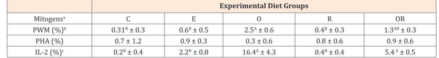
The rabbit lymphocyte proliferation assay responses, conversely, were influenced by experimental diets; indeed, at Time 1, the O group responses resulted significantly higher to PWM and IL-2 mitogens adding than those of the other diets
groups (***P<0.001) with the exception of OR group whose
response to IL-2 was as significant higher as that of O group (Table 2). Although the proliferation of rabbits PBMCs cultured in absence of mitogens revealed higher responses in the “E” and “O” groups (data not shown), the addition of mitogenic agents induced significant increased responses only in those groups where animals were supplemented with oregano.
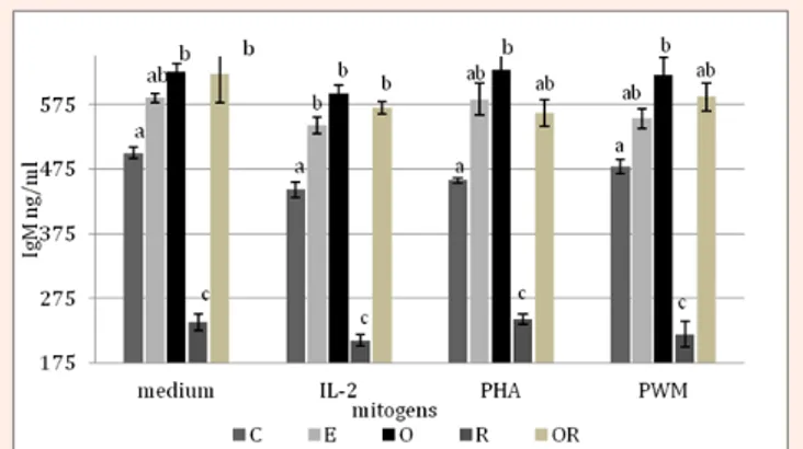
IgM in the rabbit lymphocytes culture supernatants
The titration of rabbit IgM in culture supernatants revealed a strong effect of diets (***P<0.001), with the highest Ig valuesrecorded in the O group (608.6 ± 28.2 ng/mL) followed by OR (578.4 ± 33.2 ng/mL), E (560.7 ± 23.7 ng/mL), C (461.9 ± 28.2 ng/ mL) and R (218.5 ± 24.2 ng/mL) groups. The mitogens added to the cells cultures did not particularly affect the IgM levels, indeed in some diet groups their higher values were just observed in supernatants of cells cultured without any stimulus (Figure 1). However, independently of the presence of mitogens, the O group always showed the highest values, followed by the OR group.
Table 1: PB lymphocyte subsets distribution (%) in different rabbits age or diet groups (mean ± SD).
Time 0(C) Time 1(C) Time 1(E) Time 1(O) Time 1(R) Time 1(OR)
CD4+(%) 41.6 ± 8.5 38.1 ± 4.2 37.7 ± 4.7 39.1 ± 5.0 36.2 ± 5.4 41.5 ± 4.2
CD8+(%) 16.8ab± 2.4 17.8ab± 3.2 16.8ab± 2.4 17.5ab± 2.4 15.0ac± 3.7 22.0b± 5.5
CD4+CD8+(%) 1.6 ± 0.6 1.6 ± 0.7 1.3 ± 0.5 1.5 ± 0.3 1.3 ± 0.5 1.1 ± 0.2
CD79+α(%) 20.7 ± 7.1 17.7 ± 5.6 14.8 ± 4.4 16.7 ± 4.0 20.3 ± 2.3 14.8 ± 3.8
CD4/CD8 (ratio) 2.2 ± 0.4 2.2 ± 0.4 2.3 ± 0.6 2.3 ± 0.5 2.5 ± 0.7 2.0 ± 0.8
a,b,cMeans not sharing the same superscript being significantly different at *P < 0.05 (one way ANOVA, Bonferroni Post hoc test).
SD= Standard Deviation
Table 2: Effects of dietary phytoderivates (O: oregano; R: rosemary; OR: both oregano and rosemary) on rabbits lymphocyte proliferation test, at Time
1 (mean ± SD).
Experimental Diet Groups
Mitogensa C E O R OR
PWM (%)b 0.31B ± 0.3 0.6B ± 0.5 2.5A ± 0.6 0.4B ± 0.3 1.3AB ± 0.3
PHA (%) 0.7 ± 1.2 0.9 ± 0.3 0.3 ± 0.6 0.8 ± 0.6 0.9 ± 0.6
IL-2 (%)c 0.2B ± 0.4 2.2B ± 0.8 16.4A ± 4.3 0.4B ± 0.4 5.4 A ± 0.5
*A,B.Means not sharing the same superscript being significantly different at ***P <0.001 (bBonferroni test; cTamhane test).
aValues are expressed as percentage of increased cpm vs basal cpm values (see paragraph 2.3).
SD= standard deviation.
Discussion
Rabbits have been traditionally used as experimental models in human and veterinary research for many years, however, little is known about their blood lymphocyte populations or immune responses in vitro [12,13,15-21].
In this study, the lymphocyte subsets were not influenced by animal’s age and the relative numbers of CD4+ or CD8+ T cells were
in accordance with those reported by some authors (calculated by Guerrero et al. [20]) or slightly higher than those reported by
others [12,22]. However, in rabbits, T helper lymphocytes prevail on cytotoxic T cells that, conversely, represent a minor subset in their T-lymphoid compartment. The CD4/CD8 ratio was also substantially constant over time. The mechanism determining the blood peripheral CD4/CD8 ratios seem to be species-specific, originates in the thymus and is genetically controlled [23,24]. There are controversial observations related to an increase or a decrease in CD4/CD8 ratio during maturation. A reduction in the value of this ratio seems to be a common age-related event in mammals and it was observed in mice [25], cats [26], dogs [27],
pigs [28] and rabbits [12]. However, it has been shown to possibly increase or decrease with age in humans [29-32]. Although Jeklova et al. [12] reported a rabbit CD4/CD8 ratio decreasing with age, the value they obtained from adult rabbits (20- week-old) is even slightly higher than that reported here at 90 days of age (2.8 ± 0.8 vs 2.2 ± 0.4 of the C group, Table 1). Furthermore, their value was obtained comparing 1-day-old rabbits to the adult ones. The animals studied here have been examined for a shorter period of time and they were slightly older at the beginning of the study, maybe this can explain why we did not observe a reduction in the ratio.
The relative number of double positive (DP) lymphocytes (CD4+CD8+ T cells) was found to be comparable to other reports,
while the CD79α+ B cells were lower (16.9 ± 0.8 vs 42.5 ± 4.7,
respectively) (12, pp. 636). However, our data seems to be quite similar to those calculated by the absolute values presented in the Ferrian et al. [21].
Very little is known about DP CD4+CD8+ T cell role in immune
response in vivo. Several observations in humans, chickens and pigs suggest that peripheral CD4+CD8+ T cells function as
normal T cells, respond to signals delivered by mitogens and may consist of memory cells; in humans, the presence of these cells in inflammatory sites, lamina propria of the gut and kidney allografts suggest an immunoregulatory and/or immunosurveillance function [33,34]. According to other authors [35], in pigs the DP T lymphocytes could also be defined as memory/activated CD4+CD8+T cells (T
helper). However, in contrast to what was
observed in pigs, monkeys and chickens, where the number of PB CD4+CD8+ T cells increase with age, in this study we did not
observe any changes in their percentages and this is in accordance with other studies [12].
Although the role of environmental (microbial and feed) antigens on the generation of DP lymphocytes has not been thoroughly examined in many species, exposure to antigens appears to play a role in the creation of DP cells both in vitro and in vivo [34].
Several studies demonstrated that also an intestinal DP CD4+CD8+ T cell population is present in birds and mammals
[33] and although of different origin, it has been suggested that the same mechanisms (age or antigen exposure in the intestine/ gastrointestinal flora) could induce a co-expression of CD4+ and
CD8+ both on PB and on intestinal T cell populations [33]. However,
in this study even if the diet supplemented with phytoderivates (oregano and/or rosemary) had affected the gastrointestinal flora, its effect had not been strong enough to influence the DP CD4+CD8+
T cells population in blood. A possible immunoregulatory role of phytoderivates may have been better observed in conventional rabbit herds with a major antigenic exposure (instead of these controlled conditions) or in older rabbits.
The lowest CD8+ cell values observed in R group (significantly
lower than in OR group, although within the rabbit range, Table 1) could express worse responses to signals from microbial environment, particularly from the commensal microflora of the gastrointestinal and respiratory tracts, that usually are responsible for the post-natal maturation of the immune functions [36].
A possible explanation for the lack of effectiveness of rosemary in positively modulating the rabbits immune responses in vitro, besides a lower % of CD8+ cells at 90 days of age (although it
possesses known antioxidant properties that exert a positive effect on immune functionality; 15), as already obtained in rats [37], could be found in the use of an incorrect concentration of rosemary administered. Finally, the non-detected cells to reach 100% of PBMCs (in this study between 19.3-29.4%), previously referred to as lymphocytes with CD4-, CD8-, and CD79α- phenotype,
may be a γδ T cell subpopulation as suggested by some authors [12]. Indeed, in contrast to human and mice, rabbits together with cattle, sheep, pigs and chicken belong to species with sizeable γδ peripheral T cell pool [38], about 23% of γ/+ PB T-cell for some
authors [22] or 16-48% for others [12,20]. In the first study these cells were individuated by using an anti-human TCR- /δ+mAb,
in the others these numbers were obtained/deducted by the subtraction of lymphocytes subsets recognized by CD4+, CD8+, and
CD79α+ mAbs (or a mouse anti-rabbit α-pan B) from their total
number. The current difficulty for immunological research into rabbits lies in the still limited number of commercially available monoclonal antibodies that recognize the various lymphocyte subsets antibodies [39], between these an anti-rabbit γδ mAb.
If to date there are limited data available in literature on rabbit lymphocyte phenotyping, those relative to the rabbit in vitro mitogen-induced PB lymphocyte proliferation assays are even scarcer [13] and, to the best of our knowledge, none concerning IgM titration. In this study, we observed an age-related reduction of rabbit lymphocyte proliferation responses that seems to be in contrast with what reported by other authors [40]. However, these authors referred to depressed immunologic capabilities in splenic lymphocytes of newborn rabbits; whereas the same observed a restored immune functionality over the initial 2 to 6 weeks of life. To our knowledge, there are no other studies to refer to, but since our observations began at the fourth week of rabbit life and on a lymphocyte population of a distinct district (PB) it is quite difficult to make comparisons with the aforementioned paper results. The influence of intestinal microflora on diversification of primary
Figure 1: IgM levels in the rabbits lymphocytes culture supernatants,
represented according to the diet groups (C, E, O, R, and OR) and mitogens (only medium, IL-2, PHA, and PWM.
a-cMeans not sharing the same superscript being significantly different
antibodies and selection of B cells has been intensively studied [12,41,42]. Rabbits, like chickens but unlike other species, require environmental stimuli for somatic diversification of the antibody repertoire, in particular the primary antibody repertoire that develops between 3 and 8 weeks of age. The exogenous factors that act as stimuli apparently derived from the gut microbial flora [42] and cause a somatically diversifying of the neonatal repertoire through somatic hypermutation and a somatic gene conversion-like mechanism in gut-associated lymphoid tissue (GALT).
Even if we did not observed an increase of the B cells population, the oregano (alone or in combination with rosemary) extract orally administered to rabbits probably was able to influence this process of B-cells proliferation in GALT determining a higher responsiveness of these lymphocytes in the in vitro proliferation test, in accordance with other reports on chickens [43].
These findings have been corroborated by the higher production of IgM in the supernatants of the cell cultures derived from the O, OR and E groups (both in presence or absence of mitogens).
Conclusion
Various aromatic plants and their products have been reported to have health and beneficial properties. Data of the present study show that a dietary supplementation with oregano (Origanum vulgare L.) may be able to modulate the in vitro immune response in rabbits.
Indeed, rabbits receiving oregano exhibited the highest in vitro B+ cell responsiveness together with their highest capacity
to produce IgM. These findings recognize the oregano as an aromatic plant with stimulant properties on the adaptive immune responses in rabbits.
However, further research is required to define the optimal dose and/or the combination of these aromatic plant extracts (oregano and rosemary) to improve their immune modulatory effects and to elucidate the mechanisms mediating these actions.
Ethical Standards
Animals were handled according to the principles for the care of animals in experimentation (Directive 2010/63/EU).
Acknowledgements
This study was financially supported by Ministry of Economic Development (Leader of “Industries 2015 Project PM01_00148”: HYPERLINK “http://G.I.Ma” G.I.Ma. spa). The authors acknowledge the helpful advice of Dr. Dario Zanichelli of PHENBIOX s.r.l. and Mignini & Petrini S.p.A. for their special technical assistance in preparing the experimental diets and Dott. Vittorio Bini (S. Maria della Misericordia Hospital, Perugia) for his support in statistic. )
References
1. Middleton E, Kandaswami C (1992) Effects of flavonoids on immune and inflammatory cell functions. Biochem Pharmacol 43(6): 613-619.
2. Cullen SP, Monahan FJ, Callan JJ, O’Doherty JV (2005) The effect of dietary garlic and rosemary on grower-finisher pig performance and sensory characteristics of pork. Irish J Agric Food Res 44:
57-3. Windisch W, Schedle K, Plitzner C, Kroysmayr A (2008) Use of phytogenic products as feed additives for swine and poultry. J Anim Sc 86(14 Suppl): E140-E148.
4. Adam K, Sivropoulou A, Kokkini S, Lanaras T, Arsenakis M (1998) Antifungal activities of Origanum vulgare subsp. hirtum, Mentha
spicata, Lavandula angustifolia, and Salvia fruticosa essential oils
against human pathogenic fungi. J Agri Food Chem 46(5): 1739-1745.
5. Cervato G, Carabelli M, Gervasio S, Cittera A, Cazzola R, et al. (2000) Antioxidant properties of oregano (origanum vulgare) leaf extracts. J Food Biochem 2: 453-465.
6. Florou-Paneri P, Palatos G, Govaris A, Botsoglou D, Giannenas I, et al. (2005) Oregano Herb Versus Oregano Essential Oil as Feed Supplements to Increase the Oxidative Stability of Turkey Meat. Int J Poultry Sci 411: 866-871.
7. Dorman HJD, Deans SG (2000) Antimicrobial agents from plants: Antibacterial activity of plant volatile oils. J Appl Microbiol 88(2): 308-316.
8. Daouk RK, Dagher SM, Sattout EJ (1995) Antifungal activity of the essential oil of origanum syriacum L. J Food Prot 58: 1147-1149. 9. Symeon GK, Zintilas C, Demiris N, Bizelis IA, Deligeorgis SG (2010)
Effects of oregano essential oil dietary supplementation on the feeding and drinking behaviour as well as the activity of broilers. Int J Poultry Sci 9(4): 401-405.
10. Botsoglou NA, Florou-Paneri P, Christaki E, Giannenas I, Spais AB (2004) Performance of rabbits and oxidative stability of muscle tissues as affected by dietary supplementation with oregano essential oil. Arch Anim Nutr 58(3): 209-218.
11. Faixová Z, Faix S (2008) Biological effects of rosemary (Rosmarinus
officinalis L.) essential oil. Folia Veterinaria 52(3-4): 135-139.
12. Jeklova E, Leva L, Faldyna M (2007) Lymphoid organ development in rabbits: major lymphocyte subsets. Dev Comp Immunol 31(6): 632-644.
13. Liu FC, Hoyt DB, Coimbra R, Junger WG (1996) Proliferation assays with human, rabbit, rat, and mouse lymphocytes. In Vitro Cell Dev Biol Anim 32(9): 420-523.
14. SPSS (2004) SPSS. 13.0 application guide. Chicago, IL, SPSS Inc, USA.
15. Franci O, Amici A, Margarit R, Merendino N, Piccolella E (1996) Influence of thermal and dietary tress on immune response of rabbits. J Anim Sci 74(7): 1523-1529.
16. Franci O, Ranfi F, Scaccini C, Amici A, Merendino N, et al. (1996) Differential effect of alpha-tocopherol and ascorbate on oxidative injury induced in immune cells by thermal stress. J Biol Regul Homeost 1092-3): 54-59.
17. Wells MY, Decobecq CP, Decouvelaere DM, Justice C, Guittin P (1999) Changes in clinical pathology parameters during gestation in the New Zealand white rabbit. Toxicol Pathol 27(3): 370-379. 18. Jeklova E, Leva L, Knotigova P, Faldyna M (2009) Age-related
changes in selected haematology parameters in rabbits. Res Vet Sci 86(3): 525-528.
19. Çetin N, Bekyürek T, Çetin E (2009) Effects of sex, pregnancy and season on some haematological and biochemical blood values in Angora rabbits. Scand. J Lab Anim Sci 36(2): 155-162.
20. Guerrero I, Ferrian S, Blas E, Pascual JJ, Cano JL, et al. (2011) Evolution of the peripheral blood lymphocyte populations in multiparous rabbit does with two reproductive management rhythms. Vet Immunol Immunopathol 140(1-2): 75-81.
21. Ferrian S, Guerrero I, Blas E, García-Diego FJ, Viana D, et al. (2012) How selection for reproduction or foundation for longevity could have affected blood lymphocyte populations of rabbit does under conventional and heat stress conditions. Vet Immunol Immunopathol 150(1-2): 53-60.
22. Sawasdikosol S, Hague BF, Zhao TM, Bowers FS, Simpson RM, et al.
(1993) Selection of rabbit CD4-CD8- T cell receptor γ/δcells by in
vitro transformation with human T lymphotropic virus-I. J Exp Med 178(4): 1337-1345.
23. Sim BC, Aftahi N, Reilly C, Bogen B, Schwartz RH, et al. (1998) Thymic skewing of the CD4/CD8 ratio maps the T-cell receptor α-chain locus. Curr Biol 8(12): 701-704.
24. Damoiseaux JG, Cautain B, Bernard I, Mas M, van Breda Vriesman PJ, et al. (1999) A dominant role for thymus and MCH genes in determining the peripheral CD4/CD8T cell ratio in the rat. J Immunol 163(6): 2983-2989.
25. Boersma WJA, Steinmeier FA, Haaijman JJ (1985) Age-related changes in the relative numbers of Thy-1 and Lyt-2-bearing peripheral blood lymphocytes in mice: a longitudinal approach. Cell Immunol 93(5): 417-430.
26. Heaton PR, Blount DG, Mann SJ, Devlin P, Koelsch S, et al. (2002) Assessing age-related changes in peripheral blood leukocyte phenotype in domestic shorthaired cats using flow cytometry. J Nutr 132(6 Suppl 2): 1607S-1609S.
27. Faldyna M, Sinkora J, Knotigova P, Leva L, Toman M (2005) Lymphatic organ development in dogs: major lymphocyte subsets and activity. Vet Immunol Immunopathol 104(3-4): 239-247. 28. Borghetti P, De Angelis E, Saleri R, Cavalli V, Cacchioli A, et. al. (2006)
Peripheral T lymphocyte changes in neonatal piglets: relationship with growth hormone (GH), prolactin (PRL) and cortisol changes. Vet Immunol Immunopathol 110(1-2): 17-25.
29. Aldhous MC, Raab GM, Doherty KV, Mok JY, Bird AG, et al. (1994) Age-related ranges of memory, activation, and cytotoxic markers on CD4 and CD8 cells in children. J Clinc Immunol 14(5): 289-298. 30. Amadori A, Zamarchi R, De Silvestro G, Forza G, Cavatton G, et al.
(1995) Genetic control of the CD4/CD8 T-cell ratio in humans. Nature Medicine 1: 1279-1283.
31. Miller RA (1996) The aging immune system: primer and prospectus. Science 273(5271): 70-74.
32. Petty RE, Hunt DWC (1998) Neonatal dentritic cells. Vaccine 16(14-15): 1378-1382.
33. Luthala M (1998) Chicken CD4, CD8ab, and CD8aa T Cell Co-Receptor Molecules. Poultry Sci 7: 1858-1873.
34. Zuckermann FA (1999) Extrathymic CD4/CD8 double positive T cell. Vet Immunol Immunopathol 72(1-2): 55-66.
35. Lefevre EA, Carr BV, Inman CF, Prentice H, Brown IH, et al. (2012) Immune responses in pigs vaccinated with adjuvanted and non-adjuvanted A (H1N1) pdm/09 influenza vaccines used in human immunization programmes. PLOS One 7(3): e32400.
36. Holt PG, Jones CA (2000) The development of the immune system during pregnancy and early life. Allergy 55(8): 688-697.
37. Babu US, Wiesenfeld PL, Jenkins MY (1999) Effect of dietary rosemary extract on cell-mediated immunity of young rats. Plant Foods Hum Nutr 53(2): 169-174.
38. Massari S, Ciccarese S, Antonacci R (2012) Structural and comparative analysis of the T receptor gamma (TRG) locus in
Oryctolagus cuniculus. Immunogenetics 64(10): 773-779.
39. Drouet-Viard F, Fortun-Lamothe L (2002) Review. I-The organization and functioning of the immune system: particular features of the rabbit. World Rabbit Sci 10(1): 15-23.
40. Tomai MA, Fitzgerald TJ, Froberg MK (1992) Macrophage and lymphocyte functions are down-regulated in newborn rabbits. J of Leukocyte Biology 51(2): 151-156.
41. Rhee KJ, Jasper PJ, Sethupathi P, Shanmugam M, Lanning D, et al. (2005) Positive selection of the peripheral B cell repertoire in gut-associated lymphoid tissue. J Exp Med 201(1): 55-62.
42. Lanning D. Zhu X. Zhai SK, Knight DK (2000) Development of the antibody repertoire in rabbit: gut-associated lymphoid tissue, microbes, and selection. Immunol Rev 175: 214-228.
43. Revajová V, Pistl J, Levkut M, Marcin A, Levkutová M (2010) Influence of oregano and salvia extracts on lymphocyte subpopulation and functional activity of blood fagocytes and lymphocytes in chickens. Journal of Virological Methods 21(4): 307-316.