Cancer Management and Research
Dove
press
O R I G I N A L R E S E A R C Hopen access to scientific and medical research Open Access Full Text Article
MC1R variants as melanoma risk factors
independent of at-risk phenotypic characteristics:
a pooled analysis from the M-SKIP project
Elena Tagliabue,1 Sara Gandini,2 Rino
Bellocco,3,4 Patrick Maisonneuve,2
Julia Newton-Bishop,5 David Polsky,6
DeAnn Lazovich,7 Peter A Kanetsky,8
Paola Ghiorzo,9,10 Nelleke A Gruis,11
Maria Teresa Landi,12 Chiara Menin,13
Maria Concetta Fargnoli,14 Jose
Carlos García-Borrón,15,16 Jiali Han,17
Julian Little,18 Francesco Sera,19 Sara
Raimondi2
On behalf of the M-SKIP Study Group
1Clinical Trial Center, Scientific Directorate,
Fondazione IRCCS Istituto Nazionale dei Tumori,
2Division of Epidemiology and Biostatistics,
European Institute of Oncology, Milan, Italy;
3Department of Medical Epidemiology and
Biostatistics, Karolinska Institutet, Stockholm, Sweden; 4Department of Statistics and Quantitative
Methods, University of Milano-Bicocca, Milan, Italy;
5Section of Epidemiology and Biostatistics, Institute
of Cancer and Pathology, University of Leeds, Leeds, UK; 6Ronald O. Perelman Department of
Dermatology, New York University School of Medicine, NYU Langone Medical Center, New York, NY, 7Division of Epidemiology and Community
Health, University of Minnesota, MN, 8Department
of Cancer Epidemiology, H Lee Moffitt Cancer Center and Research Institute, Tampa, FL, USA;
9Department of Internal Medicine and Medical
Specialties, University of Genoa, 10IRCCS AOU
San Martino-IST, Genoa, Italy; 11Department of
Dermatology, Leiden University Medical Center, Leiden, the Netherlands; 12Division of Cancer
Epidemiology and Genetics, National Cancer Institute, NIH, Bethesda, MD, USA; 13Immunology
and Molecular Oncology Unit, Veneto Institute of Oncology, IOV-IRCCS, Padua, 14Department
of Dermatology, University of L’Aquila, L’Aquila, Italy; 15Department of Biochemistry, Molecular
Biology, and Immunology, University of Murcia,
16IMIB-Arrixaca, Murcia, Spain; 17Department of
Epidemiology, Richard M Fairbanks School of Public Health, Melvin and Bren Simon Cancer Center, Indiana University, Indianapolis, IN, USA; 18School
of Epidemiology and Public Health, University of Ottawa, Ottawa, ON, Canada; 19Department of
Social and Environmental Health Research, London School of Hygiene and Tropical Medicine, London, UK
Purpose: Melanoma represents an important public health problem, due to its high case-fatality
rate. Identification of individuals at high risk would be of major interest to improve early diagnosis and ultimately survival. The aim of this study was to evaluate whether MC1R variants predicted melanoma risk independently of at-risk phenotypic characteristics.
Materials and methods: Data were collected within an international collaboration – the
M-SKIP project. The present pooled analysis included data on 3,830 single, primary, sporadic, cutaneous melanoma cases and 2,619 controls from seven previously published case–control studies. All the studies had information on MC1R gene variants by sequencing analysis and on hair color, skin phototype, and freckles, ie, the phenotypic characteristics used to define the red hair phenotype.
Results: The presence of any MC1R variant was associated with melanoma risk independently
of phenotypic characteristics (OR 1.60; 95% CI 1.36–1.88). Inclusion of MC1R variants in a risk prediction model increased melanoma predictive accuracy (area under the receiver-operating characteristic curve) by 0.7% over a base clinical model (P=0.002), and 24% of participants were better assessed (net reclassification index 95% CI 20%–30%). Subgroup analysis suggested a possibly stronger role of MC1R in melanoma prediction for participants without the red hair phenotype (net reclassification index: 28%) compared to paler skinned participants (15%).
Conclusion: The authors suggest that measuring the MC1R genotype might result in a
benefit for melanoma prediction. The results could be a valid starting point to guide the development of scientific protocols assessing melanoma risk prediction tools incorporating the MC1R genotype.
Keywords: pooled analysis, genetic epidemiology, cutaneous melanoma, melanocortin 1
receptor, pigmentation
Introduction
Incidence rates of malignant cutaneous melanoma (CM) continue to rise in most Euro-pean countries, whereas in other countries, rates have become rather stable in recent years.1 CM still represents an important public health problem for its high case-fatality
rate,2 and thus, identification of individuals at high risk of developing melanoma would
be of major interest to improve early diagnosis and ultimately survival.
Known risk factors for CM include sun sensitivity, sun exposure, light hair and eye color, high number of melanocytic nevi, atypical nevi, and family history of melanoma.3–5 Knowledge of risk factors for CM is the basis for the development of
risk prediction tools that may improve understanding and decision-making, leading to favorable behavior change and disease prevention.6–9 In addition to their clinical
uses, these tools can assist in planning intervention trials and prevention strategies
Correspondence: Sara Raimondi
Division of Epidemiology and Biostatistics, European Institute of Oncology, 16 Via Adamello, Milan 20139, Italy Tel +39 02 9437 2711 Fax +39 02 5748 9922 Email [email protected] Year: 2018 Volume: 10
Running head verso: Tagliabue et al
Running head recto: MC1R variants and melanoma DOI: http://dx.doi.org/10.2147/CMAR.S155283
that target particular risk groups.7–9 Clinical risk prediction
models for CM have been previously reviewed:10 their
dis-crimination ranged from fair to very good (area under the receiver-operating characteristic curve [AUC] 0.62–0.86), comparable with those obtained for other cancers.10,11 The
US Preventive Services Task Force considered the utility of these tools for population-based screening and concluded that the current evidence was insufficient to assess the bal-ance of benefits and harms of visual skin examination by a primary care clinician or patient self-examination to screen for skin cancer of any type in adults.2,12 An accompanying
editorial suggested that the Preventive Services Task Force statement should be viewed as an invitation to the scientific communities “to work together in executing well-designed studies . . . so future recommendations can be of greater public health benefit”.13 Since melanoma seems to be
deter-mined by complex interactions among host characteristics, environmental exposure, and genetic factors,14,15 the
inclu-sion and evaluation of genetic markers in risk models may be warranted and has been considered an important step for further development and testing of prediction tools before they can be used routinely with confidence.10
MC1R is the most important gene found to play a role
in predisposition to sporadic CM, and its association with CM has been replicated and confirmed by meta-analyses and genome-wide association studies.16–21 The MC1R gene
is located on chromosome 16q24.3 and is a key regulator of skin pigmentation.22 It is highly polymorphic in
popula-tions of European origin, with more than 200 coding region variants described to date23 and a prevalence of any MC1R
variant of ∼60% in healthy controls.16 Some of these variants
have been shown to reduce receptor function,24–26 result in
a quantitative shift of melanin synthesis from eumelanin to phaeomelanin,27 and determine the red hair (RH) phenotype,
characterized by the co-occurrence of fair skin, RH, freck-les, and ultraviolet (UV) radiation sensitivity (poor tanning response and solar lentigines).
Previous melanoma risk prediction models have included MC1R alongside base clinical risk factors15,28–31
and reported slight improvement in risk prediction with
MC1R inclusion. However, because of the strong
relation-ship between MC1R and phenotypic characteristics, their joint inclusion in the same model may generate biased estimates if the effect of MC1R on CM is mediated mainly by pigmentation. Therefore, before inclusion of MC1R in a risk prediction model in addition to phenotypic charac-teristics, it should be demonstrated that MC1R has a direct effect on CM development through biological pathways that
are independent of pigmentation. There is some evidence for a wider biological role, as inherited variation at the
MC1R locus has been reported to be associated with
bet-ter melanoma survival overall,32 but to reduce therapeutic
benefit from treatment with BRAF inhibitors.33 A stronger
role of MC1R variants in increasing melanoma risk in darker pigmented individuals has been suggested,16,18,34,35
but the extent to which pigmentation and nonpigmentation pathways account for the association between MC1R and CM is still not clear.
Therefore, the aims of this study were 1) to decompose the total risk estimate of MC1R on CM into two different effects: one due to the nonpigmentation pathway (direct effect) and one due to the pigmentation pathway (indirect effect); and 2) to evaluate whether the inclusion of MC1R variants in risk-prediction models increases their ability to predict CM in both the whole population and targeted subgroups of subjects with different phenotypic characteristics.
Materials and methods
Study population
Data were collected within the M-SKIP (melanocortin 1 receptor, skin cancer, and phenotypic characteristics) project, described in detail elsewhere.36 Briefly, we gathered
original individual data from studies on MC1R variants and phenotypic characteristics in patients with sporadic CM and nonmelanoma skin cancer and/or in healthy controls. Accord-ing to familial melanoma definition,37,38 sporadic melanoma
cases were defined as subjects with no more than one first-degree relative or two any-first-degree relatives with melanoma. Since 2009, of 49 investigators contacted, 38 (78%) agreed to participate and sent their data along with a signed state-ment declaring that their original study was approved by an ethics committee.
For the purpose of the present study, we excluded all the nonmelanoma skin cancer cases and included seven mela-noma case–control studies18,30,34,39–43 according to inclusion
criteria of the MC1R gene being sequenced and there being information available on hair color, skin phototype, and freckles, ie, the phenotypic characteristics used to define the RH phenotype. These phenotypic characteristics were those associated with MC1R genetic variants in our previous publication.44 The present study included data on 3,830 CM
cases and 2,619 controls (Table 1).
Statistical analysis
A complete description of statistical analysis methods is reported in the Supplementary material.
Mediation analysis
To estimate the independent contribution of MC1R variants on CM development, we performed a mediation analysis.45,46
We decomposed the overall risk estimate for CM associated with MC1R into a direct effect due to the nonpigmentation pathway and an indirect effect due to the pigmentation path-way. We estimated the direct effect of MC1R (any variant and the nine single common variants vs wild type [WT] on CM in the presence and in the absence of the RH phenotype (controlled direct effect [CDE]). Following our previous publication,44 RH phenotype was primarily defined as the
presence of at least one of the characteristics of RH, freckles, and skin type I/II. Skin type is a measure of sun sensitivity of the skin and was defined in our study according to the known Fitzpatrick classification as type I (always burns, never tans), II (usually burns, tans minimally), III (sometimes mild burns, tans uniformly), and IV (never burns, tans easily). We also estimated the natural direct effect (NDE), which essentially averages CDE over the population and finally the indirect effect of MC1R mediated by RH phenotype (natural indirect effect [NIE]). Mediation analysis was separately applied to each of the seven studies, and ORs with 95% CIs were obtained for total effect (TE), NDE, NIE, and CDE using unconditional logistic regression models with the following covariates (when available) of age, sex, intermittent and chronic sun exposure, lifetime and childhood sunburns, fam-ily history of melanoma, common nevi count, and presence of atypical nevi. Following the two-stage analysis approach, we pooled study-specific ORs with a random effects model. We calculated I2-values to assess the percentage of total
variation across studies that was attributable to heterogeneity rather than to chance.
Model comparison
We tested the prediction ability to identify CM partici-pants by adding MC1R variants to a clinical base predic-tion model. Variables included in the base model were age, sex, sunburn, number of common nevi, and RH phenotype. These covariates were available in a subset of 4,390 (68%) participants from six studies. We used unconditional logistic regression to estimate the risk of CM according to the base clinical risk model and to the model including the MC1R gene, defined as the presence of any MC1R variants versus WT, the presence of only r variants and presence of at least one R variant versus WT, and the presence of each of the nine most common
MC1R variants or rarer variants. R and r alleles have
previously been defined according to their association with RH phenotype.17,22 We compared the predictive
abil-ity of the model with MC1R over the base clinical model by receiver-operating characteristic (ROC) curves, net reclassification improvement (NRI), and decision curve analysis. Analysis was carried out with the software SAS (version 9.2) and Stata (version 11.2).
Results
The main characteristics of the studies included are sum-marized in Table 1. Three studies were performed in Italy, two in the US, one in the UK, and one in the Netherlands. All studies included more than 97% Caucasians. Two stud-ies included hospital-based controls,30,31 and five recruited
healthy controls. One study41 included an unpublished group
of sporadic melanoma cases. The study approach, control group, and genetic analysis were the same described in the corresponding published paper.
Table 1 Description of the studies included in the analysis
Study Country Cases Controls Control
typea
RH phenotypeb
in controls
Available confoundersc
Kennedy et al39 The Netherlands 115 377 Hospital 210 (56%) Sun exposure, sunburn, common and atypical nevi
Landi et al34 Italy 163 169 Healthy 83 (49%) Sun exposure, sunburn, common nevi
Bishop et al40 UK 1567 469 Hospital 314 (67%) Sunburnd
Kanetsky et al18 USA 766 322 Healthy 262 (81%) Sun exposure, sunburn, atypical nevie
Menin et al41,f Italy 118 168 Healthy 70 (42%) Sunburn, common and atypical nevi
Ghiorzo et al42 Italy 236 355 Healthy 224 (63%) Sunburnd
Penn et al30 USA 865 759 Healthy 339 (45%) Sun exposure, sunburn, common nevi
Total 3,830 2,619 1,502 (57%)
Notes: aHealthy controls were population controls, friends or partners of cases, outpatients, or hospital personnel. bDefined as presence of red hair, freckles, or skin type
I/II; cBeyond age and sex, which were available in all seven studies. Confounders with more than 20% of missing data not listed. Sun exposure includes separate information
on chronic and intermittent sun exposure. dInformation on atypical nevi was also available, but with more than 20% of subjects with missing data. eNot included in risk model
analysis because of missing data on common nevi. fIncluded an unpublished group of sporadic melanoma cases that were included in the present analysis. Study approach,
control group, and genetic analysis were the same as described in Menin et al.41
Direct and indirect effects of MC1R on
CM development
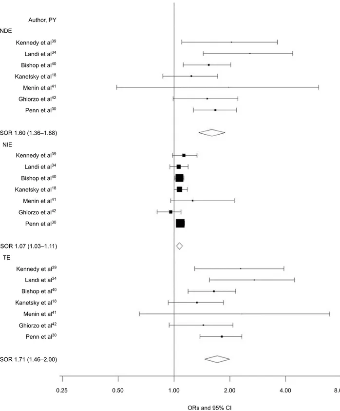
The OR (95% CI) for the TE of any MC1R variants on CM risk was 1.71 (1.46–2.00; I2=0; Figure 1). When
decom-posed, the risk was primarily due to the NDE, independent of phenotypic characteristics (OR 1.60; 95% CI 1.36–1.88;
I2=0; Figure 1); the NIE, which would be dependent on
the pigmentation pathway, was smaller (OR 1.07; 95% CI 1.03–1.11; I2=0; Figure 1). When the CDE according to
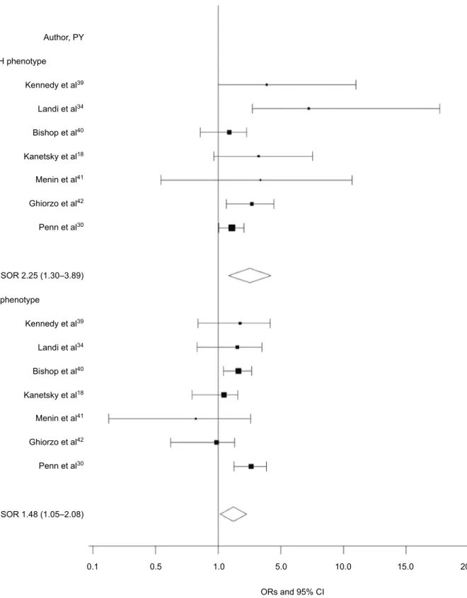
RH phenotype was examined, we found a direct, positive association between MC1R and CM in the absence of RH phenotype (OR 1.75; 95% CI 1.33–2.33; I2=0; Figure 2)
and a smaller direct association between MC1R and CM in participants with the RH phenotype (OR 1.50; 95% CI 1.19–1.89; I2=37%; Figure 2).
Looking at each of the nine most common MC1R variants (Table S1), we still found for all of them larger NDE than NIE, with significant NDE found for the R variants R142H, R151C, R160W, and D294K (ranging from OR 2.22; 95% CI 1.33–3.71 to OR 3.55; 95% CI 1.21–10.47) and significant NIE found only for the R variant R151C (OR 1.18; 95% CI 1.00–1.39). Furthermore, CDE was higher for non-RH-phenotype subjects than for RH-non-RH-phenotype subjects for the most common variants (allele frequency ≥1.5%), while it was opposite for the three rarer MC1R variants D84E, R142H, and I155T (Table S1).
Risk models for CM prediction
Table 2 reports the ORs and 95% CIs for variables included in the base clinical risk model and for MC1R variants. Hav-ing more than 30 common nevi and RH phenotype increased CM risk in our population (Table 2). Independent of other risk factors, carriers of any MC1R variant had a higher risk of CM than noncarriers (OR 1.63; 95% CI 1.40–1.90). The OR slightly decreased when the analysis was restricted to RH participants, while it increased for non-RH participants (Table 2). When we considered a distinction between MC1R r and R variants, in comparison with WT, carriers of at least one R variant had a higher risk of CM (OR 2.08; 95% CI 1.76–2.46) than carriers of only r variants (OR 1.24; 95% CI 1.04–1.47). For RH participants, carrying only MC1R r vari-ants did not increase CM risk, while the risk was increased for carriers of MC1R R variants. By contrast, both MC1R r and R variants were associated with a higher risk of CM in participants with the non-RH phenotype (Table 2). Similar results were found looking at each of the nine MC1R variants separately (Table S2).
The clinical risk model yielded an AUC of 0.706 (95% CI 0.691–0.721; Table S3). The model including any MC1R variant showed slightly greater discrimination, with an AUC of 0.713 (95% CI 0.698–0.728; P=0.002) and an NRI of 24% (95% CI 20%–30%). Differentiation between r and R vari-ants and considering each single variant further increased diagnostic accuracy by 1.5% and 1.9%, respectively, over the base clinical risk model, with an NRI of 37% (95% CI 32%–43%) and 34% (28%–39%), respectively. Subgroup analysis restricted to participants with the non-RH phenotype revealed that MC1R improved the AUC by 1.8% (from 0.678 to 0.696, P=0.0008; Figure 3; Table S3), suggesting a stronger role of MC1R in melanoma prediction for darker pigmented participants compared to RH participants. The NRI due to
MC1R inclusion for participants with a non-RH phenotype
was 28% (95% CI 19%–37%), while it was 15% (95% CI 9%–22%) for RH participants. The addiction of separate information on r and R MC1R variants and on single specific variants obtained a better model performance for both RH and non-RH participants. Decision curves showed a small increase in net benefit of MC1R testing for non-RH partici-pants over almost the entire range of threshold probabilities (Figure S1), with an average increase in net benefit of 0.003 for the model with any MC1R variant and 0.005 for the model with r or R MC1R variant over the base clinical model.
Sensitivity analysis on a subset of 2,472 (38%) par-ticipants from four studies with additional information on atypical nevi provided similar results (not shown): having more than 30 common nevi, RH phenotype, and atypical nevi increased CM risk. In this sensitivity analysis, MC1R variants increased CM risk in non-RH participants, but not in RH participants. Sensitivity analysis with different defini-tions of RH phenotype provided similar results (not shown).
Discussion
Our pooled analysis showed that the presence of any MC1R variant had a direct effect on CM, conferring a 60% higher risk to carriers versus noncarriers. The pigmentation-mediated effect of MC1R on CM was smaller with any
MC1R variant and each of the nine most common MC1R
variants. This result confirms and expands the previous sug-gestion16–18,34 of the existence of a nonpigmentation pathway
leading MC1R to CM development. Here, we give for the first time an estimate of the magnitude of total effect explained by each of the two (pigmentation and nonpigmentation) pathways. Recent studies and reviews47 have implicated
Figure 1 Forest plot for NDE, NIE, and TE of any MC1R variant on melanoma risk.
Notes: CDE estimates the direct effect of MC1R on melanoma when the mediator is controlled at level 0 (absent) or 1 (present) uniformly in the population, NDE essentially
averages CDE over the population, NIE estimates the indirect effect of MC1R mediated by RH phenotype, and TE is the overall melanoma risk estimate for MC1R carriers and in each study is the product of NDE and NIE.
Abbreviations: CDE, control direct effect; NDE, natural direct effect; NIE, natural indirect effect; PY, publication year; RH, red hair; SOR, summary OR; TE, total effect.
involved in cell-cycle control,48 apoptosis,49 and activation
of DNA-repair mechanisms and antioxidant defenses.50
Production of pheomelanin pigments seems associated with
increased oxidative DNA damage compared with synthesis of eumelanins.51 Further evidence for pheomelanin-associated
mice carrying a loss-of-function mutation of the Mc1r gene, which provided evidence in support that the pheomelanin-pigment pathway produces UV-independent carcinogenic
contributions to melanomagenesis.52 Another recent study53
found a role of germ-line MC1R variants in influencing the somatic mutational landscape of melanoma, with an expected
Figure 2 Forest plot for control direct effect of any MC1R variant on melanoma risk according to RH phenotype.*
Notes: *Defined as presence of red hair, freckles, or skin type I/II. Control direct effect estimates the direct effect of MC1R on melanoma when the mediator is controlled
at level 0 (absent) or 1 (present) uniformly in the population.
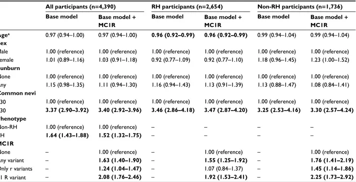
Table 2 ORs with 95% CIs for melanoma risk according to a base clinical model and the same model with inclusion of MC1R variants
All participants (n=4,390) RH participants (n=2,654) Non-RH participants (n=1,736)
Base model Base model +
MC1R
Base model Base model +
MC1R
Base model Base model +
MC1R Agea 0.97 (0.94–1.00) 0.97 (0.94–1.00) 0.96 (0.92–0.99) 0.96 (0.92–0.99) 0.99 (0.94–1.04) 0.99 (0.94–1.04)
Sex
Male 1.00 (reference) 1.00 (reference) 1.00 (reference) 1.00 (reference) 1.00 (reference) 1.00 (reference) Female 1.01 (0.89–1.16) 1.03 (0.91–1.18) 0.92 (0.77–1.09) 0.92 (0.77–1.10) 1.18 (0.96–1.45) 1.23 (1.00–1.52)
Sunburn
None 1.00 (reference) 1.00 (reference) 1.00 (reference) 1.00 (reference) 1.00 (reference) 1.00 (reference) Any 1.15 (0.98–1.35) 1.11 (0.94–1.30) 1.16 (0.94–1.43) 1.13 (0.91–1.39) 1.13 (0.88–1.47) 1.08 (0.84–1.41)
Common nevi
≤30 1.00 (reference) 1.00 (reference) 1.00 (reference) 1.00 (reference) 1.00 (reference) 1.00 (reference) >30 3.37 (2.90–3.92) 3.40 (2.92–3.96) 3.46 (2.86–4.18) 3.47 (2.87–4.20) 3.25 (2.53–4.16) 3.30 (2.57–4.24) Phenotype
Non-RH 1.00 (reference) 1.00 (reference) – – – –
RH 1.64 (1.43–1.88) 1.52 (1.32–1.75) – – – –
MC1R
None – 1.00 (reference) – 1.00 (reference) – 1.00 (reference)
Any variant – 1.63 (1.40–1.90) – 1.55 (1.25–1.92) – 1.76 (1.41–2.19)
Only r variants – 1.24 (1.04–1.47) – 1.07 (0.84–1.37) – 1.45 (1.14–1.86)
≥1 R variant – 2.08 (1.76–2.46) – 1.92 (1.53–2.41) – 2.25 (1.73–2.92)
Notes: aPer 5-year increase. Significant ORs are in bold. All models are adjusted for variables included in the table + study center. Two separate models were created for 1)
any MC1R variant vs wild type and 2) only r variants and ≥1R variant vs wild type. R and r alleles were defined basing on their stronger or weaker association with the RH phenotype for the most common variants44,67–70 and on likely pathogenicity using the algorithm proposed by Davies et al32 for the less common variants.
Abbreviation: RH, red hair.
Figure 3 ROC curve comparison between base clinical model and the same model with inclusion of MC1R variants for patients with no RH phenotype.*
Notes: (A) MC1R defined as the presence or absence of any MC1R variant and (B) as no MC1R variant, only r variants, and ≥1 R variants. *Non-RH patients defined as those
without RH and freckles and with skin type III/IV. R and r alleles were respectively defined basing on their stronger or weaker association with the RH phenotype for the most common variants44,67–70 and on likely pathogenicity using the algorithm proposed by Davies et al32 for the less common variants.
higher number of somatic C>T mutations in carriers of R alleles than those without R alleles. In this respect, it is worth noting that although the most relevant UV radiation-induced mutations are C>T transitions, highly recurrent mutations in key melanoma-driver genes, such as the V600E mutation in BRAF, are non-C>T changes. Importantly, significant increases in the rate of non-C>T changes, some of which might depend on oxidative DNA damage, have also been found in R allele carriers compared with noncarriers.53,54
Accordingly, associations of MC1R and genes frequently mutated in melanoma, such as BRAF or TERT, have been reported.55–57
We found that MC1R slightly improved risk prediction accuracy over a base clinical model, especially for non-RH participants: CM predictive accuracy increased by 1.8% and the CM risk of 28% of participants was better assessed. If a distinction is used in the model to differently score r and R variants, the benefit for the whole population increased from 24% of participants correctly reclassified with just presence/ absence of MC1R variants to 37% of participants correctly reclassified with separate information on r and R variants. Distinction between r and R alleles, however, was more apparent for RH than for non-RH participants. In the study by Cust et al,15 the R variants were responsible for most of
the improvement in risk prediction, but separate analysis for RH and non-RH participants was not performed.
Previous melanoma risk prediction models have included
MC1R with base clinical risk factors.15,28–30 Whiteman and
Green28 did not report on predictive ability. Stefanaki et al29
found no improvement in AUC with the addition of eight melanoma-associated single-nucleotide polymorphisms (SNPs) to the base model. Both Cust et al15 and Penn et al30
reported slight improvement in AUC with the inclusion of
MC1R. However, no previous paper has reported separate
results according to fairer or darker phenotypic characteris-tics. This point seems in fact extremely important, because
MC1R seems to have a stronger role in non-RH participants
in both the present paper and in previously published strati-fied analyses.16,18,34 A more precise risk assessment, therefore,
in participants with no RH, no freckles, and skin type III/ IV could potentially change individual clinical follow-up schedules and perhaps UV-exposure behavior and indoor tanning habits.
The application of risk prediction tools in cancer screen-ing has been widely discussed. In particular, there have been concerns on the impact of genetic screening in clinical decision-making. For example, in a previous review,58 genetic
screening was discussed using commercially available SNP
panel tests in prostate cancer. Conclusions were that the investigated SNP panels had poor discriminative ability and clinical validity. In our study, adding the MC1R genotype resulted in a small yet significant improvement in predictive ability over the clinical model and a substantial change in the NRI, and it is worth noting that this improvement was based on a single gene, while risk indices for both prostate and breast cancer require several genetic markers to produce increases of similar magnitude.59–62 Decreasing genotyping
costs and increasing use of genetic testing is making it more feasible to incorporate genetic risk factors into clinical risk prediction tools, and limiting testing to the non-RH partici-pants with no other risk factors may result in a cost-effective strategy via better allocation of resources. However, transla-tion into routine clinical practice requires several additransla-tional steps,63,64 and new studies are needed in order better to assess
the clinical utility of these models, taking also into account the small increase in net benefit observed in our decision curve analysis.
Our study has several strengths. We quantified for the first time the amount of total effect of MC1R on CM due to pigmentation and nonpigmentation pathways. Previous stratified analyses, including ours,16 have already suggested
that the effect of MC1R was stronger in darker pigmented participants; however, stratified analyses are not conclu-sive, especially in the presence of genotype–phenotype interaction.46,65 Precise and powerful quantification of the
effect of the two pathways was only feasible in the present analysis after inclusion of new studies.30,40,41 The large sample
and international collaborative nature of the M-SKIP project make it possible to assess various populations and ances-tries, thus providing results that are robust and consistent in different geographical areas. We were also able to create different predictive models according to the RH and non-RH phenotypes, which was not possible in previous studies. In our centralized statistical analysis, we were able to take into account all the available confounders, with a homogeneous plan of analysis and definition of covariables.
Heterogeneity among different populations is a possible limitation of our study; therefore, this tool may require adjust-ments before being applicable to each specific population.10
However, it is not easy to develop a good and precise tool for each population due to the lack of power of single stud-ies. Moreover, we did not observe any heterogeneity in risk estimates for MC1R and CM, suggesting that information on
MC1R improves CM risk prediction in different populations
of European origins. Following our previous publication,44
RH, freckles, or skin type I/II, and we are aware that other definitions may modify the results. However, in a sensitivity analysis using RH defined as a score obtained from multiple correspondence analysis,44 the results were similar.
Pheno-type misclassification is a possibility, although a previous study reported a good correlation between self-defined skin pigmentation and measured melanin density.66 In order to
minimize phenotype misclassification, we performed a sen-sitivity analysis that included only extreme categories of the RH phenotype.5 Although this analysis was underpowered,
we observed similar risk estimates to those reported for the main analysis in the present paper (results not shown). Finally, it should be noted that our analyses were performed on sporadic melanoma cases, and thus, generalization to familial melanoma is not appropriate.
Conclusion
We found a direct role of MC1R in melanoma risk inde-pendently of RH phenotype and demonstrated that add-ing the MC1R genotype to classical clinical risk factors results in a benefit for CM prediction. A change in clinical follow-up schedules and UV exposure and sun protection habits of identified at-risk individuals might favor early melanoma diagnosis and prevention. The application of risk prediction tools in cancer screening has been contro-versial, because of concerns on their impact in clinical decision-making. Our results could be a valid starting point to guide the development of scientific protocols assessing melanoma risk prediction tools incorporating the MC1R genotype, ideally with a prospective design and cost–benefit evaluation.
Acknowledgments
This work was supported by the Italian Association for Cancer Research (grant MFAG 11831). The Melanoma Susceptibility Study (PAK) was supported by the National Cancer Institute (CA75434, CA80700, CA092428). The Genoa study (PG) was supported by AIRC IG 15460. The M-SKIP study group consists of the following members: principal investigator (PI), Sara Raimondi (European Insti-tute of Oncology, Milan, Italy); advisory committee mem-bers, Philippe Autier (International Prevention Research Institute, Lyon, France), Maria Concetta Fargnoli (University of L’Aquila, Italy), José C García-Borrón (University of Murcia, Spain), Jiali Han (Indiana University, Indianapo-lis, IN, USA), Peter A Kanetsky (Department of Cancer Epidemiology, H Lee Moffitt Cancer Center and Research Institute, Tampa, FL, USA), Maria Teresa Landi (National
Cancer Institute, NIH, Bethesda, MD, USA), Julian Little (University of Ottawa, Canada), Julia Newton-Bishop (Uni-versity of Leeds, Leeds, UK), and Francesco Sera (London School of Hygiene and Tropical Medicine, London, UK); consultants, Saverio Caini (ISPO, Florence, Italy), Sara Gandini, and Patrick Maisonneuve (European Institute of Oncology, Milan, Italy); participant investigators, Albert Hofman, Manfred Kayser, Fan Liu, Tamar Nijsten, and Andre G Uitterlinden (Erasmus MC University Medical Center, Rotterdam, Netherlands), Rajiv Kumar (German Cancer Research Center, Heidelberg, Germany), Tim Bishop, Faye Elliott (University of Leeds, Leeds, UK), Eduardo Nagore (Instituto Valenciano de Oncologia, Valencia, Spain), DeAnn Lazovich (Division of Epidemiology and Community Health, University of Minnesota, MN, USA), David Pol-sky (New York University School of Medicine, New York, NY, USA), Johan Hansson, Veronica Hoiom (Karolinska Institutet, Stockholm, Sweden), Paola Ghiorzo, Lorenza Pastorino (University of Genoa, Italy), Nelleke A Gruis, Jan Nico Bouwes Bavinck (Leiden University Medical Center, Leiden, Netherlands), Ricardo Fernandez-de-Misa (Hospital Universitario Nuestra Señora de Candelaria, Santa Cruz de Tenerife, Spain), Paula Aguilera, Celia Badenas, Cristina Carrera, Pol Gimenez-Xavier, Josep Malvehy, Miriam Potrony, Susana Puig, Joan Anton Puig-Butille, Gemma Tell-Marti (Hospital Clinic, IDIBAPS, and CIBERER, Barcelona, Spain), Terence Dwyer (Murdoch Children’s Research Institute, Melbourne, Australia), Leigh Blizzard, Jennifer Cochrane (Menzies Institute for Medical Research, Hobart, Australia), Wojciech Branicki (Institute of Forensic Research, Krakow, Poland), Tadeusz Debniak (Pomeranian Medical University, Szczecin, Poland), Niels Morling, Peter Johansen (University of Copenhagen, Copenhagen, Denmark), Susan Mayne, Allen Bale, Brenda Cartmel, Leah Ferrucci (Yale School of Public Health and Medicine, New Haven, CT, USA), Ruth Pfeiffer (National Cancer Institute, NIH, Bethesda, MD, USA), Giuseppe Palmieri (Istituto di Chimica Biomolecolare, CNR, Sassari, Italy), Gloria Ribas (Fundación Investigación Clínico de Valencia Instituto de Investigación Sanitaria, INCLIVA, Spain), Chiara Menin (Veneto Institute of Oncology, IOV-IRCCS, Padua, Italy), Alexander Stratigos, Katerina Kypreou (Andreas Sygros Hospital, University of Athens, Athens, Greece), Anne Bow-cock, Lynn Cornelius, M Laurin Council (Washington Uni-versity School of Medicine, St Louis, MO, USA), Tomonori Motokawa (POLA Chemical Industries, Yokohama, Japan), Sumiko Anno (Shibaura Institute of Technology, Tokyo, Japan), Per Helsing, Per Arne Andresen (Oslo University
Hospital, Oslo, Norway), Gabriella Guida, Stefania Guida (University of Bari, Bari, Italy), Terence H Wong (University of Edinburgh, Edinburgh, UK), and the GEM study group. Participants in the GEM study group are as follows: coor-dinating center, Memorial Sloan-Kettering Cancer Center, New York, NY, USA, Marianne Berwick (PI, currently at the University of New Mexico), Colin Begg (co-PI), Irene Orlow (coinvestigator), Urvi Mujumdar (project coordinator), Amanda Hummer (biostatistician), Klaus Busam (dermato-pathologist), Pampa Roy (laboratory technician), Rebecca Canchola (laboratory technician), Brian Clas (laboratory technician), Javiar Cotignola (laboratory technician), and Yvette Monroe (interviewer); study centers; University of Sydney and Cancer Council New South Wales, Sydney (Aus-tralia), Bruce Armstrong (PI), Anne Kricker (co-PI), Melisa Litchfield (study coordinator); Menzies Institute for Medical Research, University of Tasmania, Hobart (Australia), Ter-ence Dwyer (PI), Paul Tucker (dermatopathologist), Nicola Stephens (study coordinator); British Columbia Cancer Agency, Vancouver, BC (Canada), Richard Gallagher (PI), Teresa Switzer (coordinator), Cancer Care Ontario, Toronto, ON (Canada), Loraine Marrett (PI), Beth Theis (coinvesti-gator), Lynn From (dermatopathologist), Noori Chowdhury (coordinator), Louise Vanasse (coordinator), Mark Purdue (research officer), David Northrup (manager for CATI), Centro per la Prevenzione Oncologia Torino, Piedmont (Italy), Roberto Zanetti (PI), Stefano Rosso (data manager), Carlotta Sacerdote (coordinator); University of California, Irvine, CA (USA), Hoda Anton-Culver (PI), Nancy Leighton (coordinator), Maureen Gildea (data manager); University of Michigan, Ann Arbor, MI (USA), Stephen Gruber (PI), Joe Bonner (data manager), Joanne Jeter (Coordinator); New Jersey Department of Health and Senior Services, Trenton, NJ (USA), Judith Klotz (PI), Homer Wilcox (co-PI), Helen Weiss (coordinator); University of North Carolina, Chapel Hill, NC (USA), Robert Millikan (PI), Nancy Thomas (coinvestigator), Dianne Mattingly (coordinator), Jon Player (laboratory technician), Chiu-Kit Tse (data analyst); Uni-versity of Pennsylvania, Philadelphia, PA (USA), Timothy Rebbeck (PI), Peter Kanetsky (coinvestigator), Amy Walker (laboratory technician), Saarene Panossian (laboratory technician); consultants, Harvey Mohrenweiser, University of California, Irvine, Irvine, CA (USA); Richard Setlow, Brookhaven National Laboratory, Upton, NY (USA).
Disclosure
The authors report no conflicts of interest in this work.
References
1. Erdmann F, Lortet-Tieulent J, Schuz J, et al. International trends in the incidence of malignant melanoma 1953–2008: are recent generations at higher or lower risk? Int J Cancer. 2013;132(2):385–400. 2. Wernli KJ, Henrikson NB, Morrison CC, Nguyen M, Pocobelli G, Blasi
PR. Screening for skin cancer in adults: updated evidence report and systematic review for the US Preventive Services Task Force. JAMA. 2016;316(4):436–447.
3. Gandini S, Sera F, Cattaruzza MS, et al. Meta-analysis of risk factors for cutaneous melanoma – I: common and atypical naevi. Eur J Cancer. 2005;41(1):28–44.
4. Gandini S, Sera F, Cattaruzza MS, et al. Meta-analysis of risk fac-tors for cutaneous melanoma – II: sun exposure. Eur J Cancer. 2005;41(1):45–60.
5. Gandini S, Sera F, Cattaruzza MS, et al. Meta-analysis of risk factors for cutaneous melanoma – III: family history, actinic damage and phe-notypic factors. Eur J Cancer. 2005;41(14):2040–2059.
6. Ahmed H, Naik G, Willoughby H, Edwards AG. Communicating risk.
BMJ. 2012;344:e3996.
7. Freedman AN, Seminara D, Gail MH, et al. Cancer risk prediction models: a workshop on development, evaluation, and application. J Natl
Cancer Inst. 2005;97(10):715–723.
8. Jackson A, Wilkinson C, Ranger M, Pill R, August P. Can primary prevention or selective screening for melanoma be more precisely tar-geted through general practice? A prospective study to validate a self administered risk score. BMJ. 1998;316(7124):34–39.
9. Quereux G, N’Guyen JM, Cary M, Jumbou O, Lequeux Y, Dreno B. Validation of the self-assessment of melanoma risk score for a melanoma-targeted screening. Eur J Cancer Prev. 2012;21(6):588–595. 10. Vuong K, McGeechan K, Armstrong BK, Cust AE. Risk prediction
models for incident primary cutaneous melanoma: a systematic review.
JAMA Dermatol. 2014;150(4):434–444.
11. Olsen CM, Neale RE, Green AC, et al. Independent valida-tion of six melanoma risk predicvalida-tion models. J Invest Dermatol. 2015;135(5):1377–1384.
12. US Preventive Services Task Force, Bibbins-Domingo K, Grossman DC, et al. Screening for skin cancer: US preventive services task force recommendation statement. JAMA. 2016;316(4):429–435.
13. Tsao H, Weinstock MA. Visual inspection and the US Preventive Services Task Force recommendation on skin cancer screening. JAMA. 2016;316(4):398–400.
14. Chaudru V, Chompret A, Bressac-de Paillerets B, Spatz A, Avril MF, Demenais F. Influence of genes, nevi, and sun sensitivity on melanoma risk in a family sample unselected by family history and in melanoma-prone families. J Natl Cancer Inst. 2004;96(10):785–795.
15. Cust AE, Goumas C, Vuong K, et al. MC1R genotype as a predictor of early-onset melanoma, compared with self-reported and physician-measured traditional risk factors: an Australian case-control-family study. BMC Cancer. 2013;13:406.
16. Pasquali E, Garcia-Borron JC, Fargnoli MC, et al. MC1R variants increased the risk of sporadic cutaneous melanoma in darker-pigmented Caucasians: a pooled-analysis from the M-SKIP project. Int J Cancer. 2015;136(3):618–631.
17. Raimondi S, Sera F, Gandini S, et al. MC1R variants, melanoma and red hair color phenotype: a meta-analysis. Int J Cancer. 2008;122(12):2753–2760.
18. Kanetsky PA, Panossian S, Elder DE, et al. Does MC1R genotype convey information about melanoma risk beyond risk phenotypes?
Cancer. 2010;116(10):2416–2428.
19. Chatzinasiou F, Lill CM, Kypreou K, et al. Comprehensive field synopsis and systematic meta-analyses of genetic association studies in cutaneous melanoma. J Natl Cancer Inst. 2011;103(16):1227–1235.
20. Amos CI, Wang LE, Lee JE, et al. Genome-wide association study identifies novel loci predisposing to cutaneous melanoma. Hum Mol
21. Williams PF, Olsen CM, Hayward NK, Whiteman DC. Melanocortin 1 receptor and risk of cutaneous melanoma: a meta-analysis and estimates of population burden. Int J Cancer. 2011;129(7):1730–1740. 22. Garcia-Borron JC, Sanchez-Laorden BL, Jimenez-Cervantes C.
Mela-nocortin-1 receptor structure and functional regulation. Pigment Cell
Res. 2005;18(6):393–410.
23. Garcia-Borron JC, Abdel-Malek Z, Jimenez-Cervantes C. MC1R, the cAMP pathway, and the response to solar UV: extending the horizon beyond pigmentation. Pigment Cell Melanoma Res. 2014;27(5):699–720.
24. Beaumont KA, Shekar SN, Newton RA, et al. Receptor function, domi-nant negative activity and phenotype correlations for MC1R variant alleles. Hum Mol Genet. 2007;16(18):2249–2260.
25. Beaumont KA, Shekar SN, Cook AL, Duffy DL, Sturm RA. Red hair is the null phenotype of MC1R. Hum Mutat. 2008;29(8):E88–E94. 26. Doyle JR, Fortin JP, Beinborn M, Kopin AS. Selected melanocortin 1
receptor single-nucleotide polymorphisms differentially alter multiple signaling pathways. J Pharmacol Exp Ther. 2012;342(2):318–326. 27. Duffy DL, Box NF, Chen W, et al. Interactive effects of MC1R and OCA2
on melanoma risk phenotypes. Hum Mol Genet. 2004;13(4):447–461. 28. Whiteman DC, Green AC. A risk prediction tool for melanoma? Cancer
Epidemiol Biomarkers Prev. 2005;14(4):761–763.
29. Stefanaki I, Panagiotou OA, Kodela E, et al. Replication and predictive value of SNPs associated with melanoma and pigmentation traits in a southern European case-control study. PLoS One. 2013;8(2):e55712. 30. Penn LA, Qian M, Zhang E, et al. Development of a melanoma risk
prediction model incorporating MC1R genotype and indoor tanning exposure: impact of mole phenotype on model performance. PLoS
One. 2014;9(7):e101507.
31. Dwyer T, Stankovich JM, Blizzard L, et al. Does the addition of infor-mation on genotype improve prediction of the risk of melanoma and nonmelanoma skin cancer beyond that obtained from skin phenotype?
Am J Epidemiol. 2004;159(9):826–833.
32. Davies JR, Randerson-Moor J, Kukalizch K, et al. Inherited vari-ants in the MC1R gene and survival from cutaneous melanoma: a BioGenoMEL study. Pigment Cell Melanoma Res. 2012;25(3): 384–394.
33. Guida M, Strippoli S, Ferretta A, et al. Detrimental effects of melano-cortin-1 receptor (MC1R) variants on the clinical outcomes of BRAF V600 metastatic melanoma patients treated with BRAF inhibitors.
Pigment Cell Melanoma Res. 2016;29(6):679–687.
34. Landi MT, Kanetsky PA, Tsang S, et al. MC1R, ASIP, and DNA repair in sporadic and familial melanoma in a Mediterranean population. J Natl
Cancer Inst. 2005;97(13):998–1007.
35. Guida S, Bartolomeo N, Zanna PT, et al. Sporadic melanoma in south-eastern Italy: the impact of melanocortin 1 receptor (MC1R) polymor-phism analysis in low-risk people and report of three novel variants.
Arch Dermatol Res. 2015;307(6):495–503.
36. Raimondi S, Gandini S, Fargnoli MC, et al. Melanocortin-1 receptor, skin cancer and phenotypic characteristics (M-SKIP) project: study design and methods for pooling results of genetic epidemiological studies. BMC Med Res Methodol. 2012;12:116.
37. Bergman W, Gruis NA. Management of melanoma families. Cancers
(Basel). 2010;2(2):549–566.
38. de Snoo F, Gruis N. Familial melanoma [webpage on the Internet]. 2005. Available from: http://atlasgeneticsoncology.org/Kprones/Famil-ialMelanomID10088.html. Accessed March 10, 2018.
39. Kennedy C, ter Huurne J, Berkhout M, et al. Melanocortin 1 receptor (MC1R) gene variants are associated with an increased risk for cutane-ous melanoma which is largely independent of skin type and hair color.
J Invest Dermatol. 2001;117(2):294–300.
40. Bishop DT, Demenais F, Iles MM, et al. Genome-wide association study identifies three loci associated with melanoma risk. Nat Genet. 2009;41(8):920–925.
41. Menin C, Vecchiato A, Scaini MC, et al. Contribution of susceptibility gene variants to melanoma risk in families from the Veneto region of Italy. Pigment Cell Melanoma Res. 2011;24(4):728–730.
42. Ghiorzo P, Bonelli L, Pastorino L, et al. MC1R variation and mela-noma risk in relation to host/clinical and environmental factors in CDKN2A positive and negative melanoma patients. Exp Dermatol. 2012;21(9):718–720.
43. Pastorino L, Cusano R, Bruno W, et al. Novel MC1R variants in Ligurian melanoma patients and controls. Hum Mutat. 2004;24(1):103. 44. Tagliabue E, Gandini S, Garcia-Borron JC, et al. Association of
melanocortin-1 receptor variants with pigmentary traits in humans: a pooled analysis from the M-SKIP project. J Invest Dermatol. 2016;136(9):1914–1917.
45. Vanderweele TJ, Vansteelandt S. Odds ratios for mediation analysis for a dichotomous outcome. Am J Epidemiol. 2010;172(12):1339–1348. 46. Valeri L, Vanderweele TJ. Mediation analysis allowing for
exposure-mediator interactions and causal interpretation: theoretical assumptions and implementation with SAS and SPSS macros. Psychol Methods. 2013;18(2):137–150.
47. Horrell EM, Boulanger MC, D’Orazio JA. Melanocortin 1 receptor: structure, function, and regulation. Front Genet. 2016;7:95.
48. April CS, Barsh GS. Distinct pigmentary and melanocortin 1 receptor-dependent components of cutaneous defense against ultraviolet radia-tion. PLoS Genet. 2007;3(1):e9.
49. Hauser JE, Kadekaro AL, Kavanagh RJ, et al. Melanin content and MC1R function independently affect UVR-induced DNA damage in cultured human melanocytes. Pigment Cell Res. 2006;19(4):303–314. 50. Kadekaro AL, Chen J, Yang J, et al. Alpha-melanocyte-stimulating
hormone suppresses oxidative stress through a p53-mediated signaling pathway in human melanocytes. Mol Cancer Res. 2012;10(6):778–786. 51. Wong SS, Ainger SA, Leonard JH, Sturm RA. MC1R variant allele
effects on UVR-induced phosphorylation of p38, p53, and DDB2 repair protein responses in melanocytic cells in culture. J Invest Dermatol. 2012;132(5):1452–1461.
52. Mitra D, Luo X, Morgan A, et al. An ultraviolet-radiation-independent pathway to melanoma carcinogenesis in the red hair/fair skin back-ground. Nature. 2012;491(7424):449–453.
53. Robles-Espinoza CD, Roberts ND, Chen S, et al. Germline MC1R status influences somatic mutation burden in melanoma. Nat Commun. 2016;7:12064.
54. Johansson PA, Pritchard AL, Patch AM, et al. Mutation load in mela-noma is affected by MC1R genotype. Pigment Cell Melamela-noma Res. 2017;30(2):255–258.
55. Fargnoli MC, Pike K, Pfeiffer RM, et al. MC1R variants increase risk of melanomas harboring BRAF mutations. J Invest Dermatol. 2008;128(10):2485–2490.
56. Landi MT, Bauer J, Pfeiffer RM, et al. MC1R germline variants confer risk for BRAF-mutant melanoma. Science. 2006;313(5786):521–522. 57. Nagore E, Reyes-Garcia D, Heidenreich B, Garcia-Casado Z, Requena
C, Kumar R. TERT promoter mutations associate with MC1R variants in melanoma patients. Pigment Cell Melanoma Res. 2017;30(2):273–275. 58. Little J, Wilson B, Carter R, et al. Multigene panels in prostate cancer
risk assessment: a systematic review. Genet Med. 2016;18(6):535–544. 59. Mealiffe ME, Stokowski RP, Rhees BK, Prentice RL, Pettinger M,
Hinds DA. Assessment of clinical validity of a breast cancer risk model combining genetic and clinical information. J Natl Cancer Inst. 2010;102(21):1618–1627.
60. Lindström S, Schumacher FR, Cox D, et al. Common genetic variants in prostate cancer risk prediction: results from the NCI Breast and Prostate Cancer Cohort Consortium (BPC3). Cancer Epidemiol Biomarkers
Prev. 2012;21(3):437–444.
61. Macinnis RJ, Antoniou AC, Eeles RA, et al. A risk prediction algorithm based on family history and common genetic variants: application to prostate cancer with potential clinical impact. Genet Epidemiol. 2011;35(6):549–556. 62. Chatterjee N, Park JH, Caporaso N, Gail MH. Predicting the future of genetic
risk prediction. Cancer Epidemiol Biomarkers Prev. 2011;20(1):3–8. 63. Pearson TA, Manolio TA. How to interpret a genome-wide association
study. JAMA. 2008;299(11):1335–1344.
64. Collins FS, Green ED, Guttmacher AE, Guyer MS. A vision for the future of genomics research. Nature. 2003;422(6934):835–847.
Cancer Management and Research
Publish your work in this journal
Submit your manuscript here: https://www.dovepress.com/cancer-management-and-research-journal Cancer Management and Research is an international, peer-reviewed open access journal focusing on cancer research and the optimal use of preventative and integrated treatment interventions to achieve improved outcomes, enhanced survival and quality of life for the cancer patient. The manuscript management system is completely online and includes
a very quick and fair peer-review system, which is all easy to use. Visit http://www.dovepress.com/testimonials.php to read real quotes from published authors.
Dove
press
65. Hernan MA, Clayton D, Keiding N. The Simpson’s paradox unraveled.
Int J Epidemiol. 2011;40(3):780–785.
66. Cargill J, Lucas RM, Gies P, et al. Validation of brief questionnaire measures of sun exposure and skin pigmentation against detailed and objective measures including vitamin D status. Photochem Photobiol. 2013;89(1):219–226.
67. Garcia-Borron JC, Sanchez-Laorden BL, Jimenez-Cervantes C. Mela-nocortin-1 receptor structure and functional regulation. Pigment Cell
Res. 2005;18(6):393–410.
68. Duffy DL, Box NF, Chen W, et al. Interactive effects of MC1R and OCA2 on melanoma risk phenotypes. Hum Mol Genet. 2004;13(4):447–461. 69. Box NF, Wyeth JR, O’Gorman LE, Martin NG, Sturm RA. Character-ization of melanocyte stimulating hormone receptor variant alleles in twins with red hair. Hum Mol Genet. 1997;6(11):1891–1897. 70. Sturm RA, Duffy DL, Box NF, et al. Genetic association and cellular
function of MC1R variant alleles in human pigmentation. Ann N Y Acad