Introduction
Urinary tract and kidney malformations often result in termina-tion of pregnancy after being detected on ultrasound screening, and these anomalies are also a major cause of renal failure in sur-viving children (1). While the genetic bases of kidney malforma-tions are well-recognized (2), with pathogenic variants reported in transcription and growth factors that drive metanephric dif-ferentiation, the possible genetic causes of congenital ureter and bladder anomalies are much less clear (3). A striking urinary tract phenotype is megabladder, with a first trimester prevalence of 1:330–1670 (4). Megabladder accompanied by a thinned and wrinkled abdominal wall overlying the bladder is called prune
belly syndrome (PBS). Megabladder and PBS are accompanied by kidney glomerular cysts considered to be secondary to fetal urinary flow obstruction (5). Some megabladders are associated with anatomically obstructed bladder outflow tracts, and these bladders have increased smooth muscle (SM) in their walls. Other megabladders are examples of functional outflow obstruction and have patent urethras and thin bladder walls (6). We hypothesized that mutations in genes affecting SM differentiation can cause megabladder and PBS.
Myocardin (MYOCD) is the founding member of a class of powerful transcriptional coactivators that bind to serum response factor (SRF) to activate cardiac- and SM-specific gene expression programs (7, 8). Complete loss of Myocd in mice causes embryonic lethality at midgestation due to failure of SM cell differentiation, whereas heterozygous knockout mice appear normal (9). Condi-tional mouse models subsequently defined the function of Myocd in postnatal development. Specifically, genetic ablation of Myocd in adult hearts causes heart failure due to loss of sarcomere struc-ture and increased cardiomyocyte apoptosis (10). Genetic deletion of Myocd specifically in SM revealed that Myocd maintains vascu-lar and visceral SM homeostasis postnatally (11). Despite these Myocardin (MYOCD) is the founding member of a class of transcriptional coactivators that bind the serum-response factor
to activate gene expression programs critical in smooth muscle (SM) and cardiac muscle development. Insights into the molecular functions of MYOCD have been obtained from cell culture studies, and to date, knowledge about in vivo roles of MYOCD comes exclusively from experimental animals. Here, we defined an often lethal congenital human disease associated with inheritance of pathogenic MYOCD variants. This disease manifested as a massively dilated urinary bladder, or megabladder, with disrupted SM in its wall. We provided evidence that monoallelic loss-of-function variants in MYOCD caused congenital megabladder in males only, whereas biallelic variants were associated with disease in both sexes, with a phenotype additionally involving the cardiovascular system. These results were supported by cosegregation of MYOCD variants with the phenotype in 4 unrelated families by in vitro transactivation studies in which pathogenic variants resulted in abrogated SM gene expression and by the finding of megabladder in 2 distinct mouse models with reduced Myocd activity. In conclusion, we have demonstrated that variants in MYOCD result in human disease, and the collective findings highlight a vital role for MYOCD in mammalian organogenesis.
Loss-of-function variants in myocardin cause
congenital megabladder in humans and mice
Arjan C. Houweling,1 Glenda M. Beaman,2,3 Alex V. Postma,1,4 T. Blair Gainous,5 Klaske D. Lichtenbelt,6 Francesco Brancati,7,8 Filipa M. Lopes,2,3 Ingeborg van der Made,9 Abeltje M. Polstra,1 Michael L. Robinson,10 Kevin D. Wright,10 Jamie M. Ellingford,2,3 Ashley R. Jackson,11 Eline Overwater,1 Rita Genesio,12 Silvio Romano,8 Letizia Camerota,8 Emanuela D’Angelo,8
Elizabeth J. Meijers-Heijboer,1 Vincent M. Christoffels,4 Kirk M. McHugh,11 Brian L. Black,5 William G. Newman,2,3 Adrian S. Woolf,2,3 and Esther E. Creemers9
1Department of Clinical Genetics, Amsterdam UMC, Amsterdam, Netherlands. 2School of Biological Sciences, Faculty of Biology, Medicine and Health, University of Manchester, Manchester, United Kingdom. 3Manchester Centre for Genomic Medicine and Royal Manchester Children’s Hospital, Manchester University NHS Foundation Trust, Manchester Academic Health Science Centre, Manchester, United
Kingdom. 4Department of Medical Biology, Amsterdam UMC, Amsterdam, Netherlands. 5Cardiovascular Research Institute, UCSF, San Francisco, California, USA. 6Department of Medical Genetics, University
Medical Center Utrecht, Utrecht, Netherlands. 7Laboratory of Molecular and Cell Biology, Istituto Dermopatico dell’Immacolata, IDI-IRCCS, Rome, Italy. 8Department of Life, Health and Environmental
Sciences, University of L’Aquila, Aquila, Italy. 9Department of Experimental Cardiology, Amsterdam UMC, Amsterdam, Netherlands. 10Department of Biology, Miami University, Oxford, Ohio, USA. 11Center
for Clinical and Translational Research, The Research Institute, Nationwide Children’s Hospital, Columbus, Ohio, USA. 12Department of Molecular Medicine and Medical Biotechnology, University of Naples
Federico II, Naples, Italy.
Authorship note: ACH, GMB, and AVP contributed equally to this work. WGN, ASW,
and EEC jointly supervised this work.
Conflict of interest: The authors have declared that no conflict of interest exists. Copyright: © 2019, Houweling et al. This is an open access article published under the
terms of the Creative Commons Attribution 4.0 International License.
Submitted: March 1, 2019; Accepted: September 3, 2019; Published: November 4, 2019. Reference information: J Clin Invest. 2019;129(12):5374–5380.
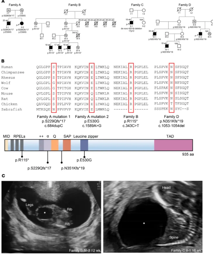
Figure 1. Identification of MYOCD variants in 4 families with congenital megabladder. (A) Pedigrees of 4 families presenting with congenital
megablad-der. Affected individuals are marked with black symbols. Available genotypes are shown beneath symbols. Slashed symbols denote deceased individuals. Gestational age is indicated above the symbol. Gray symbols denote stillbirths with external features consistent with PBS. N, normal bladder ultrasound; P with arrow, proband of the family; npe, normal prenatal echo. (B) Schematic diagram showing functional domains within MYOCD and location of the
identified mutations (7). Conservation of respective amino acid positions with the mutated residues are highlighted. (C) Ultrasound images showing
ular septal defect (VSD), a patent ductus arteriosus, and a bicus-pid aortic valve. Her brother (II-1) had been diagnosed prenatal-ly with megabladder and VSD, prompting clinical termination of pregnancy at midgestation. Autopsy revealed PBS, and histology showed disorganized SM bundles in the bladder and glomeru-lar cysts in the kidneys (Figure 2). The distal urethra was patent (Supplemental Figure 1; supplemental material available online with this article; https://doi.org/10.1172/JCI128545DS1), but the prostate and proximal urethra were not identified. A conclusion regarding anatomical obstruction was not possible because for-mal tests of urethral patency were not undertaken. The hindgut contained only a defined circular SM layer (Supplemental Figure 2) and lacked the longitudinal layer that should be present at this gestational age. In contrast, 2 normal SM layers were present in the small intestine and pulmonary artery SM appeared normal (Sup-plemental Figure 2). Ultrasonography revealed no bladder or heart abnormalities in the parents. Whole-exome sequencing (WES) in index patient II-2 and Sanger sequencing in II-1 determined that both siblings carried compound heterozygous variants in MYOCD (p.[S229Qfs*17];[E530G], respectively called family A mutation 1 and family A mutation 2 in Figure 1A). The variant p.[S229Qfs*17] is predicted to create a premature stop codon and was paternally inherited, while the missense variant p.[E530G] is located in the functional leucine zipper (LZ) of the encoded protein (12, 13) and was maternally inherited (Figure 1, A and B). Both variants were absent from over 120,000 control exomes (14). WES in the index case failed to reveal pathogenic variants in genes known to cause megabladder, including ACTG2, CHRM3, HPSE2, LRIG2, smooth muscle myosin heavy chain 11 (MYH11), and myosin light chain kinase (MYLK) (3, 15–17).
Next, we ascertained 22 additional families with megablad-der or PBS of unknown etiology, identifying 7 affected individuals from 3 unrelated families, all with heterozygous predicted loss-of-function MYOCD variants (Figure 1A and Supplemental Table 1). In family B, there were 3 male fetal deaths, all with PBS (Figure 1, A and C). WES revealed a heterozygous variant, c.343C>T, in these 3 brothers, predicted to result in a premature stop p.[R115*]. This variant was also present in the unaffected mother, unaffected maternal grandmother, and a healthy female sibling, each with normal bladder and heart imaging. The grandmother (II-3) unique and important functions of Myocd in mice, no human
genetic disorder associated with MYOCD variants has yet been defined. Here, we describe MYOCD loss-of-function variants in 9 individuals from 4 families with the megabladder/PBS spectrum, and we support these observations on urinary tract maldevelop-ment with Myocd mutant mouse models.
Results and Discussion
The index patient of family A (II-2) (Figure 1A) had a history of antenatal megabladder, and a bladder diverticulum was surgically resected in infancy. At 32 years of age, cardiac evaluation showed noncompaction cardiomyopathy and marked dilation (51 mm) of the aortic root. She also had an atrial septal defect, a
ventric-Figure 2. Bladder and kidney abnormalities in family A. (A and C) From
healthy midgestation fetuses. (B and D) From affected fetus from family
A. H&E staining from urinary bladders shows transverse sections of muscle bundles (TSM) and longitudinal sections of muscle bundles (LSM) in the healthy and affected fetuses. Note, however, that the bundles in the affected fetus appear disorganized and less compact compared with the well-defined muscle fibers in the control. (C) In a control fetal kidney,
glom-eruli (G) and tubules (T) are evident. (D) In the kidney from the affected
fetus, glomeruli are cystic, with dilated Bowman’s spaces (asterisks), a characteristic of fetal urinary flow obstruction. Scale bars: 20 μm.
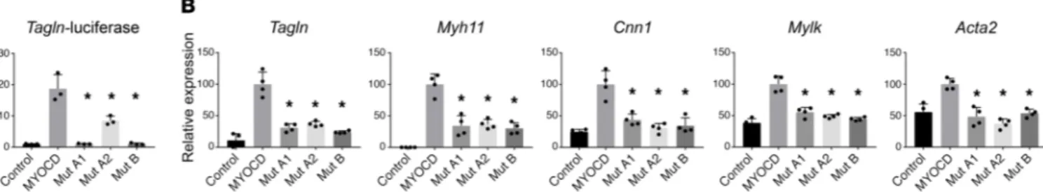
Figure 3. MYOCD mutations abrogate activation of SM cell gene expression in vitro. (A) Mouse fibroblasts were transiently transfected for 48 hours
with expression vectors for MYOCD or the indicated MYOCD mutants (mutation A1: p.S229Qfs*17; mutation A2: p.E530G; mutation B: p.R115*) and a luciferase reporter linked to the Transgelin (Sm22) promoter (n = 3/group). (B) Mouse fibroblasts were transfected with expression plasmids encoding
MYOCD or the indicated mutants (n = 4/group). An empty expression plasmid served as a control. RNA was isolated, and SM gene expression was
measured by qPCR. GAPDH was used to normalize expression. Overexpression levels of MYOCD were comparable between conditions (Supplemental Figure 3). *P < 0.01 compared with WT MYOCD according to 1-way ANOVA with Dunnett’s multiple comparison test. Shown are representative experi-ments of 2 independent repeats.
C’s first pregnancy was terminated after diagnosis of PBS in the male fetus (Figure 1, A and C). Chromosomal microarray analy-sis revealed a heterozygous de novo 420 kb deletion (chr17p12; hg19:12,172,568-12,609,597) encompassing the first 2 exons of reported a male stillbirth (III-4) of unknown cause in the third
trimester. She had 5 siblings, including 4 brothers who died ante-natally (II-5, II-6, II-7, and II-8), each with a megabladder (fur-ther details unavailable), and 1 healthy sister (Figure 1A). Family
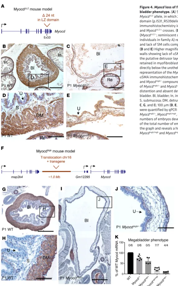
Figure 4. Myocd loss of function in mice causes the mega-bladder phenotype. (A) Schematic representation of the
MyocdΔLZ allele, in which 24 nucleotides are deleted in the LZ
domain (p.I531_R539delinsM in NP_666498.2). (B–E) αSMA
immunohistochemistry in 1-day-old neonates from Myocd+/–
and MyocdΔLZ/+ crosses. (C) Compound heterozygosity
(MyocdΔLZ/–; reminiscent of the alleles present in the affected
individuals in family A) results in wall thinning of the bladder and lack of SM cells compared with the WT bladder wall (B).
(D and E) Higher magnifications of WT and MyocdΔLZ/– bladder
walls showing lack of αSMA-expressing muscle bundles in the putative detrusor layer, although expression appeared retained in myofibroblast-like cells in the lamina propria directly below the urothelium and in the rectum. (F) Schematic
representation of the MyocdMgb allele. (G–J) Representative
αSMA immunohistochemistry in P1 bladder of WT (G and H) and Myocdmgb/– compound heterozygote (I and J) (from a cross
of Myocdmgb/+ and Myocd+/– mice). Note the severe bladder
distention and absent detrusor muscle in the Myocdmgb/–
bladder. Bl, bladder; In, intestine; Re, rectum; U, urethelium; S, submucosa; DM, detrusor muscle. Scale bars: 500 μm (B,
C, G, and I); 100 μm (D, E, H, and J). (K) Myocd mRNA levels
were quantified by qPCR using E15 bladders of WT, Myocd+/–, Myocdmgb/+, Myocdmgb/mgb, and Myocdmgb/– mice. The absolute
numbers of embryos developing megabladder as a fraction of the total number of embryos analyzed are indicated above the graph and reveals a highly penetrant phenotype in the
(Figure 4F, Supplemental Figure 5, and Supplemental Table 4), suggesting the presence of a regulatory Myocd enhancer. Next, we crossed Myocdmgb/+ with Myocd+/– mice (9) and demonstrated that compound mutant offspring (Myocdmgb/–) had megabladders, pro-viding further evidence that marked reduction of Myocd causes this phenotype (Figure 4, G–J, and Supplemental Figures 6 and 7). In addition, we observed patent ductus arteriosus in newborn Myocdmgb/– mice, but not in Myocd+/– mice with just 1 allele mutated (Supplemental Figure 8). By studying various Myocd mutants (Fig-ure 4K), we showed that a 70% to 80% reduction in Myocd mRNA in the bladder is sufficient to produce megabladder in mice. Interestingly, neonatal Myocdmgb/– mouse bladders have severely reduced transcript levels of Myocd target genes, yet in aortas and hearts of these same mice, the levels of Myocd target transcripts (apart from Myh11 in the aorta) are similar to those of heterozyous (Myocdmgb/+) controls (Supplemental Figure 9).
The bladder phenotypes in families carrying MYOCD variants are thus mimicked by 2 distinct mouse models with reduced Myocd activity. Supplemental Table 2 gives an overview of the pheno-types observed in the mouse lines. The collective results demon-strate that MYOCD plays a unique role in proper development of the bladder wall. Reduced Myocd activity resulted in little or no SM differentiation in the mouse bladders and in disorganized SM bun-dles in the human fetal bladder. These SM defects would diminish the muscular force required to void urine from the bladder, result-ing in the functional equivalent of lower urinary tract obstruction with severe bladder distension, ultimately culminating in kidney failure and death (19, 21).
Megabladder/PBS is a sex-limited trait with 95% male pre-dominance, likely the result of differences in urethra and bladder development and length differences in urethra between males and females (4, 6). Additionally, sex hormones may play a role in defin-ing the severity and progression of the disease, as clinical evidence demonstrates increased male susceptibility to acute and chronic kidney injury (22, 23). Indeed, in our study, 7 of 8 males proven to carry heterozygous loss-of-function MYOCD variants died before birth, whereas all 6 female carriers with heterozygous loss-of-function variants appeared healthy. Consistent with this obser-vation, male Myocdmgb/mgb mice were also more severely affected than females (Supplemental Table 2 and ref. 19). Notably, the only female with bladder disease in our study carried biallelic variants in MYOCD, suggesting that further reduction in MYOCD levels is needed to cause bladder phenotypes in females. This is supported by both Myocd mouse models in which compound heterozygosity (i.e., Myocdmgb/– or MyocdLZ/–) caused megabladder in either sex (Supplemental Table 2). An intriguing feature is the incomplete penetrance of bladder disease in a healthy male MYOCD mutation carrier in family D. This may be caused by allelic imbalance, where the penetrance of a dominant loss-of-function mutation is deter-mined by the expression level of the second allele, for instance, due to variants in promoter or enhancer regions of MYOCD. Alter-natively, redundancy with other MYOCD family members, such as MRTF-A, which is also expressed in developing bladders (24), may affect penetrance.
Overall, we propose that we have identified a semidominant disorder (25) in which heterozygous loss-of-function variants in MYOCD cause congenital megabladder, while biallelic loss-of-MYOCD, including the start codon. In family D, 3 males had PBS:
2 died prenatally (III-1 and IV-1), and the other (III-4) was born and underwent kidney transplantation for end-stage renal failure (Figure 1A). Sanger sequencing of MYOCD identified a heterozy-gous deletion of a single base c.1053-1054del, resulting in a pre-dicted frameshift p.[N351Kfs*19].
We tested the abilities of proteins encoded by MYOCD vari-ants from families A and B to activate the promoter of transgelin (Sm22 or transgelin [Tagln]), encoding a SM contractile protein. Western blotting revealed that the 2 predicted nonsense variants (p.[S229Qfs*17] mutation A1 from family A and p.[R115*] muta-tion B from family B) produced a truncated protein, whereas the missense variant (E530G, mutation A2 from family A) produced full-length protein (Supplemental Figure 3 and Supplemental Table 3). Neither nonsense variant resulted in activation of the Tagln-luciferase reporter, whereas the missense variant resulted in diminished activity versus WT MYOCD (Figure 3A). The above results regarding the missense variant are consistent with previ-ous reports showing that MYOCD homodimerizes through the LZ domain and that homodimerization facilitates stronger activation of SRF-dependent reporter genes (8, 12). Myocd is normally not expressed in 10T1/2 fibroblasts, but experimental overexpression in these cells activates the SM differentiation program (8). We transfected 10T1/2 cells with either WT MYOCD or one of each of the 3 variants from families A and B. As assessed by quantitative PCR (qPCR), WT MYOCD strongly induced endogenous expres-sion of the SM transcripts Tagln, Myh11, Calponin 1 (Cnn1), Mylk, and SM actin alpha 2 (Acta2). Conversely, each of the 3 MYOCD variants resulted in either a lack of increased expression or statis-tically significantly blunted responses (Figure 3B).
To assess whether reduced MYOCD activity causes mega-bladder, we took advantage of a newly generated mutant mouse line carrying an allele, MyocdΔLZ, in which critical residues within the LZ of Myocd were specifically deleted (Figure 4A and Supple-mental Figure 4A). Using primers that distinguish the WT from the LZ mutated transcript, we showed that both were detected in neonatal bladders of MyocdΔLZ/+ mice (Supplemental Figure 4B). We crossed MyocdΔLZ/+ mice with those carrying a null allele of Myocd (Myocd+/–) (18). The alleles in the compound mutant off-spring (MyocdΔLZ/–) therefore mimicked the LZ and nonsense mutant alleles in family A. In contrast to homozygous null Myocd mutants (Myocd–/–) (9), compound heterozygous mutant offspring (MyocdΔLZ/–) survived to birth. In line with the human urinary tract malformations, MyocdΔLZ/– mice developed grossly dilated bladders with little or no SM in their walls (Figure 4, A–E, and Supplemental Figure 4, C and D). In MyocdΔLZ/– mouse blad-ders, transcript levels of several Myocd target genes (i.e., Acta2, Myh11, Mylk, Tagln, and Cnn1) were blunted versus those of WTs (Supplemental Figure 4E).
To gain further insight into the potential role of Myocd gene dosage in bladder malformations, we examined the megabladder mouse (mgb) generated by random insertion and translocation of a transgene into chromosome 11 (19). Transcriptional profiling in the bladders of these mice had already revealed that levels of Myocd transcripts were significantly reduced (20). Here, we identified the translocation breakpoint (together with 4 copies of translocated chr. 16 region) approximately 500 kb upstream of the Myocd gene
tory Animals (National Academies Press, 2011). All experiments using MyocdΔLZ mice complied with federal and institutional guidelines and were reviewed and approved by the UCSF IACUC. The Mgb mouse stud-ies were approved by the IACUC of the Nationwide Children’s Hospital.
Author contributions
ACH, GMB, and AVP share first authorship, and the order in which they are listed has been determined by workload. ACH, GMB, AVP, WGN, ASW, and EEC designed the study. TBG, FML, IVDM, AMP, MLR, KDW, RG, LC, and ED performed experiments. ACH, EO, SR, KDL, and FB contributed clinical samples and clinical data. ACH, JME, GMB, AVP, and WGN performed genetic anal-ysis. ACH, GMB, AVP, KMM, ARJ, BLB, WGN, ASW, and EEC analyzed experimental data. BLB, KMM, and MLR contributed mouse lines. EJMH and VMC were instrumental in interpretation of the data. ACH, AVP, ASW, and EEC wrote the manuscript.
Acknowledgments
We acknowledge support from the Medical Research Coun-cil (MR/L002744/1 to ASW and WGN); Horizon 2020 Marie Sklodowska-Curie RENALTRACT (942937 to ASW and FML); the Newlife Foundation (to ASW, GMB, and WGN); and the NIH (EY12995 to MLR; DK70907 and DK085242 to KMM; and HL064658, HL089707, and HL136182 to BLB). We also acknowl-edge an Out-of-the-Box Grant ACS (to ACH and EEC), a CVON CONCOR genes grant (to AVP and VMC), and support from Fondi di Ateneo, University of L’Aquila, and Undiagnosed Disease Net-work Italy at Istituto Superiore di Sanità (PGR00229-919 and Far-mindustria to FB). We acknowledge Marilina Scalona and Fulvio De Simone for family B ascertainment.
Address correspondence to: Arjan C. Houweling, Departments of Clinical Genetics, Amsterdam UMC, De Boelelaan 1118, 1081HZ Amsterdam, Netherlands. Phone: 31204440150; Email: [email protected]. Or to: Esther E. Creemers, Department of Experimental Cardiology, Amsterdam UMC, Meibergdreef 15, 1105AZ Amsterdam, Netherlands. Phone: 31205663262; Email: [email protected].
function MYOCD variants also cause a cardiovascular phenotype. Both biallelic carriers developed congenital heart defects, while the affected female was found, when investigated as an adult, to have severe dilation of the aortic root. A similar association of car-diac defects was observed in the Myocdmgb/– mice (ref. 26 and Sup-plemental Figure 8). Notably, a previous study already hinted at the possible involvement of MYOCD in megabladder, as it described one sporadic case with PBS and a 1.3 Mb deletion of multiple genes, including MYOCD (27). Other SM-related genes have been impli-cated in PBS. These include variants in ACTA2, a MYOCD target gene, as well as MYH11 and MYLK, in which variants can cause vis-ceral myopathy, a phenotype encompassing megabladder (3, 16). Moreover, each of these 3 genes has been associated with inherit-ed thoracic aortic aneurysm and dissection (28). Notably, an SM- restricted deletion of Myocd in mice causes dilation of several vis-ceral organs, including the bladder, as well as dilation of the aorta (11). Hence, there is compelling evidence that reduced MYOCD levels can result in urological and cardiovascular disease.
In conclusion, we demonstrate for what we believe is the first time that variants in MYOCD result in human disease. We pro-pose that monoallelic loss-of-function variants in MYOCD cause congenital megabladder in males and that biallelic variants are associated with disease manifest in females that also involves the cardiovascular system. These findings not only have important implications for genetic counseling of families with megabladder, but also shed light on bladder development and expand the patho-physiological spectrum of inherited SM disorders.
Methods
Experimental procedures are provided in Supplemental Methods.
Study approval. Blood samples for genetic testing were obtained
upon written consent. Informed consent for DNA studies, clinical records, and use of ultrasound pictures and histological analysis of the terminated fetus of family A was obtained. Control human embryonic material, collected with maternal consent and ethical approval (REC 08/ H0906/21+5 and REC 18/NE/0290), was sourced from the MRC and Wellcome Trust Human Developmental Biology Resource. Mice were maintained according to the NIH Guide for the Care and Use of
1. Wiesel A, Queisser-Luft A, Clementi M, Bianca S, Stoll C; EUROSCAN Study Group. Prenatal detection of congenital renal malformations by fetal ultrasonographic examination: an analysis of 709,030 births in 12 European countries.
Eur J Med Genet. 2005;48(2):131–44.
2. Hildebrandt F. Genetic kidney diseases. Lancet. 2010;375(9722):1287–1295.
3. Woolf AS, Lopes FM, Ranjzad P, Roberts NA. Congenital disorders of the human urinary tract: recent insights from genetic and molecular studies. Front Pediatr. 2019;7:136. 4. Taghavi K, Sharpe C, Stringer MD. Fetal
megacystis: a systematic review. J Pediatr Urol. 2017;13(1):7–15.
5. Farrugia MK, et al. Experimental short-term fetal bladder outflow obstruction: I. Morphology and cell biology associated with urinary flow impair-ment. J Pediatr Urol. 2006;2(4):243–253. 6. Workman SJ, Kogan BA. Fetal bladder histology
in posterior urethral valves and the prune belly syndrome. J Urol. 1990;144(2 pt 1):337–339. 7. Pipes GC, Creemers EE, Olson EN. The
myo-cardin family of transcriptional coactivators: versatile regulators of cell growth, migration, and myogenesis. Genes Dev. 2006;20(12):1545–1556. 8. Wang D et al. Activation of cardiac gene
expres-sion by myocardin, a transcriptional cofactor for serum response factor. Cell. 2001;105(7):851–862. 9. Li S, Wang DZ, Wang Z, Richardson JA, Olson
EN. The serum response factor coactivator myocardin is required for vascular smooth muscle development. Proc Natl Acad Sci U S A. 2003;100(16):9366–9370.
10. Huang J, et al. Myocardin is required for cardiomyocyte survival and maintenance of heart function. Proc Natl Acad Sci U S A. 2009;106(44):18734–18739.
11. Huang J, et al. Myocardin is required for maintenance of vascular and visceral
smooth muscle homeostasis during postna-tal development. Proc Natl Acad Sci U S A. 2015;112(14):4447–4452.
12. Wang Z, Wang DZ, Pipes GC, Olson EN. Myo-cardin is a master regulator of smooth muscle gene expression. Proc Natl Acad Sci U S A. 2003;100(12):7129–34.
13. Anderson CM, et al. Cooperative activation of cardiac transcription through myocardin bridging of paired MEF2 sites. Development. 2017;144(7):1235–1241.
14. Lek M, et al. Analysis of protein-coding genetic variation in 60,706 humans. Nature. 2016;536(7616):285–291.
15. Stuart HM, et al. Urinary tract effects of HPSE2 mutations. J Am Soc Nephrol. 2015;26(4):797–804. 16. Richer J, et al. R179H mutation in ACTA2
expanding the phenotype to include prune-belly sequence and skin manifestations. Am J Med
17. Weber S, et al. Muscarinic acetylcholine receptor M3 mutation causes urinary bladder disease and a Prune-Belly-like syndrome. Am J Hum Genet. 2011;89(5):668–674.
18. Huang J, et al. Myocardin regulates expression of contractile genes in SM cells and is required for closure of the ductus arteriosus in mice. J Clin
Invest. 2008;118(2):515–525.
19. Singh S, et al. Identification of a unique trans-genic mouse line that develops megabladder, obstructive uropathy, and renal dysfunction.
J Am Soc Nephrol. 2007;18(2):461–471.
20. Singh S, et al. Transcriptional profiling of the mega-bladder mouse: a unique model of mega-bladder
dys-morphogenesis. Dev Dyn. 2008;237(1):170–186. 21. Ingraham SE, et al. Pathogenesis of renal injury
in the megabladder mouse: a genetic model of congenital obstructive nephropathy. Pediatr Res. 2010;68(6):500–507.
22. Metcalfe PD, Meldrum KK. Sex differences and the role of sex steroids in renal injury. J Urol. 2006;176(1):15–21.
23. Seliger SL, Davis C, Stehman-Breen C. Gender and the progression of renal disease. Curr Opin
Nephrol Hypertens. 2001;10(2):219–225.
24. Wang DZ, et al. Potentiation of serum response factor activity by a family of myocardin-related transcription factors. Proc Natl Acad Sci U S A.
2002;99(23):14855–60.
25. Wilkie AO. The molecular basis of genetic dominance. J Med Genet. 1994;31(2):89–98. 26. McHugh KM. Megabladder mouse model of
congenital obstructive nephropathy: genetic etiology and renal adaptation. Pediatr Nephrol. 2014;29(4):645–650.
27. Boghossian NS, et al. Rare copy number variants identified in prune belly syndrome. Eur J Med
Genet. 2018;61(3):145–151.
28. Milewicz DM, et al. Genetic basis of thoracic aortic aneurysms and dissections: focus on smooth muscle cell contractile dysfunction. Annu