Featured Article
Operationalizing mild cognitive impairment criteria in small vessel
disease: the VMCI-Tuscany Study
Emilia Salvadori
a, Anna Poggesi
a, Raffaella Valenti
a, Giovanni Pracucci
a, Francesca Pescini
b,
Marco Pasi
a, Serena Nannucci
a, Sandro Marini
a, Alessandra Del Bene
a, Laura Ciolli
a,
Andrea Ginestroni
c, Stefano Diciotti
d, Giovanni Orlandi
e, Ilaria Di Donato
f, Nicola De Stefano
f,1,
Mirco Cosottini
g, Alberto Chiti
e, Antonio Federico
f, Maria Teresa Dotti
f, Ubaldo Bonuccelli
e,
Domenico Inzitari
a,2, Leonardo Pantoni
a,b,*
,3, and on behalf of the VMCI-Tuscany Study Group
aNEUROFARBA Department, Neuroscience Section, University of Florence, Florence, Italy b
Stroke Unit and Neurology, Azienda Ospedaliero Universitaria Careggi, Florence, Italy
c
“Mario Serio” Department of Experimental and Clinical Biomedical Sciences, University of Florence, Florence, Italy
d
Department of Electrical, Electronic, and Information Engineering “Guglielmo Marconi”, University of Bologna, Cesena, Italy
e
Department of Neurosciences, University of Pisa, Pisa, Italy
f
Department of Medicine, Surgery and Neurosciences, University of Siena, Siena, Italy
gDepartment of Translational Research and New Technologies in Medicine and Surgery, University of Pisa, Pisa, Italy
Abstract Background: Mild cognitive impairment (MCI) prodromic of vascular dementia is expected to have a multidomain profile.
Methods: In a sample of cerebral small vessel disease (SVD) patients, we assessed MCI subtypes distributions according to different operationalization of Winblad criteria and compared the neuro-imaging features of single versus multidomain MCI. We applied three MCI diagnostic scenarios in which the cutoffs for objective impairment and the number of considered neuropsychological tests varied.
Results: Passing from a liberal to more conservative diagnostic scenarios, of 153 patients, 5% were no longer classified as MCI, amnestic multidomain frequency decreased, and nonamnestic single domain increased. Considering neuroimaging features, severe medial temporal lobe atrophy was more frequent in multidomain compared with single domain.
Conclusions: Operationalizing MCI criteria changes the relative frequency of MCI subtypes. Non-amnestic single domain MCI may be a previously nonrecognized type of MCI associated with SVD. ! 2015 The Alzheimer’s Association. Published by Elsevier Inc. All rights reserved.
Keywords: Cerebrovascular disease; Vascular dementia; Mild cognitive impairment; Neuropsychology; Cognitive aging
Authors’ disclosures: E.S., A.P., R.V., G.P., F.P., M.P., S.N., S.M., A.D.B., L.C., A.G., S.D., G.O., I.D.D., M.C., A.C., A.F., M.T.D., and U.B. report no disclosures.
1N.D.S. has served on scientific advisory boards and steering
commit-tees of clinical trials for Merck Serono S.A., Teva, and Novartis Pharma AG and has received support for congress participation or speaker honoraria from Biogen Idec, Merck Serono S.A., Bayer-Schering AG, Teva, Sanofi-Aventis, and Novartis Pharma AG. He received grants from the Italian MS Society, outside the submitted work.
2
D.I. serves as a member in the Editorial Board of Stroke and is Asso-ciate Editor of Neurological Sciences Journals. He has received grants for research by Bayer Italy and fees for conferences by Boeheringer Italy and Bayer Italy.
3L.P. serves on the editorial boards of Acta Neurologica Scandinavica
and Cerebrovascular Diseases and as Vascular Cognitive Impairment Section Editor for Stroke.
*Corresponding author. Tel.: 7945519; Fax: 139-055-4298461.
E-mail address:[email protected]
http://dx.doi.org/10.1016/j.jalz.2015.02.010
1. Background
Mild cognitive impairment (MCI) is an intermediate state between normal cognitive status and dementia; it is consid-ered a risk factor for dementia and has become a focus of several clinical and intervention trials. MCI is generally defined with the aid of neuropsychological tests providing evidence for object impairment with intact global cognitive functioning and activities of daily living. The criteria and the operationalization of MCI have been subjected to much debate as there is no real agreement regarding neuropsycho-logical tests, the number and/or type of cognitive domains to be assessed, and the proper use of neuropsychological cut scores[1]. The lack of a universal operational definition of MCI resulted in divergent outcomes in terms of prevalence and progression rates across studies[2].
In 2003, a multidisciplinary and international experts group proposed specific recommendations for MCI
diag-nostic criteria [3]. The definition of MCI according to
Winblad et al.’s criteria includes four clinical subtypes: amnestic MCI (A-MCI, single or multiple domain) and non-amnestic MCI (NA-MCI, single or multiple domain). It has been hypothesized that different MCI subtypes subtend
different etiologies [4,5]; amnestic MCI, either single or
multiple domain, was considered to have a degenerative etiology, whereas multiple domain MCI, either amnestic or not, a vascular etiology.
Subcortical ischemic vascular disease caused by small vessel disease (SVD) has been shown to be closely
associ-ated with cognitive impairment [6,7], particularly with
deficits in attention and executive function, and slowing of
motor performance and information processing [8–10].
The clinical spectrum of vascular cognitive impairment
(VCI) ranges from MCI to dementia [6]and a recent
pro-posal of diagnostic criteria for vascular MCI highlights the need of an objective evidence of decline using validated measures of cognitive functions and giving equal importance to several cognitive domains[11].
We aimed to study the effects of operationalizing Winblad et al.’s clinical consensus criteria on the MCI subtypes distri-butions in a sample of nondemented patients with cerebral SVD. We hypothesized that the frequency of MCI and its sub-types may be influenced by the operationalization of criteria. For example, using less restrictive criteria could increase the frequency of multidomain subtype that is, however, expected to be prominent in a sample of patients with cerebrovascular disease. The second aim was to compare the neuroimaging features across different MCI subtypes.
2. Methods
The vascular MCI (VMCI)-Tuscany study is an ongoing multicenter, prospective, observational study aimed at eval-uating predictors of the transition from VMCI (defined by the presence of moderate-to-severe white matter lesions) to
dementia [12]. The study methodology has been reported
elsewhere[12]. To be included, outpatients, referred from
neurologic or geriatric units, had to be classified as affected by MCI with SVD according to the following inclusion criteria: (1) MCI defined according to Winblad et al.’s
criteria[3]and (2) evidence on magnetic resonance imaging
(MRI) of moderate-to-severe degrees of white matter hyper-intensities (WMH) according to the modified version of the
Fazekas scale[13]. The degree of WMH severity was rated
on fluid attenuated inversion recovery sequences taking into account only deep and subcortical white matter lesions. The modified Fazekas scale is a visual scale based on a categorization into three severity classes: grade 1 (mild
WMH) 5 single lesions ,10 mm, areas of “grouped”
le-sions ,20 mm in any diameter; grade 2 (moderate
WMH)5 single lesions between 10 and 20 mm, areas of
“grouped” lesions.20 mm in any diameter, no more than
“connecting bridges” between individual lesions; and grade
3 (severe WMH)5 single lesions or confluent areas of
hy-perintensity !20 mm in any diameter. According to the study protocol, each patient underwent an extensive clinical and neuropsychological assessment and an MRI
examina-tion[12]. The study was approved by local ethics
commit-tees, and each patient gave a written informed consent. We developed a neuropsychological test battery thought to be specific for MCI due to SVD to allow automation and standardization of the scoring procedures and to obtain a cognitive profile for each patient. The development and psychometric properties of the VMCI-Tuscany neuropsy-chological battery were detailed in a methodological article
[14]. For the construction of the VMCI-Tuscany
neuropsy-chological battery, tests were selected among those
recom-mended for VCI[15] and having recent and robust norms
based on healthy Italian adult samples[16]. We took primar-ily into consideration the protocols proposed by the National Institute for Neurological Disorders and Stroke and the Canadian Stroke Network consensus conference on
harmo-nization standards for VCI[15] and selected the tests that
had received validation, correction, and evaluation norms based on healthy Italian adult samples. The review of Italian neuropsychological normative studies started from the work of Bianchi and Dai Pr!a[16], and proceeded with the analysis of the original papers. Most of these studies applied the equivalent scores (ES) methodology proposed by Capitani
and Laiacona [17]. ES methodology is a nonparametric
norming method based on percentiles and independent from the distribution form. ES is an ordinal 5-point scale (ranging from 0 to 4). The main characteristic of ES method-ology is to fix the outer tolerance limit of the left queue of the adjusted scores so that it is possible to assess, with a known risk of error (,5%), the cutoff splitting the bottom 5% of the
population and representing pathologic performance
(ES5 0). On the other end of the scale, ES 5 4 indicates
an optimal performance (!median), while the limits for ES5 1, 2, and 3 are established portioning the distribution of adjusted scores between the 5th and the 50th centiles
into equal intervals. ES 5 1 indicates a borderline
E. Salvadori et al. / Alzheimer’s & Dementia - (2015) 1-12 2
performance (an adjusted score between the outer and inner confidence limits for the 5th centile of the normal
popula-tion), whereas ES 5 2, 3 represent normal performances.
ES methodology allows to convert age and education-adjusted scores into comparable ones having the same unit of measure and to compare the performances from the various tests so as to obtain a cognitive profile of the impaired and preserved functions.
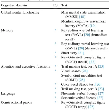
The VMCI-Tuscany neuropsychological battery in-cludes two global cognitive functioning tests and other nine tests which cover a wide range of cognitive abilities (Table 1). ES methodology was available for all the tests included in the battery except for the symbol digit modal-ities test.
Trail making test (TMT, part A and B) administration had a time limit: if patients did not complete the task in 5 minutes, the examiner stopped the administration and scored 300 sec-onds. In this case, raw scores were not adjusted for age and
education while an ES5 0 was assigned. The administration
of TMT-B had two preliminary restrictions: the completion
of the TMT-A in ,300 seconds and the knowledge of the
correct order of the alphabet letters.
Data collected were entered into a database on a
specif-ically developed Web site (www.vmci-tuscany.it). Raw
scores were automatically adjusted for demographic vari-ables using regression equations extracted by normative studies and then transformed into ES.
The diagnosis of MCI according to the Winblad et al.’s criteria [3] requires specific prerequisites: (1) patients or caregivers complaints about cognitive deficits and (2) no or minimal disability in activities of daily living (no
impair-ment at all on activities of daily living scale [28] and no
impairment or only one item compromised on instrumental activities of daily living scale[29];Fig. 1). In our operation-alization of MCI diagnostic algorithm, prerequisites’ defini-tion was maintained and we worked on the definidefini-tion of the objective cognitive impairment and the classification of cognitive domains.
The Winblad et al.’s MCI diagnostic algorithm requires the three following hierarchical steps: (1) definition of objective cognitive impairment; (2) definition of an tive impairment in memory; and (3) definition of an objec-tive cogniobjec-tive impairment in cogniobjec-tive domains other than
memory (Fig. 1). For each of the three steps, we defined:
(1) how much each score had to be below the mean to be considered impaired and (2) how many scores were impaired. We built three possible scenarios: (1) at least
one score borderline (ES5 1; corresponding to our
inclu-sion criterion); (2) at least two scores borderline; and (3) at
least one score frankly impaired (ES 5 0 or an adjusted
score lower than the 5th centile of the normal population;
Fig. 1). To this purpose, we used the 12 scores deriving
from the 9 neuropsychological tests (Table 1): the immedi-ate and delayed recalls of the Rey auditory-verbal learning test were used as two different scores, as well as the copy and the delayed reproduction of the Rey-Osterrieth com-plex figure, and the part A and B of the trail making test. As stated before, ES methodology was not available for the symbol digit modalities test and its performance was classified as “abnormal” when the adjusted score was
below the 5th centile of the normal population (ES 5 0)
or “normal” when the adjusted score was above the 5th centile (no ES was assigned).
An additional issue was the definition of cognitive do-mains. In a previous methodological article on psychometric properties of the VMCI-Tuscany neuropsychological bat-tery, a confirmatory factor analysis showed a good fit of the four theoretically assumed dimensions to empirical
data [14]. Based on those findings, we considered four
cognitive domains: memory (assessed by four cognitive scores), attention/executive functions (five cognitive scores), language (two cognitive scores), and constructional praxis
(one cognitive score; Table 1). In scenario 2, considering
constructional praxis domain, that is assessed in our battery by only one score, we applied the restricted criterion “at least one score impaired.”
The MRI baseline scans were centrally revised at the NEUROFARBA Department, University of Florence. Visual assessment of neuroimaging was performed by an experi-enced neurologist (A.P.) who was blind to clinical details and MCI classification. After the central MRI revision, of the 200 patients enrolled in the baseline VMCI-Tuscany cohort, 47 were excluded because of the evidence of
WMH of only mild degree (modified Fazekas scale 5 1).
The neuroimaging variables used in the present study
were: (1) WMH (modified Fazekas scale)[13], lacunar
in-farcts (total number in the entire brain)[30], global cortical
Table 1
The VMCI-Tuscany neuropsychological battery Cognitive domain ES Test
Global mental functioning Mini mental state examination (MMSE)[18]
Montreal cognitive assessment battery (MoCA)[19]
Memory * Rey auditory-verbal learning test (RAVL)[20](immediate recall)
* Rey auditory-verbal learning test (RAVL)[20](delayed recall)
* Short story[21]
* Rey-Osterrieth complex figure (ROCF) (recall)[22]
Attention and executive functions * Trail making test, part A[23] * Visual search[24]
Symbol digit modalities test (SDMT)[25]
* Color word Stroop test[26] * Trail making test, part B[23]
Language * Phonemic verbal fluency[27] * Semantic verbal fluency[27]
Constructional praxis * Rey-Osterrieth complex figure (ROCF) (copy)[22]
atrophy (Pasquier visual scale) [31], and medial temporal
lobe atrophy (MTA) (Scheltens scale)[32]. Forty randomly
selected scans were scored twice for the determination of the intrarater reliability, which was good (weighted Cohen’s k:
MWH 5 0.91; lacunar infarcts 5 0.82; global cortical
atrophy5 0.62; and MTA 5 0.86).
2.1. Statistical analysis
Correlations across neuropsychological tests (Pearson’s r) and the Cronbach’s a coefficients were used to verify the internal consistency of cognitive domains.
Descriptive statistics were used to show frequency distribu-tions of MCI subtypes across the three scenarios. To show the overlapping of distributions of MCI subtypes in all scenarios,
95% confidence intervals (CI) for percentages were calculated by Wilson score method with a correction for continuity[33]. Descriptive statistics were also used to show means and stan-dard deviations (SD) of mini mental state examination (MMSE) scores for each MCI subgroup, and univariate analysis of vari-ance was applied to verify significant differences in MMSE scores distributions across MCI subgroups within all scenarios. Univariate statistical analyses (Pearson’s c2test) were used to compare single and multiple domain MCI groups in terms of neuroimaging variables (WMH, lacunar in-farcts, global cortical atrophy, and MTA) in the whole sam-ple of patients classified as MCI according to scenario 1. Descriptive statistics were used to verify frequency distri-butions of neuroimaging variables in MCI subtypes in both scenarios 1 and 3 (95% CI for percentages calculated
Fig. 1. Operationalization of the MCI diagnostic algorithm according to three possible scenarios. Abbreviations: MCI, mild cognitive impairment; ADL, Activities of Daily Living; IADL, Instrumental Activities of Dailiy Living.
E. Salvadori et al. / Alzheimer’s & Dementia - (2015) 1-12 4
by Wilson score method with a correction for continuity). For statistical analysis, lacunar infarcts were coded as ab-sent or preab-sent, mean MTA of the bilateral scores was calculated and dichotomized (MTA score 0–2.5, MTA score !3), and global cortical atrophy scores were dichot-omized (global cortical atrophy score 0–2, global cortical atrophy score 3).
3. Results
Of the 153 enrolled patients, 84 (55%) were males, and the mean (6SD) age and years of education were
74.76 6.9 and 7.9 6 4.2, respectively. Mean age and
educa-tion level were not significantly different among MCI sub-types in any of the three scenarios (data not shown). Concerning vascular risk factors distributions, of the 153 pa-tients, 125 (82%) had hypertension, 91 (60%) had hypercho-lesterolemia, 22 (14%) had diabetes, 67 (44%) reported smoking habits, 57 (37%) had history of stroke, and 46 (30%) consumed alcohol.
As shown in Table 2, across neuropsychological tests
of the same cognitive domain, all Pearson’s correlation coefficients resulted statistically significant and Cron-bach’s a were .0.650 showing a good internal
consis-tency of each domain. No measure of internal
consistency could be calculated for the constructional praxis domain (assessed by only the immediate copy of the Rey-Osterrieth complex figure). Nevertheless, this test resulted significantly although moderately correlated with the delayed reproduction of the Rey-Osterrieth
com-plex figure (r5 0.217, P , .01), the TMT-A (r 5 0.201,
P , .05), and the phonemic verbal fluency (r 5 0.162,
P, .05).
Percentage distributions of subjects categorized accord-ing to different ES values for all the 12 cognitive scores used in the operationalization of MCI diagnostic criteria
are shown in the Online Supplemental Table. Percentages
of patients with at least a borderline performance were approximately 50% for all tests included in the memory domain except the short story test that resulted sparsely impaired. In the attention/executive domain, percentages of patients with at least a borderline performance were be-tween 40% and 60% in all tests. The Rey-Osterrieth complex figure resulted the most difficult test for the patients (66% with abnormal performances and 3% with borderline perfor-mances), whereas language tests resulted normal in approx-imately two-third of our sample.
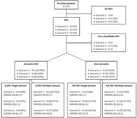
The application of the three scenarios led to the following distributions of MCI subtypes (Fig. 2).
3.1. Scenario 1 (at least one score borderline)
This was the inclusion MCI criteria in our study, and consequently all the 153 enrolled patients were classified as MCI. The A-MCI type prevailed (78%; 95% CI, 70–84) and 86% of patients resulted to be of the multiple domain type (72% A-MCI; 95% CI, 64–79; 14% NA-MCI; 95% CI, 9–21).
3.2. Scenario 2 (at least two scores borderline)
Applying this intermediate criterion, of the 153 enrolled patients, 3 (2%) resulted cognitively normal. For further four MCI patients, we were not able to define the MCI sub-type because they had two scores borderline but only one in the memory domain. All these seven patients fell into the A-MCI group (three single domain and four multiple domain) in scenario 1.
Passing from scenario 1 to scenario 2, of the 153 MCI pa-tients, 119 were classified in the same subtypes, 20 moved from the A-MCI multiple domain group to the other sub-types (11 NA-MCI multiple domain, 7 NA-MCI single domain, and 2 A-MCI single domain), and 7 moved within NA-MCI from multiple to single domain group.
3.3. Scenario 3 (at least one score impaired)
Applying this restricted criterion, of the 153 enrolled pa-tients, 7 (5%) resulted cognitively normal, 59% (95% CI, 50–67) were A-MCI and 73% resulted to be of multiple domain type (53% A-MCI; 95% CI, 44–61; 20% NA-MCI; 95% CI, 14–28).
The distribution of MCI subtypes was almost the same for both the intermediate and restricted criterion. Passing from scenario 2 to scenario 3, of the 146 MCI patients, 9 moved from the A-MCI multiple domain group to other subtypes (4 NA-MCI multiple domain, 4 NA-MCI single domain, and 1 A-MCI single domain).
Table 2
Internal consistency of cognitive domains
Cognitive domain
Memory (Cronbach’s a 5 0.671)
RAVL (immediate) RAVL (delayed) Short story RAVL (delayed) 0.678**
Short story 0.337** 0.352**
ROCF (recall) 0.289** 0.191* 0.225**
Cognitive domain
Attention and executive functions (Cronbach’s a 5 0.761)
TMT-A Visual search SDMT Stroop test Visual search 0.509** SDMT 0.515** 0.388** Stroop test 0.287** 0.362** 0.255** TMT-B 0.553** 0.446** 0.513** 0.415** Language (Cronbach’s a 5 0.651) Semantic fluency Phonemic fluency 0.331**
Abbreviations: RAVL, Rey auditory-verbal learning test; ROCF, Rey-Osterrieth complex figure; TMT-A, trail making test part A; SDMT, symbol digit modalities test; TMT-B, trail making test part B.
*Pearson’s r coefficient significant at P, .05. **Pearson’s r coefficient significant at P, .01.
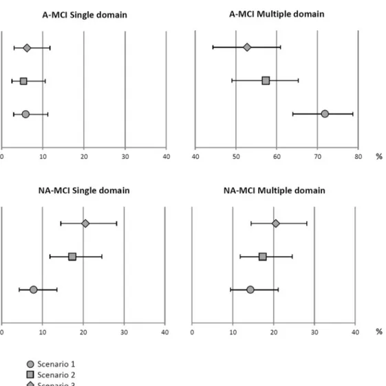
In comparison to scenario 1, applying scenarios 2 and 3 produced a decrease in percentages of multiple domain A-MCI (from 86% to 77% and 73%, respectively) and an increase in percentages of single domain NA-MCI (from 8% to 18% and 20%, respectively). Ninety-five percent CI for percentages of MCI subtypes in all
sce-narios are shown in Fig. 3 using a forest plot. The
95% CI distribution of percentages of diagnoses made according to scenarios 1 and 3 for the subtypes A-MCI multiple domain and NA-MCI single domain were not overlapping.
Mean MMSE scores and SD for each MCI subgroup are shown in Fig. 2. In all scenarios, significant differences in MMSE scores distributions were found across MCI
sub-groups (scenario 1: F 5 5.49, P , .01; scenario 2:
F 5 8.43, P , .01; and scenario 3: F 5 8.04, P , .01).
The mean MMSE scores of A-MCI multiple domain group always resulted lower compared with the other MCI sub-types, and post hoc tests (Bonferroni) showed significant differences between A-MCI multiple domain group and NA-MCI groups, either single or multiple domain, in all sce-narios (data not shown).
3.4. Neuroimaging characterization of single and multiple domain MCI
Of the 153 enrolled patients, 82 (54%) had a severe de-gree of WMH, 103 (67%) at least one lacunar infarct, 28 (18%) a severe degree of global cortical atrophy, and 94 (61%) a mean MTA score !3.
Using Pearson’s c2test, only MTA showed a statistically significant association with multiple domain MCI (68% vs.
38% multiple vs. single domain MCI; c2 5 6.82,
P5 .009). Global cortical atrophy (20% vs. 10% multiple
vs. single domain), WMH (55% vs. 43%), and lacunar in-farcts (68% vs. 67%) were not significantly associated with single or multiple domain MCI.
The 95% CI distribution of percentages of neuroimaging variables were largely overlapping between scenarios 1 and 3 for all MCI subtypes (data not shown). Comparing neuro-imaging variables that could characterize those MCI sub-types whose distributions of diagnoses differed between the scenarios, the A-MCI multiple domain group resulted al-ways in high percentages of both lacunar infarcts (66% vs. 69%, scenario 1 vs. 3) and mean MTA score !3 (70% vs.
Fig. 2. Distributions of MCI subtypes according to three possible scenarios. Definitions of scenarios are as follows: Scenario 1: at least one test borderline; Scenario 2: at least two tests borderline; and Scenario 3: at least one test impaired. Percentages refer to the total number of MCI patients in each scenario. Ab-breviations: MCI, mild cognitive impairment; A-MCI, amnestic mild cognitive impairment; NA-MCI, nonamnestic mild cognitive impairment; MMSE, mini mental state examination.
E. Salvadori et al. / Alzheimer’s & Dementia - (2015) 1-12 6
68%, scenario 1 vs. 3), whereas the NA-MCI single domain group showed high percentages of lacunar infarcts (73% vs. 73%, scenario 1 vs. 3;Fig. 4).
4. Discussion
This study represents the first attempt to assess the effect of the operationalization of MCI consensus criteria in terms of subtypes distribution in a sample of patients with SVD. We found that the application of differently operationalized criteria led to minimal changes in the total number of pa-tients diagnosed as MCI but to more marked differences in the frequency of MCI subtypes. Most of our patients were classified as multiple-domain A-MCI in line with the Win-blad et al.’s hypothesis. However, about one-fifth showed a domain profile. Finally, in comparison with single-domain MCI patients, multiple-single-domain patients showed higher frequency of severe MTA.
Multiple-domain MCI was highly prevalent in our sample across all scenarios and this is in line with the hypothesis that MCI subtypes characterized by impairment in nonmemory
domains, such as executive function and visuospatial skills,
may have a vascular etiology[5,34–36].
The fact that when using more restrictive criteria to diagnose MCI, a certain number of our patients were diagnosed with single NA domain MCI supports the hypothesis that MCI patients with SVD might have spe-cific patterns of cognitive impairments in domains other
than memory [34,35]. This would expand the clinical
spectrum of vascular MCI.
Recent studies have examined empirically derived sub-types of MCI based on patterns of neuropsychological defi-cits in clinic- and community-based samples and most of them had identified homogenous subgroups that were consistent across studies and could reflect a common etiol-ogy (e.g., memory impaired group, multidomain amnestic
group, and dysexecutive group) [1,34,36]. In particular,
Delano-Wood et al.[34] found significantly greater levels
of white matter changes burden on neuroimaging in their empirically derived dysexecutive MCI subgroup, consis-tently with the hypothesis of the association of cerebrovas-cular lesions with this pattern of deficits.
Fig. 3. Ninety-five percent confidence intervals of percentage distributions of MCI subtypes in three scenarios. Abbreviations: MCI, mild cognitive impairment; A-MCI, amnestic mild cognitive impairment; NA-MCI, nonamnestic mild cognitive impairment.
Most of our patients fell in the A-MCI group across different scenarios. This is likely a result of the fact that in Winblad et al.’s criteria for MCI memory deficits are hierarchically prevailing over other cognitive domains in the diagnostic algorithm. As a result, patients with mild memory deficits and severe deficits in other domains are nonetheless classified as amnestic. Taking into account the mentioned aspect and applying the three different sce-narios, we had to decide how to classify those patients with borderline performances in memory domain and frankly abnormal performances in other cognitive do-mains. We decided to classify as A-MCI only those pa-tients who had at least one memory score borderline and no frankly impaired scores in other cognitive
do-mains; otherwise patients were assigned to NA-MCI. For MCI subtyping, it seems advisable to take into ac-count the overall neuropsychological profile of patients without attributing to memory a prominent role. This is in line with the recent proposal of redefinition of VMCI diagnostic criteria which, according to a comprehensive and neuropsychological approach, excludes the prevailing position of memory impairment and gives equal
impor-tance to other cognitive domains[11].
Previous reports are conflicting on the nature and extent
of brain changes associated with MCI subtypes [37].
According to Winblad et al.’s criteria, VMCI should be
characterized by a multidomain profile [5]. However,
be-tween one-sixth and one-fourth of our patients were
Fig. 4. Ninety-five percent confidence intervals of percentage distributions of neuroimaging variables in A-MCI multiple domain and NA-MCI single domain subtypes between scenarios 1 and 3. Abbreviations: WMH, white matter hyperintensities; A-MCI, amnestic mild cognitive impairment; NA-MCI, nonamnestic mild cognitive impairment; GCA, global cortical atrophy; MTA, medial temporal lobe atrophy.
E. Salvadori et al. / Alzheimer’s & Dementia - (2015) 1-12 8
classified as single domain. To test whether this latter group differed in neuroimaging terms from the multido-main group, for example for an overrepresentation of degenerative aspects, we compared MRI findings and found that instead neurodegenerative features, such as MTA, were more prevalent in the multidomain group, particularly in the A-MCI multi domain. On the other hand, the main neuroimaging characteristics emerged in the NA-MCI single domain group was the presence of lacunar infarcts.
Limitations of our study need to be considered. The main limitation is that each cognitive domain included a different number of tests and scores. Theoretically, having more cognitive scores increases the likelihood of finding a deficit in that specific domain. The memory impairment was eval-uated taking into account four cognitive scores, whereas the attention/executive impairment was based on five scores and this difference is likely to influence the decrease in pro-portion of A-MCI, and the resulting increase of NA-MCI, when using more restrictive criteria. Distribution of cogni-tive performances confirmed that attention-execucogni-tive dysfunction was one of the prominent features, but impair-ments in memory and high-level visuoconstructional abili-ties were also observed in our sample despite the lower number of available scores.
Another consequence of different number of tests and scores is that language and constructional praxis impair-ments might have been underestimated in comparison with memory and attention/executive functions deficits. To verify the impact of different number of tests and scores in each cognitive domain on MCI subtypes distributions, we explored also an operationalization based on three cognitive domains: memory and executive functions (as described mentioned), and a third “mixed” cognitive domain that pooled the two language tests and the constructional praxis test. Applying this three-domain strategy, distributions of MCI subtypes according to three possible scenarios were basically the same of our original analysis. In all scenarios, only one patient, classified as NA-MCI multiple domain in the four-domain analysis, moved to the NA-MCI single domain group in the three-domain analysis. Also, 95% CI for percentages of MCI subtypes in all scenarios remained the same. Furthermore, our results showed a good internal consistency of cognitive domains in the four-domain approach. We therefore decided to use the model confirmed also in the previous methodological article on the psycho-metric properties of the VMCI-Tuscany neuropsychological battery[14].
A second limitation is the use of the number of impaired cognitive scores, as opposed to that of an overall summary score, for the determination of cognitive impairment. A recent study found that summary scores, such as averaging of z-scores and item response theory score, provided a more accurate determination of the prevalence of cognitive impairment in a very large sample of 461 patients and 724
controls [38]. Although the use of the number of impaired
cognitive scores has been demonstrated to be less sensitive than summary scores, the relatively small sample in our study did not allow the use of such sophisticated methods; that, however, will have to be implemented in further studies on the optimization of operationalization of criterion of mild cognitive impairment.
A third limitation is that the multiple-domain MCI group was notably larger than the single-domain MCI group and this reduced the statistical power of comparative analyses.
Another possible limitation may be the lack of cerebro-spinal fluid biomarkers and positron emission tomography assessments of markers of Alzheimer disease to better define the etiology of our sample. This, however, reflects the current situation in most centers. On the other hand, the lack of an association between cerebrovascular burden and MCI subtypes may be due also to the quantification of WMH according to a visual rating scale, rather than a more objective and metric methodology. Therefore, we cannot be completely sure that our sample was composed of patients with pure vascular MCI. Yet, this patient sam-ple likely represents what is encountered in clinical prac-tice. At the end of the ongoing follow-up, data will be available concerning the incidence of dementia and its subtypes and their possible association with baseline neu-ropsychological patterns of deficits. Finally, it is impor-tant to note that our results and conclusions refer to a sample of patients with MCI and SVD and not to the global MCI population.
Cognitive profiling of MCI subtypes is important from the clinical, research, and epidemiologic points of view. In this sense, the hierarchical approach used in the Winblad et al.’s criteria, based on the presence or absence of memory deficits, could not be optimal to identify other specific pat-terns of cognitive impairment, particularly in patients with cerebrovascular diseases thought to have domains other than memory mainly affected. A more comprehensive eval-uation of the cognitive profile, based on several hierarchical-ly equivalent cognitive domains, should guide the classification, and future studies in this regard are warranted. Acknowledgment
The authors thank Dr David Libon (Department of Neurology, Drexel University College of Medicine, Phila-delphia, PA) for useful comments and criticism on this article.
The VMCI-Tuscany study is funded by Tuscany region. E.S. is currently supported by a project funded by Tuscany region and Health Ministry (Bando Ricerca Finalizzata 2010, grant
number: RF-2010-2321706, ClinicalTrials.gov identifier:
NCT02033850, PI L.P.). Supplementary data
Supplementary data related to this article can be found at
RESEARCH IN CONTEXT
1. Systematic review: The authors reviewed the litera-ture using standard databases (e.g., PubMed). The topic is in expansion, and relevant articles related to the debate on best criteria for MCI diagnosis and their operationalization are appropriately cited. 2. Interpretation: Our findings suggest that the
hierar-chical approach used in current MCI criteria could not be optimal to identify specific patterns of cogni-tive impairment in patients with cerebrovascular dis-eases who have domains other than memory mainly affected.
3. Future directions: Our study provides a framework for further studies on operationalization of criteria for MCI in patients with cerebrovascular disease: (1) studies on MCI diagnostic criteria based on a comprehensive cognitive evaluation without a hierar-chical approach; (2) studies on comparisons between clinically and empirically derived subtypes of MCI based on patterns of neuropsychological deficits; and (3) studies on the reliability of different MCI diagnostic approaches on the progression to demen-tia and its subtypes.
References
[1] Bondi MW, Smith GE. Mild cognitive impairment: A concept and diagnostic entity in need of input from neuropsychology. J Int Neuro-psychol Soc 2014;20:129–34.
[2] Christa Maree Stephan B, Minett T, Pagett E, Siervo M, Brayne C, McKeith IG. Diagnosing mild cognitive impairment (MCI) in clinical trials: A systematic review. BMJ Open 2013;3.
[3] Winblad B, Palmer K, Kivipelto M, Jelic V, Fratiglioni L, Wahlund LO, et al. Mild cognitive impairment—Beyond contro-versies, towards a consensus: Report of the International Working Group on mild cognitive impairment. J Intern Med 2004;256:240–6. [4] Petersen RC, Doody R, Kurz A, Mohs RC, Morris JC, Rabins PV, et al.
Current concepts in mild cognitive impairment. Arch Neurol 2001; 58:1985–92.
[5] Gauthier S, Reisberg B, Zaudig M, Petersen RC, Ritchie K, Broich K, et al. Mild cognitive impairment. Lancet 2006;367:1262–70. [6] O’Brien JT, Erkinjuntti T, Reisberg B, Roman G, Sawada T, Pantoni L,
et al. Vascular cognitive impairment. Lancet Neurol 2003;2:89–98. [7] Pantoni L, Poggesi A, Inzitari D. The relation between white-matter
lesions and cognition. Curr Opin Neurol 2007;20:390–7.
[8] Sachdev PS, Brodaty H, Looi JC. Vascular dementia: Diagnosis, man-agement and possible prevention. Med J Aust 1999;170:81–5. [9] Lamar M, Price CC, Giovannetti T, Swenson R, Libon DJ. The
dysex-ecutive syndrome associated with ischaemic vascular disease and related subcortical neuropathology: A Boston process approach. Be-hav Neurol 2010;22:53–62.
[10] Pantoni L. Cerebral small vessel disease: From pathogenesis and clin-ical characteristics to therapeutic challenges. Lancet Neurol 2010; 9:689–701.
[11] Sachdev P, Kalaria R, O’Brien J, Skoog I, Alladi S, Black SE, et al. Diagnostic criteria for vascular cognitive disorders: A VASCOG state-ment. Alzheimer Dis Assoc Disord 2014;28:206–18.
[12] Poggesi A, Salvadori E, Pantoni L, Pracucci G, Cesari F, Chiti A, et al. Risk and determinants of dementia in patients with mild cognitive impairment and brain subcortical vascular changes: A study of clinical, neuroimaging, and biological markers—The VMCI-Tuscany study: Rationale, design, and methodology. Int J Alzheimers Dis 2012;608013. [13] Pantoni L, Basile AM, Pracucci G, Asplund K, Bogousslavsky J, Chabriat H, et al. Impact of age-related cerebral white matter changes on the transition to disability—The LADIS study: Rationale, design and methodology. Neuroepidemiology 2005;24:51–62.
[14] Salvadori E, Poggesi A, Pracucci G, Inzitari D, Pantoni L. Develop-ment and psychometric properties of a neuropsychological battery for mild cognitive impairment with small vessel disease: The VMCI-Tuscany study. J Alzheimers Dis 2014;43:1313–23.
[15] Hachinski V, Iadecola C, Petersen RC, Breteler MM, Nyenhuis DL, Black SE, et al. National Institute of Neurological Disorders and Stroke-Canadian Stroke Network vascular cognitive impairment harmonization standards. Stroke 2006;37:2220–41.
[16] Bianchi A, Dai Pr!a M. Twenty years after Spinnler and Tognoni: New instruments in the Italian neuropsychologist’s toolbox. Neurol Sci 2008;29:209–17.
[17] Capitani E, Laiacona M. Composite neuropsychological batteries and demographic correction: Standardization based on equivalent scores, with a review of published data. The Italian Group for the Neuropsycho-logical Study of Ageing. J Clin Exp Neuropsychol 1997;19:795–809. [18] Measso G, Cavarzeran F, Zappala G, Lebowitz B, Crook TC,
Pirozzolo FJ, et al. The Mini Examination: Normative study of an Ital-ian random sample. Dev Neuropsychol 1993;9:77–85.
[19] Nasreddine ZS, Phillips NA, B"edirian V, Charbonneau S, Whitehead V, Collin I, et al. The Montreal Cognitive Assessment, MoCA: A brief screening tool for mild cognitive impairment. J Am Geriatr Soc 2005;53:695–9.
[20] Carlesimo GA, Caltagirone C, Gainotti G. The mental deterioration battery: Normative data, diagnostic reliability and qualitative analyses of cognitive impairment. The group for the standardization of the mental deterioration battery. Eur Neurol 1996;36:378–84.
[21] Novelli G, Papagno C, Capitani E, Laiacona M, Cappa SF, Vallar G. Tre test clinici di memoria a lungo termine. Archivio di Psicologia. Neurologia e Psichiatria 1986;47:278–96.
[22] Caffarra P, Vezzadini G, Dieci F, Zonato F, Venneri A. Rey-Osterrieth complex figure: Normative values in an Italian population sample. Neurol Sci 2002;22:443–7.
[23] Giovagnoli AR, Del Pesce M, Mascheroni S, Simoncelli M, Laiacona M, Capitani E. Trail making test: Normative values from 287 normal adult controls. Ital J Neurol Sci 1996;17:305–9. [24] Della Sala S, Laiacona M, Spinnler H, Ubezio C. A cancellation test:
Its reliability in assessing attentional deficit in Alzheimer’s disease. Psychol Med 1992;22:885–901.
[25] Nocentini U, Giordano A, Di Vincenzo S, Panella M, Pasqualetti P. The Symbol digit modalities test—Oral version: Italian normative data. Funct Neurol 2006;21:93–6.
[26] Caffarra P, Vezzadini G, Dieci F, Zonato F, Venneri A. Una versione abbreviata del test di Stroop. Dati normativi nella popolazione italiana. Nuova Rivista di Neurologia 2002;12:111–5.
[27] Novelli G, Papagno C, Capitani E, Laiacona M, Vallar G. Tre test clin-ici di ricerca e produzione lessicale. Taratura su soggetti normali. Ar-chivio di Psicologia, Neurologia e Psichiatria 1986;47:477–506. [28] Katz S, Ford AB, Moskowitz RW, Jackson BA, Jaffe MW. Studies of
illness in the aged. The index of ADL: A standardized measure of bio-logical and psychosocial function. JAMA 1963;185:914–9. [29] Lawton MP, Brody EM. Assessment of older people: Self-maintaining
and instrumental activities of daily living. Gerontologist 1969; 9:179–86.
[30] Gouw AA, van der Flier WM, Fazekas F, van Straaten EC, Pantoni L, Poggesi A, et al. Progression of white matter hyperintensities and
E. Salvadori et al. / Alzheimer’s & Dementia - (2015) 1-12 10
incidence of new lacunes over a 3-year period: The leukoaraiosis and disability study. Stroke 2008;39:1414–20.
[31] Pasquier F, Leys D, Weerts JG, Mounier-Vehier F, Barkhof F, Scheltens P. Inter- and intraobserver reproducibility of cerebral atro-phy assessment on MRI scans with hemispheric infarcts. Eur Neurol 1996;36:268–72.
[32] Scheltens P, Leys D, Barkhof F, Huglo D, Weinstein HC, Vermersch P, et al. Atrophy of medial temporal lobes on MRI in “probable” Alz-heimer’s disease and normal ageing: Diagnostic value and neuropsy-chological correlates. J Neurol Neurosurg Psychiatry 1992;55:967–72. [33] Newcombe R. Two-sided confidence intervals for the single
propor-tion: Comparison of seven methods. Stat Med 1998;17:857–72. [34] Delano-Wood L, Bondi MW, Sacco J, Abeles N, Jak AJ, Libon DJ,
et al. Heterogeneity in mild cognitive impairment: Differences in neu-ropsychological profile and associated white matter lesion pathology. J Int Neuropsychol Soc 2009;15:906–14.
[35] Rasquin SM, Lodder J, Visser PJ, Lousberg R, Verhey FR. Predictive accuracy of MCI subtypes for Alzheimer’s disease and vascular de-mentia in subjects with mild cognitive impairment: A 2-year follow-up study. Dement Geriatr Cogn Disord 2005;19:113–9.
[36] Clark LR, Delano-Wood L, Libon DJ, McDonald CR, Nation DA, Bangen KJ, et al. Are empirically-derived subtypes of mild cognitive impairment consistent with conventional subtypes? J Int Neuropsychol Soc 2013;19:635–45.
[37] Jak AJ, Bangen KJ, Wierenga CE, Delano-Wood L, Corey-Bloom J, Bondi MW. Contributions of neuropsychology and neuroimaging to understanding clinical subtypes of mild cognitive impairment. Int Rev Neurobiol 2009;84:81–103.
[38] Godefroy O, Gibbons L, Diouf M, Nyenhuis D, Roussel M, Black S, et al. Validation of an integrated method for determining cognitive ability: Implications for routine assessments and clinical trials. Cortex 2014;54:51–62.
Appendix. List of participating centers and personnel in the VMCI-Tuscany
University of Florence: (coordinating center): Domenico Inzitari (study coordinator), Rosanna Abbate, Maria Boddi, Francesca Cesari, Laura Ciolli, Mirella Coppo, Alessandra Del Bene, Stefano Diciotti (currently at University of Bologna), Andrea Ginestroni, Betti Giusti, Anna Maria Gori, Sandro Marini, Mario Mascalchi, Serena Nannucci, Leonardo Pantoni, Marco Pasi, Francesca Pescini, Anna Poggesi, Giovanni Pracucci, Emilia Salvadori, and Raffaella Valenti.
University of Pisa: Ubaldo Bonuccelli, Paolo Cecchi, Alberto Chiti, Mirco Cosottini, Giovanni Orlandi, Cristina Pagni, Gabriele Siciliano, and Gloria Tognoni.
University of Siena: Antonio Federico, Nicola De Stefano, Ilaria Di Donato, Maria Teresa Dotti, Patrizia Formichi, Claudia Gambetti, Antonio Giorgio, Francesca Rossi, Laura Stromillo, and Enza Zicari.
Tuscany region: Arezzo (Paolo Zolo, Alessandro Tiezzi); Empoli (Elisabetta Bertini, Stefania Brotini, Leonello Guidi, Maria Lombardi, Stefania Mugnai, Antonella Notarelli); Florence (Laura Bracco, Massimo Cadelo, Renzo Cisbani, Luciano Gabbani, Guido Gori, Lorella Lambertucci, Luca Massacesi, Enrico Mossello, Marco Paganini, Maristella Piccininni, Francesco Pinto, Claudia Pozzi, Sandro Sorbi, Gaetano Zaccara); Grosseto (Tiziano Borgogni, Mario Man-cuso, Roberto Marconi); Lucca (Monica Mazzoni, Marco Vista); Livorno (Giuseppe Meucci, Giovanna Bellini); Massa Carrara (Luciano Gabrielli); Pisa (Cristina Frittelli, Renato Galli, Gianna Gambaccini); Pistoia (Stefano Barto-lini, Carlo Biagini, Veronica Caleri, Paola Vanni); Prato (Donatella Calvani, Carla Giorgi, Stefano Magnolfi, Pas-quale Palumbo, Carlo Valente); Siena (Alessandro Rossi, Rossana Tassi, Stefania Boschi); and Viareggio (Filippo Baldacci).
E. Salvadori et al. / Alzheimer’s & Dementia - (2015) 1-12 12