R E S E A R C H
Open Access
The extremely halotolerant black yeast
Hortaea werneckii - a model for intraspecific
hybridization in clonal fungi
Polona Zalar
1*, Jerneja Zupan
čič
1, Cene Gostin
čar
1,2, Janja Zajc
3, G. Sybren de Hoog
4,5, Filomena De Leo
6,
Armando Azua-Bustos
7,8and Nina Gunde-Cimerman
1Abstract
The polymorphic black yeast Hortaea werneckii (Capnodiales, Ascomycota) is extremely halotolerant (growth from 0 to 30% [w/v] NaCl) and has been extensively studied as a model for halotolerance in Eukaryotes for over two decades. Its most frequent sources are hypersaline environments and adjacent sea-water habitats in temperate, subtropical and tropical climates. Although typically saprobic, H. werneckii can also act as a commensal coloniser on human skin, causing tinea nigra on hands and soles. Here, we report that addition of NaCl to culture media expands the growth range of H. werneckii to 37 °C, which explains its colonisation of human skin, with its increased salinity. The morphological and physiological plasticity/ versatility of H. werneckii indicate that a species complex might be involved. This was investigated in this polyphasic taxonomic analysis based on the global diversity of H. werneckii strains collected from hypersaline environments, and from humans and animals. Analysis of D1/ D2domains of 28S and internal transcribed spacer rDNA revealed 10 and 17 genotypes, respectively, that were not always compliant. The genotypes have global distributions. Human and environmental strains with the same genotypes are intermingled. Due to the limited number of phylogenetically informative characters in the ribosomal DNA dataset, the partial genes encoding for β-tubulin (BTB) and mini-chromosome maintenance protein (MCM7) were also sequenced. The use of these genes was hampered by ambiguous sequences obtained by Sanger sequencing, as a consequence of the diploid and highly heterozygous genome of many H. werneckii strains. Analysis of the BTB and MCM7 genes showed that in some cases two copies of the gene from the same genome are positioned in distant phylogenetic clusters of the intraspecific gene tree. Analysis of whole-genome sequences of selected H. werneckii strains generally confirmed the phylogenetic distances estimated on the basis of ribosomal genes, but also showed substantial reticulation within the phylogenetic history of the strains. This is in line with the hypothesis that the diploid genomes of H. werneckii were formed by hybridizations, which have sometimes occurred between relatively divergent strains.
Keywords: Morphology, Clustering, NaCl tolerant enyzmes, ITS, D1/D2 rDNA, MCM7, BTB, Genomes INTRODUCTION
The ascomycetous black yeast Hortaea werneckii (Capno-diales, Teratosphaeriaceae) is the most extremely halotoler-ant fungus known, and as such, it has become an importhalotoler-ant model organism for the study of halotolerance in Eukarya (Plemenitaš et al.2008,2014, Gunde-Cimerman et al.2018). Its primary ecological niche are natural hypersaline
environments worldwide. It has been isolated from thalasso-haline brines in salterns on three continents, with numerous locations ranging from temperate (Mediterra-nean) (Gunde-Cimerman et al. 2000, Cabañes et al. 2012, Gunde-Cimerman & Zalar2014, Elsayed et al.2016) to sub-tropical and sub-tropical (Mexico, Puerto Rico) (Díaz-Muñoz & Montalvo-Rodríguez 2005, Cantrell et al. 2006) climate zones, although never in subpolar and polar regions. Hor-taea werneckii is more frequently found in eutrophic thalasso-haline waters at moderate temperatures, while it is not known to occur in oligotrophic or © The Author(s). 2019 Open Access This article is distributed under the terms of the Creative Commons Attribution 4.0
International License (http://creativecommons.org/licenses/by/4.0/), which permits unrestricted use, distribution, and reproduction in any medium, provided you give appropriate credit to the original author(s) and the source, provide a link to the Creative Commons license, and indicate if changes were made. The Creative Commons Public Domain Dedication waiver (http://creativecommons.org/publicdomain/zero/1.0/) applies to the data made available in this article, unless otherwise stated.
* Correspondence:[email protected]
1Department of Biology, Biotechnical Faculty, University of Ljubljana, Večna
pot 111, SI-1000 Ljubljana, Slovenia
athalasso-haline hypersaline waters, and only rarely in hypersaline waters with elevated temperatures (Plemenitaš & Gunde-Cimerman2005). Occasionally, H. werneckii has been found in seawater (Iwatsu & Udagawa 1988), on rocks adjacent to seawater (Ruibal et al.2009), and inhabit-ing sea sponges (Brauers et al.2001), corals (Amend et al. 2012, Xu et al. 2014), marine fish (Todaro et al. 1983), salted freshwater fish (Mok et al. 1981), beach soil (de Hoog & Guého 1998), saltern microbial mats (Cantrell et al. 2006), and salt marsh plants (Formoso et al. 2015), and as an endophyte in mangrove plants (Chen et al. 2012). Recently, it was discovered in an indoor dust sample collected in Hawaii (Humphries et al.2017). In agreement with the extremotolerant nature, it was isolated from 2500 m in depth in the Mediterranean Sea (De Leo et al.2018), from sediments 5000 m below sea level in the Central Indian Basin (Singh et al.2012), and in sediments 4000 m below sea level in the East India Ocean (Zhang et al.2014). Using internal transcribed spacer (ITS) clone libraries, H. werneckiioperational taxonomic units were detected in the South China Sea in methane-hydrate-bearing deep-sea sediments (Lai et al.2007). In spite of the wide distribution in marine environments, brines in solar salterns are con-sidered the primary ecological niche, where at salinities above 20% (w/v) NaCl, H. werneckii represents from 70 to 80% of all fungal isolates (Gunde-Cimerman et al. 2000; Butinar et al.2005), with densities of up to 1400 CFU/L.
Hortaea werneckii is the only fungus that can grow across almost the whole range of NaCl concentrations, from 0 to 30% NaCl, with a broad optimum between 6 and 14% NaCl (Butinar et al. 2005). As well as NaCl, H. werneckii can also tolerate up to 2 M concentrations of the chaotropic salt MgCl2, and up to 1.7 M CaCl2, and it
has been isolated from bitterns, i.e. MgCl2-rich leftover
brines after harvesting of NaCl (Zajc et al. 2014). The species also grows on osmotic media with 10% NaCl with 12% glucose (e.g. malt extract, yeast extract, 10% NaCl, 12% glucose agar) and with 50% glucose (e.g., malt extract, yeast extract, 50% glucose agar) (Gunde-Cimer-man et al.2000, Humphries et al.2017).
Until 2000, H. werneckii was primarily known as the aetiological agent of a skin disorder known as tinea nigra, a superficial infection of the human hands and feet that can occur in warmer climates (de Hoog et al. 2000, Perez et al. 2005, Bonifaz et al. 2008). Infections have been associated with superficially abraded skin ex-posed to hypersaline tidal waters (de Hoog & Gerrits van der Ende 1992) in combination with hyperhydrosis (Bonifaz et al.2008, Paneque Rodríguez et al.2015). The species has been associated with animal bites (Rossetto et al. 2014). The pathogenic potential of H. werneckii has, however, remained unclear, as it was generally con-sidered that the species could not grow at 37 °C (de Hoog et al.2000, Ng et al.2005).
Under extremely saline conditions, H. werneckii has an extremophilic ecotype, which is characterised by en-hanced melanization, changes in cell size and meristem-atic growth, and changes in colony appearance (Kogej et al. 2007). At the molecular level, the responses of H. werneckii to high concentrations of toxic sodium ions have involved changes in the expression of genes associ-ated with the synthesis of compatible solutes, regulation of intracellular alkali-metal cation concentrations, modi-fications of cell membrane lipid composition, and changes in cell-wall ultrastructure and morphology (Ple-menitaš et al. 2014). Due to these adaptations, H. wer-neckiihas an outstanding ability to overcome the turgor loss and sodium toxicity that are typical of hypersaline environments (Gostinčar et al.2011).
Genome sequencing of the H. werneckii strain EXF-2000 (Lenassi et al.2013, Sinha et al.2017) has re-vealed that with almost 50 Mb, the H. werneckii genome is larger than the genomes of other sequenced represen-tatives of Capnodiales. The H. werneckii genome con-tained close to 16,000 genes, of which 90% were present in two copies, with an average of 5% amino-acid se-quence divergence between the predicted proteins. Genomic data fit well with the molecular studies on in-dividual genes performed earlier, as all of the genes stud-ied to this point were duplicated and coded for two functionally identical proteins (Vaupotič & Plemenitaš 2007, Vaupotič et al.2008).
Recent analyses of the whole genome sequences of 11 additional strains revealed that two were haploid and the remaining nine were diploid. Differences between the subgenomes of individual strains and their detailed phylogenetic analyses have shown that the diploid ge-nomes were most likely produced by hybridization be-tween relatively divergent strains (Gostinčar et al.2018). At the same time, apart from identification of a hetero-thallic mating locus, the comparative genomic analyses have failed to find any traits that can be linked to sexual reproduction.
In summary, the global presence of H. werneckii in hypersaline environments, its role as the most important model for halotolerance in Eukarya, its extensive physio-logical plasticity and adaptability, the availability of numerous strains collected from various osmotic environ-ments, and its particular combination of hybridization with an exclusively or primarily clonal reproduction mode (as revealed by comparative genomics) make H. werneckii an attractive model organism.
The present study compared 98 strains, of which a se-lection was subjected to classical taxonomic analyses (macro-, microscopy, assimilation tests), and was tested for halotolerance and thermotolerance and the produc-tion of selected pathogenicity-related enzymes (prote-ases, esterases), both with and without the addition of
salt. Taxonomic analyses have been supported by com-parative analyses of the most frequently used barcodes, ITS and D1/D2 28S rDNA. Indeed, apart from the ribo-somal regions, almost all of the other genes were present in two different copies per strain, which prevented clas-sical taxonomic phylogenetic approaches based on ana-lyses of household genes. Therefore, in addition to ambiguous Sanger sequences and adjusted algorithms to work with such sequences, sequences derived from whole-genome sequencing of 11 strains were also con-sidered. The taxonomic study presented was aimed at definition of the variability in H. werneckii, the descrip-tion of potentially new species, and the definidescrip-tion of ge-notypes that might correlate to particular ecological habitats.
METHODS
This study presents a taxonomic analysis of 98 strains (Table 1) that were mostly isolated from brine or bit-terns of three Mediterranean salbit-terns (47 strains), adja-cent sea and deep sea environments (10 strains), and air above hypersaline ponds (1). Five strains were also in-cluded from patients living in the Mediterranean area (i.e., Italy, Portugal, France), as well as strains from ani-mals (7) and plants (3). One strain was food-borne (it formed a black biofilm on cheese brine), one origi-nated from the air in Japan, and one was of unknown source. Twenty-two strains originated from a coastal cave located in the Coastal Range of the Atacama Desert (Chile). These strains were isolated from spider webs at-tached to the cave wall, from a newly described species of microalga (Dunaliella atacamensis; Azúa-Bustos et al. 2010), and directly from the cave walls and from sand underneath the spider webs.
Isolation of strains and sampling sites
The Hortaea werneckii strains studied are listed in Table 1, and were collected over a 20-year period. They are maintained in the Microbial Culture Collection Ex of the Infrastructural Centre Mycosmo, MRIC UL, Slovenia (http://www.ex-genebank.com), in the Department of Biology, Biotechnical Faculty, University of Ljubljana (Slovenia). Most of these strains originated from brine and bitterns of the Sečovlje seasonal solar salterns on the northern Adriatic coast (Zalar et al. 1999, Gunde-Cimerman et al.2000, Butinar et al.2005), at the border between Slovenia and Croatia (sub-Mediterranean cli-mate). Brines from salterns in Spain (i.e. Santa Pola and Ebro River Delta) were sampled in 1999 and 2000, and in Namibia (i.e. Skeleton coast) in 2003. Isolations were performed as described in Gunde-Cimerman et al. (2000), by filtration of water and placing the filters on hypersaline culture media. Strains from bitterns were isolated with additional enrichment in half-strength
liquid media: malt extract (pH 7, 3.5), yeast nitrogen base (YNB), and corn steep liquid supplemented with 50 mg/L chloramphenicol. Enrichments were carried out in 500 mL Erlenmeyer flasks with 50 mL medium and 50 mL original bittern. After 7 d incubation on a rotary shaker at 25 °C at 180 rpm, 100μL of the broth was cul-tured on DG18, malt extract agar (MEA) + 3 M NaCl and MEA + 1.5 M MgCl2.
The strains from Puerto Rico salterns originate from the study of Cantrell et al. (2006). Seawater and deep sea strains were collected in 2013 during the DEEP-PRES-SURE Cruise on board the R/V Urania (Smedile et al. 2015), from water columns 25–2500 m in depth, sam-pled at four stations in the Mediterranean Sea: Station Vector in the Tyrrhenian Sea (39°32′00.6″N 13°22′28.5″ E, Station KM3 in the Ionian Sea (36°30′61.3″N 15°40′ 59.9″E), and above the anoxic hypersaline lakes L’Ata-lante (35°18′92″N 21°23′92″E) and Medee (34°24′00″N 22°26′99″E), in the central and south Mediterranean Sea, respectively (de Leo et al.2018). Strains from corals in the Pacific Ocean (China) were isolated by Luo et al. (2017). A large number of strains were collected in 2009 and 2017 by Azúa-Bustos from soil, sand and spider webs collected inside Huanillos cave, which is located under an ancient guano deposit that is rich in ammo-nium nitrate, urate and phosphates. This cave is in the Coastal Range hills of the Atacama Desert, south of the city of Iquique, Chile (Azúa-Bustos et al. 2010), as also described by Martinelli et al. (2017).
All of the strains were single colony isolates after plate streaking. All of the human-, animal-, and plant-related strains were obtained from the Westerdijk Institute for Fungal Biodiversity culture collection (CBS; http://www. westerdijkinstitute.nl/Collections/).
DNA extraction and sequencing
DNA extraction was performed according to van den Ende & de Hoog (1999). About 50 ng template DNA was used in the PCRs. The complete ITS and the D1/D2 domain of 28S rDNA were amplified with the primers ITS4 and ITS5 (White et al. 1990) or NL1 and NL4 (O’Donnell 2000). A fragment of the β-tubulin gene (BTB) was amplified with primers BT2a and BT2b (Glass & Donaldson 1995), and a fragment of DNA replication licensing factor required for DNA replication initiation and cell proliferation (MCM7) was amplified with primers Mcm7-709for and Mcm7-1348rev (Schmitt et al. 2009). The same primers were also used in the se-quencing reactions. Sequences were generated through Sanger sequencing (16 ABI 3730xl) using a commercial provider (Microsynth, Wien, Austria). The sequence chromatograms were manually checked for quality using the FinchTV software (Geospiza, Los Altos, CA) and assembled and edited using SeqMan 3.61 (DNAStar,
Table 1 Hortaea werneckii strains treated in this study
Strain no. ORIGIN (sample, collection date, location) GenBank acc. No.
EXF- Other collections Genotype
(28S/ITS)
D1/D2 28S rDNA ITS rDNA Hypersaline brine and bittern in salterns
9 brine, 11/1998, Salinas de la Trinitat (Ebre Delta), Spain D/8 MH327587 MH327684
64 brine, 7/1998, Salinas de la Trinitat (Ebre Delta), Spain B/7 MH327569 MH327666
96 brine, 11/1998, Salinas de la Trinitat, Spain D/8 MH327585 MH327682
120 brine, pond 3, 11/1998, Salinas Santa Pola, Spain B/7 MH327575 MH327672
132 brine, pond 5, 11/1998, Salinas Santa Pola, Spain B/7 MH327576 MH327673
152 brine, pond 1, 9/1996, Sečovlje salterns, Slovenia B/7 MH327577 MH327674
153 brine, pond 1, 5/1996, Sečovlje, Slovenia B/7 MH327597 MH327694
154 brine, pond 1, 9/1996, Sečovlje, Slovenia B/7 MH327581 MH327678
225 brine, pond 1, 8/1996, Sečovlje, Slovenia B/7 MH327568 MH327665
241 brine, pond 1, 6/1996, Sečovlje, Slovenia A/1 MH327539 MH327636
247 brine, pond 1, 9/1996, Sečovlje, Slovenia A/1 MH327541 MH327638
269 brine, 11/1998, Santa Pola, Spain B/7 MH327579 MH327676
489 brine, pond 3, 11/1998, Salinas de la Trinitat, Spain C/9 MH327592 MH327689
537 brine, pond 1, 8/1996, Sečovlje, Slovenia B/7 MH327571 MH327668
554 brine, pond 1, 8/1996, Sečovlje, Slovenia A/1 MH327532 MH327629
561 brine, 3/2000, Salterns at the Skeleton coast (Atlantic), Namibia J/13 MH327618 MH327626
562 brine, 3/2000, Salterns at the Skeleton coast (Atlantic), Namibia E/4 MH327563 MH327660
631 brine, 11/1998, Santa Pola, Spain D/8 MH327586 MH327683
647 brine, 11/1998, Santa Pola, Spain D/7 MH327564 MH327661
2516 brine, 12/2004, Candelaria, Puerto Rico I/11 MH327546 MH327643
2685 CBS 100456 brine, 1998, Sečovlje, Slovenia B/7 MH327590 MH327687
2782 brine, pond 4, 8/1996, Sečovlje, Slovenia B/7 MH327598 MH327695
2783 brine, pond 2, 8/1996, Sečovlje, Slovenia D/8 MH327596 MH327693
2785 brine, pond 1, 5/1996, Sečovlje, Slovenia C/9 MH327593 MH327690
2787 immersed wood in brine, pond 1, 1/1997, Sečovlje, Slovenia B/7 MH327580 MH327677
2788 brine, pond 2, 7/1996, Sečovlje, Slovenia D/8 MH327584 MH327681
3506 PR 1105-73 brine, Candelaria, Puerto Rico E/5 MH327550 MH327647
3846 PR08-Candel.12 brine, 3/2008, Candelaria, Puerto Rico A/16 MH327545 MH327642
4642 brine, pond 1, 9/1996, Sečovlje, Slovenia B/7 MH327572 MH327669
4662 brine, pond 1, 9/1996, Sečovlje, Slovenia B/7 MH327573 MH327670
4667 brine, pond 1, 7/1996, Sečovlje, Slovenia B/7 MH327574 MH327671
4717 brine, pond 1, 8/1996, Sečovlje, Slovenia B/10 MH327591 MH327688
7620 brine, reservoir, 12/2009, Sečovlje, Slovenia B/7 MH327578 MH327675
7637 brine, reservoir, 12/2009, Sečovlje, Slovenia B/7 MH327566 MH327663
7638 brine, reservoir 12/2009, Sečovlje, Slovenia B/7 MH327565 MH327662
10304 brine, 11/2015, Sečovlje salterns, Slovenia B/7 MH327600 MH327698
10813 bittern, 11/2016, Sečovlje salterns, Slovenia B/7 MH327601 MH327699
10820 bittern, 11/2016, Sečovlje salterns, Slovenia B/7 MH327582 MH327679
10828 brine, 11/2016, Sečovlje salterns, Slovenia A/1 MH327602 MH327700
10830 brine, 11/2016, Sečovlje salterns, Slovenia D/8 MH327588 MH327685
Table 1 Hortaea werneckii strains treated in this study (Continued)
Strain no. ORIGIN (sample, collection date, location) GenBank acc. No.
EXF- Other collections Genotype
(28S/ITS)
D1/D2 28S rDNA ITS rDNA
10834 brine, 11/2016, Sečovlje salterns, Slovenia B/7 MH327604 MH327702
10842 brine, 11/2016, Sečovlje salterns, Slovenia C/9 MH327595 MH327692
10843 brine, 11/2016, Sečovlje salterns, Slovenia B/9 MH327605 MH327703
10957 bittern, 11/2016, Sečovlje salterns, Slovenia B/7 MH327607 MH327705
10974 brine, 11/2016, Sečovlje salterns, Slovenia C/9 MH327608 MH327706
10975 brine, 11/2016, Sečovlje salterns, Slovenia C/9 MH327609 MH327707
Sea water and related habitats
166 CBS 100496 sea water-sprayed marble, Delos, Greece A/1 MH327538 MH327635
2684 sea water, 1998, Slovenia C/9 MH327594 MH327691
2686 CBS 373.92 beach sand, 1992, La Palma, Spain H/1 MH327530 MH327627
10508 AT 25f Mediterranean sea, depth 25 m, 12/2013,“Atalante” station E/4 MH327555 MH327652
10509 KM3 200r Mediterranean sea, depth 200 m, 12/2013,“KM3” station E/4 MH327556 MH327653
10510 M 94 Mediterranean sea, depth 94 m, 12/2013,“Medee” station E/4 MH327557 MH327654
10511 V 25 a Mediterranean sea, depth 25 m, 12/2013,“Vector” station E/4 MH327558 MH327655
10512 V 25c Mediterranean sea, depth 25 m, 12/2013,“Vector” station E/4 MH327559 MH327656
10513 V 2500b Mediterranean sea, depth 2 500 m, 12/2013,“Vector” station E/4 MH327560 MH327657
10853 no. 445345 beach sand, Portugal B/7 MH327606 MH327704
Animal related strains
156 CBS 116.90 fish kantar (Spondyliosoma cantharus), eye infection, unknown B/7 MH327570 MH327667
157 CBS 115.90 frog, kidney, Brazil E/4 MH327554 MH327651
2683 CBS 117.90 fish Osteoglossum bicirrhosum, Brazil A/1 MH327542 MH327639
4625 CBS 100455 red coral (Corallium rubrum), 8/1996, Ugljan, Croatia B/7 MH327567 MH327664
12619 MCCC 3A00558 corals, Pacific Ocean, China A/5 MH327619 MH327714
12620 MCCC 3A00680 corals, Pacific Ocean, China E/15 MH327620 MH327716
12684 MCCC 3A00555 corals, Pacific Ocean, China A/5 MH327621 MH327715
Plant related strains
161 CBS 706.76 unknown tree leaf, Senegal D/8 MH327583 MH327680
2688 CBS 255.96 Casuarina equisetifolia, Canarian Islands, Spain B/12 MH327589 MH327686
2690 CBS 707.76 sooty mould, Sri Lanca F/4 MH327552 MH327649
Human related strains
151 CBS 107.67T man, tinea nigra, Portugal G/1 MH327544 MH327641
155 CBS 359.66 man, unknown A/1 MH327543 MH327640
171 CBS 111.31 man, keratomycosys, Brazil E/4 MH327551 MH327648
177 CBS 705.76 man, tinea nigra, France A/1 MH327540 MH327637
2682 CBS 126.35 man, trichomycosys nigra, 9/1935, Italy F/6 MH327561 MH327658
Strains from arid environments
6651 spiderweb with (w) algae, 1/2010, Huanillos cave, Atacama, Chile A/1 MH327533 MH327630
6652 spiderweb w algae, 1/2010, Huanillos cave, Atacama, Chile A/1 MH327534 MH327631
6653 spiderweb w algae, 1/2010, Huanillos cave, Atacama, Chile A/1 MH327535 MH327632
6654 spiderweb without (wo) algae, 1/2010, Huanillos cave, Atacama, Chile A/3 MH327549 MH327646
6655 spiderweb wo algae, 1/2010, Huanillos cave, Atacama, Chile A/1 MH327536 MH327633
Madison, QI). PCR products that resulted in Sanger-derived BTB sequences that showed ambiguous signals were first purified from 1% agarose gels with Gene-JET Gel extraction kits (ThermoFisher Scientific, Wal-tham, MA), and then cloned into the pJET vector and transformed into XL1Blue Escerichia coli using Transfor-mAid bacterial transformation kits (ThermoFisher Scientific). Transformants were selected after overnight growth on Luria-Bertani agar medium supplemented with ampicillin. Plasmids were extracted from 20 transformant colonies using GeneJET plasmid miniprep kits (Thermo-Fisher Scientific) and the cloned BTB fragments were se-quenced as described above. The DNA sequence data have been deposited with GenBank (Table1).
ITS and 28S rDNA genotypes and genome variants
ClustalW aligned ITS and 28S rDNA Sanger sequences were checked for single nucleotide polymorphisms and overall similarities using the unweighted pair group method with arithmetic averages (UPGMA) for cluster analyses. MEGA 5.2 was also used to select the nucleotide substitution model used in the UPGMA runs. Other set-tings included the use of gaps or missing data as partial deletions with a site coverage cut-off of 95%, with
nearest-neighbour-interchange used as a heuristic method. Internal branch support was assessed on the basis of 1000 bootstrapped datasets. Genotypes encountered in the ITS were numbered 1–17, and those for the 28S rDNA A–J. Genotypes that were based on both loci were charac-terised by using first the letter code for the 28 rDNA followed by a slash and then the number code of the ITS. The ITS (28S rDNA) alignment started with ATCATTA (ACGGCGA).
Tandem repeated ITS and 28S rDNA fragments from whole genome sequences were aligned with the ITS and 28S rDNA reference sequences from strain EXF-2000 (= EXF-225) with BWA-MEM (Li & Durbin2009). Vari-ant calling was performed with Samtools 1.5 (Li et al. 2009) in the diploid mode, after the sequences were sorted and deduplicated.
Phylogenetic analyses based on protein-encoding genes and whole genomes
The two protein-encoding genes were separately analysed phylogenetically after their sequences were aligned with MAFFT, in automatic mode (Katoh & Toh 2008). Sanger sequences of BTB not showing ambiguous bases, which included those with clean chromatograms
Table 1 Hortaea werneckii strains treated in this study (Continued)
Strain no. ORIGIN (sample, collection date, location) GenBank acc. No.
EXF- Other collections Genotype
(28S/ITS)
D1/D2 28S rDNA ITS rDNA
6658 spiderweb wo algae, 1/2010, Huanillos cave, Atacama, Chile A/1 / MH327722
6663 spiderweb w algae, 1/2010, Huanillos cave, Atacama, Chile A/2 MH327622 MH327718
6664 spiderweb w algae, 1/2010, Huanillos cave, Atacama, Chile A/1 MH327531 MH327628
6665 spiderweb w algae, 1/2010, Huanillos cave, Atacama, Chile A/3 MH327548 MH327645
6666 spiderweb w algae, 1/2010, Huanillos cave, Atacama, Chile A/2 MH327623 MH327719
6667 spiderweb w algae, 1/2010, Huanillos cave, Atacama, Chile A/17 MH327625 MH327723
6668 spiderweb w algae, 1/2010, Huanillos cave, Atacama, Chile A/2 MH327624 MH327720
6669 spiderweb w algae, 1/2010, Huanillos cave, Atacama, Chile A/1 MH327537 MH327634
11528 sand, 2/2017, Huanillos cave, Atacama, Chile A/1 MH327610 MH327721
11531 sand, 2/2017, Huanillos cave, Atacama, Chile A/2 MH327611 MH327708
11537 sand, 2/2017, Huanillos cave, Atacama, Chile A/14 MH327612 MH327709
11538 sand, 2/2017, Huanillos cave, Atacama, Chile A/2 MH327613 MH327710
11539 sand, 2/2017, Huanillos cave, Atacama, Chile A/2 MH327614 MH327711
11540 sand, 2/2017, Huanillos cave, Atacama, Chile A/2 MH327615 MH327712
11547 sand, 2/2017, Huanillos cave, Atacama, Chile A/2 MH327616 /
11548 sand, 2/2017, Huanillos cave, Atacama, Chile A/1 MH327617 MH327713
2687 CBS 410.51 air, 2/1951, Japan F/4 MH327553 MH327650
4493 air, 1993, abandoned salterns Sečovlje, Slovenia nd/10 / MH327696
Other
4661 CBS 122.32 unknown F/6 MH327562 MH327659
obtained after cloning, were aligned with BTB fragments obtained from whole genomes. Alignments of the MCM7sequences were based on sequences without am-biguous signals and also with those showing amam-biguous signals, from which the estimated haplotypes were gen-erated using PHASE 2.1 (Stephens et al. 2001), using 10 haplotype pairs from whole genome sequences as known phases (Gostinčar et al. 2018). The resulting haplotype pairs that showed identical tree positions in preliminary phylogenetic analyses were collapsed into one sequence. The MCM7 alignments also included sequences derived from sequenced genomes. Best suiting substitution models were determined using jModelTest 2.1.10 (Dar-riba et al. 2012) and phylogenetic trees were generated using PhyML 3.1 (Guindon et al. 2010). The alpha par-ameter of the gamma distribution of substitution rate categories and the proportion of invariable sites were es-timated using PhyML. Branch supports were eses-timated using aLRT, as Chi2-based supports.
A whole genome phylogenetic network was calculated as described by Gostinčar et al. (2018). In short, the se-quencing reads (Genbank BioProject accession code PRJNA42832) were mapped to the genome of strain EXF-562 using BWA-MEM and sorted and deduplicated (Li & Durbin2009). Variant calling was performed using Genome Analysis Toolkit 3.8 (Alkan et al.2011) accord-ing to‘GATK Best Practices’ with the ‘hard filtering’ op-tion. After discarding all of the variants with depth of coverage outside the range of 700–900, the resulting sin-gle nucleotide polymorphisms were used to calculate the dissimilarity distance matrix with the R package Poppr (Kamvar et al. 2015; R Development Core Team 2017). The Neighbour-Net algorithm as implemented in the R package Phangorn (Schliep et al.2017) was used for con-struction of the phylogenetic network, with Hortaea thailandicaas an outgroup.
All of the alignments have been submitted to Tree-BASE, and are accessible at the following address:http:// purl.org/phylo/treebase/phylows/study/TB2:S24174
Halotolerance and thermotolerance tested on solid and in liquid media
The strains characterised in this study were line-streak cultivated on MEA medium supplemented with NaCl (0, 5, 10, 15, 20, 25, and 30%; w/v), and incubated at 25 °C for up to 9 weeks. The colonies were then measured and the colony structures characterised, as yeast (Y), filamentous (F), or yeast-filamentous (Y-F). The temperature tolerance of the strains was tested on MEA medium incubated at 5, 10, 15, 25 and 37 °C for up to 2 weeks. Growth was re-corded as good, weak, or no growth.
For testing of the growth at different NaCl concentra-tions, cells of H. werneckii were grown in defined YNB medium (ForMedium, Norfolk, UK): 0.17% (w/v) yeast
nitrogen base, 0.5% (w/v) ammonium sulphate, 2.0% (w/ v) glucose, in deionised water, with pH adjusted to 7.0, and supplemented with 5, 10, 15, 20, and 25% w/v NaCl, at 24 °C and 37 °C. The tests were performed in 96-well microtiter plates, in a 200μL volume, with 16 replicates per strain. Each well was inoculated with 10μL cell sus-pension (107cells/mL) obtained from 5-day-old cultures grown on Yeast Extract Peptone Dextrose (YPD) agar (1% yeast extract, 2% peptone, 2% D-glucose, 1.6% agar). The microtiter plates were sealed with parafilm and in-cubated for up to 2 weeks. Growth was followed daily by measuring the optical density at 590 nm.
Morphological studies
Strains were streak plated from single colonies onto MEA without NaCl or supplemented with 10% NaCl (MEA + 10% NaCl), onto potato dextrose agar (PDA), and onto oatmeal agar (OA), and incubated at 24 °C for up to 2 weeks. The diameters of the colonies and their colours and structures were recorded after 2 weeks of in-cubation. The colour codes used to describe the colony colours were from Kornerup and Wanscher (1978). For the microscopy study, slides were prepared from 2-week-old cultures using the media indicated above, from the colony centre and the colony margin, and were mounted in 60% lactic acid. The preparations were ob-served under light microscopy (BX-51, Olympus, Tokyo) with interference contrast optics. Micrographs were re-corded with a digital camera (DP73, Olympus). Conidia (one celled, two celled) and hyphae were measured using the micro-imaging software cellSens (Olympus). These measurements are given as means ±standard deviation from≥ 50 conidia and ≥ 10 hyphae.
Use of carbon and nitrogen sources tested with a Biolog system
For phenotypic assessment of five strains of Hortaea werneckii(EXF-151, EXF-225, EXF-2788, EXF-6651, and EXF-10513) three different sets of phenotypic microar-rays were used (Biolog Inc., Hayward, CA): PM1 Micro-Plate and PM2A MicroMicro-Plate for carbon metabolism, and PM3B MicroPlate for nitrogen metabolism. The cultures were grown on YPD medium for 5 days at 24 °C.
The cells were then resuspended in sterile water to obtain homogenous suspensions the turbidities of which were adjusted to an absorbance of 0.2 at 590 nm, as de-scribed previously (Viti et al.2015). The inoculation sus-pensions were prepared by adding 0.25 mL cell suspension in minimal medium (IFY-0, 1.2×) that lacked sources of either carbon (for PM1, PM2A) or nitrogen (for PM3B), and one of the two corresponding phenotypic microarray (12×) additives and tetrazolium dye mix (DyeMixE; Biolog Inc., Hayward, CA). The phenotypic microarray additives were prepared as sterile 12× concentrated solutions: for
PM1 and PM2A, the NPS additive (nitrogen/ phosphorus/ sulphur; 60 mM L-glutamic acid monosodium, 60 mM po-tassium phosphate monobasic anhydrous, pH 6.0, 24 mM sodium sulphate); and for PM3B, the CPS additive (car-bon/ phosphorus/ sulphur; 1200 mM D-glucose, 60 mM potassium phosphate monobasic anhydrous, pH 6.0, 24 mM sodium sulphate). The phenotypic microarray plates were wrapped with parafilm and incubated at 24 °C for > 2 weeks, and the absorbance was measured at 590 nm (A590) on CytationI3 Imaging reader employing Gen5
Mi-croplate Reader and Imager Software, both from BioTek Instruments (Bad Friedrichshall, Germany). The absorb-ance readings were taken after 0, 2, 7, and 17 days incuba-tion, and the measurements were compared to the negative control (i.e. inoculated well without carbon and nitrogen source, for PM1–2 and PM3, respectively) and to the measurement of the individual wells before incubation of the phenotypic microarray. The phenotypic microarray tests were interpreted as positive for A590> 0.2, weak for
A590 0.1 to 0.2, and negative for A590< 0.1. Additionally,
the plates were inspected visually to ensure that the change in absorbance corresponded to the change of colour due to the reduction of the tetrazolium dye.
Enzyme activity tests
The extracellular enzyme activities were tested on solid 2% agar media (Paterson & Bridge 1994, Strauss et al. 2001, Brizzio et al. 2007) without NaCl and with addition of 10 and 20% NaCl. Conidial suspensions of 7-day-old cultures grown at 24 °C on MEA were used for three-point inoculation of the culture medium. The cultures were incubated in the dark at 25 °C for 2 weeks and 4 weeks. Proteolytic activity was determined on 0.5% skimmed milk agar medium with 0.1% yeast extract (Biolife). A clear zone in the otherwise opaque medium indicated a positive reaction (Brizzio et al. 2007). Ester-ase activity was determined on Tween 80 agar medium that consisted of 1% Tween 80 (polyoxyethylene-sorbi-tan-monooleate), 1% peptone (Merck), and 0.01% CaCl2× 2H2O. The pH indicator bromocresol blue (12.5
mg/L) was added to the medium before adjusting the pH to 5.4. After incubation, the cleavage of the ester bonds was detected as a precipitate and a purple halo around colonies in the otherwise yellowish-green medium (Brizzio et al.2007). The basic medium for the testing β-glucosidase was 0.67% YNB (Difco), with the addition of 1% aesculin (Sigma). If not stated otherwise, the pH of all of the media was adjusted to 5.5 before pouring. To determineβ-glucosidase activity, filter steri-lised 1% ammonium ferric citrate solution was added to the medium, to a final concentration of 0.02%. Enzym-atic activity was recognised by dark brown coloration of the medium (Strauss et al.2001). The activity of gelati-nases was determined in 5 mL nutrient gelatine tubes
with 12% gelatine (Sigma-Aldrich, Munich, Germany), 0.3% beef extract (Becton Dickenson, Franklin Lakes, NJ), and 0.5% peptone (Merck, Darmstadt, Germany). After the incubation, the tubes were placed at 4 °C for 30 min, to allow liquefaction of the gelatine, which was interpreted as a positive reaction (Hankin & Anagnosta-kis1975).
Statistical analysis
The Biolog data were tested for similarity using the SIM-PER test (Euclidean distance index) and the PAST soft-ware, version 3.13. The phenotypic data (temperature, ESTs) were subjected to multivariant ordination princi-pal component analysis using the PAST software, version 3.13 (Hammer et al.2001).
RESULTS
Cluster analysis
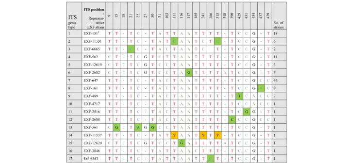
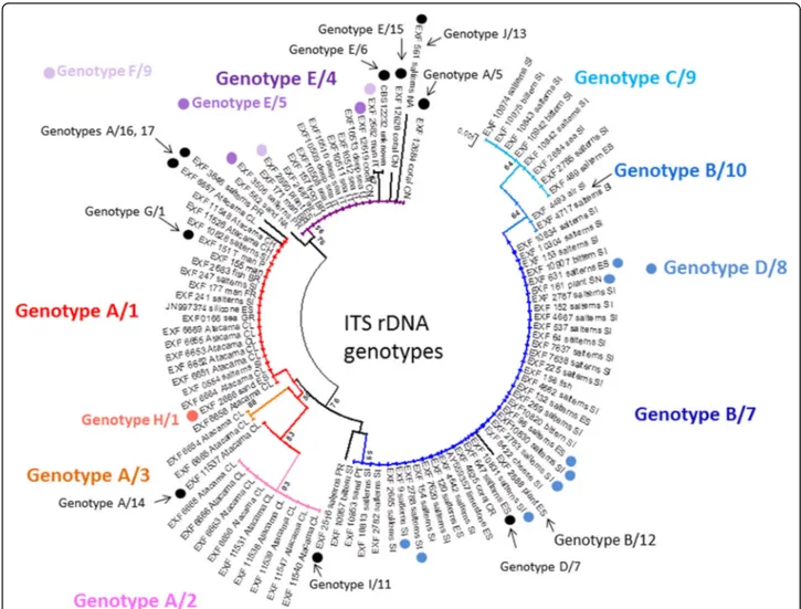
All of these strains studied were identified as Hortaea werneckii based on ITS sequences and the closest Gen-Bank hits. The ITS sequences were of good quality, whereby the chromatograms had clear peaks and no background noise, with a single exception (EXF-11537), which had three ambiguous positions in both strands. The variant calling for ITS and large subunit (LSU) re-gions for the strains with sequenced genomes showed that a single genome contained a single variant of the ITS and D1/D2 rDNA sequence (Additional file1: Table S1). In the D1/D2 domains of 28S rDNA, 10 genotypes (A–J) were recognised with the manual inspection of the sequence alignment, as also seen in the UPGMA analysis (Fig. 1). Within this dataset, eight variable sites were identified, which constituted 1.5% dissimilarity in the 521-nucleotide-long alignment (Table2). The ITS rDNA sequence alignment was 457 nucleotides long, and the number of variable nucleotides was 23 (dissimilarity 5%), defining 17 different genotypes (Table3). UPGMA ana-lysis of the 28S rDNA alignment fully reflected the observed genotypes (Fig. 1), but in the ITS rDNA ana-lysis (Fig. 2), the genotypes D/7 (EXF-647) and D/8 (161, 2788, 96, 631, 9, EXF-10830, EXF-2783) were intermixed with genotype B/7, despite single nucleotide differences. On the other hand, according to D1/D2, the H. werneckii ex-type strain (CBS 107.67; EXF-151) formed a single strain genotype J, but for the ITS sequence it belonged to genotype 1. The phylogenetic analyses of the same datasets (i.e. ITS, D1/D2 28S rDNA) with maximum parsimony resulted in overall poorly supported clades (data not shown). Sev-eral genotypes showed paraphyletic relationships and not monophyletic groups. Although analyses of the household genes BTB and MCM7 resulted in overall good quality of the sequences, at specific nucleotide po-sitions in the chromatograms for the majority of the
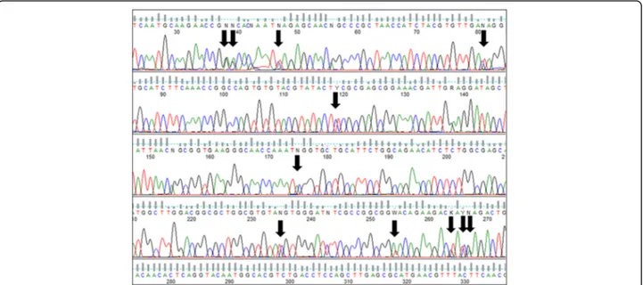
strains, double peaks were found in both strands, which led to ambiguity (Fig.3). On the other hand, unambigu-ous sequences of both loci were obtained by Sanger se-quencing for a number of the strains. The situation was not necessarily the same at both loci, as different strains had, for example, ambiguous sequences in MCM7, but unambiguous sequences in BTB.
For the MCM7 gene, these ambiguous sequences were used for computational estimation of the haplo-types, and the estimated haplotypes in most cases matched well to known haplotypes recovered by whole-genome sequencing. Haplotypes from the same strain were positioned in different parts of the intra-specific phylogeny (Fig. 4). The estimated haplotype pairs of strains EXF-2000 (=EXF-225), EXF-4667, EXF-7620, EXF-4662, EXF-152 and EXF-2685 were nearly identical: they had the same phylogenetic position and were collapsed into one leaf per strain in the final tree. Twenty-eight sequences did not contain any ambiguous nucleotide positions. Large
Fig. 1 Unweighted pair group method with arithmetic averages analysis of Hortaea werneckii D1/D2 28S rDNA sequences defines 10 genotypes
Table 2 Variable sites in Hortaea werneckii alignment of partial D1/D2 28S rDNA sequences
Legend: Green marked nucleotides represent nucleotide change in a single group or strain
phylogenetic clusters largely corresponded to clusters of MCM7 genes (Fig. 4, clusters marked ‘a’ and ‘b’).
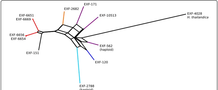
The distances between the strains calculated from single nucleotide polymorphism data and used for the construction of the phylogenetic network (Fig. 5) in general matched the distances estimated using the ITS and D1/D2 rDNA sequences. However, the ana-lysis also showed a substantial reticulation within the phylogenetic history of the strains.
Halotolerance
Halotolerance was expressed as colony diameters of strains on solid MEA media, without NaCl and supple-mented with 10, 20, 25 and 30% (w/v) NaCl. This was measured after 9 weeks of incubation, and the data are given in Additional file 1: Table S2. All of the strains grew without NaCl and at up to 20% NaCl within these 9 weeks, with the exception of two strains (i.e., EXF-3846, EXF-2687), which did not grow at 20% NaCl.
The growth optimum was at 10% NaCl, which was typically seen as filamentous colonies, with yeast-like cells in the central part. EXF-120 grew as yeasts without NaCl and at 20% NaCl, but showed filament-ous immersed growth with 10% NaCl. All other strains were either filamentous on all of the media (representative strain EXF-4717), or formed numer-ous filamentnumer-ous sectors within initial yeast colonies without NaCl (representative strains EXF-96, EXF-467), or only filamentous growth without sectors on media with NaCl (representative strains EXF-467, EXF-96) (Fig. 6).
Over the 9 weeks of incubation, only a few of the strains (EXF-247, EXF-554, EXF-156, EXF-269, EXF-4667, EXF-489, EXF-647, EXF-3506) grew at 30% NaCl, to pro-duce colonies of 1–2 mm diam. Some additional strains grew at 30% NaCl after longer incubation periods (data not shown). The largest differences identified by SIMPER tests between the genotypes were in the colony diameters on MEA + 20% NaCl, where the group of strains belong-ing to LSU/ITS genotype E/4 developed colonies that were 4-fold larger than for the other genotypes. Other strains from this group grew more slowly and did not at-tain such colony sizes.
Thermotolerance
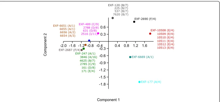
The growth of the tested strains on solid MEA media without NaCl and supplemented with 10% NaCl and at different temperatures (5, 15, 25, 37 °C) varied among the strains (Additional file 1: Table S3). Here, some patterns could be correlated with some geno-types, as shown by principal component analysis (Fig. 7). For example, the sea and deep-sea strains from Italy (genotype E/4) grew at 5 °C to 37 °C regardless of the addition of NaCl. Their growth at 5 °C was slow, while it was rapid at 37 °C, and comparable to their growth at 25 °C. However, a human-associated strain (EXF-171) from the same genotype (E/4) did not grow at 5 °C and at 37 °C, which might have been due to its degeneration dur-ing subcultivation, as it was originally isolated in 1935. Strains from Atacama, Chile, that belonged to genotypes A/1 to A/3 grew at 5 °C, but only with the addition of 10% NaCl, while they did not grow at 37 °C. The strains
Table 3 Variable sites in Hortaea werneckii alignment of partial ITS rDNA sequences
Legend: Green marked nucleotides represent nucleotide change in a single group or strain. Yellow marked nucleotides (Y) represent ambiguous peaks in the sequence, being either Cytosine (C) or Thymine (T)
assigned to genotype B/7 grew at 15 °C to 25 °C, without and with NaCl, while they grew at 37 °C only with the addition of NaCl. The optimum temperature for all tested strains was at 15 °C to 25 °C.
Strains were also tested for temperature tolerance in liquid media (YNB without and with addition of 5, 10, 15, 20, 25% [w/v] NaCl). All of these strains grew faster at 25 °C, and grew at 37 °C with the addition of at least 5% NaCl to the medium (Fig.8), although they reached lower optical densities at this higher temperature (37 °C, OD590 0.3; 25 °C, OD590 3.0). In agreement with the
growth tests on solid media, strain EXF-10813 from the deep sea grew well at 37 °C even without NaCl, although it grew faster with the addition of NaCl (OD5901.5).
Morphological studies
The colony morphologies differed substantially among strains and across the culture media. Among 34 strains grown for 2 weeks on MEA, nine different colony
morphologies were recorded, which differed in terms of colour, structure and size (Additional file 1: Table S4). The colony colours (according to the Methuen Hand-book of Colour) were most often olive-brown (4E7), but also honey-yellow (5D6), mustard-brown (5E6), yellow-ish grey (2B2), olive yellow (2D7) and blond greyyellow-ish yel-low (4C4). Colony diameters were 2–7 mm, as yeast-like, filamentous, or mixtures of both forms (Figs. 9 and 10, columns A).
In comparison to MEA, the colonies on MEA + 10% NaCl were initially yeast-like, but developed marginal immersed budding mycelium that were visible as filament-ous growth, and some became exclusively filamentfilament-ous (EXF-6656, EXF-3846), while some appeared filamentous regardless of their growth medium (EXF-161, EXF-171), and reached diameters from 0.5–10 mm. The colony col-ours here were most often olive-brown (4D, E, F), or olive (3E, F4) to golden brown, to yellowish brown (5D, E), dull green (30D3), or even yellowish grey (4B2).
On OA media, the cultures were dark green (30F5) to black, and reached diameters of 2.5–13.0 mm. The dif-ferent colonies of the same strain were mostly similar, and were composed of a yeast-like small centre that was surrounded by a wider superficial aerial filamentous margin, although in some cases yeast and yeast/ fila-mentous colonies developed from a single strain on the same agar plate.
The colonies on PDA were mostly yeast-like and shiny, and reached diameters of 1–9 mm in 14 days. These were yellowish brown (5F7) to olive-brown (4F4–6), with many darker, or they were coloured similar to those grown on OA (e.g. EXF-161).
The informations on their microscopic structures on these culture media are shown in Fig. 10. In general, the conidial size, shape, and pigmentation depended on the culture medium, and varied among the strains. On all of the culture media, the colonies started to grow as yeasts, and many of them later developed superficial, aerial, or immersed mycelia. The different kinds of hyphae mea-sured from 2.5–15.0 μm diam. The thin hyphae were either composed of long cells (length, 5–15 μm), or were seen as pseudomycelia that were composed of concatenated two-celled conidia that were constricted at the septa (width, 7μm). The thick hyphae were additionally longitudinally septate (Fig. 11). The mycelia had lateral proliferating openings that were either almost sessile or developed short single conidiogenous cells, or even complex conidiophores that developed into synnema-like structures.
On the OA medium, even hyphal clusters that resem-bled sterile ‘pseudo’ fruiting bodies were seen (Fig. 12). Conidia developed in annellidic conidiogenesis, and were typically one or two-celled and melanized, with
bi-polar budding (Fig. 13). On the OA medium they differed in size and pigmentation. One-celled conidia from the colony centres measured 5.0–8.0 × 2.0– 4.5μm, and from the colony margins 5.5–8.0 × 2.0– 4.0μm. Typical two-celled conidia that aggregated in the colony centres as yeast colonies measured 7.5– 15.0 × 3.0–6.0 μm, and for some of the strains these were also seen in the marginal parts of the colonies, where they measured 9.0–12.0 × 3.0–8.5 μm (Additional file 1: Table S5). The mean conidial measurements from the centres of the colonies (±standard deviations) are shown in Fig.14.
Growth in the form of meristematic clusters was evi-dent for almost all of the strains growing on MEA at 37 °C, and this also appeared in the centres of 14-d-old colonies on PDA and MEA, and for some of the strains also on MEA with 10 to 30% NaCl (Fig.15).
Use of carbon and nitrogen sources
The 190 carbon assimilation tests were divided into 10 groups according to their main roles in fungal metabolism: pentose catabolic pathway; galactose pathway; starch and su-crose metabolism; uronic acid metabolism; glycolysis and branches; pyruvate metabolism; citrate cycle; amino-acid and protein metabolism; compatible solutes; and aromatic pathways. An additional group of‘other carbon sources’ was as defined previously by Nai et al. (2013). Some substrates involved in more than one metabolic pathway were classi-fied into two groups (e.g. sugar alcohols, with D-sorbitol, D-arabitol and L-arabitol classified both as the pentose cata-bolic pathway and as metabolism of compatible solutes).
Of the five strains tested, good growth was detected in 52 of the carbon assimilation tests (27.4%) that defined the
pentose catabolic pathway, galactose pathway, starch and sucrose metabolism, glycolysis and branches, pyruvate metabolism, citrate cycle, compatible solutes, uronic acid metabolism, and other carbon sources. The most efficiently catabolised substrates were those from the pentose (D-ri-bose, D-xylose, L-arabinose, L-arabitol) and galactose (D-galactose, D-melibiose, D-raffinose) catabolic pathways, glycolysis and branches (D-glucose, D-mannose, arbutin, gentiobiose, laminarin), and metabolism of compatible solutes (D-mannitol, D-trehalose). None of the strains cata-bolised 46 of the substrates (24.2%), a large proportion of which belonged to amino-acid and protein metabolism (34.8%; 16/46) and other carbon sources (32.6%; 15/46). The use of the remaining amino-acid and protein metabol-ism sources and the aromatic pathways varied between the strains, and ranged from good to weak, and negative (see Additional file1: Table S6 for details).
The catabolism of nitrogen was tested on 95 nitrogen sources that were divided into eight groups: simple ni-trogen sources; amino acids and derivates; primary amines; amides; amino sugars; nucleobases and deri-vates; aminated alkenes; and dipeptides. The strains grew well on the majority of these nitrogen substrates (from
68 to 90% of the tests), except for EXF-2788, which grew well only on 31 (32.6%) of these nitrogen substrates, and did not grow on 40 (42.1%) of them (Additional file 1: Table S7). Also, the SIMPER test revealed that the lack of catabolism of numerous nitrogen sources by this strain (e.g. methylamine, D-glutamic acid, L-histidine, N-butylamine, uric acid, D-alanine, ethylamine, cytosine, cytidine, D,L-α-amino-N-butyric acid, L-methionine, uri-dine) contributed the most to its separation from the remaining strains. All of the strains showed little or no catabolism of amino-acid D-lysine, primary amine ethy-lenediamin, amino sugars N-acetyl-D-mannosamine and N-acetyl-D-galactosamine, and nucleobases thymidine, thymine and uracil.
Enzymatic tests
The results of the enzymatic tests are given in Additional file 1: Table S8. The proteolytic activities on casein were positive or weak for around 60% of the strains, and became weaker or were lost with addition of 5 and 10% NaCl at 25 °C; at 37 °C, only 30% of the strains were positive. The proteolytic ac-tivities on gelatine were generally positive and became
(See figure on previous page.)
Fig. 4 Maximum likelihood phylogenies based on the MCM7 and BTB genes. a The MCM7 gene. The amplicons were amplified by PCR and sequenced by Sanger sequencing. Sequence names without ambiguous nucleotides after sequencing are shown in green. Haplotype estimation was used to separate haplotypes from sequences with ambiguous nucleotides (in blue). Estimated haplotypes with identical phylogenetic positions were collapsed into one sequence. Haplotype sequences recovered from the whole-genome sequences are included for comparison (in red). Haplotype pairs from the same genomes are connected by yellow lines. b The BTB gene. Only sequences without ambiguous nucleotides and sequences obtained by cloning the amplicons and sequencing individual clones were included in the analysis. Haplotype sequences recovered from the whole-genome sequences are included for comparison (in red). The large phylogenetic clusters observed in both the MCM7 and BTB phylogenies are indicated with‘a’ and ‘b’
Fig. 5 Phylogenetic network of the Hortaea werneckii strains with sequenced genomes. The network was reconstructed with the Neighbour-Net algorithm based on the dissimilarity distance matrix calculated from the single nucleotide polymorphism data
stronger with the addition of NaCl. The esterase ac-tivities were positive for all of the strains at 25 °C, even in the presence of 5 and 10% NaCl, while at 37 °C, addition of 10% NaCl (but not 5% NaCl) largely inhibited the esterase activity.
DISCUSSION
Black yeasts are a polyphyletic group of melanized poly-morphic fungi that can grow in yeast-like and filament-ous forms. They can express an ‘extremophilic ecotype’ (Gunde-Cimerman et al. 2000) that is characterised by thick melanized cell walls, slow, often meristematic growth, and proliferation with endoconidiation. Globally, black yeasts populate different extreme environments, from hypersaline coastal salterns worldwide (Gunde-Ci-merman & Zalar 2014), to surfaces and subsurfaces of rocks at high or low temperatures (Wollenzien et al. 1995, Selbmann et al. 2005) and Arctic glacial ice (Gun-de-Cimerman et al. 2003). Many are involved in opportunistic infections, which demonstrates a certain
coherence between extremotolerance and opportunism (de Hoog et al.2005, Teixeira et al.2017).
Hortaea werneckiiis a cosmopolitan, extremely haloto-lerant, black yeast without a known sexual morph. Due to its phenotypic plasticity, historically, H. werneckii was placed in numerous genera (e.g. Cladosporium, Exo-phiala, Dematium, Pullularia, Aureobasidium, Sarcino-myces, Pheoannellomyces) before being ascribed to a new genus, due to its special method of conidiogenesis (Nishimura & Miyaji 1984), of which it was designated the type species. Two additional, so far rarely isolated, species have been described in the last decade: H. acido-phila from lignite (Hölker et al. 2004) and acidic saline soils (Hujslova et al.2010); and H. thailandica originally described from plant material in Thailand (Crous et al. 2009), and was later also found in subglacial ice on Svalbard (Zalar & Gunde-Cimerman, unpubl.). However, H. acidophilawas later transferred to the genus Neohor-taea in Capnodiales based on its phylogeny for nuclear ribosomal and five household genes (Quaedvlieg et al. 2014).
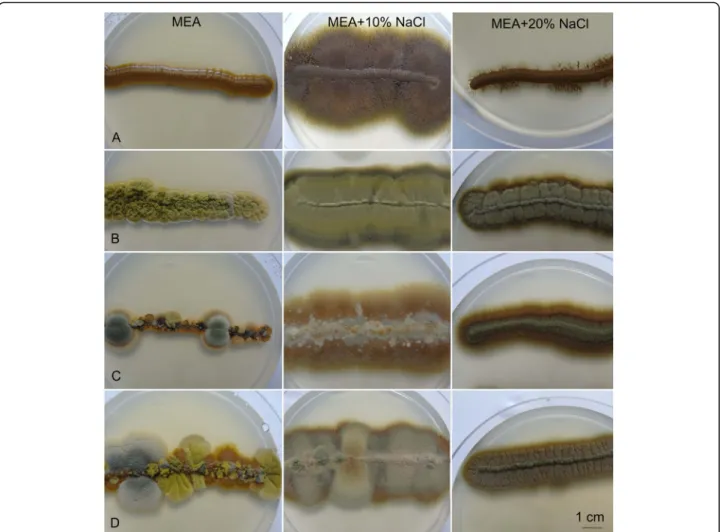
Fig. 6 Morphology of Hortaea werneckii cultures on malt extract agar (MEA) without and with added NaCl. Left: MEA; middle: MEA + 10% NaCl; right: MEA + 20% NaCl. a EXF-120. b EXF-4717. c EXF-467. d EXF-96. Bar = 1 cm (for all)
Hortaea werneckiiis the causative agent of tinea nigra, and its primary habitat is hypersaline environments and seawater (Gunde-Cimerman & Zalar 2014). It can also grow in other osmotic environments, as recently demon-strated by its discovery in house dust in Hawaii (Humphries et al. 2017) and on spider webs, walls and sand in arid Atacama caves that have been exposed to marine aerosols (Azúa-Bustos et al. 2010), as indicated above. In the present study, 98 strains were studied that were collected worldwide from natural hypersaline envi-ronments and Atacama Desert, and from human, animal and plant sources.
All tested H. werneckii strains were halotolerant and grew from 0 to 25% NaCl, and some strains even grew at 30% NaCl (Fig. 6, Additional file 1: Table S2). In agreement with other studies (Gunde-Cimerman et al. 2000, de Hoog et al. 2000, Butinar et al. 2005), these strains grew best from 15 to 25 °C, although some strains grew at 5 °C and 37 °C, without or with addition of at least 5 to 10% NaCl (Fig.8, Additional file1: Table S3). This indicated their previously underestimated pathogenic potential (de Hoog et al.2000).
The morphological data (except for the conidial size) were in agreement with previous descriptions (Nishimura & Miyaji 1984, de Hoog et al. 2000). In particular, the two-celled conidia were larger than stated in the species description (7.0–9.5 × 3.5–4.5 μm) and varied considerably across the different culture media (7.5–15.0 × 2.5–6.5 μm; Additional file1: Table S5).
The study of the use of carbon and nitrogen sources for five selected H. werneckii strains is the most comprehensive study to date (Additional file1: Tables S6–S7), and the data
are in line with previous data obtained by traditional methods (Zalar et al. 1999, de Hoog et al. 2000). These strains showed various carbon and nitrogen assimilation patterns, with the exception of the haploid strain EXF-2788, which did not catabolise numerous nitrogen substrates.
The extracellular enzymatic activities of four H. wer-neckiistrains, which were recently evaluated by Formoso et al. (2015) and Elsayed et al. (2016), were dominated by plant degrading enzymes, including amylase, lipase, ester-ase, pectinase and/or cellulester-ase, and no animal-related enzymes, like albuminase, keratinase, phospholipase and DNAse. All of the tested strains were urease positive, while caseinase, gelatinase and laccase were variable. The data are in agreement with both of the previous studies (Additional file1: Table S8), although the enzymatic activ-ities varied considerably between the strains.
H. werneckii strains show a unique natural ecology and pathogenic potential, along with high phenotypic and genotypic variability. These aspects were indicated previously and are defined further in this extensive study, and they have raised the question whether a spe-cies complex might be concerned (Boekhout et al.1993, Uijthof et al. 1994, de Hoog et al., 1999, Zalar et al. 1999, Cabañes et al.2012, Formoso et al.2015, Elsayed et al. 2016, Marchetta et al. 2018). This also prompted our phylogenetic analysis here. All of the previous stud-ies were based on smaller sets of strains (i.e. from three to 67), and used different genotypic markers and methods, like karyotyping, different PCR fingerprinting techniques, and ITS/28S rDNA sequence variability.
In the present study, the 98 worldwide strains were analysed according to alignments of D1/D2 and ITS
Fig. 7 Principal component analysis of Hortaea werneckii growth at different temperatures on solid malt extract agar. Strains with the same temperature patterns are coloured the same
Fig. 8 Growth curves of selected Hortaea werneckii strains grown in YNB liquid media without and with added 5, 10, 15, 20, and 25% NaCl, incubated at 25 °C and 37 °C
rDNA sequences. These analyses allowed the definition of 10 and 17 genotypes, respectively, that are based on 1.5 and 5% dissimilarities, respectively (Figs.1,2). In the ITS analysis, several phylogenetically non-informative
single point mutations were seen, which resulted in nu-merous paraphyletic groups in the phylogenetic analysis. The majority of strains that belong to genotype A were derived from tropical, subtropical and southern
Fig. 9 Macromorphology of Hortaea werneckii strains grown on malt extract agar (MEA; column 1), MEA + 10% NaCl (column 2), oatmeal agar (column 3) and potato dextrose agar (column 4), for 14 days at 25 °C. a EXF-151T. b 3846. c 489. d 225. e 161. f 2788. g EXF-171. Bar = 1 cm (for all)
hemisphere hypersaline environments, while genotypes B to E contained more strains from Mediterranean re-gions. Although the majority of the strains originated from the Mediterranean area, they were spread (with few excep-tions) among almost all of the recognized genotypes. Al-though genotype B included some strains from animals (i.e. fish, corals) and plants, animal-related and plant-related strains were also found in genotype A. The five strains from humans grouped in the 28S/ITS genotypes A/ 1 (France, unknown), E/4 (Brazil), F/9 (Italy) and G/1 (type strain from Portugal), and not in genotype B. Interestingly,
all of the strains from the Atacama Desert grouped into genotype A for 28S rDNA, but based on ITS, they were di-vided into four genotypes: A/1, A/2, A/3, and A/14. For these strains only, variability was also noted for conidial size: strains of genotype A/1 (EXF-6651, EXF-6655, EXF-6669) had double the conidial width in comparison to strains of genotype A/3 (EXF-6654, EXF-6656) (Figs.13o-s, 14). Additionally, random representatives of all of the ge-notypes were highly variable. This intraspecific variability indicates a potentially problematic division into genotypes, as exemplified by the strains associated with tinea nigra.
Fig. 10 Micromorphology of Hortaea werneckii strains grown on malt extract agar (MEA) (column 1), MEA + 10% NaCl (column 2), oatmeal agar (column 3) and potato dextrose agar (column 4) for 7 days at 25 °C. a EXF-151T. b 3846. c 489. d 225. e 161. f 2788. g EXF-171. Bar = 10μm (for all)
The taxonomy of a potential H. werneckii species com-plex was further resolved by Sanger sequencing of the standard taxonomic markers; i.e. the partial BTB and MCM7genes. Most of the resulting sequences contained ambiguities (Fig.3). This problem was initially explained by the finding of genome duplication in the single strain where the genome was sequenced (Lenassi et al. 2013, Sinha et al. 2017). A more recent study by Gostinčar et al. (2018) analysed an additional 11 H. werneckii ge-nomes, and showed that the majority of the strains were diploid and highly heterozygous. The large divergences within the estimated MCM7 haplotype pairs of individ-ual H. werneckii strains matched well the proposed for-mation of hybrids between heterozygous strains of H. werneckii – even though comparative genomics indi-cated that H. werneckii is clonal (Gostinčar et al.2018). In the analysis here of the MCM7 sequences that in-cluded the whole-genome sequence-derived and compu-tationally estimated haplotypes, in three cases the pairs of haplotypes of four or more strains shared the same phylogenetic history (Fig. 4), which might have resulted from the same hybridization event. In addition to this, several such events that involved smaller numbers of strains were indicated. In several cases the phylogenetic distances between the haplotypes were substantial.
The situation shown by the phylograms of the BTB and MCM7 genes depicted in Fig. 4 illustrates the difficulties encountered when investigating intraspecific phylogeny of H. werneckii using standard approaches of sequencing molecular markers. In some cases, both cop-ies of the marker genes were amplified. If they did not
contain insertions/ deletions, Sanger sequencing pro-duced useful results, although with ambiguous nucleo-tides in positions that differed between the copies (Fig. 3). After careful investigation of the sequencing chromatograms, the resulting sequence was used for es-timation of both haplotypes, a process that was some-times relatively accurate (e.g. EXF-120), and somesome-times unsuccessful (e.g. with the production of two nearly identical haplotypes; such as for EXF-2000/ EXF-225), as seen from the comparisons with the true haplotypes de-rived from the corresponding whole-genome sequences. In some cases, the sequencing produced good quality se-quences with no ambiguities. Theoretically, this can occur if the strain is indeed haploid (e.g. EXF-2788, and possibly other strains in the same cluster), if the two copies of the gene in a diploid genome are identical, or if PCR favours amplification of one copy over the other (e.g. EXF-6669). This last case can lead to an incomplete result– the apparently clear positioning of the species in the intraspecific phylogeny, although the other gene copy might be positioned in a very different part of the gene tree. If both copies are amplified, it is possible to sequence each copy separately by cloning the amplicon and sequencing several clones, as was achieved for β-tubulin (Fig.4; strain EXF-6655). However, if the amp-lification is strongly biased towards one of the copies, this approach is not feasible, as all of the sequenced clones will contain the same copy of the gene.
Interestingly, sequencing the ITS and D1/D2 rDNA markers was not hampered by the heterozygosity that resulted in mixed sequences with ambiguous nucleotide
Fig. 11 Different kinds of non-sporulating mycelium produced by Hortaea werneckii cultures after 14 days at 25 °C. a EXF-561: PDA, immersed margin. b EXF-161: oatmeal agar (OA), aerial margin. c EXF-171: potato dextrose agar (PDA), aerial. d EXF-4493: malt extract agar (MEA), immersed centre. e EXF-11537: OA, immersed margin. f EXF-2682: PDA, centre. g EXF-2690: OA, margin. h. EXF-2682: MEA + 10% NaCl, centre. Bar = 20μm (a, f), 5μm (b-e, g-h)
positions (Additional file 1: Table S1), as for all of the other genes of H. werneckii analysed here. This can be explained by concerted evolution, a process that is re-sponsible for sequence homogenisation of different gene copies in the same genome (Liao 1999). If this is the case, concerted evolution in H. werneckii appears to be happening more efficiently in ribosomal sequences than in other genes. A reason for this might be the organisa-tion of the ribosomal DNA in tandem repeats, and the resulting increased opportunity for unequal
crossing-over, which has been linked to concerted evolution of the ITS region in fungi (Naidoo et al. 2013). ITS and D1/D2 rDNA phylogenetic markers can thus be used to discern the intraspecific phylogeny of H. werneckii with-out the technical problems that prevent the use of other markers, as discussed above. However, it has to be noted that the phylogeny estimated based on ITS and D1/D2 rDNA markers is a simplified approximation of the real evolutionary history of the species, as it ignores the sub-stantial reticulation of the H. werneckii phylogeny (as
Fig. 12 Sporulating mycelium of Hortaea werneckii strains in cultures after 14 days at 25 °C. a 4493: malt extract agar (MEA), centre. b EXF-2682: oatmeal agar (OA), margin. c EXF-489: OA. d EXF-647: MEA + 20% NaCl. e EXF-151: MEA + 10% NaCl. f EXF-2687: OA, centre. g EXF-171: potato dextrose agar. h EXF-6655: MEA + 10% NaCl, margin. i EXF-6654: OA. j EXF-247: MEA + 10% NaCl. k EXF-562: OA, margin. l EXF-225: MEA. m EXF-2788: OA. Bar = 20μm (d, i), 10 μm (a-c, e-h, j-m)
demonstrated in Fig. 5), which is a consequence of hybridization into persistent and highly heterozygous diploids (Gostinčar et al.2018).
Hybridization events and changes in ploidy in fungi have been described in other species, but with distinct differ-ences compared to these observations for H. werneckii. In Saccharomyces cerevisiae a whole genome duplication event occurred through an ancient hybridization, which
was then followed by loss of most of the duplicated regions (Marcet-Houben & Gabaldón2015). Apart from this, vari-ations in ploidy and aneuploidy are relatively frequent and are thought to be important for rapid adaptation to envir-onmental changes (Sunshine et al. 2015). Interspecies hy-brids have been described in fungi as well, and while most of these are in species only known as asexual morphs, Zygosaccharomyces parabailiiwas shown to have regained
Fig. 13 Conidia of Hortaea werneckii strains grown on oatmeal agar (OA) for 14 days at 25 °C. a 120. b 157. c 161. d 171. e 247. f 562. g 631. h 2516. i 2687. j 2690. k 2785. l 2788. m 3506. n 4625. o 6651. p 6654. r EXF-6656. s EXF-6669. t EXF-7620. u EXF-10508. v EXF-10512. w EXF-10513. x EXF-11537. Bar = 10μm (for all)
fertility by inactivation of one mating-type locus (Ortiz--Merino et al.2017). Another well-studied example is Can-dida albicans, a predominantly diploid species that nevertheless has a high degree of genome plasticity, where frequent losses of heterozygosity and gross chromosomal rearrangements might result in aneuploidy (McManus & Coleman 2014). Although reproduction in C. albicans is predominantly clonal, this species can also use a parasex-ual cycle that involves the formation of tetraploid progeny from the mating of diploid parents, the former of which subsequently revert to diploidy by concerted chromosome loss (Bennett & Johnson2003). While double peaks on se-quencing chromatograms can be easily dismissed as a re-sult of suboptimal sequencing quality, they have been reported in at least one other case – in the yeast genus Ogataea. Here, such double peaks were reported to be an indication of heterozygous diploidy or of the existence of gene paralogues that were both/all amplified with the pri-mer sequences used (Čadež et al.2013).
The failure to recognise hybrids is not only a danger in terms of serious taxonomic misunderstandings, but can also have implications beyond taxonomy. Inderbitzin et al. (2011a) indicated such implications in the management of
pathogenic fungi. In this field, the existence of hybrids was first inferred from credible morphological and genetic evi-dence, and later based on molecular phylogenetics (Inder-bitzin et al. 2011a). Hybrids are known in basidiomycete rust genera (e.g. Tilletia, Melampsora, Cronartium, Pucci-nia), ascomycetous plant pathogens (e.g. Botrytis, Fusar-ium, Ophiostoma, Verticillium), yeasts (e.g. Saccharomyces, Candida, Cryptococcus), and endophytic fungi. Hybridiza-tions or recombination of introduced fungal pathogens with related resident fungi often results in strains that can infect an expanded range of hosts (Brasier, 2000). By whole genome sequencing of two strains, Fogelqvist et al. (2018) confirmed the findings of Inderbitzin et al. (2011b) for Verticillium longisporum. Their compara-tively large draft genomes compared to other asco-mycete fungi studied were composed of two parts, where one lineage was more ancient than the part that was more closely related to V. dahliae.
The pattern of intraspecific hybridization seen here in H. werneckii, which is otherwise a clonal species, represents an unusual strategy that to the best of our knowledge has not been described in other fungi to date. Enriching the genomic toolbox with two different copies of the same
Fig. 14 Measurements of the 1-celled and 2-celled conidia of Hortaea werneckii strains. Data are means ±standard deviation. 28S rDNA genotypes are indicated as letters (genotype A, red; genotype B, C, D, blue; genotype E, violet; genotype F, orange; miscellaneous LSU genotypes, black), and ITS genotypes as numbers 1–16
gene, or combining traits that are otherwise combined via sexual reproduction, might promote survival under ex-treme environmental conditions. However, the possible benefits of heterozygous diploid genomes for the survival of H. werneckii remain to be investigated. The presence of four ITS genotypes in a small area was seen for the Ata-cama cave strains, where some diploid strains were almost identical in one haploid subgenome, but differed substan-tially in the other haploid subgenome (Gostinčar et al. 2018). This makes H. werneckii an intriguing extremotoler-ant experimental system with persistent stable haploid and diploid strains and little, if any, noise introduced by sexual recombination.
CONCLUSIONS
Ongoing recent trends in fungal taxonomy that are sup-ported by powerful phylogenetic multi-locus analyses encourage descriptions of new species, which can often be based on only a single strain, or a few strains, and/or
a few nucleotide differences. When using a limited set of strains, the decisions to recognise cryptic species have to be taken with great caution, as these analyses can be ser-iously misleading. The present study that is based on nu-merous geographically and time-spread strains is characterised by a highly plastic and unusual fungal spe-cies that challenges this concept. In the majority of these H. werneckii strains, on the one hand, there were am-biguous positions for the household genes routinely used in taxonomic studies, and on the other hand, there were the unambiguous sequences of the ribosomal genes. Ac-cording to our estimates, around 20% of these H. wer-neckii strains were haploid, while the others were diploid. This situation prevented standard species delin-eation and requires a restrained and adjusted taxonomic approach. Analysis of a smaller number of geographic-ally separated strains or a restricted number of pheno-typic and clustering analyses might have resulted in the description of several new species. We believe that
Fig. 15 Growth of Hortaea werneckii strains after 21 days at 37 °C on solid malt extract agar (MEA) medium supplemented with 10% NaCl (if not indicated otherwise). a, b EXF-120. c, d EXF-151T. e EXF-151Tin liquid YNB. f EXF-157. g EXF-225. h EXF-269. i EXF-537. j EXF-561. k EXF-631. l EXF-2690. m EXF-3506. n EXF-6651. o, p EXF-7620. q EXF-10508 (on MEA). r EXF-105012 (on MEA). s EXF-10513. t EXF-11537 (on MEA). Bar =5μm (a, b, d-f, h-m, o-t), 20 μm (c, g, n)